User login
The dangers of desonide
In a previous column, I warned about the high cost of generic desonide. This month, I alert you to the many potential dangers of this drug. By the time I’m done, you may not want to go near the stuff.
To approve e-scribe refills, we all need to acknowledge warnings and dangers and click “Benefit outweighs risk” or “Previously tolerated” or some other option. But some of these warnings make me wonder who on earth writes them.
Desonide comes with more warnings than almost any other medicine I prescribe electronically. I counted 21 such warnings. Here are some examples:
1. Desonide External Cream 0.05% should be used cautiously in Bacterial Infection, especially in Systemic Bacterial Infection. Since Folliculitis is a specific form of Bacterial Infection, the same precaution may apply.
I confess that I never thought of prescribing desonide for Bacterial Infection, Systemic or otherwise. Have you? (By the way, what’s with the excess use of capital letters?)
The second warning is even more dramatic.
2. Desonide External Cream 0.05% should be used cautiously in Viral Infection, especially in Systemic Viral Infection.
What makes this even more curious is the Viral Infections the warnings go on to enumerate.
2a. Since Actinic Keratosis is a specific form of Viral Infection, the same precaution may apply.
Actinic Keratosis is a Viral Infection? I didn’t know that.
3. Since Actinic Keratosis of the Hands and Arms is a specific form of Viral Infection, the same precaution may apply.
Now we learn of different subgroups of Actinic Keratoses that are Viral Infections. Did they teach you these in Dermatology School? (Please see Warnings 6-10, below.)
4. This warning refers to a specific Bacterial Infection called Folliculitis Nares Perforans. I don’t know what that is, but it sounds bad. Glad they warned me.
5. Since Pseudofolliculitis Barbae is a specific form of Bacterial Infection, the same precaution may apply.
I never used much desonide for pseudofolliculitis, cautiously or otherwise.
Warnings 6-10 describe more specific forms of Viral Infection: (6) Non-Hyperkeratotic Actinic Keratosis, (7) Actinic Keratosis of Face and Anterior Scalp, (8) Non-Hyperkeratotic Non-Pigmented Actinic Keratosis, (9) Non-Hyperkeratotic Face and Scalp Actinic Keratosis, (10) Pigmented Actinic Keratosis.
This is most disturbing. What Systemic Viral Infections did they leave me to use desonide on? Hyperkeratotic Non-Pigmented Actinic Keratoses of the Posterior Scalp?
Warning 11 is another specific Bacterial Infection: Local Folliculitis. What is the opposite of Local Folliculitis? Express Folliculitis?
Warning 12 is Perioral Dermatitis. Steroids on rosacea? Really? Maybe a cheaper one.
I will now skip to warning 16: Hirsutism has been associated with Desonide External Cream 0.05%. Since Hair Disease is a more general form of Hirsutism, it may also be considered a drug-related medical condition.
Did you know that desonide causes unwanted hair growth? Or realize that Hair Disease is a more general form of Hirsutism? I myself have male-pattern baldness. (Sorry, Male-Pattern BALDNESS.) Since Baldness is a Hair Disease, is it also a more general form of Hirsutism? Instead of having too little hair, do I now have too much?
The same is true for warning 17, which is identical to 16, except that it substitutes “Hypertrichosis” for “Hirsutism.”
Okay, colleagues, it’s time for a personal reckoning. You trained, practiced, took CME, but you didn’t know about any of these risks, did you? You’ve just been just heedlessly, incautiously, throwing around desonide, producing hairy patients with Systemic Bacterial and Viral Infections. And on “Non-Hyperkeratotic Non-Pigmented Actinic Keratosis,” no less. Aren’t you disappointed in yourselves?
When I first read warnings like these, I wrote my EMR provider to ask who puts together this stuff, and which consultants vet it. They never answered. It is very hard to believe that a dermatologist was involved at any stage of developing these warnings, with their irrelevant caveats and absurd classification schemes.
Who would develop electronic prescribing guidelines without at least consulting the physicians who do the prescribing? Why would they want to?
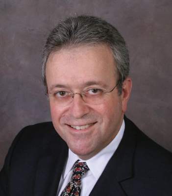
Dr. Rockoff practices dermatology in Brookline, Mass., and is a longtime contributor to Dermatology News. He serves on the clinical faculty at Tufts University, Boston, and has taught senior medical students and other trainees for 30 years.
In a previous column, I warned about the high cost of generic desonide. This month, I alert you to the many potential dangers of this drug. By the time I’m done, you may not want to go near the stuff.
To approve e-scribe refills, we all need to acknowledge warnings and dangers and click “Benefit outweighs risk” or “Previously tolerated” or some other option. But some of these warnings make me wonder who on earth writes them.
Desonide comes with more warnings than almost any other medicine I prescribe electronically. I counted 21 such warnings. Here are some examples:
1. Desonide External Cream 0.05% should be used cautiously in Bacterial Infection, especially in Systemic Bacterial Infection. Since Folliculitis is a specific form of Bacterial Infection, the same precaution may apply.
I confess that I never thought of prescribing desonide for Bacterial Infection, Systemic or otherwise. Have you? (By the way, what’s with the excess use of capital letters?)
The second warning is even more dramatic.
2. Desonide External Cream 0.05% should be used cautiously in Viral Infection, especially in Systemic Viral Infection.
What makes this even more curious is the Viral Infections the warnings go on to enumerate.
2a. Since Actinic Keratosis is a specific form of Viral Infection, the same precaution may apply.
Actinic Keratosis is a Viral Infection? I didn’t know that.
3. Since Actinic Keratosis of the Hands and Arms is a specific form of Viral Infection, the same precaution may apply.
Now we learn of different subgroups of Actinic Keratoses that are Viral Infections. Did they teach you these in Dermatology School? (Please see Warnings 6-10, below.)
4. This warning refers to a specific Bacterial Infection called Folliculitis Nares Perforans. I don’t know what that is, but it sounds bad. Glad they warned me.
5. Since Pseudofolliculitis Barbae is a specific form of Bacterial Infection, the same precaution may apply.
I never used much desonide for pseudofolliculitis, cautiously or otherwise.
Warnings 6-10 describe more specific forms of Viral Infection: (6) Non-Hyperkeratotic Actinic Keratosis, (7) Actinic Keratosis of Face and Anterior Scalp, (8) Non-Hyperkeratotic Non-Pigmented Actinic Keratosis, (9) Non-Hyperkeratotic Face and Scalp Actinic Keratosis, (10) Pigmented Actinic Keratosis.
This is most disturbing. What Systemic Viral Infections did they leave me to use desonide on? Hyperkeratotic Non-Pigmented Actinic Keratoses of the Posterior Scalp?
Warning 11 is another specific Bacterial Infection: Local Folliculitis. What is the opposite of Local Folliculitis? Express Folliculitis?
Warning 12 is Perioral Dermatitis. Steroids on rosacea? Really? Maybe a cheaper one.
I will now skip to warning 16: Hirsutism has been associated with Desonide External Cream 0.05%. Since Hair Disease is a more general form of Hirsutism, it may also be considered a drug-related medical condition.
Did you know that desonide causes unwanted hair growth? Or realize that Hair Disease is a more general form of Hirsutism? I myself have male-pattern baldness. (Sorry, Male-Pattern BALDNESS.) Since Baldness is a Hair Disease, is it also a more general form of Hirsutism? Instead of having too little hair, do I now have too much?
The same is true for warning 17, which is identical to 16, except that it substitutes “Hypertrichosis” for “Hirsutism.”
Okay, colleagues, it’s time for a personal reckoning. You trained, practiced, took CME, but you didn’t know about any of these risks, did you? You’ve just been just heedlessly, incautiously, throwing around desonide, producing hairy patients with Systemic Bacterial and Viral Infections. And on “Non-Hyperkeratotic Non-Pigmented Actinic Keratosis,” no less. Aren’t you disappointed in yourselves?
When I first read warnings like these, I wrote my EMR provider to ask who puts together this stuff, and which consultants vet it. They never answered. It is very hard to believe that a dermatologist was involved at any stage of developing these warnings, with their irrelevant caveats and absurd classification schemes.
Who would develop electronic prescribing guidelines without at least consulting the physicians who do the prescribing? Why would they want to?
Dr. Rockoff practices dermatology in Brookline, Mass., and is a longtime contributor to Dermatology News. He serves on the clinical faculty at Tufts University, Boston, and has taught senior medical students and other trainees for 30 years.
Resident debt is ruining medicine
Most physicians accumulate near $300,000 in debt by the time they finish residency and go to work. This debt is crushing, and it is having negative effects.
College and medical school tuition has soared in the last 20 years, keeping pace with federal loan availability. There is no clear relationship with increased quality of the educational experience or with increased knowledge of the students. Colleges are part of the problem, but, in my opinion, the medical schools are even more egregious. After all, how much does it cost to rent a lecture hall for 2 years and provide some lecturers?
I will never forget when I complained about the increasing expense of medical school to a dean once. He explained that all of the faculty’s salary is considered an expense to the medical school, and that students are only paying a fraction of the true cost.
I must object! If a professor makes a guest appearance for an afternoon or two, the medical students are expected to pay him for a year of work?
I will never forget the goofy physiology professor who gave us two afternoons of demonstrations and lectures. He hooked live frogs to electrodes and made waves on a monitor. It was interesting, but his salary is $200,000 a year. Was it worth $2,000 a student (a class of 100 medical students) to watch him make frogs twitch two afternoons? I think not.
Caribbean medical schools charge about the same as those in North America and make a large profit. Medical students have become a “profit center.” This is occurring in an age when medical students write and share much of their own educational content in an interactive environment. This cannot be sustained.
For the last 2 years of medical school, the students are turned loose on the hospital wards and become slaves. This is called running scut, and it includes running specimens to the lab, wheeling patients to x-ray, drawing blood, fetching lab results, doing much chart work (completing the chart is the most important thing), and learning a lot in spite of the grunt work. The older physicians do teach on rounds, and there is always a resident physician around.
Again, these practicing physicians (they bill for their services) would be there anyway for the residents, and if there were not residents, would have to be there for their patients. The medical students cannot possibly add much additional cost, but these attending physicians and residents salaries are included in the educational cost justification.
The debt introduces a toxic calculus to specialty selection. Residencies are chosen for their fiscal attractiveness, which is not a correct or sustaining reason in a long hard career. Also, it may become economically impossible to practice in a lower-paying specialty.
Let me tell you about Razor Rick, a college student I mentored for several years. I hire these kids in their college summers and breaks to do not much but make a little money, and in exchange they let me pontificate and smile at me. Well, Rick got through med school and then dropped out of his primary care residency, owing several hundred thousand dollars. I was stunned when his parents called me, and I insisted he come in to talk to me.
He patiently explained that his board scores were passing but not good enough to get into to a well-paying specialty. He logically explained that he would have a better life, and have a better return on his investment, if he simply went into pharma with his expensive degree.
I helped him find some interviews.
Some criticize this generation of doctors for not being engaged, for not joining organized medicine, and for avoiding the glorious heart of difficult medicine. I don’t blame them for being distracted. They have, on average, 300,000 good reasons to be more self-concerned.
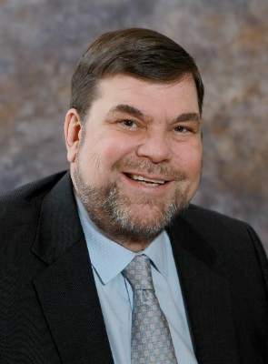
Dermatology News is proud to introduce the inaugural column of Cold Iron Truth by Dr. Brett Coldiron. Dr. Coldiron is a past president of the American Academy of Dermatology. He is currently in private practice, but maintains a clinical assistant professorship at the University of Cincinnati. He cares for patients, teaches medical students and residents, and has several active clinical research projects. Dr. Coldiron is the author of over 80 scientific letters, papers, and several book chapters, and he speaks frequently on a variety of topics.
Most physicians accumulate near $300,000 in debt by the time they finish residency and go to work. This debt is crushing, and it is having negative effects.
College and medical school tuition has soared in the last 20 years, keeping pace with federal loan availability. There is no clear relationship with increased quality of the educational experience or with increased knowledge of the students. Colleges are part of the problem, but, in my opinion, the medical schools are even more egregious. After all, how much does it cost to rent a lecture hall for 2 years and provide some lecturers?
I will never forget when I complained about the increasing expense of medical school to a dean once. He explained that all of the faculty’s salary is considered an expense to the medical school, and that students are only paying a fraction of the true cost.
I must object! If a professor makes a guest appearance for an afternoon or two, the medical students are expected to pay him for a year of work?
I will never forget the goofy physiology professor who gave us two afternoons of demonstrations and lectures. He hooked live frogs to electrodes and made waves on a monitor. It was interesting, but his salary is $200,000 a year. Was it worth $2,000 a student (a class of 100 medical students) to watch him make frogs twitch two afternoons? I think not.
Caribbean medical schools charge about the same as those in North America and make a large profit. Medical students have become a “profit center.” This is occurring in an age when medical students write and share much of their own educational content in an interactive environment. This cannot be sustained.
For the last 2 years of medical school, the students are turned loose on the hospital wards and become slaves. This is called running scut, and it includes running specimens to the lab, wheeling patients to x-ray, drawing blood, fetching lab results, doing much chart work (completing the chart is the most important thing), and learning a lot in spite of the grunt work. The older physicians do teach on rounds, and there is always a resident physician around.
Again, these practicing physicians (they bill for their services) would be there anyway for the residents, and if there were not residents, would have to be there for their patients. The medical students cannot possibly add much additional cost, but these attending physicians and residents salaries are included in the educational cost justification.
The debt introduces a toxic calculus to specialty selection. Residencies are chosen for their fiscal attractiveness, which is not a correct or sustaining reason in a long hard career. Also, it may become economically impossible to practice in a lower-paying specialty.
Let me tell you about Razor Rick, a college student I mentored for several years. I hire these kids in their college summers and breaks to do not much but make a little money, and in exchange they let me pontificate and smile at me. Well, Rick got through med school and then dropped out of his primary care residency, owing several hundred thousand dollars. I was stunned when his parents called me, and I insisted he come in to talk to me.
He patiently explained that his board scores were passing but not good enough to get into to a well-paying specialty. He logically explained that he would have a better life, and have a better return on his investment, if he simply went into pharma with his expensive degree.
I helped him find some interviews.
Some criticize this generation of doctors for not being engaged, for not joining organized medicine, and for avoiding the glorious heart of difficult medicine. I don’t blame them for being distracted. They have, on average, 300,000 good reasons to be more self-concerned.
Dermatology News is proud to introduce the inaugural column of Cold Iron Truth by Dr. Brett Coldiron. Dr. Coldiron is a past president of the American Academy of Dermatology. He is currently in private practice, but maintains a clinical assistant professorship at the University of Cincinnati. He cares for patients, teaches medical students and residents, and has several active clinical research projects. Dr. Coldiron is the author of over 80 scientific letters, papers, and several book chapters, and he speaks frequently on a variety of topics.

Lawsuits against skilled nursing facilities
Question: Grandma finally checked into a skilled nursing facility (SNF), a member of a national chain of for-profit SNFs, after her progressive dementia prevented her from performing the basic activities of daily living. Unfortunately, the staffing was inadequate, and there were lapses in attention toward her nutrition, medications, and body hygiene. She even fell from her bed on a couple of occasions. Other residents have registered similar complaints. Which of the following legal recourses is available?
A. A lawsuit against the SNF, alleging neglect and abuse.
B. A class action suit against the SNF and its corporate owners.
C. A lawsuit against the attending physician and/or the medical director.
D. A, B, and C.
E. Only A and B.
Answer: D. According to the Wall Street Journal, more than 1.4 million people live in U.S. nursing homes, 69% of which are run by for-profit entities.1 In contrast to a malpractice complaint, lawsuits against nursing homes and SNFs typically involve allegations of a pattern of neglect and abuse rather than any single incident of negligence. The terms nursing home and SNF are often used interchangeably, but do differ somewhat in that the former deals with non-Medicare regulated custodial care, whereas Medicare regulates and certifies all SNFs, which provide both custodial and medical care. Federal and state statutes, e.g., 42 CFR §483 and Centers for Medicare and Medicaid Services guidelines in the State Operations Manual, prescribe the requisite standards. Collectively referred to as the OBRA standards, their violations are frequently at the heart of a plaintiff’s allegations. These may include short staffing, inattention to body hygiene, skin infections, pressure ulcers, improper use of restraints, poor nutrition and hydration, and failure to monitor or supervise, including failure to administer prescription medications and prevention of falls and violent acts from other residents.

In the past 2 decades, nursing homes and SNFs have experienced soaring numbers of lawsuits, with Texas and Florida being especially vulnerable.2 Runaway jury verdicts can result even where the elderly victim has incurred little or no economic loss. Noneconomic losses such as pain and suffering as well as punitive damages explain these huge awards. A recent widely publicized case is illustrative: On Sept. 4, 2009, 87-year-old Dorothy Douglas, an Alzheimer’s patient, was admitted to Heartland Nursing Home in Charleston, W.Va. Still cognitive, she was able to ambulate with an assistive device and was well nourished. However, within 19 days of admission, she became barely responsive, dehydrated, and bedridden, and had fallen numerous times, injuring her head. She died shortly thereafter. Her son sued the owner of Heartland and those responsible for its operations, claiming, among other things, medical and corporate negligence. A jury found in his favor, awarding $11.5 million in compensatory damages and $80 million in punitive damages. On appeal, the West Virginia Supreme Court affirmed in part the trial court’s order, although it reduced the punitive damages from $80 million to $32 million (termed a remittitur).3
There are other sizable verdicts, such as a $29 million lawsuit against a Rocklin, Calif., facility in 2010. Another, possibly the largest on record, was a 2013 Florida jury award of $110 million in compensatory damages and $1.0 billion in punitive damages against Auburndale Oaks Healthcare Center. However, this may not have been the final negotiated amount. Increasingly popular is the use of class action lawsuits, where representative plaintiffs assert claims on behalf of a large class of similarly injured members. Typically, they allege grossly substandard care and understaffing in violation of Medicare and/or other statutory rules. New York’s first nursing home class action suit,4 which dragged on for some 9 years, ended up with a settlement sum of only $950,000 for its 22 class members. The suit alleged, among other things, inedible food, inadequate heat, and squalid conditions. A more recent example: In 2010, a Humboldt County, Calif., jury returned a $677 million verdict (Lavender v. Skilled Healthcare Group Inc.) against one of the nation’s largest nursing home chains for violating California’s Health and Safety Code in its 22 statewide facilities. The case later settled for $62.8 million on behalf of the 32,000 residents.
What about physician liability? Many doctors attend to SNF patients and a number act as medical directors. Liability exists in both roles. The first is governed by the usual tort action of malpractice. The latter is infinitely trickier. Medicare mandates all SNFs to have a medical director, and federal law [42 CFR 483.75 (i)] requires the medical director to be responsible for implementation of resident care policies and the coordination of medical care in the facility. Although their duties are administrative in nature, medical directors are not infrequently named as codefendants in SNF lawsuits. Allegations against the medical director may include negligent supervision of staff, and/or the failure to set standards, policies, and procedures, especially if they have been made aware of citations by auditing agencies. Because a doctor’s professional liability policy typically excludes coverage for such work, it behooves all medical directors to insist on being a named insured in the institution’s general liability policy (to include tail coverage), and to be informed in a timely fashion should there be a relevant change or cancellation of coverage. Their contract should stipulate that the facility would indemnify them for all lawsuits arising out of their work. More and more nursing homes are dropping their insurance to bypass legal exposure, leaving the attending physician and/or medical director at increased risk. To avoid a serious gap in coverage, medical directors should consider purchasing a specific medical director policy.5 Medical directors should also be aware of potential Stark Law violations, such as treating private patients without paying fair rent or receiving compensation in exchange for referrals.
Importantly, elder abuse judgments, as opposed to malpractice awards, may negate restrictions on attorney fees and noneconomic damages such as California’s $250,000 cap. The jury may also levy punitive damages, which are not covered by professional insurance. The plaintiff will need to prove, by clear and convincing evidence, something more than simple or gross negligence such as malice, fraud, oppression, or recklessness.6 Under California’s elder abuse and dependent Adult Civil Protection Act, an appellate court has held that a plaintiff may mount an elder abuse claim directed at physicians and not just facilities with “custodial” duties.7This important issue is currently under appeal before the California Supreme Court.
References
1. The Wall Street Journal, Oct. 3, 2014.
2. Stevenson, DG and DM Studdert, The Rise Of Nursing Home Litigation: Findings From A National Survey Of Attorneys. Health Affairs 2003; 22:219-29.
3. Manor Care Inc. v. Douglas, 763 N.E.2d 73 (W. Va. 2014).
4. Fleming v. Barnwell Nursing Hone and Health Facilities Inc., 309 A.D.2d 1132 (N.Y. App. Div. 2003).
5. See the American Medical Directors Association’s (AMDA) offering at http://locktonmedicalliabilityinsurance.com/amda/.
6. Delaney v. Baker, 20 Cal. 4th 23 (1999).
7. Winn v. Pioneer Medical Group Inc., 216 Cal. App. 4th 875 (2013).
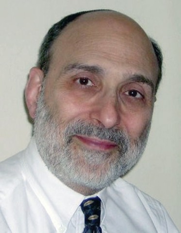
Dr. Tan is emeritus professor of medicine and former adjunct professor of law at the University of Hawaii. He currently directs The St. Francis International Center for Healthcare Ethics in Honolulu. This article is meant to be educational and does not constitute medical, ethical, or legal advice. For additional information, readers may contact the author at [email protected].
Question: Grandma finally checked into a skilled nursing facility (SNF), a member of a national chain of for-profit SNFs, after her progressive dementia prevented her from performing the basic activities of daily living. Unfortunately, the staffing was inadequate, and there were lapses in attention toward her nutrition, medications, and body hygiene. She even fell from her bed on a couple of occasions. Other residents have registered similar complaints. Which of the following legal recourses is available?
A. A lawsuit against the SNF, alleging neglect and abuse.
B. A class action suit against the SNF and its corporate owners.
C. A lawsuit against the attending physician and/or the medical director.
D. A, B, and C.
E. Only A and B.
Answer: D. According to the Wall Street Journal, more than 1.4 million people live in U.S. nursing homes, 69% of which are run by for-profit entities.1 In contrast to a malpractice complaint, lawsuits against nursing homes and SNFs typically involve allegations of a pattern of neglect and abuse rather than any single incident of negligence. The terms nursing home and SNF are often used interchangeably, but do differ somewhat in that the former deals with non-Medicare regulated custodial care, whereas Medicare regulates and certifies all SNFs, which provide both custodial and medical care. Federal and state statutes, e.g., 42 CFR §483 and Centers for Medicare and Medicaid Services guidelines in the State Operations Manual, prescribe the requisite standards. Collectively referred to as the OBRA standards, their violations are frequently at the heart of a plaintiff’s allegations. These may include short staffing, inattention to body hygiene, skin infections, pressure ulcers, improper use of restraints, poor nutrition and hydration, and failure to monitor or supervise, including failure to administer prescription medications and prevention of falls and violent acts from other residents.
In the past 2 decades, nursing homes and SNFs have experienced soaring numbers of lawsuits, with Texas and Florida being especially vulnerable.2 Runaway jury verdicts can result even where the elderly victim has incurred little or no economic loss. Noneconomic losses such as pain and suffering as well as punitive damages explain these huge awards. A recent widely publicized case is illustrative: On Sept. 4, 2009, 87-year-old Dorothy Douglas, an Alzheimer’s patient, was admitted to Heartland Nursing Home in Charleston, W.Va. Still cognitive, she was able to ambulate with an assistive device and was well nourished. However, within 19 days of admission, she became barely responsive, dehydrated, and bedridden, and had fallen numerous times, injuring her head. She died shortly thereafter. Her son sued the owner of Heartland and those responsible for its operations, claiming, among other things, medical and corporate negligence. A jury found in his favor, awarding $11.5 million in compensatory damages and $80 million in punitive damages. On appeal, the West Virginia Supreme Court affirmed in part the trial court’s order, although it reduced the punitive damages from $80 million to $32 million (termed a remittitur).3
There are other sizable verdicts, such as a $29 million lawsuit against a Rocklin, Calif., facility in 2010. Another, possibly the largest on record, was a 2013 Florida jury award of $110 million in compensatory damages and $1.0 billion in punitive damages against Auburndale Oaks Healthcare Center. However, this may not have been the final negotiated amount. Increasingly popular is the use of class action lawsuits, where representative plaintiffs assert claims on behalf of a large class of similarly injured members. Typically, they allege grossly substandard care and understaffing in violation of Medicare and/or other statutory rules. New York’s first nursing home class action suit,4 which dragged on for some 9 years, ended up with a settlement sum of only $950,000 for its 22 class members. The suit alleged, among other things, inedible food, inadequate heat, and squalid conditions. A more recent example: In 2010, a Humboldt County, Calif., jury returned a $677 million verdict (Lavender v. Skilled Healthcare Group Inc.) against one of the nation’s largest nursing home chains for violating California’s Health and Safety Code in its 22 statewide facilities. The case later settled for $62.8 million on behalf of the 32,000 residents.
What about physician liability? Many doctors attend to SNF patients and a number act as medical directors. Liability exists in both roles. The first is governed by the usual tort action of malpractice. The latter is infinitely trickier. Medicare mandates all SNFs to have a medical director, and federal law [42 CFR 483.75 (i)] requires the medical director to be responsible for implementation of resident care policies and the coordination of medical care in the facility. Although their duties are administrative in nature, medical directors are not infrequently named as codefendants in SNF lawsuits. Allegations against the medical director may include negligent supervision of staff, and/or the failure to set standards, policies, and procedures, especially if they have been made aware of citations by auditing agencies. Because a doctor’s professional liability policy typically excludes coverage for such work, it behooves all medical directors to insist on being a named insured in the institution’s general liability policy (to include tail coverage), and to be informed in a timely fashion should there be a relevant change or cancellation of coverage. Their contract should stipulate that the facility would indemnify them for all lawsuits arising out of their work. More and more nursing homes are dropping their insurance to bypass legal exposure, leaving the attending physician and/or medical director at increased risk. To avoid a serious gap in coverage, medical directors should consider purchasing a specific medical director policy.5 Medical directors should also be aware of potential Stark Law violations, such as treating private patients without paying fair rent or receiving compensation in exchange for referrals.
Importantly, elder abuse judgments, as opposed to malpractice awards, may negate restrictions on attorney fees and noneconomic damages such as California’s $250,000 cap. The jury may also levy punitive damages, which are not covered by professional insurance. The plaintiff will need to prove, by clear and convincing evidence, something more than simple or gross negligence such as malice, fraud, oppression, or recklessness.6 Under California’s elder abuse and dependent Adult Civil Protection Act, an appellate court has held that a plaintiff may mount an elder abuse claim directed at physicians and not just facilities with “custodial” duties.7This important issue is currently under appeal before the California Supreme Court.
References
1. The Wall Street Journal, Oct. 3, 2014.
2. Stevenson, DG and DM Studdert, The Rise Of Nursing Home Litigation: Findings From A National Survey Of Attorneys. Health Affairs 2003; 22:219-29.
3. Manor Care Inc. v. Douglas, 763 N.E.2d 73 (W. Va. 2014).
4. Fleming v. Barnwell Nursing Hone and Health Facilities Inc., 309 A.D.2d 1132 (N.Y. App. Div. 2003).
5. See the American Medical Directors Association’s (AMDA) offering at http://locktonmedicalliabilityinsurance.com/amda/.
6. Delaney v. Baker, 20 Cal. 4th 23 (1999).
7. Winn v. Pioneer Medical Group Inc., 216 Cal. App. 4th 875 (2013).
Dr. Tan is emeritus professor of medicine and former adjunct professor of law at the University of Hawaii. He currently directs The St. Francis International Center for Healthcare Ethics in Honolulu. This article is meant to be educational and does not constitute medical, ethical, or legal advice. For additional information, readers may contact the author at [email protected].

A wish list for a better doctor visit
I heard somewhere that doctors are judged by patients as soon as we walk in the door. We are held to high standards of behavior and knowledge – and rightly so. After all, we are given this incredible responsibility of keeping people healthy. But our ability to do our job is limited by regulations that make up the business of medicine: time constraints, insurance hurdles, meaningful use requirements, and coding issues. Few of us would count those as part of the good doctor’s toolbox, but these are the hidden forces that shape how we conduct our jobs.
With that in mind, here is a wish list of sorts for how patients might do their part in making the interaction pleasant and productive:

• I wish patients arrived on time. I often hear the counter argument that we doctors do not run on time, but that’s not the same. When we run late it is often for reasons that are beyond our control. On the other hand, patients coming on time indicates a respect for the doctor’s time and a commitment to their own health and well-being.
• It would be helpful if patients brought a medication list and, along with that, a list of their medical conditions. A list of their doctors is helpful as well. Not infrequently, when I ask a patient how they came to need a rheumatologist, the answer is “It should be in my records.” This is problematic, in part because what counts for records these days is actually an auto-filled document containing pages upon pages of repetitive information that is not germane to the problem at hand.
• If your practice is anything like mine, patients often bring up another problem or two at the end of the visit. Twenty minutes into the visit, as they are stepping off the exam table, they throw in “one last question” or an “oh, by the way.” This can run the gamut from chest pain or weight loss to disability paperwork or medical marijuana. Our patients may not be aware that we can only afford to spend 15-20 minutes with them.
It is also difficult for patients to identify which problems are related to their rheumatologic condition and which are not, leading them to expect us to weigh in on issues that are perhaps better discussed with their primary care physicians. And because we are physicians and want to do what we can for them, we also feel obligated to address all of their concerns. It would be helpful if patients came to the visit prepared with the issues they want to discuss in order of priority. Otherwise, we will never be able to satisfactorily address all of their concerns and still run an efficient practice.
• I have seen many patients who were unhappy with their previous rheumatologist. I have also had unhappy patients fire me and seek care elsewhere. I completely support this system as it leads to a self-selection of sorts. This works out for everyone involved, particularly for the patients. Some patients, though, feel the need to denigrate their other doctors to me in a conspiratorial manner, as if waiting for me to confirm their opinions. This makes me very uncomfortable. I discourage this behavior by reminding patients that symptoms evolve and that the previous doctor, in all likelihood, already did some of the preliminary work that allows the next doctor to seem brilliant in comparison.
• My assistant is fantastic. She can almost read my mind. My mentor at the University of Massachusetts, Dr. Kathy Upchurch, used to say that she saw her assistant more than she saw her husband. This is probably true for most of us who work full time. Our staffs have the unenviable job of keeping both patients and doctors happy, and they are largely responsible for the seamless operations that we run. It is very important to me that my patients treat our staff with respect.
In the end it comes down to this: In this age of managed care, the business of medicine has gotten in the way of the art of medicine. But, as with any human interaction, courtesy and respect make the whole enterprise much more rewarding.
Dr. Chan practices rheumatology in Pawtucket, R.I.
I heard somewhere that doctors are judged by patients as soon as we walk in the door. We are held to high standards of behavior and knowledge – and rightly so. After all, we are given this incredible responsibility of keeping people healthy. But our ability to do our job is limited by regulations that make up the business of medicine: time constraints, insurance hurdles, meaningful use requirements, and coding issues. Few of us would count those as part of the good doctor’s toolbox, but these are the hidden forces that shape how we conduct our jobs.
With that in mind, here is a wish list of sorts for how patients might do their part in making the interaction pleasant and productive:
• I wish patients arrived on time. I often hear the counter argument that we doctors do not run on time, but that’s not the same. When we run late it is often for reasons that are beyond our control. On the other hand, patients coming on time indicates a respect for the doctor’s time and a commitment to their own health and well-being.
• It would be helpful if patients brought a medication list and, along with that, a list of their medical conditions. A list of their doctors is helpful as well. Not infrequently, when I ask a patient how they came to need a rheumatologist, the answer is “It should be in my records.” This is problematic, in part because what counts for records these days is actually an auto-filled document containing pages upon pages of repetitive information that is not germane to the problem at hand.
• If your practice is anything like mine, patients often bring up another problem or two at the end of the visit. Twenty minutes into the visit, as they are stepping off the exam table, they throw in “one last question” or an “oh, by the way.” This can run the gamut from chest pain or weight loss to disability paperwork or medical marijuana. Our patients may not be aware that we can only afford to spend 15-20 minutes with them.
It is also difficult for patients to identify which problems are related to their rheumatologic condition and which are not, leading them to expect us to weigh in on issues that are perhaps better discussed with their primary care physicians. And because we are physicians and want to do what we can for them, we also feel obligated to address all of their concerns. It would be helpful if patients came to the visit prepared with the issues they want to discuss in order of priority. Otherwise, we will never be able to satisfactorily address all of their concerns and still run an efficient practice.
• I have seen many patients who were unhappy with their previous rheumatologist. I have also had unhappy patients fire me and seek care elsewhere. I completely support this system as it leads to a self-selection of sorts. This works out for everyone involved, particularly for the patients. Some patients, though, feel the need to denigrate their other doctors to me in a conspiratorial manner, as if waiting for me to confirm their opinions. This makes me very uncomfortable. I discourage this behavior by reminding patients that symptoms evolve and that the previous doctor, in all likelihood, already did some of the preliminary work that allows the next doctor to seem brilliant in comparison.
• My assistant is fantastic. She can almost read my mind. My mentor at the University of Massachusetts, Dr. Kathy Upchurch, used to say that she saw her assistant more than she saw her husband. This is probably true for most of us who work full time. Our staffs have the unenviable job of keeping both patients and doctors happy, and they are largely responsible for the seamless operations that we run. It is very important to me that my patients treat our staff with respect.
In the end it comes down to this: In this age of managed care, the business of medicine has gotten in the way of the art of medicine. But, as with any human interaction, courtesy and respect make the whole enterprise much more rewarding.
Dr. Chan practices rheumatology in Pawtucket, R.I.

Cash pay is an unworkable proposition for most patients
We hear a lot about the pros and cons of cash-pay medicine. I’ve written about it in the past. So far, I haven’t made the leap of faith it would require in my own practice.
But recently I developed a gradually worsening toothache. For a while I ignored it, hoping it would go away. Like most doctors, my first thought was “I don’t have time for this.” But, as it progressed, I knew I didn’t have a choice.
I didn’t have a dentist, either, and have never had dental insurance. Now what?
So I called a family friend who’s a dentist. His actual charge for the visit was around $200, but he kindly charged me only $50. I sent him a $100 gift card to thank him for his help, and I’m well aware most folks don’t have the benefit of knowing a dentist on nonprofessional terms.
Unfortunately, he found he couldn’t help me. I had an upper molar that was busily reabsorbing itself, and needed to come out.
So I went to an oral surgeon. The first visit (“limited exam and counseling”) was $95. Two days later, I went back to get the tooth yanked for good. That was $275. I paid both in full by credit card.
Do I think any of these charges were unreasonable? Absolutely not. The transaction is done; the tooth is out. His office doesn’t have to bill my insurance, hope to collect part of it, and then bill me for the rest. I don’t have to worry about being billed for something my insurance didn’t pay, or spend an hour (or more) on hold to ask questions of an insurance representative as to why my claim was denied. And I get to put the $520 down on my 2015 taxes as a medical deduction.
It’s pretty simple, isn’t it? The guy pulls my tooth, and I pay a fair amount for his service. This is the same business relationship I have with a grocery store, car mechanic, or office landlord.
So why doesn’t this catch on for most of medicine? It would be nice if it were that simple.
I’m fortunate to be able to afford the total of $520 I’ve spent on the tooth. Many people don’t have that luxury, and have to rely on insurance coverage; $520 is also a pittance, compared with what other things may run. Multiple MRIs? An electromyogram/nerve conduction velocity test? Multiple sclerosis drugs? Chemotherapy? Neurosurgery? A hospital stay? In that group, you’re talking about things that can range from $1,000 to $100,000 (or more) – a far cry from what I spent on my tooth, but still medically necessary for many.
Some will argue these medical costs should all be cash pay, too. If patients can’t afford multiple sclerosis drugs out of pocket ($50,000/year and up for some), then the market will force the drug companies to lower their prices to where people will buy them. That may be, but the price an average person can likely afford isn’t going to support what the company spent to bring the drug to market or pay for trials of the next generation of treatments. So in the long run, it hurts people more than it helps.
Now if I apply this to my practice, I’m sure there are many who could afford my cash rates if I dropped their plan, but since there are other neurologists taking insurance in the area, people will generally go to whoever is best for their wallet. And a copay is going to trump my cash price for most – not to mention the costs of tests that may be needed.
For an office visit, cash pay would likely work for most. It’s simple, quick, and straightforward. But it’s the high costs of modern medicine – advanced tests, hospital stays, and upper-line pharmaceuticals (some patients don’t have other options) – that make it an unworkable proposition for many.
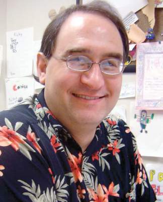
Dr. Block has a solo neurology practice in Scottsdale, Ariz.
We hear a lot about the pros and cons of cash-pay medicine. I’ve written about it in the past. So far, I haven’t made the leap of faith it would require in my own practice.
But recently I developed a gradually worsening toothache. For a while I ignored it, hoping it would go away. Like most doctors, my first thought was “I don’t have time for this.” But, as it progressed, I knew I didn’t have a choice.
I didn’t have a dentist, either, and have never had dental insurance. Now what?
So I called a family friend who’s a dentist. His actual charge for the visit was around $200, but he kindly charged me only $50. I sent him a $100 gift card to thank him for his help, and I’m well aware most folks don’t have the benefit of knowing a dentist on nonprofessional terms.
Unfortunately, he found he couldn’t help me. I had an upper molar that was busily reabsorbing itself, and needed to come out.
So I went to an oral surgeon. The first visit (“limited exam and counseling”) was $95. Two days later, I went back to get the tooth yanked for good. That was $275. I paid both in full by credit card.
Do I think any of these charges were unreasonable? Absolutely not. The transaction is done; the tooth is out. His office doesn’t have to bill my insurance, hope to collect part of it, and then bill me for the rest. I don’t have to worry about being billed for something my insurance didn’t pay, or spend an hour (or more) on hold to ask questions of an insurance representative as to why my claim was denied. And I get to put the $520 down on my 2015 taxes as a medical deduction.
It’s pretty simple, isn’t it? The guy pulls my tooth, and I pay a fair amount for his service. This is the same business relationship I have with a grocery store, car mechanic, or office landlord.
So why doesn’t this catch on for most of medicine? It would be nice if it were that simple.
I’m fortunate to be able to afford the total of $520 I’ve spent on the tooth. Many people don’t have that luxury, and have to rely on insurance coverage; $520 is also a pittance, compared with what other things may run. Multiple MRIs? An electromyogram/nerve conduction velocity test? Multiple sclerosis drugs? Chemotherapy? Neurosurgery? A hospital stay? In that group, you’re talking about things that can range from $1,000 to $100,000 (or more) – a far cry from what I spent on my tooth, but still medically necessary for many.
Some will argue these medical costs should all be cash pay, too. If patients can’t afford multiple sclerosis drugs out of pocket ($50,000/year and up for some), then the market will force the drug companies to lower their prices to where people will buy them. That may be, but the price an average person can likely afford isn’t going to support what the company spent to bring the drug to market or pay for trials of the next generation of treatments. So in the long run, it hurts people more than it helps.
Now if I apply this to my practice, I’m sure there are many who could afford my cash rates if I dropped their plan, but since there are other neurologists taking insurance in the area, people will generally go to whoever is best for their wallet. And a copay is going to trump my cash price for most – not to mention the costs of tests that may be needed.
For an office visit, cash pay would likely work for most. It’s simple, quick, and straightforward. But it’s the high costs of modern medicine – advanced tests, hospital stays, and upper-line pharmaceuticals (some patients don’t have other options) – that make it an unworkable proposition for many.
Dr. Block has a solo neurology practice in Scottsdale, Ariz.

The facts don’t speak for themselves
Whenever I meet families who oppose vaccination, my first response is to flood them with more evidence, reciting the pro-vaccine fact sheet that every doctor knows by heart. I can’t help myself. The evidence is so self-explanatory that it should put any question to rest.
But this approach may just exacerbate the problem. In fact, studies suggest that attacking parents’ beliefs can backfire, and that “attempts to increase concerns about communicable diseases or correct false claims about vaccines may be … counterproductive.” We can inform parents that vaccines prevent thousands of deaths and millions of cases of disease while saving billions in health care costs, but this will not change the mind of someone who does not trust the data.
One major problem is that doctors and parents sometimes speak different languages. As trainees, we are raised on nomograms, algorithms, and clinical trials. When a problem presents itself, we instinctively seek out a rational, data-driven response. This is how doctors are built, but studies suggest that vaccine-hesitant parents may be built differently, driven more by fears and negative emotions than by data. Their decisions seem to be based not on a misunderstanding of the facts, but rather a mistrust of the facts.
As pediatric trainees, this is the world we are inheriting. The more successful vaccines become at preventing disease, the harder it will become to convince parents of the serious risks of nonvaccination. It is a perpetual uphill battle.
To make the future better for our patients (and to contribute to our future sanity), we need to work past frustrations and focus on developing pragmatic solutions. There is surely no easy or perfect answer, and different parents may require different approaches, but the status quo is not working. To find the solutions, we need to develop a robust evidence base to guide our good intentions. Perhaps the answer will come from tailoring communication strategies, refocusing public outreach efforts, creating legal mandates, or maybe something completely different.
And if we are successful, maybe we can close the communications gap between vaccine-resisting parents and doctors.
Dr. Sisk is a pediatrics resident at St. Louis Children’s Hospital. E-mail him at [email protected].

Duped
While folks my age are sometimes referred to as “elderly” victims in newspaper stories about phone and Internet scams, I resist and object to the implication that I am less than sharp and worrisomely vulnerable to being duped.
I hang up when a stranger calls to warn me that I am about to be audited by the Internal Revenue Service and asks for my Social Security number. I double-delete e-mails purported to come from my Internet provider that ask for my e-mail address and password. I’m no pushover.
However, I fear that over the last 20 years of my practice career, I was duped by several of my adolescent patients on more than one occasion. I hope that I had a reputation in town as one of the physicians least likely to leap to the diagnosis of attention-deficit/hyperactivity disorder (ADHD) and even less likely to pull out my pad and prescribe stimulants.
I was particularly hesitant to make the diagnosis of ADHD in an adolescent whose academic career and behavior in grade school had been unremarkable. But from time to time I was presented with a case that included a combination of apparently reliable teachers’ reports, parental pleas, and patient complaints that was hard to ignore. Having ruled out anxiety, depression, learning disabilities, and severe sleep deprivation (all my teenage patients were sleep deprived to some degree) I would reluctantly agree to a trial of stimulant medication.
As you can imagine, assessing success or failure took time because we were usually looking for improvement in academic performance. For adolescents, this often means waiting to the end of the semester or grading period. If the academic improvement was less than dramatic as it was in the usual scenario, I was left relying on the patient’s report of his subjective observations and waiting another 6 months for more information from the school. Occasionally, the patient would report that the medication made him feel weird and that he wanted to stop it. More often, the patient would report that he was able to pay attention in class more easily, even though he had difficulty pointing to a documented improvement in his performance.
So what does one do? Sometimes I could convince the patient and his family that the trial had failed and that we should stop the medication and work harder to find a better match between his learning style, study habits, and the demands of the school. In other cases, I would adjust dosages and switch medications. The results were seldom dramatic. However, if the patient continued to claim a benefit, I would continue to prescribe the stimulant. I would make phone assessments with every refill, and face-to-face visits at least once a year.
While it may be that a few of those adolescents without clearly demonstrable benefit were indeed being helped by the stimulants, I am now convinced that I was being duped more often than I cared to admit then. I know there were stimulants available on the streets and in the school hallways and parking lots because some of my patients told me that they were easy to find and had tried them. I have to believe that some of those pills on the street were ones I had prescribed. I worry when I consider how many.
The national statistics are staggering and embarrassing. In 2013, the federal Substance Abuse and Mental Health Services Administration reported that ED visits associated with the nonmedical use of prescribed stimulants among adults aged 18-34 years had tripled from 2005 to 2011. (“Workers Seeking Productivity in a Pill Are Abusing ADHD Drugs,” by Alan Schwarz, New York Times, April 18, 2015). How many of the pills associated with those visits were originally prescribed for adolescent who didn’t have ADHD?
How many of my patients were just trying to be good friends by sharing their pills and how many were selling them? How many of the pills I prescribed were fueling all-night parties, and how many were being used as performance-enhancing drugs by students who needed to finish a term paper on time?
I don’t know. But I do know that although I miss practicing pediatrics, I am glad I no longer have to face the dilemma of the adolescent with ADHD-like complaints, because I hate being duped.
Surviving a meaningful use audit
Sixty-six years after George Orwell penned his prophetic vision of 1984, many physicians are encountering tangible evidence that “Big Brother” is alive and well: Meaningful use audits are here, and we’ve heard from a lot of our readers that they are time consuming and disconcerting, if not a bit scary. We, too, have encountered Stage II audits and have helped physicians navigate audit mysteries, so we thought it would be a good idea to offer our insight into the process.

First, a disclaimer: The best advice for passing an audit should come from a consultant familiar with your unique situation and audit request. There are, however, a few general principles that everyone should adopt to make the process as smooth as possible. We will highlight them here, beginning with the following axiom:
Prepare, but don’t panic
The most important piece of advice we can offer to providers attesting for meaningful use is to plan carefully for an audit, even if an audit request never comes. The best way to do this is to maintain attestation documentation so it is readily available when needed. This might mean screenshots, “dashboard” reports, letters of intent from collaborating registries, or records of successful data transmission. If you are not sure what to prepare, your EHR vendor should be able to provide you with guidance; every EHR product needs to be certified for meaningful use, so vendors must have documentation on how you can prove you’ve achieved it.
It’s also worth pointing out that audits may encompass a multistep process. Once you respond to the request, the auditor may require additional documentation. This may become a two-way conversation and allow you to clarify what is needed, but preparation will make the process go much more smoothly. Thus, if an audit request materializes, you’ll have already done all of the hard work and will need only to provide it to the third-party auditor. A word of caution, though: Don’t believe that once you’ve received your meaningful use payment that you are completely out of the woods, because ...
Audits may come before or after payment
There are several types of audit requests. Auditors may simply ask you to prove you are using a certified EHR, or they may ask for a whole lot more. Furthermore, they may perform “prepayment” or “postpayment” audits. In the case of a prepayment audit, they will hold any incentive payments until after the audit requests are satisfied. Audits after payment is issued can be a bit trickier; if you don’t “pass” a postpayment audit, the Centers for Medicare & Medicaid Services will ask for the money back and typically won’t be too friendly about it. The appeal process isn’t easy and may involve lawyers and other complications, so make sure to take the audit request quite seriously, and cover yourself by remembering that when it comes to a Meaningful Use audit …
There is no such thing as TMI (too much information)
When submitting supporting documentation to an auditor, try to be as exhaustive as possible to avoid making the process lengthier than it needs to be. It is critical to make your case clear, and screenshots depicting the measure, source, and date can be one great way to do this.
For example, if you are sending attainment numbers from scored measures (e.g., ePrescribing, CPOE, or Clinical Summaries), be sure to include some evidence that the numbers were obtained from your certified EHR’s scorecard or dashboard (such as the software’s logo, etc.). If sending evidence of compliance for a yes/no measure (e.g., patient lists by condition or clinical support rule activation), be sure the date you generated it is clear so you can prove it was active during the reporting period. Finally, be sure to provide clear documentation that you are actively submitting data to immunization and disease registries (or at least have done your due diligence to set it up), and whatever you do ...
Don’t neglect the security risk analysis
This is one area that seems to become a focus point in every audit. It’s also something that may be taken for granted by providers, as it is a yes/no measure that can be easily overlooked. Don’t fall into this trap; the CMS takes data security very seriously and so should you. If you receive an audit request from them, they will want to know not only that the analysis was performed but also what potential risks were identified, what type of data was collected, and what your practice is doing to improve upon any deficiencies. If you need guidance on how to do this, you’ll find help here.
This leads us to one final important piece of advice:
Make sure you follow up
It is not uncommon for providers to submit information to auditors and then wait a little while for a response, but we have also heard of communications being lost to spam filters and misspelled email addresses. It’s therefore critical to follow up carefully on any audit request, as there are deadlines attached and financial incentives at risk. In our experience, auditors are not offended by providers “checking in” on the status of their audit, nor are they opposed to clarifying questions. So even though “Big Brother” may be watching, in this case he (or she) has a name and an e-mail address and is willing to answer your questions, should any arise. In summary, audits will happen, but with some preparation as described above, the process will be less intimidating and (hopefully) a lot less work.
Dr. Notte is a family physician and clinical informaticist for Abington (Pa.) Memorial Hospital. He is a partner in EHR Practice Consultants, a firm that aids physicians in adopting electronic health records. Dr. Skolnik is associate director of the family medicine residency program at Abington Memorial Hospital and professor of family and community medicine at Temple University in Philadelphia.
Sixty-six years after George Orwell penned his prophetic vision of 1984, many physicians are encountering tangible evidence that “Big Brother” is alive and well: Meaningful use audits are here, and we’ve heard from a lot of our readers that they are time consuming and disconcerting, if not a bit scary. We, too, have encountered Stage II audits and have helped physicians navigate audit mysteries, so we thought it would be a good idea to offer our insight into the process.
First, a disclaimer: The best advice for passing an audit should come from a consultant familiar with your unique situation and audit request. There are, however, a few general principles that everyone should adopt to make the process as smooth as possible. We will highlight them here, beginning with the following axiom:
Prepare, but don’t panic
The most important piece of advice we can offer to providers attesting for meaningful use is to plan carefully for an audit, even if an audit request never comes. The best way to do this is to maintain attestation documentation so it is readily available when needed. This might mean screenshots, “dashboard” reports, letters of intent from collaborating registries, or records of successful data transmission. If you are not sure what to prepare, your EHR vendor should be able to provide you with guidance; every EHR product needs to be certified for meaningful use, so vendors must have documentation on how you can prove you’ve achieved it.
It’s also worth pointing out that audits may encompass a multistep process. Once you respond to the request, the auditor may require additional documentation. This may become a two-way conversation and allow you to clarify what is needed, but preparation will make the process go much more smoothly. Thus, if an audit request materializes, you’ll have already done all of the hard work and will need only to provide it to the third-party auditor. A word of caution, though: Don’t believe that once you’ve received your meaningful use payment that you are completely out of the woods, because ...
Audits may come before or after payment
There are several types of audit requests. Auditors may simply ask you to prove you are using a certified EHR, or they may ask for a whole lot more. Furthermore, they may perform “prepayment” or “postpayment” audits. In the case of a prepayment audit, they will hold any incentive payments until after the audit requests are satisfied. Audits after payment is issued can be a bit trickier; if you don’t “pass” a postpayment audit, the Centers for Medicare & Medicaid Services will ask for the money back and typically won’t be too friendly about it. The appeal process isn’t easy and may involve lawyers and other complications, so make sure to take the audit request quite seriously, and cover yourself by remembering that when it comes to a Meaningful Use audit …
There is no such thing as TMI (too much information)
When submitting supporting documentation to an auditor, try to be as exhaustive as possible to avoid making the process lengthier than it needs to be. It is critical to make your case clear, and screenshots depicting the measure, source, and date can be one great way to do this.
For example, if you are sending attainment numbers from scored measures (e.g., ePrescribing, CPOE, or Clinical Summaries), be sure to include some evidence that the numbers were obtained from your certified EHR’s scorecard or dashboard (such as the software’s logo, etc.). If sending evidence of compliance for a yes/no measure (e.g., patient lists by condition or clinical support rule activation), be sure the date you generated it is clear so you can prove it was active during the reporting period. Finally, be sure to provide clear documentation that you are actively submitting data to immunization and disease registries (or at least have done your due diligence to set it up), and whatever you do ...
Don’t neglect the security risk analysis
This is one area that seems to become a focus point in every audit. It’s also something that may be taken for granted by providers, as it is a yes/no measure that can be easily overlooked. Don’t fall into this trap; the CMS takes data security very seriously and so should you. If you receive an audit request from them, they will want to know not only that the analysis was performed but also what potential risks were identified, what type of data was collected, and what your practice is doing to improve upon any deficiencies. If you need guidance on how to do this, you’ll find help here.
This leads us to one final important piece of advice:
Make sure you follow up
It is not uncommon for providers to submit information to auditors and then wait a little while for a response, but we have also heard of communications being lost to spam filters and misspelled email addresses. It’s therefore critical to follow up carefully on any audit request, as there are deadlines attached and financial incentives at risk. In our experience, auditors are not offended by providers “checking in” on the status of their audit, nor are they opposed to clarifying questions. So even though “Big Brother” may be watching, in this case he (or she) has a name and an e-mail address and is willing to answer your questions, should any arise. In summary, audits will happen, but with some preparation as described above, the process will be less intimidating and (hopefully) a lot less work.
Dr. Notte is a family physician and clinical informaticist for Abington (Pa.) Memorial Hospital. He is a partner in EHR Practice Consultants, a firm that aids physicians in adopting electronic health records. Dr. Skolnik is associate director of the family medicine residency program at Abington Memorial Hospital and professor of family and community medicine at Temple University in Philadelphia.
Telemedicine and healing touch
The practice of medicine is a sacred event between a doctor and a patient – two individuals who have met each other in the flesh. This is the sentiment of many, and soon to be the law in the great state of Texas. It was also a popular topic of conversation at the American Telemedicine Association meeting in Los Angeles.
As a physician, I’m passionate about the profession of medicine, and as a soon-to-be MBA, I’m passionate about the practice of medicine. In this case, these ideals are pulling in opposite directions. I’m writing this dialectic as much to help me form my opinion as to help you with yours.
Should the practice of medicine be reserved only for those patients whom you have met in person? This is, after all, our history. All of us who practice medicine know the laying on of hands heals. It is a core human interaction that conveys care and concern, warmth and protection. It is the most powerful tool we have as clinicians to diagnose and heal. Touching patients can only be done in the flesh, and any interaction between a doctor and a patient that lacks this is, in one way, inferior. Not only does seeing a patient in person ensure that we won’t miss the obvious melanoma not visualized in the photo or video, but it also enables us to better connect this visit with every other visit and with the entirety of the patient’s record.
Having access to a patient’s medical record improves the quality and efficiency of care, a value conspicuously missing from one-off-dial-a-doc interactions from an online doctor who cares nothing of patient history or future.
Dr. Jack Resneck, a plenary presenter at ATA this year, told a cautionary tale of an acne patient who was treated online with an antibiotic that caused him to develop Stevens-Johnson syndrome, a life-threatening condition requiring extreme medicine and extraordinary costs. Clearly, digital medicine has shortcomings.
On the other hand, should we yield to the demands of our patients who want convenient care? Leaving work (or family) to endure traffic, to find a parking space, to sit in your waiting room, then your exam room, to have a chance to touch you once and see you for 10 minutes is not convenient. Given the option to receive care by the phone, online, or in person, patients increasingly are choosing the more convenient channels. And if you wrote a prescription for trimethoprim-sulfamethoxazole for an acne patient with no history of medication allergy and he developed Stevens-Johnson syndrome, would seeing him in person have helped? Perhaps, but probably not.
We are reluctant to call our patients “customers” for good reasons. I respect that, and I caution us to consider the truth that the trade of service for a fee is a business transaction. No matter how much patients love and respect us as doctors, if the price of our services grew exorbitantly high, or if it became impossible to see us, then they would abandon us. They would do so with remorse, yet they would do so.
Ideally, a person with a rash makes an appointment to see a dermatologist. In reality, patients turn to many services and products for this need. They choose primary care doctors, physician assistants, nurses, naturopaths, moisturizers, coconut oil, pharmacists, Dr. Oz, WebMD, customer care agents at WalMart, Google, and even God (see Book of Job). Our patients have many choices, and they choose us because we are the best value for their money. This could change.
I’m concerned that in a digital world where customers can book a trip to Italy, buy a house, and even get married without ever having to leave their phone, (yes, the last is legal in some states, but not in Texas), we risk irrelevance. By forcing our patients to use an inconvenient channel, we risk losing our one and only customer, our patient.
Dr. William Osler said, “In seeking the truth, we aim at the unattainable, and must be content with finding broken portions.” Telemedicine may not offer the warmth of our hand, but it does offer the service our modern patients desire. The question is not whether or not we practice medicine digitally, but rather how we will maintain the quality, trust and convenience that our patients ask of us.
Please discuss.
Dr. Benabio is a partner physician in the department of dermatology of the Southern California Permanente Group in San Diego, and a volunteer clinical assistant professor at the University of California, San Diego. Dr. Benabio is @dermdoc on Twitter.

Sunshine Act – another reminder
I’ve written about the Physician Payment Sunshine Act several times since it became law in 2013. My basic opinion – that it is a tempest in a teapot – has not changed. Nonetheless, now is the time to review the 2014 data reported under your name – and if necessary, initiate a dispute – before the information is posted publicly on June 30.
A quick review: The Sunshine Act, known officially as the “Open Payments Program,” requires all manufacturers of drugs, devices, and medical supplies covered by federal health care programs to report to the Centers for Medicare & Medicaid Services any financial interactions with physicians and teaching hospitals.
Reportable interactions include consulting, food, ownership or investment interest, direct compensation for speakers at education programs, and research. Compensation for clinical trials must be reported but is not made public until the product receives FDA approval, or until 4 years after the payment, whichever is earlier. Payments for trials involving a new indication for an approved drug are posted the following year.
Exemptions include CME activities funded by manufacturers and product samples for patient use. Medical students and residents are exempted entirely.
You are allowed to review your data and request corrections before information is posted publicly. You will have an additional 2 years to pursue corrections after the content goes live at the end of June, but any erroneous information will remain online until the next scheduled update, so you should find and fix errors as promptly as possible.
If you don’t see drug reps, accept sponsored lunches, or give sponsored talks, don’t assume that you won’t be on the website. Check anyway: You might be indirectly involved in a compensation that you were not aware of, or you might have been reported in error.
To review your data, register at the CMS Enterprise Portal (https://portal.cms.gov/wps/portal/unauthportal/home/) and request access to the Open Payments system.
The question remains as to what effect the law might be having on research, continuing education, or physicians’ relationships with the pharmaceutical industry. The short answer is that no one knows. The first data posting this past September came and went with little fanfare, and no repercussions directly attributable to the program have been reported as of this writing.
Sunshine laws have been in effect for several years in six states: California, Colorado, Massachusetts, Minnesota, Vermont, and West Virginia, plus the District of Columbia. (Maine repealed its law in 2011.) Observers disagree on their impact. Studies in Maine and West Virginia showed no significant public reaction or changes in prescribing patterns, according to a 2012 article in the Archives of Internal Medicine (now JAMA Internal Medicine).
Reactions from the public are equally inscrutable. Do citizens think less of doctors who accept the occasional industry-sponsored lunch for their employees? Do they think more of doctors who speak at meetings, or conduct industry-sponsored clinical research? There are no objective data. Anecdotally, I haven’t heard a peep – positive, negative, or indifferent – from any of my patients, nor have any other physicians that I’ve asked.
As of now, I stand by my initial prediction that attorneys, activists, and the occasional reporter will data-mine the information for various purposes, but few patients will bother to visit. Of course, that doesn’t mean you should ignore it as well. As always, I suggest you review the accuracy of anything posted about you, in any form or context, on any venue. This year’s data (reflecting all 2014 reports) have been available for review since April 6. You can initiate a dispute at any time over the next 2 years, before or after public release on June 30, but the sooner the better. Corrections are made each time CMS updates the system.
Maintaining accurate financial records has always been important, but it will be even more important now to support your disputes. CMS won’t simply take your word for it. A free app is available to help you track payments and other reportable industry interactions; search for “Open Payments” at your favorite app store.
Dr. Eastern practices dermatology and dermatologic surgery in Belleville, N.J. He is the author of numerous articles and textbook chapters, and is a longtime monthly columnist for Dermatology News.