User login
MDedge conference coverage features onsite reporting of the latest study results and expert perspectives from leading researchers.
Halting active inflammation key in treating PIH
CHICAGO –
Dr. Desai, clinical assistant professor in the department of dermatology at the University of Texas Southwestern Medical Center, Dallas, spoke at the Pigmentary Disorders Exchange Symposium, provided by MedscapeLive!
Like all dermatologists, he said at the meeting, he sees lots of acne cases. However, PIH is often the presenting reason for the visit in his practice, which focuses predominantly on skin of color.
“Most of my patients come in not even worried about the acne,” he said. “They come in wanting me to fix the dark spots.”
Inflammation persists
Dermatologists, Dr. Desai said, should educate patients with active PIH resulting from acne or other diseases that even though the condition has been labeled post- inflammatory hyperpigmentation, the inflammation continues to be a problem.
He said, while patients may think PIH is “just scars,” the inflammation is still active and the condition needs to be treated from a skin-lightening perspective but, more importantly, with a focus on halting the inflammation. “If you were to biopsy the areas of hyperpigmentation, you would find a high density of active inflammatory behaviors still present in the skin,” he said.
When treating patients, it’s critical to first treat the underlying skin condition aggressively, he said. “Things like topical retinoids and azelaic acid mechanistically actually make a lot more sense for PIH than even hydroquinone, in some cases, because these therapies are actually anti-inflammatory for many of the diseases we treat.”
Dr. Desai noted that, in patients with darker skin tones, even diseases like seborrheic dermatitis and plaque psoriasis can result in PIH, while in patients with lighter skin tones, the same diseases may leave some residual postinflammatory erythema.
“I think it’s very important, particularly when you’re treating a darker skin–toned patient, to arrest the erythema early on to prevent that further worsening of hyperpigmentation,” he said.
Biopsies important
In cases of PIH, determining the best treatment requires finding out where the pigment is and how deep it is, Dr. Desai said.
He noted dermatologists are often worried about doing biopsies, particularly in patients with darker skin, because of the risk of scarring and keloid formation for those more prone to keloids. The preference is also for a therapeutic effect without using invasive procedures.
“But particularly with PIH, in patients who have been therapeutically challenging, I don’t hesitate to do very small biopsies – 2- and 3-mm punch biopsies – particularly if they are from the head and neck area.”
He suggests doing biopsies on part of the ear, lower jaw line, or the neck area, as these areas tend to heal nicely. “You don’t have to be so concerned about the scarring if you counsel appropriately,” he said.
The biopsy can be valuable in determining whether a very expensive treatment will reach the intended target.
Topical retinoids play an important role as anti-inflammatories for PIH, Dr. Desai said.
He gave an example of a patient with Fitzpatrick skin type IV or V with chronic acne and extensive PIH. “Are you going to effectively tell that patient to apply 4% hydroquinone triple-combination compound across 30 different areas of PIH on their face? The answer is that’s really not very efficient or effective.”
That’s why therapies, such as retinoids, that target the pathogenesis of PIH, particularly the inflammatory component, are important, he added.
Psychological burden
PIH comes with significant stigma and loss of quality of life loss that can last many years.
During another presentation at the meeting, Susan C. Taylor, MD, professor and vice chair of diversity, equity and inclusion in the department of dermatology, at the University of Pennsylvania, Philadelphia, pointed out that in a 2016 study of 324 patients in seven Asian countries, acne-related PIH lasted longer than 1 year in 65.2% of patients and 5 years or longer in 22.3%, significantly affecting their quality of life.
Dr. Desai added that, in a paper recently published in the British Journal of Dermatology, on the impact of postacne hyperpigmentation in patients, the authors pointed out that the reported prevalence of PIH in patients with acne ranges between 45.5% and 87.2%, depending on skin phototype, and that in most cases, PIH takes more than a year to fade.
“Studies have demonstrated that patients with acne and resulting scarring often face stigmatization, leading to quality of life impairment, social withdrawal and body image disorders, which can further contribute to higher risk for depression and social anxiety,” the paper’s authors wrote.
Dr. Desai reported no financial disclosures relevant to his talk.
AT THE MEDSCAPELIVE! PIGMENTARY DISORDERS SYMPOSIUM
MS relapse rates similar between anti-CD20 mAbs and switching to fumarates
DENVER – according to a retrospective study presented at the annual meeting of the Consortium of Multiple Sclerosis Centers. Those who switched did, however, experience a lower rate of inpatient infection-related health care visits per year than those who continued anti-CD20 mAbs.
“As MS is a chronic disease requiring long-term treatment, switching between disease-modifying therapies [DMTs] is a common clinical strategy to optimize individual patient outcomes,” lead author Aliza Ben-Zacharia, PhD, DNP, RN, an assistant professor at the Phillips School of Nursing at Mount Sinai and Hunter College, both in New York, and colleagues reported. They noted that anti-CD20 mAbs are considered high-efficacy DMTs while fumarates are considered moderate-efficacy DMTs.
The researchers used data from the Komodo Health Sentinel Claims Database to track and compare 108 patients who were clinically stable on anti-CD20 mAbs and then switched to fumarates with 540 patients who remained on anti-CD20 mAbs for a follow-up period of approximately 1 year.
The study included adults with a diagnosis of MS between January 2015 and August 2022, and only those with a gap of no more than 9 months between anti-CD20 mAbs and fumarates were included as switchers. The researchers also required that switchers had not had any relapses in the previous year on anti-CD20 mAbs before switching, and had to have been on fumarates for at least 3 months after switching.
Women made up 70% of both groups, and both had an average age of 49 years. The racial/ethnic demographics were similar in both groups, and the average MS severity score was 5.5 in the switching group and 5.6 in the staying group. Most patients had been taking or remained on ocrelizumab (93.5%) with a smaller proportion on rituximab (5.6%). Just over a third of those who switched therapy took diroximel fumarate while 64% took dimethyl fumarate.
The researchers noted that patients who stayed on anti-CD20 mAbs had “slightly higher use of other mAbs prior to anti-CD20 mAbs.” Further, “a higher proportion of patients were DMT naive prior to anti-CD20 mAb initiation, compared with those in the switchers group.”
Patients who switched had been on anti-CD20 mAbs an average 730 days before switching to fumarates, and the average time between their last anti-CD20 mAbs dose and starting fumarates was 274 days. Average exposure to fumarates was 341 days.
The 10.2% of patients who relapsed during follow-up after switching to fumarates was not significantly statistically different than the 6.7% of patients who relapsed while remaining on anti-CD20 mAbs (P = .17). A relapse was considered “an MS-related inpatient claim with a primary diagnosis of MS or an outpatient MS-related diagnosis and a prescription claim for an intravenous steroid, adrenocorticotropic hormone, total plasma exchange, or a high-dose oral corticosteroid 7 days or sooner after the outpatient visit,” the researchers explained. The researchers could not track mild relapses that didn’t involve a health care interaction.
There was also no significant difference in overall average health care encounters between those who switched (7.85 encounters) and those who stayed (8.08; P = .57). Further, average health care costs were statistically similar between those who switched ($22,512) and those who stayed ($20,634; P = 0.59).
The likelihood of having more than one infection-related health care encounter was greater for those who remained on anti-CD20 mAbs, but the difference was not statistically significant. The annual rate of infection-related health care encounters was also not statistically different for outpatient and ED visits, but those who switched did have a statistically lower rate of annual infection-related inpatient visits (P = .03).
Among those who switched, 2.8% were hospitalized for infections, compared with 6.5% who stayed on anti-CD20 mAbs. Urinary tract infections, sepsis, and Escherichia coli were the most common infections among those who switched to fumarates, compared with COVID-19, sepsis, and pneumonia, among those who stayed on anti-CD20 mAbs.
The research was sponsored by and funded by Biogen. Six of the authors are Biogen employees who hold stock options in the company. The other three authors reported combined consulting fees from Biogen, EMD Serono, Greenwich Biosciences, TG Therapeutics, Bristol-Myers Squibb, Horizon, and Novartis; research funding from Genentech and Novartis; and stock options in Pfizer.
AT CMSC 2023
High Lp(a) tied to higher coronary plaque volume, progression
MANNHEIM, GERMANY – , an observational imaging study shows.
This could explain the greater risk for major adverse cardiovascular events seen in patients with high Lp(a) levels, suggests the research, presented during the annual European Atherosclerosis Society Congress.
The team performed follow-up coronary CT angiography (CCTA) on almost 275 patients who had undergone imaging approximately 10 years earlier, finding that almost one-third had high Lp(a) levels.
At baseline, per cent plaque volumes were 1.8 times greater in high Lp(a) patients versus those with low levels of the protein. After 10 years, plaque volumes were 3.3 times larger in patients with high Lp(a) levels.
Over this period, the rate of increase of plaque volume was 1.9 times greater in patients with high Lp(a) levels.
Study presenter Nick S. Nurmohamed, MD, PhD candidate, department of vascular medicine, Amsterdam University Medical Centers, also showed that high Lp(a) levels were associated with a 2.1-fold increase in rates of MACE.
He said in an interview that this finding could be related to Lp(a) increasing inflammatory signaling in the plaque, “making it more prone to rupture, and we saw that on the CCTA scans,” where high Lp(a) levels were associated with the presence of more high-risk plaques.
He added that in the absence of drugs that target Lp(a) levels directly, the results underline the need to focus on other means of lipid-lowering, as well as “creating awareness that Lp(a) is associated with plaque formation.”
Dr. Nurmohamed said that “for the moment, we have to treat patients with high Lp(a) with other risk-lowering therapies, such as low-density lipoprotein [LDL] cholesterol–lowering drugs, and the management of other risk factors.”
However, he noted that “there are a couple of Lp(a)-lowering medications in trials,” with results expected in the next 2-3 years.
“Then we will have the means to treat those patients, and with CCTA we can identify the patients with the biggest risk,” Dr. Nurmohamed added.
Plaque burden
Philippe Moulin, MD, PhD, head of endocrinology and professor of human nutrition at Faculté Lyon Est, Claude Bernard Lyon (France) 1 University, said that the association between Lp(a) and plaque burden has been seen previously in the literature in a very similar study but with only 1-year follow-up.
Similarly, registry data have suggested that Lp(a) is associated with worsening plaque progression over time.
“Here, with 10-year follow-up, [the study] is much more interesting,” due to its greater statistical power, he said in an interview. It is also “well-documented” and uses an “appropriate” methodology.
But Dr. Moulin underlined that the number of patients with high Lp(a) levels included in the study is relatively small.
Consequently, the researchers were not able to look at the level and rate of progression of atherosclerosis between different quartiles of Lp(a), “so you have no dose-response analysis.”
It also does not “establish causality,” as it remains an observational study, despite being longitudinal, “well done, and so on.”
Dr. Moulin added that the study nevertheless adds “one more stone” to the construct of the idea of high risk around high Lp(a) levels, and “prepares the ground” for the availability of two drugs to decrease Lp(a) levels, expected in 2026 and 2027.
These are expected to substantially reduce Lp(a) levels and achieve a reduction in cardiovascular risk of around 20%-40%, “which would be interesting,” especially as “we have patients who have Lp(a) levels four times above the upper normal value.”
Crucially, they may already have normal LDL cholesterol levels, meaning that, for some patients, “there is clearly a need for such treatment, as long as it is proven that it will decrease cardiovascular risk.”
For the moment, however, the strategy for managing patients with high Lp(a) remains to increase the dose of statin and to have more stringent targets, although Dr. Moulin pointed out that, “when you give statins, you raise slightly Lp(a) levels.”
Dr. Nurmohamed said in an interview that “we know from largely genetic and observational studies that Lp(a) is causally associated with atherosclerotic cardiovascular disease.”
What is less clear is the exact underlying mechanism, he said, noting that there have been several imaging studies in high and low Lp(a) patients that have yielded conflicting results in terms of the relationship with plaque burden.
To investigate the impact of Lp(a) levels on long-term coronary plaque progression, the team invited patients who had taken part in a previous CCTA study to undergo repeat CCTA, regardless of their underlying symptoms.
In all, 299 patients underwent follow-up imaging a median of 10.2 years after their original scan. Plaque volumes were quantified and adjusted for vessel volumes, and the patients were classified as having high (≥ 70 nmol/L) or low (< 70 nmol/L) Lp(a) levels.
After excluding patients who had undergone coronary artery bypass grafting, the team analyzed 274 patients with a mean age at baseline of 57 years. Of these, 159 (58%) were men. High Lp(a) levels were identified in 87 (32%) patients.
The team found that at baseline, patients with high Lp(a) levels had significantly larger percent atheroma volumes than those with low levels, at 3.92% versus 2.17%, or an absolute difference of 1.75% (P = .013).
The difference between the two groups was even greater at the follow-up, when percent atheroma volumes reached 8.75% in patients with high Lp(a) levels versus 3.90% for those with low levels, or an absolute difference of 4.85% (P = .005).
Similar findings were seen when looking separately at percentage of noncalcified and calcified plaque volumes as well as when analyzing for the presence of low-density plaques.
Multivariate analysis taking into account clinical risk factors, statin use, and CT tube voltage found that high Lp(a) levels were associated with a greater percent atheroma volume at baseline, at an odds ratio versus low Lp(a) of 1.83 (95% confidence interval, 0.12-3.54; P = .037).
High Lp(a) levels were also linked to a larger percent atheroma volume on follow-up imaging, at an odds ratio of 3.25 (95% CI, 0.80-5.71; P = .010), and a significantly greater change in atheroma volume from baseline to follow-up imaging, at an odds ratio of 1.86 (95% CI, 0.59-3.14; P = .005)
Finally, the team showed that, after adjusting for clinical risk factors, high baseline Lp(a) levels were associated with an increased risk of MACE during the follow-up period, at a hazard ratio versus low Lp(a) levels of 2.10 (95% CI, 1.01-4.29, P = .048).
No funding was declared. Dr. Nurmohamed is cofounder of Lipid Tools. Other authors declare relationships with Amgen, Novartis, Esperion, Sanofi-Regeneron, Ackee, Cleerly, GW Heart and Vascular Institute, Siemens Healthineers, and HeartFlow.
A version of this article first appeared on Medscape.com.
AT EAS 2023
Regular, optimal sleep tied to lower mortality risk
INDIANAPOLIS –
In a diverse group of older adults, those with regular and optimal sleep had about a 40% lower risk of dying of any cause during follow-up compared with peers who had irregular and insufficient sleep.
“If sleep were an 8-hour pill, it would be beneficial to take the full dose at regular times consistently,” lead researcher Joon Chung, PhD, of Harvard Medical School and Brigham and Women’s Hospital, Boston, said in a news release.
The findings were presented at the annual meeting of the Associated Professional Sleep Societies.
Broad adverse health effects
“Evidence is mounting that irregular sleep is associated with pretty broad adverse health outcomes, most prominently cardiometabolic disease, obesity, and cardiovascular disease,” Dr. Chungsaid in an interview.

In the current study, the researchers estimated the association of regular sleep of optimal sleep duration with all-cause mortality using data from 1,759 adults the Multi-Ethnic Study of Atherosclerosis Sleep Study.
Sleep regularity and duration were classified using 7 days of data gathered by wrist actigraphy. Adults were categorized as “regular-optimal” sleepers (n = 1,015) or “irregular-insufficient” sleepers (n = 744).
During 7 years of follow-up, 176 people died. In the fully adjusted model, the regular-optimal group had a 39% lower mortality risk compared with the irregular-insufficient sleep group (hazard ratio, 0.61;95% confidence interval [CI], 0.45-0.83). The findings were robust in sensitivity analyses.
The regular and optimal duration sleep pattern maps behaviorally to regular bed and wake times, suggesting potential health benefits of adherence to recommended sleep practices, the researchers noted.
“Results suggest benefits of expanding the public conversation on getting ‘a good night’s sleep’ and broadening this goal to getting many good nights of sleep, in a row, on weekdays and weekends,” Dr. Chung said in the release.
He further said that “getting adequate, regular sleep seems to be something that is good for all. I don’t know of anyone who wouldn’t benefit.”
Fariha Abassi-Feinberg, MD, spokesperson for the American Academy of Sleep Medicine and sleep specialist with the Millennium Physician Group, Fort Myers, Fla., agreed.
“We know our bodies have an internal clock, known as the circadian rhythm, which regulates various biological processes, including sleep-wake cycles. Sticking to a consistent sleep schedule allows your body to align its natural rhythm with the external day-night cycle. This synchronization promotes better sleep quality and therefore better health,” said Dr. Abassi-Feinberg, who wasn’t involved in the study.
“The AASM recommends adults try to aim for at least 7 hours of sleep and I often tell my patients that keeping a regular routine is best for your sleep and health,” she said in an interview.
Funding for the study was provided by the American Academy of Sleep Medicine Foundation and the National Institutes of Health. Dr. Chung and Dr. Abassi-Feinberg report no relevant financial relationships.
A version of this article originally appeared on Medscape.com.
INDIANAPOLIS –
In a diverse group of older adults, those with regular and optimal sleep had about a 40% lower risk of dying of any cause during follow-up compared with peers who had irregular and insufficient sleep.
“If sleep were an 8-hour pill, it would be beneficial to take the full dose at regular times consistently,” lead researcher Joon Chung, PhD, of Harvard Medical School and Brigham and Women’s Hospital, Boston, said in a news release.
The findings were presented at the annual meeting of the Associated Professional Sleep Societies.
Broad adverse health effects
“Evidence is mounting that irregular sleep is associated with pretty broad adverse health outcomes, most prominently cardiometabolic disease, obesity, and cardiovascular disease,” Dr. Chungsaid in an interview.
In the current study, the researchers estimated the association of regular sleep of optimal sleep duration with all-cause mortality using data from 1,759 adults the Multi-Ethnic Study of Atherosclerosis Sleep Study.
Sleep regularity and duration were classified using 7 days of data gathered by wrist actigraphy. Adults were categorized as “regular-optimal” sleepers (n = 1,015) or “irregular-insufficient” sleepers (n = 744).
During 7 years of follow-up, 176 people died. In the fully adjusted model, the regular-optimal group had a 39% lower mortality risk compared with the irregular-insufficient sleep group (hazard ratio, 0.61;95% confidence interval [CI], 0.45-0.83). The findings were robust in sensitivity analyses.
The regular and optimal duration sleep pattern maps behaviorally to regular bed and wake times, suggesting potential health benefits of adherence to recommended sleep practices, the researchers noted.
“Results suggest benefits of expanding the public conversation on getting ‘a good night’s sleep’ and broadening this goal to getting many good nights of sleep, in a row, on weekdays and weekends,” Dr. Chung said in the release.
He further said that “getting adequate, regular sleep seems to be something that is good for all. I don’t know of anyone who wouldn’t benefit.”
Fariha Abassi-Feinberg, MD, spokesperson for the American Academy of Sleep Medicine and sleep specialist with the Millennium Physician Group, Fort Myers, Fla., agreed.
“We know our bodies have an internal clock, known as the circadian rhythm, which regulates various biological processes, including sleep-wake cycles. Sticking to a consistent sleep schedule allows your body to align its natural rhythm with the external day-night cycle. This synchronization promotes better sleep quality and therefore better health,” said Dr. Abassi-Feinberg, who wasn’t involved in the study.
“The AASM recommends adults try to aim for at least 7 hours of sleep and I often tell my patients that keeping a regular routine is best for your sleep and health,” she said in an interview.
Funding for the study was provided by the American Academy of Sleep Medicine Foundation and the National Institutes of Health. Dr. Chung and Dr. Abassi-Feinberg report no relevant financial relationships.
A version of this article originally appeared on Medscape.com.
AT SLEEP 2023
Macular dermal hyperpigmentation: Treatment tips from an expert
CHICAGO – based on cases she has treated in her practice.
Heather Woolery-Lloyd, MD, director of the skin of color division in the dermatology department at University of Miami, provided three general pointers.
- When in doubt, biopsy.
- For inflammatory disorders, always treat the inflammation in addition to the hyperpigmentation.
- Avoid long-term hydroquinone use in these patients.
Dr. Woolery-Lloyd also reviewed examples of what she has found successful in treating her patients with these conditions.
Lichen planus pigmentosus (LPP)
“It’s one of the hardest things that we treat,” said Dr. Woolery-Lloyd, who often sees cases of LPP in patients in their 30s, 40s, and 50s.
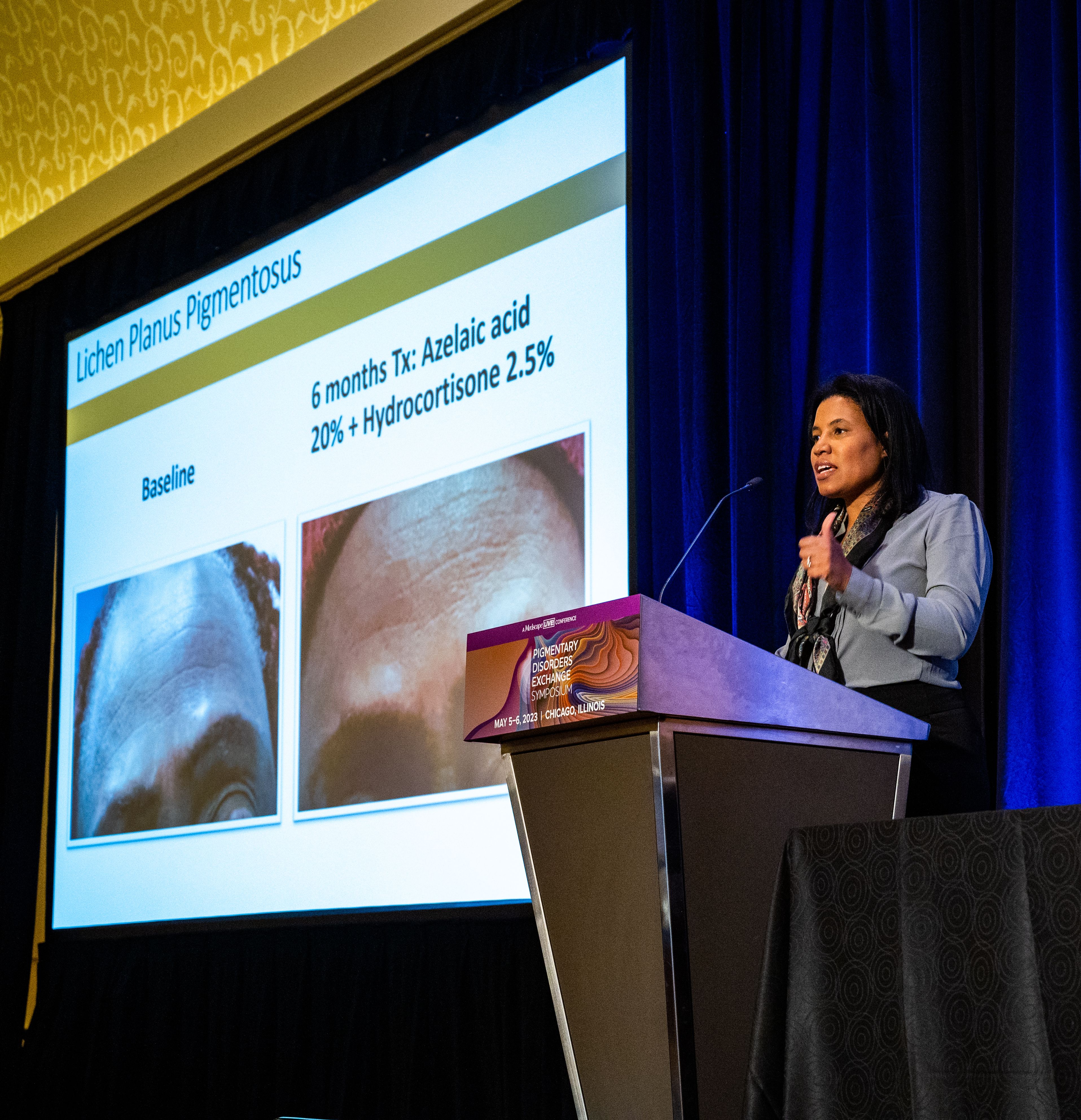
Lesions first appear as small, ill-defined oval-to-round macules, which later become confluent and form large areas of pigmentation. In different patients, the pigment on the face and neck, and sometimes on the forearms can be slate gray or brownish black.
In 2013, dermatologist N.C. Dlova, MD, at the University of KwaZulu‐Natal, Durban, South Africa, reported a link between frontal fibrosing alopecia and LPP in the British Journal of Dermatology. “I definitely see this connection in my practice,” said Dr. Woolery-Lloyd, noting that “both conditions often result in the loss of both eyebrows.”
She recommends always using a topical anti-inflammatory that is safe for the face. One combination she uses is azelaic acid 20% plus hydrocortisone 2.5%.
“We do use a lot of azelaic acid in my practice because it’s affordable,” she said, at the meeting, provided by MedscapeLive! She added that the hardest area to treat in women is around the chin.
Two other conditions, ashy dermatosis and erythema dyschromicum perstans (EDP), are similar. Ashy dermatosis mimics LPP but occurs more prominently on the trunk and extremities. EDP often has a preceding ring of erythema.
Dr. Woolery-Lloyd said the term EDP is often used to cover both EDP and ashy dermatosis in North America because “ashy” can have a negative connotation.
She noted there is no consensus on effective therapy for LPP, ashy dermatosis, or EDP.
A review of the literature on EDP, which included 16 studies on treatment outcomes, found the following:
- Narrow-band ultraviolet B and tacrolimus were effective treatments with minimal side effects.
- Clofazimine was effective, but had side effects, which, ironically, included pigmentary changes.
- Griseofulvin, isotretinoin, and dapsone were comparatively ineffective as lesions recurred after discontinuation.
- Lasers were largely ineffective and can also result in postinflammatory hyperpigmentation and fibrosis.
Ochronosis
Dr. Woolery-Lloyd said she may see one to two patients a year with ochronosis, which is characterized by paradoxical darkening of the skin with long-term hydroquinone use. It usually starts with redness followed by blue-black patches on the face where hydroquinone is applied. In severe cases, blue-black papules and nodules can occur.
“When I give a patient hydroquinone, I always say: ‘I don’t want to see any redness,’” Dr. Woolery-Lloyd said. “If you have any redness, please stop because ochronosis is typically preceded by this redness.”
But, she noted, “people will come in actively using hydroquinone, will have the dark brown or deep black papules or macules on their face, and then this background of redness because they are so inflamed.”
She said that ochronosis can occur in any skin type, not just in patients with darker skin tones. Dr. Woolery-Lloyd advised: “Do not hesitate to biopsy the face if ochronosis is suspected. I always biopsy ochronosis.”
There are two reasons for doing so, she explained. It can help with the diagnosis but it will also provide the patient with an incentive to stop using hydroquinone. “People who are using hydroquinone are addicted to it. They love it. They don’t want to stop. They keep using it despite the fact that their face is getting darker.” When they see a biopsy report, they may be convinced to stop.
Dr. Woolery-Lloyd said she does a 2-mm punch biopsy in the crow’s feet area because there’s almost always ochronosis in that area and it does not leave an obvious scar.
Eventually, she said, if the person stops using hydroquinone, it will clear up, “but it will take years.” Again, here she has had success with her “special formula” of azelaic acid 20% plus hydrocortisone 2.5%
“Don’t tell patients there’s no treatment. That’s the take-home,” she said.
Drug-induced facial hyperpigmentation
“I see this all the time in my African American patients,” Dr. Woolery-Lloyd said. The condition usually is characterized by dark brown hyperpigmentation on the face.
In this situation, the first question to ask is whether the patient is taking medication for hypertension, and the second question is whether it is “HCTZ.” It’s important to use the abbreviation for hydrochlorothiazide – the most common cause of drug-induced facial hyperpigmentation – because that’s what a patient sees on the bottle.
If they are taking HCTZ or another blood pressure medication associated with photosensitivity, they need to switch to a nonphotosensitizing antihypertensive agent (there are several options) and they should start treatment with a topical anti-inflammatory, Dr. Woolery-Lloyd said. Then, she suggests introducing hydrocortisone 2.5% cream and a hydroquinone-free skin brightener (azelaic acid, for example).
Importantly, with any of these conditions, Dr Woolery-Lloyd said, dermatologists should talk with patients about realistic expectations. “It takes a long time for dermal pigment to clear,” she emphasized.
Dr. Woolery-Lloyd has been a speaker for Ortho Dermatologics, L’Oreal, and EPI; has done research for Pfizer, Galderma, Allergan, Arcutis, Vyne, Merz, and Eirion; and has been on advisory boards for L’Oreal, Allergan, Ortho Dermatologics, Pfizer, and Merz.
CHICAGO – based on cases she has treated in her practice.
Heather Woolery-Lloyd, MD, director of the skin of color division in the dermatology department at University of Miami, provided three general pointers.
- When in doubt, biopsy.
- For inflammatory disorders, always treat the inflammation in addition to the hyperpigmentation.
- Avoid long-term hydroquinone use in these patients.
Dr. Woolery-Lloyd also reviewed examples of what she has found successful in treating her patients with these conditions.
Lichen planus pigmentosus (LPP)
“It’s one of the hardest things that we treat,” said Dr. Woolery-Lloyd, who often sees cases of LPP in patients in their 30s, 40s, and 50s.
Lesions first appear as small, ill-defined oval-to-round macules, which later become confluent and form large areas of pigmentation. In different patients, the pigment on the face and neck, and sometimes on the forearms can be slate gray or brownish black.
In 2013, dermatologist N.C. Dlova, MD, at the University of KwaZulu‐Natal, Durban, South Africa, reported a link between frontal fibrosing alopecia and LPP in the British Journal of Dermatology. “I definitely see this connection in my practice,” said Dr. Woolery-Lloyd, noting that “both conditions often result in the loss of both eyebrows.”
She recommends always using a topical anti-inflammatory that is safe for the face. One combination she uses is azelaic acid 20% plus hydrocortisone 2.5%.
“We do use a lot of azelaic acid in my practice because it’s affordable,” she said, at the meeting, provided by MedscapeLive! She added that the hardest area to treat in women is around the chin.
Two other conditions, ashy dermatosis and erythema dyschromicum perstans (EDP), are similar. Ashy dermatosis mimics LPP but occurs more prominently on the trunk and extremities. EDP often has a preceding ring of erythema.
Dr. Woolery-Lloyd said the term EDP is often used to cover both EDP and ashy dermatosis in North America because “ashy” can have a negative connotation.
She noted there is no consensus on effective therapy for LPP, ashy dermatosis, or EDP.
A review of the literature on EDP, which included 16 studies on treatment outcomes, found the following:
- Narrow-band ultraviolet B and tacrolimus were effective treatments with minimal side effects.
- Clofazimine was effective, but had side effects, which, ironically, included pigmentary changes.
- Griseofulvin, isotretinoin, and dapsone were comparatively ineffective as lesions recurred after discontinuation.
- Lasers were largely ineffective and can also result in postinflammatory hyperpigmentation and fibrosis.
Ochronosis
Dr. Woolery-Lloyd said she may see one to two patients a year with ochronosis, which is characterized by paradoxical darkening of the skin with long-term hydroquinone use. It usually starts with redness followed by blue-black patches on the face where hydroquinone is applied. In severe cases, blue-black papules and nodules can occur.
“When I give a patient hydroquinone, I always say: ‘I don’t want to see any redness,’” Dr. Woolery-Lloyd said. “If you have any redness, please stop because ochronosis is typically preceded by this redness.”
But, she noted, “people will come in actively using hydroquinone, will have the dark brown or deep black papules or macules on their face, and then this background of redness because they are so inflamed.”
She said that ochronosis can occur in any skin type, not just in patients with darker skin tones. Dr. Woolery-Lloyd advised: “Do not hesitate to biopsy the face if ochronosis is suspected. I always biopsy ochronosis.”
There are two reasons for doing so, she explained. It can help with the diagnosis but it will also provide the patient with an incentive to stop using hydroquinone. “People who are using hydroquinone are addicted to it. They love it. They don’t want to stop. They keep using it despite the fact that their face is getting darker.” When they see a biopsy report, they may be convinced to stop.
Dr. Woolery-Lloyd said she does a 2-mm punch biopsy in the crow’s feet area because there’s almost always ochronosis in that area and it does not leave an obvious scar.
Eventually, she said, if the person stops using hydroquinone, it will clear up, “but it will take years.” Again, here she has had success with her “special formula” of azelaic acid 20% plus hydrocortisone 2.5%
“Don’t tell patients there’s no treatment. That’s the take-home,” she said.
Drug-induced facial hyperpigmentation
“I see this all the time in my African American patients,” Dr. Woolery-Lloyd said. The condition usually is characterized by dark brown hyperpigmentation on the face.
In this situation, the first question to ask is whether the patient is taking medication for hypertension, and the second question is whether it is “HCTZ.” It’s important to use the abbreviation for hydrochlorothiazide – the most common cause of drug-induced facial hyperpigmentation – because that’s what a patient sees on the bottle.
If they are taking HCTZ or another blood pressure medication associated with photosensitivity, they need to switch to a nonphotosensitizing antihypertensive agent (there are several options) and they should start treatment with a topical anti-inflammatory, Dr. Woolery-Lloyd said. Then, she suggests introducing hydrocortisone 2.5% cream and a hydroquinone-free skin brightener (azelaic acid, for example).
Importantly, with any of these conditions, Dr Woolery-Lloyd said, dermatologists should talk with patients about realistic expectations. “It takes a long time for dermal pigment to clear,” she emphasized.
Dr. Woolery-Lloyd has been a speaker for Ortho Dermatologics, L’Oreal, and EPI; has done research for Pfizer, Galderma, Allergan, Arcutis, Vyne, Merz, and Eirion; and has been on advisory boards for L’Oreal, Allergan, Ortho Dermatologics, Pfizer, and Merz.
AT THE MEDSCAPELIVE! PIGMENTARY DISORDERS SYMPOSIUM
Shingles infection rates higher in patients with MS
DENVER – , according to research presented at the annual meeting of the Consortium of Multiple Sclerosis Centers. “Herpes zoster and its complications are associated with increased health care cost and decreased quality of life,” lead author Nikita Stempniewicz, MSc, director of U.S. Health Outcomes & Epidemiology at GSK Vaccines, Alexandria, Va., reported.
“The take-home finding is that herpes zoster incidence is high among people with MS overall,” Mr. Stempniewicz said in an interview. “We also found that herpes zoster incidence is numerically higher among MS patients with higher levels of baseline immunosuppression, so another conclusion is that herpes zoster prevention may be warranted among this population given the high level of immunosuppression and the high risk of developing herpes zoster infection.” GSK manufactures Shingrix, the only currently approved and recommended herpes zoster vaccine available in the United States
Lawrence Steinman, MD, a professor of neurology and neurological sciences, pediatrics, and genetics at Stanford (Calif.) Medicine, was not involved in the research but said in an interview that the findings “raise the issue of whether not enough individuals with MS are getting Shingrix, and also whether there is a need for rapid intervention with an antiviral, for those individuals who develop shingles.”
Real-world data
For the study, researchers analyzed U.S. administrative claims data from the Optum Research Database between October 2015 and March 2022 to compare shingles infections between adults with MS (and no other immunocompromising conditions) and a random sample of one million people without any immunocompromising conditions. The study excluded anyone who had been vaccinated against herpes zoster or diagnosed with it in the year before October 2015.
Among the 42,185 adults with MS included in the cohort, just over half (53%) were commercially insured, and 47% had Medicare Advantage. Their average age was 53, and 75% were female. Just over half the cohort (55%) had no immunosuppression because of medications while 35% had low immunosuppression from MS therapy and 10% had high immunosuppression from therapy. High suppression meant patients were taking fingolimod, siponimod, ozanimod, ponesimod, cladribine, or a monoclonal antibody except natalizumab. Those with low suppression were taking natalizumab, fumarates, IVIG, glatiramer acetate, interferon beta or a related drug, teriflunomide, azathioprine, methotrexate, or mycophenolate mofetil.
The rate of shingles infections in the MS patient population was 13.8 per 1,000 people per year, compared with 5.6 infections per 1,000 immunocompetent people per year (adjusted incident rate ratio, 1.69; 95% confidenceinterval, 1.58-1.81. When broken down by age, younger adults aged 18-49 with MS were more than three times more likely to develop shingles (incidence rate, 11.6 per 1,000 people per year) than immunocompetent younger adults (IR, 3.5). The gap was narrower for those age 50 and older, where adults with MS had a rate of 15.2 infections per 1,000 people per year versus 8.6 per 1,000 immunocompetent people per year.
Although MS patients with a higher baseline level of immunosuppression from therapy had higher herpes zoster infection rates (18 cases per 1,000 people per year) than those with low immunosuppression (14 cases per 1,000 people per year) or no immunosuppression from medication (13 cases per 1,000 people per year), rates for all three remained higher than for the immunocompetent population.
Herpes and MS: Some questions still unanswered
“We’ve known that herpes zoster is more common in people with MS, and we’ve known that it is seen in people on MS therapies,” Robert Fox, MD, a staff neurologist at the Mellen Center for MS and vice-chair for research at the Neurological Institute at the Cleveland Clinic, said in an interview. “What we haven’t known is just how much more common it is in people with MS than the rest of the adult population and whether it truly is more common in people taking MS therapies than people not taking MS therapies. This study puts real, population-based numbers on the incidence rates.”
Dr. Fox, who was not involved in the research, noted that a limitation of the study was the inability to know the risk of shingles according to specific MS therapies since all the therapies were grouped together.
”So I can’t say to a patient that their particular therapy increases their risk,” Dr. Fox said. “Similarly with the MS therapies listed in the ‘high’ immunosuppression category: We don’t know that each of the therapies listed do in fact increase the rate of herpes zoster. We just know that the group of MS therapies bunched into the ‘high’ category, on the whole, increase the rate of herpes zoster.”
The study does not provide any information about the impact of Shingrix vaccination, he added, since vaccinated individuals were excluded from the analysis.
Timing the vaccination with MS therapy
Dr. Steinman said in an interview that he recommends herpes zoster vaccination to his patients with MS.
“The mistake that people make with MS is that they don’t want to take the [herpes zoster] vaccine, and they should be taking it,”
Dr. Steinman said. “In a perfect world, they would get it before they went on their [immunosuppressive] drug. But now we’ll have a lot of people who didn’t take the vaccine; they can get it while they’re on their drug.” Although it depends on the particular therapy they’re taking, Dr. Steinman said that most people can get the shingles vaccine while continuing their medication.
The Centers for Disease Control and Prevention recommends that adults who are or will be immunodeficient or immunosuppressed because of a disease or therapy get two doses of the Shingrix vaccine against herpes zoster, regardless of whether they have previously been vaccinated with Zostavax or have ever had shingles. The agency has also issued detailed clinical guidance regarding how to administer the vaccine to individuals taking immunosuppressive therapy, including the option to administer the second dose 1-2 months after the first instead of 2-6 months to “facilitate avoiding vaccination during periods of more intense immunosuppression,” the agency wrote.
The research was sponsored, funded, and analyzed by GSK, which manufactures the shingles vaccine Shingrix, and Mr. Stempniewicz is a GSK employee. Two other authors are GSK employees, and three authors are employees of Optum who received fees from GSK for this study. Dr. Steinman and Dr. Fox reported no relevant disclosures.
AT CMSC 2023
More support for thrombectomy in large-core strokes: TESLA, MAGNA
Although not quite meeting its primary endpoint,
And a new meta-analysis (MAGNA) of previous studies in a similar population has provided more detailed estimates of the treatment benefit of thrombectomy in these patients.
The TESLA trial, which included patients with large-core infarcts (ASPECTS score 2-5) within 24 hours of symptom onset, showed encouraging trends towards a benefit with thrombectomy for the primary outcome of 90-day utility-weighted scores on the modified Rankin scale (mRS), but this did not reach the prespecified Bayesian superiority threshold.
Several secondary efficacy endpoints also showed suggestions of benefits with thrombectomy.
“The interventional group had higher mean or average utility-weighted mRS scores than the control group which means that their functional recovery at 90 days was trending for better outcome and less disability,” lead TESLA investigator, Osama Zaidat, MD, neuroscience & stroke director at Mercy St. Vincent Medical Center, Toledo, Ohio, said in an interview. “They also showed better neurological improvement and a higher chance of achieving a good outcome (mRS 0-3).”
These patients with large-core infarct strokes were not included in the initial trials of endovascular therapy in patients presenting in the late time window, up to 24 hours, as it was thought they would not benefit. However, three recent trials (RESCUE-Japan LIMIT; ANGEL ASPECT; and SELECT 2) have shown that patients with large core infarcts can still benefit from endovascular thrombectomy.
While these three previous trials used sophisticated imaging techniques (MRI or CT perfusion) to select patients, and restricted patients included to those with an ASPECTS score of 3-5, the TESLA study had a more pragmatic design, using just noncontrast CT scan evaluation without advanced imaging to select patients, and extending the inclusion criteria to patients with an ASPECTS score of 2.
“Noncontrast CT scans are available at all stroke centers so this study is more practical, highly generalizable, and more applicable globally,” Dr. Zaidat commented.
“However, our results suggest that when using noncontrast CT only to select patients, the gain or treatment effect of thrombectomy seems to be smaller than when using sophisticated advanced imaging to make the decision to go for thrombectomy or not as in the other trials,” he added.
The TESLA trial results were presented at the recent European Stroke Organisation Conference, held in Munich.
The study included 300 stroke patients with anterior circulation large‐vessel occlusion (NIHSS of 6 or more) with a large‐core infarction (investigator read ASPECTS Score 2-5), selected on the basis of noncontrast CT scan, who were randomized to undergo intra-arterial thrombectomy or best medical management (control) up to 24 hours from last known well.
The trial had a Bayesian probabilities design, with a primary endpoint of the 90-day utility-weighted mRS (uw-mRS), a relatively new patient-centered outcome used in stroke trials, which includes a quality-of-life measurement. Utilities represent preferences for mRS health states and range from 0 (death) to 1 (perfect health), so in contrast to the traditional mRS scores, a higher uw-mRS score is better.
The 90-day uw-MRS scores were 2.93 in the thrombectomy group vs. 2.27 in the control group.
The Bayesian probability of thrombectomy superiority was 0.957, which Dr. Zaidat said was “similar” to a P value of .043, but this was less than the prespecified superiority probability of > .975 to declare efficacy.
A separate analysis in a population of patients selected by core-lab read noncontrast CT scan, showed a Bayesian probability of benefit with thrombectomy of 0.98, “similar” to one-sided P value of .02.
In terms of secondary endpoints, there were also some encouraging trends, including a suggestion of benefit in the 90-day mRS ordinal shift (odds ratio 1.40; P = .06).
The number of patients achieving functional independence (mRS 0-2) was 14% in the thrombectomy groups vs. 9% in the control group (P = .09); and a good functional outcome (mRS 0-3) was achieved in 30% of thrombectomy patients vs. 20% of those in the control group (P = .03).
Major neurological improvement (NIHSS scale of 0-2 or improvement of 8 points or more) occurred in 26% of thrombectomy patients vs. 13% of controls (P = .0008).
Quality of life, measured by the EuroQol 5-Dimension 5-Level survey, also showed a trend towards improvement in the thrombectomy group with mean scores of 53 vs. 46 (P = .058).
In terms of safety, all-cause mortality was similar in the two groups (35% thrombectomy and 33% control) and symptomatic intracerebral hemmorhage (ICH) occurred in 3.97% of thrombectomy vs. 1.34% of control patients (relative risk, 2.96).
“Cost-effective analysis and additional subgroup studies will provide more insight about the training needs to read the CT scan and if there is any value to treat patients with an ASPECTS score of 2,” Dr. Zaidat concluded.
“Larger pooled analysis will also be very useful in understanding the threshold of brain volume with irreversible damage beyond which thrombectomy wouldn’t be helpful,” he added.
Meta-analysis of previous studies: MAGNA
Another presentation at the ESOC meeting reported an individual patient data meta-analysis (MAGNA) of the three previous trials suggesting benefit of thrombectomy in patients with large-core ischemic strokes of the anterior circulation up to 24 hours of last known well.
The RESCUE Japan Limit trial was conducted in Japan; the SELECT-2 trial in North America, Europe, Australia, and New Zealand; and the ANGEL ASPECT trial in China.
In total, the meta-analysis included 1,009 patients, half of whom received thrombectomy and half received medical management only.
Results showed that in the whole population in the three trials, the use of thrombectomy improved functional outcomes, with an adjusted odds ratio of 1.78 (P < .001).
Functional independence (mRS 0-2) was also increased (23% vs. 9%; adjusted risk ratio, 2.62; P < .001); as was independent ambulation (mRS, 0-3; 41% vs. 24%; aRR, 1.76; P < .001).
But early neurological worsening was more frequent with thrombectomy (aRR 1.42, 1.09-1.84, P = .010).
No difference in mortality was identified between thrombectomy (27%) and medical management (28%) or in rates of symptomatic ICH (1.8% thrombectomy vs. 1.6% medical management).
“The results from the previously published large-core trials and from this pooled dataset provide unequivocal evidence on the efficacy and safety of endovascular thrombectomy in patients with large-core infarcts,” lead author of the MAGNA meta-analysis, Amrou Sarraj, MD, professor of neurology at University Hospitals Cleveland Medical Center, affiliate of Case Western Reserve University in Cleveland, concluded.
“The benefit persists across the spectrum of age, clinical severity, and time, with clear benefit up to an estimated ischemic core volume of 150 mL,” he added. “We have great hopes that these results will lead to more patients being treated and achieving improved functional outcomes.”
On how the TESLA results fit in with the previous three trials, Dr. Sarraj pointed out to this news organization that the TESLA trial was conducted in the United States and enrolled patients based on ASPECTS 2-5 on noncontrast CT.
“The primary outcome for intention-to-treat analysis did not reach the prespecified threshold for efficacy, but the results were largely in the same direction as shown in SELECT2, ANGEL ASPECT, and RESCUE Japan Limit,” he said. “These findings further emphasize the efficacy and safety of thrombectomy in patients with large ischemic core, at the same time reinforcing the need to provide results from pooled data from all large-core trials.”
He noted that results from two further trials of thrombectomy in large core strokes, TENSION and LASTE – both of which have now been stopped early because of the positive findings from the previous studies – are expected soon, and the MAGNA meta-analysis will be updated to include data from all six trials.
“This will increase the accuracy of the estimation of the treatment effect and will give even more power to look further into the details related to subgroups and selection imaging modalities,” Dr. Sarraj added.
The research team hopes that this joint effort will eventually set the pathway for selection algorithms and treatment boundaries in patients with large-vessel occlusion.
TESLA was an investigator-initiated study funded by unrestricted grants from Cerenovus, Penumbra, Medtronic, Stryker, and Genentech. Dr. Zaidat is a consultant for Stryker, Cerenovus, Penumbra, and Medtronic.
A version of this article first appeared on Medscape.com.
FROM ESOC 2023
Tips, contraindications for superficial chemical peels reviewed
CHICAGO – Heather Woolery-Lloyd, MD, says she’s generally “risk averse,” but when it comes to superficial chemical peels, she’s in her comfort zone.
Superficial peeling is “one of the most common cosmetic procedures that I do,” Dr. Woolery-Lloyd, director of the skin of color division in the dermatology department at the University of Miami, said at the Pigmentary Disorders Exchange Symposium.
In her practice, .
Contraindications are an active bacterial infection, open wounds, and active herpes simplex virus. “If someone looks like they even have a remnant of a cold sore, I tell them to come back,” she said.
Setting expectations for patients is critical, Dr. Woolery-Lloyd said, as a series of superficial peels is needed before the desired results are evident.
The peel she uses most is salicylic acid, a beta-hydroxy acid, at a strength of 20%-30%. “It’s very effective on our acne patients,” she said at the meeting, provided by MedscapeLIVE! “If you’re just starting with peels, I think this is a very safe one. You don’t have to time it, and you don’t have to neutralize it,” and at lower concentrations, is “very safe.”
Dr. Woolery-Lloyd provided these other tips during her presentation:
- Even superficial peels can be uncomfortable, she noted, so she keeps a fan nearby to use when needed to help with discomfort.
- Find the peel you’re comfortable with, master that peel, and don’t jump from peel to peel. Get familiar with the side effects and how to predict results.
- Stop retinoids up to 7 days before a peel. Consider placing the patient on hydroquinone before the chemical peel to decrease the risk of hyperpigmentation.
- Before the procedure, prep the skin with acetone or alcohol. Applying petrolatum helps protect around the eyes, alar crease, and other sensitive areas, “or anywhere you’re concerned about the depth of the peel.”
- Application with rough gauze helps avoid the waste that comes with makeup sponges soaking up the product. It also helps add exfoliation.
- Have everything ready before starting the procedure, including (depending on the peel), a neutralizer or soapless cleanser. Although peels are generally safe, you want to be able to remove one quickly, if needed, without having to leave the room.
- Start with the lowest concentration (salicylic acid or glycolic acid) then titrate up. Ask patients about any reactions they experienced with the previous peel before making the decision on the next concentration.
- For a peel to treat hyperpigmentation, she recommends one peel about every 4 weeks for a series of 5-6 peels.
- After a peel, the patient should use a mineral sunscreen; chemical sunscreens will sting.
Know your comfort zone
Conference chair Pearl Grimes, MD, director of The Vitiligo & Pigmentation Institute of Southern California in Los Angeles, said superficial peels are best for dermatologists new to peeling until they gain comfort with experience.
Superficial and medium-depth peels work well for mild to moderate photoaging, she said at the meeting.
“We know that in darker skin we have more intrinsic aging rather than photoaging. We have more textural changes, hyperpigmentation,” Dr. Grimes said.
For Fitzpatrick skin types I-III, she said, “you can do superficial, medium, and deep peels.” For darker skin types, “I typically stay in the superficial, medium range.”
She said that she uses retinoids to exfoliate before a superficial peel but added, “you’ve got to stop them early because retinoids can make a superficial peel a medium-depth peel.”
Taking photos is important before any procedure, she said, as is spending time with patients clarifying their outcome expectations.
“I love peeling,” Dr. Grimes said. “And it’s cost effective. If you don’t want to spend a ton of money, it’s amazing what you can achieve with chemical peeling.”
When asked by a member of the audience whether they avoid superficial peels in women who are pregnant or breastfeeding, both Dr. Woolery-Lloyd and Dr. Grimes said they do avoid them in those patients.
Dr. Grimes said she tells her patients, especially in the first trimester, “I am the most conservative woman on the planet. I do nothing during the first trimester.”
Dr. Woolery-Lloyd has been a speaker for Ortho Dermatologics, Loreal and EPI, and has done research for Pfizer, Galderma, Allergan, Arcutis, Vyne, Merz, and Eirion. She has been on advisory boards for Loreal, Allergan, Ortho Dermatologics, Pfize,r and Merz. Dr. Grimes reports grant/research Support from Clinuvel Pharmaceuticals, Incyte, Johnson & Johnson, LASEROPTEK, L’Oréal USA, Pfizer, Procter & Gamble, skinbetter science, and Versicolor Technologies, and is on the speakers bureau/receives honoraria for non-CME for Incyte and Procter & Gamble; and is a consultant or is on the advisory board for L’Oréal USA and Procter & Gamble. She has stock options in Versicolor Technologies.
AT THE MEDSCAPE LIVE! PIGMENTARY DISORDERS SYMPOSIUM
`Remarkable’: CAR T therapy for CLL/SLL
The phase 1/2 TRANSCEND CLL 004 trial represents “the first pivotal multicenter trial to evaluate a CAR T-cell therapy in heavily pretreated patients with relapsed or refractory chronic lymphocytic leukemia or small lymphocytic lymphoma,” first author Tanya Siddiqi, MD, associate professor in the division of lymphoma, City of Hope National Medical Center, Duarte, Calif., said in a press statement in connection with her presentation at the annual meeting of the American Society of Clinical Oncology.
“The durable complete responses observed with liso-cel in the TRANSCEND CLL 004 trial are remarkable and represent a major step in bringing a personalized, T cell–based treatment approach delivered as a one-time infusion into clinical practice for a complex and historically incurable disease,” she said.
Real-world evidence shows that patients with CLL or SLL who have relapsed or are refractory to treatment with BTKi therapy can have progressively worse outcomes. Moreover, with few other treatment options, research shows that the median time from dual discontinuation of BTKi and venetoclax to subsequent treatment failure or death is just 5.6 months.
“We are seeing a subset of patients now who are progressing on BTK inhibitors and venetoclax, and there is a high, unmet medical need for new, more effective treatments in this patient population,” Dr. Siddiqi said.
With liso-cel showing efficacy in the treatment of large B-cell lymphoma and receiving approval from the Food and Drug Administration for the indication, the multicenter TRANSCEND CLL 004 trial was launched to investigate the therapy’s effects in r/r CLL/SLL.
In a safety set of 117 patients with r/r CLL or SLL who received at least two prior lines of therapy, including a BTKi, patients received a single target dose of either 50 (n=9) or 100 × 106 (n = 87) CAR-positive T cells.
The primary efficacy analysis set included 49 patients who were treated with the target dose of 100 x 106 CAR-positive viable T cells of liso-cel.
With a median on-study follow-up of 21.1 months, the primary endpoint of a complete response (CR) and complete response was achieved among 18.4% (n = 9; P = .0006).
Among patients achieving a complete response, no disease progression or deaths were reported, with a median duration of response that was not reached.
The undetectable minimal residual disease (MRD) rate was 63.3% in blood and 59.2% in bone marrow, which was associated with progression-free survival.
The overall response rate was 42.9%, which was not statistically significant, and the median duration of an objective response was 35.3 months (95% confidence interval, 11.01 to not reached).
The median time to first response was 1.2 months, and the median time to first complete response was 3.0 months.
The results were consistent in the broader safety set of 117 patients, including those who were heavily pretreated with a median of five prior lines of therapy (range, 2-12) and high-risk disease, with a CR rate of 18.4%.
In terms of safety, no new safety signals were observed, and the treatment’s safety profile was manageable, the authors noted.
Cytokine release syndrome (CRS), common with CAR T-cell therapy, occurred in 85% of patients; however, most cases were low grade; 9% of cases were grade 3, and there were no grade 4 or 5 cases.
Neurologic events occurred among 45%, including grade 3 in 17.9% and grade 4 in 0.9%, with no cases of grade 5.
For treatment of the CRS, 69.2% of patients received tocilizumab and/or corticosteroids for the cases of CRS and neurological events.
Of 51 deaths that occurred while on the study, 43 occurred following liso-cel infusion, including 5 caused by treatment-emergent adverse events occurring within 90 days of liso-cel infusion.
One death was determined to be related to liso-cel, involving macrophage activation syndrome–hemophagocytic lymphohistiocytosis.
“The safety profile was manageable, with low rates of grade 3 or higher CRS and neurotoxicity,” Dr. Siddiqi said.
She noted that, as encouraging as the results are, work should continue regarding further improving survival for patients.
“We need to look at this population more closely to see how we can make it even better for them,” she said in her talk.
For instance, “do we need to add maintenance, or do we need to do something else with CAR T therapy? Because one shot of CAR T is buying them a lot of time – 6 or 12 months of progression-free survival, but maybe we can make it even better.”
Dr. Siddiqi noted that she has “a lot of patients” who received CAR T-cell therapy who have not progressed or relapsed after as long as 4 years.
“I also have some patients who did relapse at 3 or 3 and 1/2 years, but everybody is so thankful for having that time of several years without any treatment; without the need for continuous therapy or continuous doctors’ visits. It is actually priceless,” she said.
Largest data set to date
Commenting on the study, Jakub Svoboda, MD, agreed that the findings suggest an important role of liso-cel among the growing numbers of patients who progress despite standard therapies.
“This is an important study and the [results] are very relevant as there is a growing population of patients with CLL/SLL who stopped responding to both BTKi and venetoclax and have limited options,” Dr. Svoboda, a medical oncologist at Penn Medicine, and associate professor of medicine at the Hospital of the University of Pennsylvania, both in Philadelphia, said in an interview.
“Many of my CLL/SLL patients benefited from BTK inhibitors and venetoclax for years, but it is clear that these are not curative agents, and ultimately our patients need other effective therapeutic options,” he said. “We have seen reports of smaller single-site studies with different anti-CD19 CAR T-cell products used in CLL/SLL in the past, but this multisite study using liso-cel represents the largest data set in over 100 patients with median follow-up of 21 months.”
Liso-cel, like other CAR T-cell treatments – which are derived from patients’ own cells that are then reengineered and delivered via a one-time infusion – has a 4-1BB costimulatory domain. This has the effect of enhancing the expansion and persistence of the CAR T cells.
Significantly, the study establishes that CAR T-cell manufacturing in CLL/SLL patients is feasible on a large scale, “which is important, considering the unique T-lymphocyte biology in CLL/SLL,” Dr. Svoboda remarked.
In terms of efficacy, “I have been mostly impressed by the high degree of undetectable minimal residual disease and the duration of response in the cohort of patients who previously failed both BTKi and venetoclax,” he added. “While there are a few agents used or being developed for patients failing both BTKi and venetoclax, it appears that CAR T-cell therapy has the unique potential to achieve long-term remissions in a subset of these patients.”
Discussant Carolyn Owen, MD, an associate professor in the division of hematology and hematological malignancies, University of Calgary (Alta.), and hematologist at the Tom Baker Cancer Centre, also in Calgary, also expressed enthusiasm over the encouraging results.
“The results of this study are very exciting,” she said during her discussion in the session.
“What is really important is that, even though this may be a small proportion of all of the patients, if we start offering this therapy a little bit earlier, and don’t wait for people to become completely refractory, we could increase the proportion of patients who are [not relapsing].”
Furthermore, “what’s most groundbreaking about this study is that patients could indeed have a really durable remission,” Dr. Owen added. “Hopefully not relapsing even beyond this 20-month follow up, which we haven’t seen with any of our other therapies.”
The results were also published in The Lancet.
The study was sponsored by Juno Therapeutics. Dr. Siddiqi disclosed relationships with Acerta Pharma, Ascentage Pharma, AstraZeneca, BeiGene, Bristol-Myers Squibb/Sanofi, Celgene, Juno Therapeutics, Kite, Oncternal Therapeutics, Pharmacyclics, and TG Therapeutics. Dr. Svoboda reported ties with Bristol-Myers Squibb. Dr. Owen disclosed relationships with Janssen, AstraZeneca, Roche Canada, AbbVie, Novartis Canada Pharmaceuticals, BeiGene, Merck, Incyte, and Seagen.
FROM ASCO 2023
Long COVID risk not higher with rheumatic diseases
MILAN – presented at the annual European Congress of Rheumatology.
Although more patients with inflammatory rheumatic diseases (iRD) report symptoms resembling long COVID, the data suggest that many of these symptoms can be attributed to the underlying rheumatic disease. “Overall, we find the data quite reassuring,” said Laura Boekel, Amsterdam Rheumatology and Immunology Center, Amsterdam University Medical Center.
The results were also published in The Lancet Rheumatology.
The risk of developing long COVID after infection with the Omicron variant appeared to be higher in patients with iRD, with 21% meeting the criteria set by the World Health Organization, compared with 13% of healthy individuals (odds ratio, 1.58; P = .037). Fatigue and loss of fitness were the most common long COVID symptoms reported by both iRD patients and controls. However, the difference in risk decreased after accounting for factors that are significantly associated with an increased risk for long COVID, such as body mass index and the severity of the acute COVID-19 infection (adjusted OR, 1.46; P = .081). The duration of symptoms did not show a statistically significant difference.
Kim Lauper, MD, University of Geneva, who chaired the session in which Ms. Boekel reported the study, said in an interview that the data should be interpreted with caution. “The data demonstrate that rheumatic disease itself is not a risk factor for long COVID. However, patients with rheumatic diseases are at a higher risk of severe disease, which in turn increases the likelihood of long COVID. Therefore, as a population, these patients are more susceptible to long COVID overall.”
Moreover, irrespective of their previous COVID-19 infection status, iRD patients often exhibit symptoms similar to those of long COVID even without a prior COVID-19 infection. (There was no history of COVID-19 in 21% of iRD patients vs. 11% of controls.) This suggests that some of the reported long COVID symptoms may actually be clinical manifestations of the underlying rheumatic disease, thereby complicating the diagnosis of long COVID in this population. The study employed the WHO definition of long COVID, which includes persistent symptoms lasting at least 8 weeks, beginning within 3 months of a confirmed SARS-CoV-2 infection, and that cannot be attributed to an alternative diagnosis. However, the data presented in Milan indicate that the WHO definition “is not well suited for patients with iRD due to significant overlap in symptoms and features,” Ms. Boekel concluded.
The cases of Omicron COVID-19 were identified during Jan. 1–April 25, 2022, among iRD patients recruited from the Amsterdam Rheumatology and Immunology Center. The population with confirmed SARS-CoV-2 Omicron infection during this period was monitored for long COVID. The total number of patients included in the study consisted of 77 iRD patients and 23 healthy controls. When asked about the potential risk of selection bias in the survey, Ms. Boekel stated that only approximately 8% of participants declined to respond, and the nonresponders were comparable with the respondents. She concluded that “the risk of selection bias is minimal.”
In an editorial published in The Lancet Rheumatology, Leonard H. Calabrese, DO, Cleveland Clinic, provided his insights on the findings. He emphasized that, “at present, long COVID remains an important reality that significantly impacts the lives of millions of individuals, yet it remains incompletely defined. ... These limitations in defining cases should not in any way undermine the experiences of those suffering from long COVID. Instead, they should serve as a reminder that, at this stage of the pandemic, we unfortunately still lack validated classification criteria for long COVID. It is crucial to include non–SARS-CoV-2–infected controls in all studies to further enhance our understanding.”
Ms. Boekel and coauthors, as well as Dr. Lauper and Dr. Calabrese, reported no relevant financial relationships.
A version of this article originally appeared on Medscape.com.
AT EULAR 2023