User login
Laser Treatment of Scars and Keloids
Hypertrophic Scars and Keloids, Part 1: Conventional Treatments
Cyst Removal: Punch Incision Leaves Smaller Scar
RALEIGH, N.C. – Punch incision epidermal inclusion cysts located on the trunk leaves a significantly smaller scar than does elliptical excision with a similarly low recurrence rate, according to the results of a randomized trail.
Procedure time was essentially the same for the two techniques, at around 13 minutes. Although punch incision and its wound closure can be easier, it took a fair amount of time to squeeze the cyst contents through the small punch opening and remove the cyst lining using a curette, Dr. Justin T. Cheeley explained at the annual meeting of the Society for Investigative Dermatology.
He reported on 40 consecutive patients with one or more truncal epidermal inclusion cysts 1-3 cm in diameter who were randomized to elliptical excision or punch incision in a head-to-head comparative trial.
The primary study end point – cyst recurrence during 16 months of prospective follow-up – occurred in three patients in the punch incision group and two in the elliptical excision group. Predictors of cyst recurrence were sought, but none could be identified, according to Dr. Cheeley of Emory University, Atlanta.
Most secondary end points were similar for the two study arms, including early and late complication rates, as well as improvement in skin-specific quality of life and patient satisfaction as measured by change in Skindex-16 scores.
There was, however, a significant difference between the two study groups in terms of average scar length. In the punch incision group, average scar length was 1.1 cm, compared with 1.8 cm in the elliptical excision group.
The investigators employed a 4-mm punch for the most part, although they turned to a 6-mm punch in treating larger cysts. Punch incision wounds were closed with a single nylon suture. Closure of the elliptical excision sites required more extensive suturing.
Audience member Dr. Eric L. Simpson complimented Dr. Cheeley and his coinvestigators for conducting a study with important cost implications given how often epidermal inclusion cysts are encountered in practice.
“The difference between punch incision and elliptical excision with an intermediate-level repair is probably 10-fold in terms of cost,” said Dr. Simpson of Oregon Health and Science University, Portland.
Dr. Cheeley reported having no financial conflicts.
FROM THE ANNUAL MEETING OF THE SOCIETY FOR INVESTIGATIVE DERMATOLOGY
Major Finding: In the punch incision group, average scar length was 1.1 cm, compared with 1.8 cm in the elliptical excision group.
Data Source: This was a randomized trial of 40 consecutive patients.
Disclosures: Dr. Cheeley reported having no financial conflicts.
Onion Extract Improved Scars by 36%
RALEIGH, N.C. – A new once-daily topical gel containing a proprietary onion extract resulted in a 36% improvement in the appearance of recent postsurgical dermal scars at 8 weeks, according to the results of a randomized, controlled trial.
The over-the-counter product, Merz Pharmaceuticals’ Mederma Advanced Scar Gel, was studied in 44 adults, each of whom underwent surgical shave removal of two similar-size seborrheic keratoses on the chest. At 2 weeks, after the wounds had reepithelialized, patients were randomly assigned to apply the nonprescription onion extract gel once daily to one scar and no treatment to the other.
Blinded investigator assessment was carried out after 2, 4, and 8 weeks of once-daily therapy. Each scar was graded on a 0-3 scale for improvement over baseline for overall appearance and for more specific individual domains of texture, redness, and softness. Patients independently carried out the same assessments, explained Dr. Zoe D. Draelos, a clinical dermatologist and researcher in High Point, N.C.
At week 8, investigators rated the onion extract–treated scars as demonstrating a mean 2.6-point improvement over baseline in terms of overall appearance, with comparable improvements noted in texture, redness, and scar softness. These were significantly better outcomes than was the mean 2.1-point improvement in the overall appearance of untreated control scars, she noted.
The patients rated the onion extract gel–treated scars as showing a mean 2.0-point improvement at week 8, significantly better than the 1.5-point improvement noted in the control scars.
Although optimal results were seen at week 8, the topical gel–treated scars showed a significant advantage in appearance scores, compared with control scars, as early as week 4, with a nonsignificant favorable trend noted at week 2.
The chief advantage that the transparent onion extract gel offers over other scar treatment products is the convenience of once-daily application, noted Dr. Draelos.
Merz announced the launch of Mederma Advanced Scar Gel in the spring. It is available in the first-aid section of pharmacies nationwide at a retail price of about $20 for a 20-g tube and $32 for 50 g, according to the company.
Other Merz products containing Cepalin, the proprietary onion extract, include Mederma Scar Cream plus SPF 30, Mederma for Kids, and Mederma Stretch Marks Therapy.
Dr. Draelos received research funding from Merz to conduct the clinical trial.
FROM THE ANNUAL MEETING OF THE SOCIETY FOR INVESTIGATIVE DERMATOLOGY
Major Finding: An 8-week regimen of an onion extract–based topical gel led to a 36% greater improvement in the overall appearance of new postsurgical dermal scars, compared with no treatment.
Data Source: A randomized, controlled study of 44 patients who underwent surgical removal of two similar-sized seborrheic keratoses on their chest.
Disclosures: Dr. Draelos received research funding from Merz to conduct the clinical trial.
Hyperbaric Oxygen Improves Diabetic Ulcers Regardless of Glycemia
PHILADELPHIA – Diabetic lower-extremity wound response to hyperbaric oxygen treatment was unaffected by pretreatment glycemic control in a multicenter, prospective cohort study of 22 adults with lower-extremity ulcers.
The finding suggests that hyperbaric oxygen treatment should not be delayed in patients whose glycemic control is suboptimal at the time that the therapy is prescribed, said Dr. Owaise Mansuri, an endocrinology fellow at Southern Illinois University, Springfield.
Hyperbaric oxygen is increasingly used as an adjunct to antibiotics, debridement, and revascularization for therapy of chronic nonhealing wounds associated with diabetes mellitus. The treatment enhances wound healing pathways including phagocyte function, collagen synthesis, and angiogenesis. Results have been mixed, but studies show increased rates of ulcer healing (Diabetes Care 2003;26:2378-82) and decreased amputation rates (Wound Rep. Regen. 2008;16:513-9). The impact of glycemic control at time of treatment has not been studied, Dr. Mansuri said.
There were 12 patients with hemoglobin A1c values less than 7.5% ("controlled") and 10 with values of 7.5% or above ("uncontrolled"). Other than mean HbA1c (6.5% vs. 8.8%), there were no significant demographic or disease characteristic differences between the two groups.
Patients received 20 hyperbaric oxygen sessions and routine wound care over a 1-month period. No ulcers were superficial; all were Wagner grade 2-4.
Wound volume was reduced by 65% in the controlled group and 71% in the uncontrolled group, a nonsignificant difference. Wound healing also was unaffected by presence or absence of peripheral artery disease, hypertension, tobacco use, weight, duration of diabetes, or ulcer duration, Dr. Mansuri said.
Ulcer area and depth were similarly unaffected by glycemic status, with both groups experiencing an area reduction of 46% and a depth reduction of 47% for the "controlled" group and 48% for the "uncontrolled" patients. Similar numbers of patients from both groups experienced a 50% or greater reduction in ulcer size (4 and 3, respectively).
Asked how to reconcile these findings with those from numerous previous studies suggesting that hyperglycemia delays wound healing, Dr. Mansuri said "We suspect that the effect of hyperbaric oxygen therapy was potent enough to overcome the negative effect of hyperglycemia."
This study was funded by Nevada Idea Network of Biomedical Research Excellence. Dr. Mansuri had no other disclosures.
FROM THE ANNUAL MEETING OF THE AMERICAN ASSOCIATION OF CLINICAL ENDOCRINOLOGISTS
Major Finding: Wound volume was reduced by 65% among the 12 patients with hemoglobin A1c less than 7.5%, and by 71% among the 10 with Hb A1c 7.5% or above, a nonsignificant difference.
Data Source: The findings come from a multicenter, prospective cohort study of 22 adults with lower extremity ulcers.
Disclosures: This study was funded by Nevada Idea Network of Biomedical Research Excellence. Dr. Mansuri had no other disclosures.
Ablative Fractional Resurfacing for the Treatment of Traumatic Scars and Contractures
Nathan S. Uebelhoer, DO, E. Victor Ross, MD, and Peter R. Shumaker, MD
After a decade of military conflict, thousands of wounded warriors have suffered debilitating and cosmetically disfiguring scars and scar contractures. Clearly, there is a need for effective scar treatment regimens to assist in the functional and cosmetic rehabilitation of these patients. Traditional treatments, including aggressive physical and occupational therapy and dedicated wound care, are essential. Adjunctive treatments with established laser technologies, such as vascular lasers and full-field ablative lasers, have had a somewhat limited role in scar contractures due to modest efficacy and/or an unacceptable side effect profile in compromised skin. Refractory scar contractures often require surgical
revision, which can be effective, but is associated with additional surgical morbidity and a significant risk of recurrence. Furthermore, current scar treatment paradigms often dictate scar maturation for approximately a year to allow for spontaneous improvement before surgical intervention. Since 2009, the Dermatology Clinic at the Naval Medical Center San Diego has been treating scars and scar contractures in wounded warriors and others using ablative fractionated laser technology. Although traditionally associated with the rejuvenation of aged and photo-damaged skin, our clinical experience and a handful of early reports indicate that laser ablative fractional resurfacing demonstrates promising efficacy and an excellent side effect profile when applied to the functional and cosmetic enhancement of traumatic scars and contractures. This article discusses our clinical experience with ablative fractional resurfacing and its potential prominent role in rehabilitation from traumatic injuries, including a possible shift in scar treatment paradigms toward earlier procedural intervention. Potential benefits include the optimization of scar trajectory and higher levels of full or adapted function in a more favorable time course.
*For a PDF of the full article, click on the link to the left of this introduction.
Laser Treatment of Postprocedural Scars
Lasers Emerging as Effective Burn Scar Therapy
Dr. Jill S. Waibel, a private practice physician in Miami, and her colleagues are studying the efficacy of ablative fractional lasers for treating burn scars.
A study of 11 patients treated with a 2,940 Er:YAG laser revealed that "through the induction of unique histologic ablation zones, fractional technology leads to an organized wound healing response leading to improved clinical appearance of scars," according to Dr. Waibel and colleagues.
In an interview with reporter Naseem S. Miller at the American Society for Laser Medicine and Surgery in Kissimmee, Fla., Dr. Waibel said that treating the scars and improving the patients' range of motion has been very rewarding.
Blog: Dermatologist Joins Military at Age 59
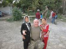
Physicians can learn a great deal after they join the military, such as how to approach an army general and suggest potentially life-saving medical changes on a base, said Dr. Dore J. Gilbert, a dermatologist on the faculty at the University of California Irvine.
A week into Lt. Col. Gilbert's deployment as a brigade surgeon on a base in Kabul, Afghanistan, he identified areas where necessary changes needed to be made and scheduled an appointment with the General.
Dr. Gilbert determined there was insufficient blood stored on the base in the event of an emergency. He suggested a walk-in blood bank with the General as the first volunteer. The General, concerned with the potential consequences, strongly supported the idea of the blood bank.
Next, Dr. Gilbert requested training for the 16 medics on the base and for 14 more at outlying bases once a month for 2 hours. "They were not getting any training," he said. The General again agreed.
"We have these unbelievable mannequins that actually bleed," Dr. Gilbert said. "They are a great way to train people. Sometimes we blow smoke so it's difficult to see the mannequins or do [medic training] at night so they have to put on their head lamps," Dr. Gilbert said during the South Beach Symposium in Miami.
When he raised a third issue, the General said, "Just do it." Dr. Gilbert increased the screening of local Afghan cafeteria employees for endemic intestinal parasites. Their contract only stipulated testing once a year on a base that serves 3,000 soldiers.
And Dr. Gilbert, as a dermatologist, also ran a skin cancer screening clinic. He treated other patients as well, which was part of the multitasking everyone did on the base. "On the second day, I was peeling old shrapnel out of a guy's face."
"I got up every day at 5 am to go to they gym and then work until 9 or 10 at night. I worked 12 to 14 hours a day, 7 days a week," he rcalled.
All his initiative impressed the General, and he invited Dr. Gilbert to become part of the everyday army operations on the base. "I went from being a simple doc to being a part of every general staff meeting," he said. Dr. Gilbert also was called in to the command center every time there was a terrorist-related attack in downtown Kabul. He went out on about 20 missions, most of which were to support medical personnel at other bases.
"I got to do things that 99.9% of doctors do not get to do when they are overseas or here in the United States because of a happenstance meeting I had with the General," Dr. Gilbert said.
Perhaps the most impressive part of the tale is Dr. Gilbert joined and started basic training at Fort Sam Houston at the age of 59 years. "I was not going to allow people to say I was too old. Because there is such a dire need for physicians in the army they are willing to look at people who are slightly more mature."
One day he was doing a 5 mile ruck march with 35 pounds on his back. As he passed one of the younger soldiers, Dr. Gilbert said, "Pick it up. I'm older than your father." The soldier later approached him and said, "Sir, with all due respect, you are not older than my father, you are way older than my father."
Dr. Gilbert always wanted to serve and was inspired by his son Kevin, a corporal in the Marine Corps. "I was happy to see him home with his arms and legs. His battalion had about 25% casualties."
When he finished reflecting on his personal experiences as a dermatologist serving overseas, Dr. Gilbert said: "It was a great experience. I was honored to be able to serve my country."
--Damian McNamara @MedReporter on Twitter
Wound Healing Update
Laurel M. Morton, MD, and Tania J. Phillips, MD
The management of acute and chronic wounds has drastically changed within the past 20 years. This update focuses on the most recent recommendations for acute wound care as well as new technologies that are available for chronic wounds.
*For a PDF of the full article, click on the link to the left of this introduction.


