User login
Interpregnancy weight gain ups stillbirth risk
The risk of stillbirth increases linearly with interpregnancy weight gain, according to a Swedish population-based cohort study.
The researchers found that between-pregnancy weight gain greater than four body mass index units – about 11 kg or 24 lbs – was associated with more than a 50% increase in stillbirth risk in a second pregnancy, even after controlling for confounders such as maternal education and smoking.

The Swedish study of 456,711 women who had their first and second singleton births between 1992 and 2012 showed an increased risk of infant mortality for women who had a healthy weight in their first pregnancy and then gained weight. For those that gained between two and four body mass index (BMI) units, the relative risk was 27%, compared with stable weight women. There was a 60% increased risk of infant mortality among women who gained more than four BMI units, according to the findings published online in the Lancet on Dec. 2.
Each BMI unit corresponds to about 2.8 kg (6 lbs) in an average height woman (167 cm, or 5 feet 5 inches).
Already overweight women did not show an increased risk of infant mortality with weight gain, but did show significantly reduced infant mortality with weight loss greater than two BMI units between pregnancies (relative risk 0.49; 95% confidence interval, 0.27-0.88). But that same weight loss in healthy-weight women increased the risk of infant mortality (Lancet. 2015 Dec 2. doi: 10.1016/S0140-6736(15)00990-3).
“Obesity and weight gain are associated with inflammatory upregulation, and inflammation has been proposed as one mechanism for the associations between maternal overweight and obesity and several adverse pregnancy outcomes,” wrote Dr. Sven Cnattingius, from the Karolinska Institute, Stockholm, and Dr. Eduardo Villamor from the University of Michigan, Ann Arbor.
The study was supported by the Swedish Research Council for Health, Working Life and Welfare, and the Karolinska Institute. The researchers reported having no financial disclosures.
The risk of stillbirth increases linearly with interpregnancy weight gain, according to a Swedish population-based cohort study.
The researchers found that between-pregnancy weight gain greater than four body mass index units – about 11 kg or 24 lbs – was associated with more than a 50% increase in stillbirth risk in a second pregnancy, even after controlling for confounders such as maternal education and smoking.
The Swedish study of 456,711 women who had their first and second singleton births between 1992 and 2012 showed an increased risk of infant mortality for women who had a healthy weight in their first pregnancy and then gained weight. For those that gained between two and four body mass index (BMI) units, the relative risk was 27%, compared with stable weight women. There was a 60% increased risk of infant mortality among women who gained more than four BMI units, according to the findings published online in the Lancet on Dec. 2.
Each BMI unit corresponds to about 2.8 kg (6 lbs) in an average height woman (167 cm, or 5 feet 5 inches).
Already overweight women did not show an increased risk of infant mortality with weight gain, but did show significantly reduced infant mortality with weight loss greater than two BMI units between pregnancies (relative risk 0.49; 95% confidence interval, 0.27-0.88). But that same weight loss in healthy-weight women increased the risk of infant mortality (Lancet. 2015 Dec 2. doi: 10.1016/S0140-6736(15)00990-3).
“Obesity and weight gain are associated with inflammatory upregulation, and inflammation has been proposed as one mechanism for the associations between maternal overweight and obesity and several adverse pregnancy outcomes,” wrote Dr. Sven Cnattingius, from the Karolinska Institute, Stockholm, and Dr. Eduardo Villamor from the University of Michigan, Ann Arbor.
The study was supported by the Swedish Research Council for Health, Working Life and Welfare, and the Karolinska Institute. The researchers reported having no financial disclosures.
FROM THE LANCET
Key clinical point: The risk of stillbirth increases linearly with interpregnancy weight gain.
Major finding: Women who gained more than four BMI units after their first pregnancy had a more than 50% increased risk of stillbirth in their second pregnancy.
Data source: A population-based cohort study in 456,711 women.
Disclosures: The study was supported by the Swedish Research Council for Health, Working Life and Welfare, and the Karolinska Institute. The researchers reported having no financial disclosures.

CPAP, oral devices reduced blood pressure in sleep apnea
Continuous positive airway pressure (CPAP) and mandibular advancement devices (MADs) both achieved similar reductions in blood pressure in individuals with obstructive sleep apnea, compared with inactive controls.
In a systematic review and meta-analysis of 51 studies involving 4,888 patients, researchers found that CPAP use was associated with a significant mean systolic blood pressure reduction of 2.5 mm Hg and mean diastolic reduction of 2 mm Hg, compared with inactive controls. Each 1-hour increase in mean CPAP use was associated with a significant additional 1.5 mm Hg systolic and 0.9 mm Hg diastolic blood pressure reduction.

Similarly, MADs were associated with a significant 2.1 mm Hg reduction in systolic pressure and 1.9 mm Hg reduction in diastolic pressure, compared with inactive controls.
“This is partly in contrast to a previous meta-analysis, which did not find a beneficial association with MADs, perhaps due to including only two [randomized controlled trials] and thus having inadequate power to detect a difference,” wrote Daniel J. Bratton, Ph.D., of the department of pulmonology, University Hospital, Zurich, and coauthors (JAMA. 2015 Dec 1;314:2280-93).
Overall, the authors found no significant differences between CPAP and MADs in the associated changes in systolic or diastolic blood pressure, although they noted that CPAP showed the strongest association with systolic blood pressure reductions.
The Swiss National Science Foundation and the University of Zurich supported the study. The authors declared no conflicts of interest.
Continuous positive airway pressure (CPAP) and mandibular advancement devices (MADs) both achieved similar reductions in blood pressure in individuals with obstructive sleep apnea, compared with inactive controls.
In a systematic review and meta-analysis of 51 studies involving 4,888 patients, researchers found that CPAP use was associated with a significant mean systolic blood pressure reduction of 2.5 mm Hg and mean diastolic reduction of 2 mm Hg, compared with inactive controls. Each 1-hour increase in mean CPAP use was associated with a significant additional 1.5 mm Hg systolic and 0.9 mm Hg diastolic blood pressure reduction.
Similarly, MADs were associated with a significant 2.1 mm Hg reduction in systolic pressure and 1.9 mm Hg reduction in diastolic pressure, compared with inactive controls.
“This is partly in contrast to a previous meta-analysis, which did not find a beneficial association with MADs, perhaps due to including only two [randomized controlled trials] and thus having inadequate power to detect a difference,” wrote Daniel J. Bratton, Ph.D., of the department of pulmonology, University Hospital, Zurich, and coauthors (JAMA. 2015 Dec 1;314:2280-93).
Overall, the authors found no significant differences between CPAP and MADs in the associated changes in systolic or diastolic blood pressure, although they noted that CPAP showed the strongest association with systolic blood pressure reductions.
The Swiss National Science Foundation and the University of Zurich supported the study. The authors declared no conflicts of interest.
FROM JAMA
Key clinical point: Continuous positive airway pressure and mandibular advancement devices both achieve similar reductions in blood pressure in individuals with obstructive sleep apnea.
Major finding: CPAP use was associated with a mean systolic blood pressure reduction of 2.5 mm Hg, and MADs were associated with a 2.1 mm Hg reduction, compared with inactive controls.
Data source: A systematic review and meta-analysis of 51 studies involving 4,888 patients.
Disclosures: The Swiss National Science Foundation and the University of Zurich supported the study. The authors declared no conflicts of interest.

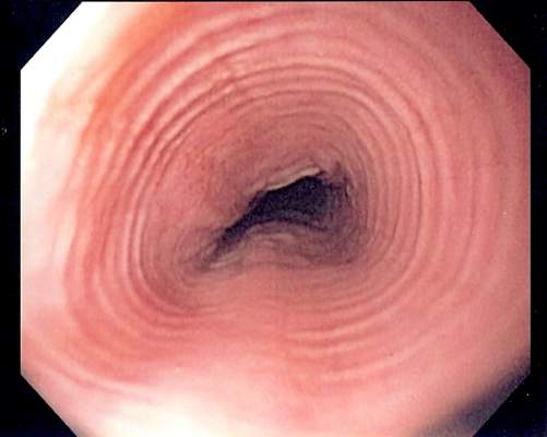
EREFS value has diagnostic utility for eosinophilic esophagitis
The Eosinophilic Esophagitis Endoscopic Reference Score, or EREFS, is not only highly predictive of eosinophilic esophagitis (EoE) but also its responsiveness to treatment, which suggests it may be used as an outcome measure, researchers say.
A prospective study of 211 adults undergoing upper endoscopy to investigate symptoms of esophageal dysfunction compared the EREFS with consensus guidelines for diagnosis of eosinophilic esophagitis.

The guidelines approach identified 67 cases of eosinophilic esophagitis and 144 control subjects without eosinophilic esophagitis. When these patients were assessed via the EREFS, researchers found multiple, highly significant differences between the cases and controls, with a mean total EREFS of 3.88 for cases and 0.42 for controls, according to a paper published online in Clinical Gastroenterology and Hepatology.
“On ROC [receiver operator characteristic] analysis, a model that contained all 5 components of the EREFS system as categorical variables had an AUC [area under the curve] of 0.946, indicating an excellent ability to predict EoE case status based on endoscopic findings alone,” wrote Dr. Evan S. Dellon and colleagues of the University of North Carolina at Chapel Hill.
In this model, a score of 2.0 or above showed a sensitivity of 88%, specificity of 92%, positive predictive value of 84%, negative predictive value of 94%, and accuracy of 91%.
Most of the score’s predictive ability was attributed to its inflammatory component, and less from the fibrostenotic score, which the authors suggested was due to the high prevalence of strictures in the control group.
The EREFS also improved significantly after treatment, in conjunction with endoscopic findings.
Total EREFS significantly decreased from 3.88 to 2.01, the inflammatory score decreased from 2.41 to 1.22, and the fibrostenotic score decreased from 1.46 to 0.89.
Histologic responders to treatment showed much more significant decreases in EREFS compared with nonresponders (Clin Gastro Hepatol. 2015, Sept. 12 [http://dx.doi.org/10.1016/j.cgh.2015.08.040]).
Researchers also examined the impact of weighing the various features of EREFS differently.
“The iterative analysis investigating weighing the EREFS features differently showed that increasing the weight of the exudate, rings, and edema score modestly increased the predictive power when the change in eosinophil counts was treated continuously and that increasing the weight of exudates and rings was beneficial with a threshold eosinophil count (less than 15 eosinophil/hpf) for response,” they reported.
Based on this finding, the researchers created a set of EREFS scores using these varied weights, and showed that doubling the exudates, rings, and edema scores achieved the score’s maximum responsiveness while still keeping the weighting system simple, although these changes did not alter the score’s overall predictive ability.
The EREFS score was developed as a way to standardize the description, recognition, and reporting of eosinophilic esophagitis, but its diagnostic utility and responsiveness to treatment were unknown, the authors said.
“This prospective study found that the EREFS classification has diagnostic utility for EoE,” they wrote. “Moreover, the score is responsive to treatment, decreasing significantly in histologic responders, and can be used as an outcome measure.”
The National Institutes of Health and the University of North Carolina Center for Gastrointestinal Biology and Disease funded the study. No conflicts of interest were declared.
The Eosinophilic Esophagitis Endoscopic Reference Score, or EREFS, is not only highly predictive of eosinophilic esophagitis (EoE) but also its responsiveness to treatment, which suggests it may be used as an outcome measure, researchers say.
A prospective study of 211 adults undergoing upper endoscopy to investigate symptoms of esophageal dysfunction compared the EREFS with consensus guidelines for diagnosis of eosinophilic esophagitis.
The guidelines approach identified 67 cases of eosinophilic esophagitis and 144 control subjects without eosinophilic esophagitis. When these patients were assessed via the EREFS, researchers found multiple, highly significant differences between the cases and controls, with a mean total EREFS of 3.88 for cases and 0.42 for controls, according to a paper published online in Clinical Gastroenterology and Hepatology.
“On ROC [receiver operator characteristic] analysis, a model that contained all 5 components of the EREFS system as categorical variables had an AUC [area under the curve] of 0.946, indicating an excellent ability to predict EoE case status based on endoscopic findings alone,” wrote Dr. Evan S. Dellon and colleagues of the University of North Carolina at Chapel Hill.
In this model, a score of 2.0 or above showed a sensitivity of 88%, specificity of 92%, positive predictive value of 84%, negative predictive value of 94%, and accuracy of 91%.
Most of the score’s predictive ability was attributed to its inflammatory component, and less from the fibrostenotic score, which the authors suggested was due to the high prevalence of strictures in the control group.
The EREFS also improved significantly after treatment, in conjunction with endoscopic findings.
Total EREFS significantly decreased from 3.88 to 2.01, the inflammatory score decreased from 2.41 to 1.22, and the fibrostenotic score decreased from 1.46 to 0.89.
Histologic responders to treatment showed much more significant decreases in EREFS compared with nonresponders (Clin Gastro Hepatol. 2015, Sept. 12 [http://dx.doi.org/10.1016/j.cgh.2015.08.040]).
Researchers also examined the impact of weighing the various features of EREFS differently.
“The iterative analysis investigating weighing the EREFS features differently showed that increasing the weight of the exudate, rings, and edema score modestly increased the predictive power when the change in eosinophil counts was treated continuously and that increasing the weight of exudates and rings was beneficial with a threshold eosinophil count (less than 15 eosinophil/hpf) for response,” they reported.
Based on this finding, the researchers created a set of EREFS scores using these varied weights, and showed that doubling the exudates, rings, and edema scores achieved the score’s maximum responsiveness while still keeping the weighting system simple, although these changes did not alter the score’s overall predictive ability.
The EREFS score was developed as a way to standardize the description, recognition, and reporting of eosinophilic esophagitis, but its diagnostic utility and responsiveness to treatment were unknown, the authors said.
“This prospective study found that the EREFS classification has diagnostic utility for EoE,” they wrote. “Moreover, the score is responsive to treatment, decreasing significantly in histologic responders, and can be used as an outcome measure.”
The National Institutes of Health and the University of North Carolina Center for Gastrointestinal Biology and Disease funded the study. No conflicts of interest were declared.
FROM CLINICAL GASTROENTEROLOGY AND HEPATOLOGY
Key clinical point: The Eosinophilic Esophagitis Endoscopic Reference Score is highly predictive of eosinophilic esophagitis and responsiveness to treatment.
Major finding: A model containing all five components of the EREFS system as categorical variables had an AUC of 0.946.
Data source: A prospective study of 211 adults undergoing upper endoscopy to investigate esophageal dysfunction.
Disclosures: The National Institutes of Health and the University of North Carolina Center for Gastrointestinal Biology and Disease funded the study. No conflicts of interest were declared.
Increased prevalence of pancreatic cysts due to MRI improvements
The apparent increase in the prevalence of incidental pancreatic cysts in recent years may be tied to improvements in MRI scanning technology, results of a study suggest.
Researchers conducted a retrospective analysis of data from 500 patients who underwent an MRI for nonpancreatic indications at a single center between 2005 and 2014; 50 were sampled from each year in chronological order.

A total of 208 patients (41.6%) were found to have an incidental cyst, of which less than a quarter were described in the original MRI report, according to a paper published online in Clinical Gastroenterology and Hepatology.
Analysis showed a very strong association between the type of imaging hardware and software, and the presence of cysts; older hardware found pancreatic cysts in 30.3% of patients and the newest hardware found cysts in 56.3% of patients.
However, MRI field strength was not associated with the frequency of lesion discovery (Clin Gastro Hepatol. 2015 Sep 11. doi: 10.1016/j.cgh.2015.08.038).
Most cysts were relatively small, with a median size of 4 mm. Nearly half of the patients with a cyst only had one described.
Nearly two-thirds of these cysts (62%) had an uncertain diagnosis, but 35% of patients were diagnosed with an intraductal papillary mucinous neoplasm, and one patient showed radiologic evidence of subacute pancreatitis.
When compared to the rest of the cohort, individuals with pancreatic cysts were more likely to be older, have diabetes mellitus, or have a personal history of cancer, particularly nonmelanoma skin cancer and hepatocellular carcinoma.
“Our study demonstrates the relationship between the higher trend of incidental pancreatic cysts observed in the recent years and the improvements in the technical features of MRIs,” wrote Dr. Maria Moris and colleagues from the Mayo Clinic, Jacksonville.
The authors said the real prevalence of pancreatic cystic lesions is estimated to range from 0.2% to 44.7%.
Their finding of a prevalence of 41.6% was higher than that found in similar imaging studies, but the authors suggested some of this may be due to the lack of a size cutoff in their study, as opposed to a 5-mm cutoff used in one earlier study that found a prevalence of 10%.
“Moreover, we believe that we may have even underestimated the real prevalence because of the absence of magnetic resonance cholangiopancreatography sequences (only 19% of the examinations), and the lack of 3-T studies (6% of the MRIs),” they wrote.
The median size of the lesions was lower than those found in previous studies.
“This smaller size was unexpected because, as a result of the exclusion criteria applied, the technical features of the MRIs were not the most specific for [pancreatic cystic lesion] PCL visualization,” the authors wrote, suggesting that this may have been due to the radiologist’s experience in this field.
The study was funded by the Joyce E. Baker Foundation for Research at Mayo Clinic in Jacksonville. One author disclosed grants or travel support from Olympus, Boston Scientific, and Cosmo Pharmaceuticals. There were no other conflicts of interest declared.
The apparent increase in the prevalence of incidental pancreatic cysts in recent years may be tied to improvements in MRI scanning technology, results of a study suggest.
Researchers conducted a retrospective analysis of data from 500 patients who underwent an MRI for nonpancreatic indications at a single center between 2005 and 2014; 50 were sampled from each year in chronological order.
A total of 208 patients (41.6%) were found to have an incidental cyst, of which less than a quarter were described in the original MRI report, according to a paper published online in Clinical Gastroenterology and Hepatology.
Analysis showed a very strong association between the type of imaging hardware and software, and the presence of cysts; older hardware found pancreatic cysts in 30.3% of patients and the newest hardware found cysts in 56.3% of patients.
However, MRI field strength was not associated with the frequency of lesion discovery (Clin Gastro Hepatol. 2015 Sep 11. doi: 10.1016/j.cgh.2015.08.038).
Most cysts were relatively small, with a median size of 4 mm. Nearly half of the patients with a cyst only had one described.
Nearly two-thirds of these cysts (62%) had an uncertain diagnosis, but 35% of patients were diagnosed with an intraductal papillary mucinous neoplasm, and one patient showed radiologic evidence of subacute pancreatitis.
When compared to the rest of the cohort, individuals with pancreatic cysts were more likely to be older, have diabetes mellitus, or have a personal history of cancer, particularly nonmelanoma skin cancer and hepatocellular carcinoma.
“Our study demonstrates the relationship between the higher trend of incidental pancreatic cysts observed in the recent years and the improvements in the technical features of MRIs,” wrote Dr. Maria Moris and colleagues from the Mayo Clinic, Jacksonville.
The authors said the real prevalence of pancreatic cystic lesions is estimated to range from 0.2% to 44.7%.
Their finding of a prevalence of 41.6% was higher than that found in similar imaging studies, but the authors suggested some of this may be due to the lack of a size cutoff in their study, as opposed to a 5-mm cutoff used in one earlier study that found a prevalence of 10%.
“Moreover, we believe that we may have even underestimated the real prevalence because of the absence of magnetic resonance cholangiopancreatography sequences (only 19% of the examinations), and the lack of 3-T studies (6% of the MRIs),” they wrote.
The median size of the lesions was lower than those found in previous studies.
“This smaller size was unexpected because, as a result of the exclusion criteria applied, the technical features of the MRIs were not the most specific for [pancreatic cystic lesion] PCL visualization,” the authors wrote, suggesting that this may have been due to the radiologist’s experience in this field.
The study was funded by the Joyce E. Baker Foundation for Research at Mayo Clinic in Jacksonville. One author disclosed grants or travel support from Olympus, Boston Scientific, and Cosmo Pharmaceuticals. There were no other conflicts of interest declared.
FROM CLINICAL GASTROENTEROLOGY AND HEPATOLOGY
Key clinical point: The apparent increase of the prevalence of incidental pancreatic cysts in recent years may be tied to improvements in MRI scanning technology.
Major finding: Older MRI hardware found pancreatic cysts in 30.3% of patients and the newest hardware found cysts in 56.3% of patients.
Data source: A retrospective analysis of data from 500 patients who underwent an MRI for nonpancreatic indications.
Disclosures: The study was funded by the Joyce E. Baker Foundation for Research at Mayo Clinic in Jacksonville, Fla. One author disclosed grants or travel support from Olympus, Boston Scientific, and Cosmo Pharmaceuticals. There were no other conflicts of interest declared.

Mechanical thrombectomy improves stroke outcomes
Mechanical removal of a thrombus in addition to usual care is associated with significantly better functional outcomes than usual care in patients who have experienced an acute ischemic stroke, according to a meta-analysis published online Nov. 30 in Journal of the American College of Cardiology.
The analysis of nine trials of usual care with or without mechanical thrombectomy in 2,410 patients showed that those undergoing mechanical thrombectomy had a 45% greater likelihood of achieving a good functional outcome and a 67% greater chance of excellent functional outcome, defined respectively as a modified Rankin scale score of 0-2 or 0-1.

Researchers observed a nonsignificant trend toward lower all-cause mortality in the mechanical thrombectomy patients, which was also linked with improved recanalization, compared with usual care alone (risk ratio, 1.57; 95% confidence interval, 1.11-2.23; P = .01).
Mechanical thrombectomy did show a nonsignificant increase in the risk of recurrent stroke at 90 days, but this was largely driven by one study, and when that was excluded, the risk was similar between mechanical thrombectomy and no mechanical thrombectomy (J Am Coll Cardiol. 2015;66:2498-505).
“Although mechanical thrombectomy is beneficial, this procedure requires specialized centers of excellence; therefore, the widespread application of this therapy for acute ischemic stroke patients will likely remain limited for the foreseeable future,” wrote Dr. Islam Y. Elgendy of the University of Florida, Gainesville, and coauthors.
One author declared advisory board membership and research funding from private industry, but there were no other conflicts of interest declared.
In recent trials, removable devices consisting of self-expanding, clot-retrieving stents achieved higher rates of recanalization than did earlier methods of thrombus extraction, representing the first effective new treatment for stroke in nearly 20 years.
With absolute benefits substantially greater than systemic intravenous thrombolysis alone, the combination of intravenous tissue plasminogen activator and endovascular therapy have improved outcomes for selected patients who receive endovascular treatment within 6 hours of symptom onset, and further efforts to shorten the interval between emergency department arrival and treatment hold the promise of even better outcomes.
Dr. Gregory W. Albers of the Stanford Stroke Center at Stanford (Calif.) University, and Dr. Jonathan L. Halperin of the Cardiovascular Institute at the Mount Sinai Medical Center, New York, made these comments in an accompanying editorial (J Am Coll Cardiol. 2015;66:2506-9). Dr. Halperin has served as a consultant to Boston Scientific, Medtronic, and Johnson & Johnson. Dr. Albers has an equity interest in iSchemaView and is a consultant for iSchemaView and Medtronic.
Mechanical removal of a thrombus in addition to usual care is associated with significantly better functional outcomes than usual care in patients who have experienced an acute ischemic stroke, according to a meta-analysis published online Nov. 30 in Journal of the American College of Cardiology.
The analysis of nine trials of usual care with or without mechanical thrombectomy in 2,410 patients showed that those undergoing mechanical thrombectomy had a 45% greater likelihood of achieving a good functional outcome and a 67% greater chance of excellent functional outcome, defined respectively as a modified Rankin scale score of 0-2 or 0-1.
Researchers observed a nonsignificant trend toward lower all-cause mortality in the mechanical thrombectomy patients, which was also linked with improved recanalization, compared with usual care alone (risk ratio, 1.57; 95% confidence interval, 1.11-2.23; P = .01).
Mechanical thrombectomy did show a nonsignificant increase in the risk of recurrent stroke at 90 days, but this was largely driven by one study, and when that was excluded, the risk was similar between mechanical thrombectomy and no mechanical thrombectomy (J Am Coll Cardiol. 2015;66:2498-505).
“Although mechanical thrombectomy is beneficial, this procedure requires specialized centers of excellence; therefore, the widespread application of this therapy for acute ischemic stroke patients will likely remain limited for the foreseeable future,” wrote Dr. Islam Y. Elgendy of the University of Florida, Gainesville, and coauthors.
One author declared advisory board membership and research funding from private industry, but there were no other conflicts of interest declared.
FROM JOURNAL OF THE AMERICAN COLLEGE OF CARDIOLOGY
Key clinical point: Mechanical thrombectomy and usual care are associated with significantly better functional outcomes after acute ischemic stroke than is usual care alone.
Major finding: Patients undergoing mechanical thrombectomy had a 67% greater likelihood of excellent functional outcome than patients having usual care alone.
Data source: Meta-analysis of nine trials of usual care with or without mechanical thrombectomy in 2,410 patients.
Disclosures: One author declared advisory board membership and research funding from private industry, but there were no other conflicts of interest declared.

Methylphenidate evidence in ADHD ‘very low quality’
The evidence for a benefit from methylphenidate in the treatment of attention-deficit/hyperactivity disorder is of very low quality, a meta-analysis showed.
The Cochrane Review of 38 parallel-group trials in 5,111 participants and 147 crossover trials in 7,134 participants found that while methylphenidate significantly reduced teacher-rated attention-deficit/hyperactivity disorder (ADHD) symptoms by 23%, compared with placebo, the evidence was of very low quality.
The change in teacher-rated symptoms corresponded to a mean difference of 9.6 points on the ADHD Rating Scale, which the authors said represented a modest improvement in ADHD symptoms.

“Methylphenidate may improve ADHD symptoms, general behavior, and quality of life in children and adolescents aged 18 years and younger with ADHD, [but] we rated the evidence to be of very low quality, and, as a result, we cannot be certain about the magnitude of the effects from the meta-analyses,” Dr. Ole Jakob Storebø of the child and adolescent psychiatry department, Region Zealand, Roskilde, Denmark, and coauthors wrote (Cochrane Database Syst Rev. 2015 Nov 25. doi: 10.1002/14651858.CD009885.pub2).
Methylphenidate also significantly reduced independent assessor-rated ADHD symptoms, although the trials were all judged to be at high risk of bias.
Researchers found that methylphenidate significantly reduced parent-rated ADHD symptoms, compared with placebo, with an impact corresponding to a mean difference of –8.2 points on the ADHD Rating Scale, which was independent of factors including treatment duration, dose, and medication status before randomization.
There also was significantly improved quality of life with methylphenidate treatment, compared with placebo, by a measure equivalent to a mean difference of 8 points on the Child Health Questionnaire, although the trials that found this were judged to be at high risk of bias.
Researchers also examined the effects of methylphenidate on general behavior, and found a significant impact on both teacher-rated and parent-rated general behavior.
One trial showed that the impact of methylphenidate was significantly greater in individuals with the inattentive subtype, compared with the combined subtype.
However, treatment with methylphenidate was associated with an increased risk of adverse events that were not serious, including a 60% increase in the incidence of sleep problems and a greater than threefold increase in decreased appetite.
“This review highlights two major issues concerning the overall completeness and applicability of the evidence of the benefits and harms of methylphenidate for children with ADHD: the dearth of trials conducted in children and adolescents in low- and middle-income countries, and the lack of follow-up beyond 6 months,” Dr. Storebø and associates wrote.
They called for better-designed trials to assess the benefits of methylphenidate. “Given the frequency of nonserious adverse events associated with methylphenidate, the particular difficulties for blinding of participants and outcome assessors point to the advantage of large, ‘nocebo tablet’ controlled trials.”
The review was funded by Region Zealand Psychiatry, Region Zealand Research Foundation, and Copenhagen University Hospital. One author declared doing speaking engagements for Novartis.
The evidence for a benefit from methylphenidate in the treatment of attention-deficit/hyperactivity disorder is of very low quality, a meta-analysis showed.
The Cochrane Review of 38 parallel-group trials in 5,111 participants and 147 crossover trials in 7,134 participants found that while methylphenidate significantly reduced teacher-rated attention-deficit/hyperactivity disorder (ADHD) symptoms by 23%, compared with placebo, the evidence was of very low quality.
The change in teacher-rated symptoms corresponded to a mean difference of 9.6 points on the ADHD Rating Scale, which the authors said represented a modest improvement in ADHD symptoms.
“Methylphenidate may improve ADHD symptoms, general behavior, and quality of life in children and adolescents aged 18 years and younger with ADHD, [but] we rated the evidence to be of very low quality, and, as a result, we cannot be certain about the magnitude of the effects from the meta-analyses,” Dr. Ole Jakob Storebø of the child and adolescent psychiatry department, Region Zealand, Roskilde, Denmark, and coauthors wrote (Cochrane Database Syst Rev. 2015 Nov 25. doi: 10.1002/14651858.CD009885.pub2).
Methylphenidate also significantly reduced independent assessor-rated ADHD symptoms, although the trials were all judged to be at high risk of bias.
Researchers found that methylphenidate significantly reduced parent-rated ADHD symptoms, compared with placebo, with an impact corresponding to a mean difference of –8.2 points on the ADHD Rating Scale, which was independent of factors including treatment duration, dose, and medication status before randomization.
There also was significantly improved quality of life with methylphenidate treatment, compared with placebo, by a measure equivalent to a mean difference of 8 points on the Child Health Questionnaire, although the trials that found this were judged to be at high risk of bias.
Researchers also examined the effects of methylphenidate on general behavior, and found a significant impact on both teacher-rated and parent-rated general behavior.
One trial showed that the impact of methylphenidate was significantly greater in individuals with the inattentive subtype, compared with the combined subtype.
However, treatment with methylphenidate was associated with an increased risk of adverse events that were not serious, including a 60% increase in the incidence of sleep problems and a greater than threefold increase in decreased appetite.
“This review highlights two major issues concerning the overall completeness and applicability of the evidence of the benefits and harms of methylphenidate for children with ADHD: the dearth of trials conducted in children and adolescents in low- and middle-income countries, and the lack of follow-up beyond 6 months,” Dr. Storebø and associates wrote.
They called for better-designed trials to assess the benefits of methylphenidate. “Given the frequency of nonserious adverse events associated with methylphenidate, the particular difficulties for blinding of participants and outcome assessors point to the advantage of large, ‘nocebo tablet’ controlled trials.”
The review was funded by Region Zealand Psychiatry, Region Zealand Research Foundation, and Copenhagen University Hospital. One author declared doing speaking engagements for Novartis.
FROM THE COCHRANE DATABASE OF SYSTEMATIC REVIEWS
Key clinical point: Evidence for a benefit from methylphenidate in the treatment of attention-deficit/hyperactivity disorder is of low quality.
Major finding: Methylphenidate reduced teacher-rated ADHD symptoms by 23%, compared with placebo.
Data source: A meta-analysis of 185 trials and 12,245 participants.
Disclosures: The review was funded by Region Zealand Psychiatry, Region Zealand Research Foundation, and Copenhagen University Hospital. One author declared doing speaking engagements for Novartis.

Global HIV treatment goals face resource challenges
The World Health Organization’s “90-90-90” treatment targets for HIV, which aim to have 81% of people living with HIV on antiretroviral therapy by 2020, may be financially unachievable, according to modeling that predicts a potential resources shortfall of up to US$25 billion.
A modeling analysis of the financial resources needed to scale up antiretroviral therapy (ART) in 97 countries to meet the WHO targets for 2015-2020 suggests that, under the 90-90-90 scenario, 30.4 million adults and 1.68 million children could receive treatment by 2020, which would require an estimated US$52.5 billion, according to a paper published Nov. 24 in PLoS Medicine.

When current funding trends were taken into account, the analysis predicted financing gaps ranging from US$19.8 billion to US$25 billion, with up to a US$16.8 billion gap for ART alone (PLoS Med. 2015 Nov 24;12[11]. doi:10.1371/journal.pmed.1001907).
The analysis estimated the number of individuals eligible for and receiving treatment and the cost of providing antiretroviral therapy, but it also looked at the full spectrum of costs in delivering therapy, including diagnostic supplies, and factored in predictions for shifts in epidemics. The 90-90-90 treatment scenario envisions 90% of all people living with HIV knowing their status, 90% of all people diagnosed with HIV infection receiving antiretroviral therapy, and 90% of all people receiving ART having robust viral suppression.
“Our estimates of the resource requirements for ART suggest that the financial sustainability of a scaled-up global ART response, where the scale-up itself is cost-effective, may be at risk without either additional resource mobilization or efficiency and effectiveness gains,” wrote Arin Dutta, senior technical director at Palladium in Washington, D.C., and his coauthors.
The study was funded by the U.S. Agency for International Development. No conflicts of interest were declared.
The World Health Organization’s “90-90-90” treatment targets for HIV, which aim to have 81% of people living with HIV on antiretroviral therapy by 2020, may be financially unachievable, according to modeling that predicts a potential resources shortfall of up to US$25 billion.
A modeling analysis of the financial resources needed to scale up antiretroviral therapy (ART) in 97 countries to meet the WHO targets for 2015-2020 suggests that, under the 90-90-90 scenario, 30.4 million adults and 1.68 million children could receive treatment by 2020, which would require an estimated US$52.5 billion, according to a paper published Nov. 24 in PLoS Medicine.
When current funding trends were taken into account, the analysis predicted financing gaps ranging from US$19.8 billion to US$25 billion, with up to a US$16.8 billion gap for ART alone (PLoS Med. 2015 Nov 24;12[11]. doi:10.1371/journal.pmed.1001907).
The analysis estimated the number of individuals eligible for and receiving treatment and the cost of providing antiretroviral therapy, but it also looked at the full spectrum of costs in delivering therapy, including diagnostic supplies, and factored in predictions for shifts in epidemics. The 90-90-90 treatment scenario envisions 90% of all people living with HIV knowing their status, 90% of all people diagnosed with HIV infection receiving antiretroviral therapy, and 90% of all people receiving ART having robust viral suppression.
“Our estimates of the resource requirements for ART suggest that the financial sustainability of a scaled-up global ART response, where the scale-up itself is cost-effective, may be at risk without either additional resource mobilization or efficiency and effectiveness gains,” wrote Arin Dutta, senior technical director at Palladium in Washington, D.C., and his coauthors.
The study was funded by the U.S. Agency for International Development. No conflicts of interest were declared.
Key clinical point: The World Health Organization’s 90-90-90 treatment targets for HIV may face a resources shortfall of up to US$25 billion.
Major finding: Scaling up to the 90-90-90 treatment target for HIV by 2020 may require an estimated US$52.5 billion, with potential financing gaps up to US$25 billion.
Data source: Economic modeling and analysis of data from various third-party sources, including government agencies, watchdog organizations, and external funding organizations.
Disclosures: The study was funded by the U.S. Agency for International Development. No conflicts of interest were declared.

ACOG: Think of menses as a vital sign in adolescents
The American College of Obstetricians and Gynecologists wants physicians who treat adolescent girls to consider a new vital sign: menstruation.
Once girls begin menstruating, physicians should ask about the first day of the last menstrual period and the pattern of menses at every preventive care or comprehensive visit, according to an opinion from ACOG’s Committee on Adolescent Health Care, released on Nov. 23. This information can help identify early health concerns such as polycystic ovary syndrome, thyroid disease, eating disorders, coagulopathies, or even hepatic failure or malignancy.

“By including an evaluation of the menstrual cycle as an additional vital sign, clinicians reinforce its importance in assessing overall health status for patients and caretakers,” the ACOG committee members wrote. “Just as abnormal blood pressure, heart rate, or respiratory rate may be key to diagnosing potentially serious health conditions, identification of abnormal menstrual patterns in adolescence may improve early identification of potential health concerns for adulthood.”
The American Academy of Pediatrics endorsed the committee opinion.
Physicians also need to educate girls and their parents about normal and abnormal menstruation, ACOG urged.
Although there are variations, the median age at menarche has been stable for the last 30 years, starting between ages 12 and 13. Studies have shown that higher gain in body mass index during childhood is associated with an earlier onset of puberty (Obstet Gynecol. 2015;126:e143-6).
Most girls will bleed for 2-7 days during their first menses and though it’s normal for menstrual cycles to be irregular in adolescence, 90% of cycles will range from 21 to 45 days, according to the committee opinion. By the third year after the start of menstruation, 60%-80% of cycles are 21-34 days long.
Abnormalities that might signal health problems include menstruation that hasn’t started within 3 years of breast budding, menstruation that hasn’t started by age 14 with signs of hirsutism, menstruation that hasn’t started by age 14 with a history or exam suggestive of excessive exercise or an eating disorder, and menses that are heavy and linked to a history of excessive bruising or bleeding.
“It is important for clinicians to have an understanding of the menstrual patterns of adolescent girls, the ability to differentiate between normal and abnormal menstruation, and the skill to know how to evaluate the adolescent girl patient,” the committee wrote.
The American College of Obstetricians and Gynecologists wants physicians who treat adolescent girls to consider a new vital sign: menstruation.
Once girls begin menstruating, physicians should ask about the first day of the last menstrual period and the pattern of menses at every preventive care or comprehensive visit, according to an opinion from ACOG’s Committee on Adolescent Health Care, released on Nov. 23. This information can help identify early health concerns such as polycystic ovary syndrome, thyroid disease, eating disorders, coagulopathies, or even hepatic failure or malignancy.
“By including an evaluation of the menstrual cycle as an additional vital sign, clinicians reinforce its importance in assessing overall health status for patients and caretakers,” the ACOG committee members wrote. “Just as abnormal blood pressure, heart rate, or respiratory rate may be key to diagnosing potentially serious health conditions, identification of abnormal menstrual patterns in adolescence may improve early identification of potential health concerns for adulthood.”
The American Academy of Pediatrics endorsed the committee opinion.
Physicians also need to educate girls and their parents about normal and abnormal menstruation, ACOG urged.
Although there are variations, the median age at menarche has been stable for the last 30 years, starting between ages 12 and 13. Studies have shown that higher gain in body mass index during childhood is associated with an earlier onset of puberty (Obstet Gynecol. 2015;126:e143-6).
Most girls will bleed for 2-7 days during their first menses and though it’s normal for menstrual cycles to be irregular in adolescence, 90% of cycles will range from 21 to 45 days, according to the committee opinion. By the third year after the start of menstruation, 60%-80% of cycles are 21-34 days long.
Abnormalities that might signal health problems include menstruation that hasn’t started within 3 years of breast budding, menstruation that hasn’t started by age 14 with signs of hirsutism, menstruation that hasn’t started by age 14 with a history or exam suggestive of excessive exercise or an eating disorder, and menses that are heavy and linked to a history of excessive bruising or bleeding.
“It is important for clinicians to have an understanding of the menstrual patterns of adolescent girls, the ability to differentiate between normal and abnormal menstruation, and the skill to know how to evaluate the adolescent girl patient,” the committee wrote.
FROM OBSTETRICS AND GYNECOLOGY

Stroke risk boosted by adult congenital heart disease
Adult congenital heart disease significantly increases the risk of both hemorrhagic and ischemic stroke, particularly in individuals under 55 years of age, new data suggests.
A retrospective study of 29,638 adults aged 18-64 years with adult congenital heart disease (ACHD) showed that women aged 15-54 years with the disease were more than 12 times as likely to experience an ischemic stroke compared to the general population, while men had a nine-fold increase in risk.
Women aged over 55 years with ACHD had a four-fold higher risk of ischemic stroke, and men had a two-fold increase in risk, compared to the general population, according to a study published Nov. 23 in Circulation.
In the case of hemorrhagic stroke, women aged under 55 had a five-fold greater risk and men had a more than six-fold greater risk of ischemic stroke, while the risk for those older than 55 years was 2-3 times higher (Circulation 2015, November 23 [doi: 10.1161/CIRCULATIONAHA.115.011241]).
The risk of ischemic stroke increased significantly with heart failure, diabetes, or a recent myocardial infarction, and overall, 8.9% of men and 6.8% of women with ACHD who reached the age of 18 years had at least one stroke before age 65.
“Whether subgroups of patients with heart failure and sinus rhythm could benefit from an antithrombotic treatment is a matter of ongoing research in the general population and based on our findings may warrant further investigation in ACHD-patients,” wrote Dr. Jonas Lanz, from the McGill Adult Unit for Congenital Heart Disease Excellence, and co-authors.
The study was funded by the Heart and Stroke Foundation of Québec, the Fonds de Recherche en Santé Québec and the Canadian Institute of Health Research. There were no conflicts of interest declared.
FROM CIRCULATION
Key clinical point:Adult congenital heart disease significantly increases the risk of both hemorrhagic and ischemic stroke, particularly in younger patients.
Major finding: Younger women with adult congenital heart disease have a 12-fold higher risk of ischemic stroke than the general population.
Data source: A retrospective study of 29,638 adults aged 18-64 years with adult congenital heart disease.
Disclosures: Authors were funded by the Heart and Stroke Foundation of Québec, the Fonds de Recherche en Santé Québec and the Canadian Institute of Health Research. There were no conflicts of interest declared.
Artificial pancreas improved glycemia after islet cell transplant
A closed-loop insulin pump with continuous glucose monitor produced significantly better blood glucose control in patients who have received islet cell transplants after pancreatectomy, compared with regular insulin injections, a pilot study has found.
Fourteen adults who received auto-islet transplants after pancreatectomy for chronic pancreatitis were randomized either to receive a closed-loop insulin pump system or the usual treatment of multiple insulin injections for 72 hours after transition from intravenous to subcutaneous insulin following surgery.

Researchers observed a significantly lower mean serum glucose in the insulin pump group, compared with the control group, with the highest average serum glucose level in individual patients in the pump group still being lower than the lowest average in the control group.
These improvements in glycemia were not associated with an increased risk of hypoglycemia in the closed-loop pump group and patients in the closed-loop pump group also required a significantly lower total daily insulin dose than did the control group, according to a paper published Nov. 20 in the American Journal of Transplantation.
“Success of islet engraftment is heavily dependent on maintenance of narrow-range euglycemia in the post-transplant period,” wrote Dr. Gregory P. Forlenza of the University of Minnesota Medical Center, Minneapolis, and his coauthors (Am J Transplant. 2015 Nov 20. doi: 10.1111/ajt.13539).
“This technology was shown in this study to provide some statistically and clinically significant improvements in glycemic parameters in adults after TP [total pancreatectomy] with IAT [intraportal islet autotransplantation] without producing associated increased episodes of hypoglycemia or adverse events.”
The study was funded by the Vikings Children’s Research Fund and the University of Minnesota. Medtronic Diabetes provided supplies as part of an investigator-initiated grant. No conflicts of interest were declared.
A closed-loop insulin pump with continuous glucose monitor produced significantly better blood glucose control in patients who have received islet cell transplants after pancreatectomy, compared with regular insulin injections, a pilot study has found.
Fourteen adults who received auto-islet transplants after pancreatectomy for chronic pancreatitis were randomized either to receive a closed-loop insulin pump system or the usual treatment of multiple insulin injections for 72 hours after transition from intravenous to subcutaneous insulin following surgery.
Researchers observed a significantly lower mean serum glucose in the insulin pump group, compared with the control group, with the highest average serum glucose level in individual patients in the pump group still being lower than the lowest average in the control group.
These improvements in glycemia were not associated with an increased risk of hypoglycemia in the closed-loop pump group and patients in the closed-loop pump group also required a significantly lower total daily insulin dose than did the control group, according to a paper published Nov. 20 in the American Journal of Transplantation.
“Success of islet engraftment is heavily dependent on maintenance of narrow-range euglycemia in the post-transplant period,” wrote Dr. Gregory P. Forlenza of the University of Minnesota Medical Center, Minneapolis, and his coauthors (Am J Transplant. 2015 Nov 20. doi: 10.1111/ajt.13539).
“This technology was shown in this study to provide some statistically and clinically significant improvements in glycemic parameters in adults after TP [total pancreatectomy] with IAT [intraportal islet autotransplantation] without producing associated increased episodes of hypoglycemia or adverse events.”
The study was funded by the Vikings Children’s Research Fund and the University of Minnesota. Medtronic Diabetes provided supplies as part of an investigator-initiated grant. No conflicts of interest were declared.
FROM THE AMERICAN JOURNAL OF TRANSPLANTATION
Key clinical point: A closed-loop insulin pump with a continuous glucose monitor offers significantly better blood glucose control in patients who have received islet cell transplants after pancreatectomy.
Major finding: Closed-loop insulin pumps were associated with a significantly lower mean serum glucose, compared with multiple daily insulin injections.
Data source: Randomized, controlled pilot study in 14 patients receiving auto-islet transplants after pancreatectomy.
Disclosures: The study was funded by the Vikings Children’s Research Fund and the University of Minnesota. Medtronic Diabetes provided supplies as part of an investigator-initiated grant. No conflicts of interest were declared.