User login
Bringing you the latest news, research and reviews, exclusive interviews, podcasts, quizzes, and more.
div[contains(@class, 'header__large-screen')]
div[contains(@class, 'read-next-article')]
div[contains(@class, 'main-prefix')]
div[contains(@class, 'nav-primary')]
nav[contains(@class, 'nav-primary')]
section[contains(@class, 'footer-nav-section-wrapper')]
footer[@id='footer']
section[contains(@class, 'nav-hidden')]
div[contains(@class, 'ce-card-content')]
nav[contains(@class, 'nav-ce-stack')]
div[contains(@class, 'view-medstat-quiz-listing-panes')]
div[contains(@class, 'pane-article-sidebar-latest-news')]
Why doctors are losing trust in patients; what should be done?
This transcript has been edited for clarity.
Hi. I’m Art Caplan. I’m at the division of medical ethics at New York University.
I want to talk about a paper that my colleagues in my division just published in Health Affairs.
As they pointed out, there’s a large amount of literature about what makes patients trust their doctor. There are many studies that show that, although patients sometimes have become more critical of the medical profession, in general they still try to trust their individual physician. Nurses remain in fairly high esteem among those who are getting hospital care.
What isn’t studied, as this paper properly points out, is, what can the doctor and the nurse do to trust the patient? How can that be assessed? Isn’t that just as important as saying that patients have to trust their doctors to do and comply with what they’re told?
What if doctors are afraid of violence? What if doctors are fearful that they can’t trust patients to listen, pay attention, or do what they’re being told? What if they think that patients are coming in with all kinds of disinformation, false information, or things they pick up on the Internet, so that even though you try your best to get across accurate and complete information about what to do about infectious diseases, taking care of a kid with strep throat, or whatever it might be, you’re thinking, Can I trust this patient to do what it is that I want them to do?
One particular problem that’s causing distrust is that more and more patients are showing stress and dependence on drugs and alcohol. That doesn’t make them less trustworthy per se, but it means they can’t regulate their own behavior as well.
That obviously has to be something that the physician or the nurse is thinking about. Is this person going to be able to contain anger? Is this person going to be able to handle bad news? Is this person going to deal with me when I tell them that some of the things they believe to be true about what’s good for their health care are false?
I think we have to really start to push administrators and people in positions of power to teach doctors and nurses how to defuse situations and how to make people more comfortable when they come in and the doctor suspects that they might be under the influence, impaired, or angry because of things they’ve seen on social media, whatever those might be – including concerns about racism, bigotry, and bias, which some patients are bringing into the clinic and the hospital setting.
We need more training. We’ve got to address this as a serious issue. What can we do to defuse situations where the doctor or the nurse rightly thinks that they can’t control or they can’t trust what the patient is thinking or how the patient might behave?
It’s also the case that I think we need more backup and quick access to security so that people feel safe and comfortable in providing care. We have to make sure that if you need someone to restrain a patient or to get somebody out of a situation, that they can get there quickly and respond rapidly, and that they know what to do to deescalate a situation.
It’s sad to say, but security in today’s health care world has to be something that we really test and check – not because we’re worried, as many places are, about a shooter entering the premises, which is its own bit of concern – but I’m just talking about when the doctor or the nurse says that this patient might be acting up, could get violent, or is someone I can’t trust.
My coauthors are basically saying that it’s not a one-way street. Yes, we have to figure out ways to make sure that our patients can trust what we say. Trust is absolutely the lubricant that makes health care flow. If patients don’t trust their doctors, they’re not going to do what they say. They’re not going to get their prescriptions filled. They’re not going to be compliant. They’re not going to try to lose weight or control their diabetes.
It also goes the other way. The doctor or the nurse has to trust the patient. They have to believe that they’re safe. They have to believe that the patient is capable of controlling themselves. They have to believe that the patient is capable of listening and hearing what they’re saying, and that they’re competent to follow up on instructions, including to come back if that’s what’s required.
Everybody has to feel secure in the environment in which they’re working. Security, sadly, has to be a priority if we’re going to have a health care workforce that really feels safe and comfortable dealing with a patient population that is increasingly aggressive and perhaps not as trustworthy.
That’s not news I like to read when my colleagues write it up, but it’s important and we have to take it seriously.
Dr. Caplan disclosed that he has served as a director, officer, partner, employee, adviser, consultant, or trustee for Johnson & Johnson’s Panel for Compassionate Drug Use (unpaid position), and is a contributing author and adviser for Medscape. A version of this article first appeared on Medscape.com.
Cognitive behavioral therapy app lowers A1c in type 2 diabetes
CHICAGO – A smartphone app that delivers nutritional cognitive behavioral therapy (CBT) to people with type 2 diabetes produced an average 0.29 percentage point drop in hemoglobin A1c during 180 days of use compared with controls, and an average 0.37 percentage point reduction in A1c compared with baseline values in a randomized, pivotal trial with 669 adults.
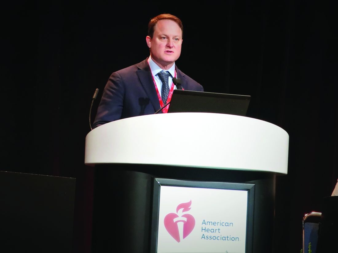
Use of the app for 180 days also significantly linked with a reduced need for additional medications, reduced weight and blood pressure, and improved patient-reported outcomes, and it led to fewer adverse effects than seen in control subjects, Marc P. Bonaca, MD, reported at the American Heart Association scientific sessions.

The findings also showed a clear dose-response relationship: The more CBT lessons a person completed with the app, the greater the A1c reduction.
The results suggest that the app, called BT-001, “potentially provides a scalable treatment option for patients with type 2 diabetes,” concluded Dr. Bonaca.
On the basis of the results from this trial, also called BT-001, the company developing the app, Better Therapeutics, announced in September 2022 that it had filed a classification request with the Food and Drug Administration that would allow marketing authorization for the BT-001 app. Better Therapeutics envisions that once authorized by the FDA, the app would be available to people with type 2 diabetes by prescriptions written by health care providers and that the cost for the app would be covered by health insurance, explained a company spokesperson.
A ‘modest positive impact’
“CBT is an empirically supported psychotherapy for a variety of emotional disorders, and it has been adapted to target specific emotional distress in the context of chronic illness,” said Amit Shapira, PhD, a clinical psychologist at the Joslin Diabetes Center in Boston who has not been involved in the BT-001 studies. A CBT protocol designed for diabetes, CBT for Adherence and Depression, “has been shown to have a positive impact on depression symptoms and glycemic control in adults with type 2 diabetes,” Dr. Shapira said in an interview.
Based on published results, the BT-001 app “seems to have a modest positive impact on glycemic control, especially among people who completed more than 10 [lesson] modules.” The evidence appears to suggest that the app “might be a good supplement to working with a behavioral health counselor.”
The BT-001 trial enrolled 669 adults with type 2 diabetes for an average of 11 years and an A1c of 7%-10.9% with an average level of 8.2%. Participants had to be on a stable medication regimen for at least 3 months but not using insulin, and their treatment regimens could undergo adjustment during the trial. At baseline, each subject was on an average of 2.1 antidiabetes medications, including 90% on metformin and 42% on a sulfonylurea. The researchers also highlighted that the enrolled cohort of people with type 2 diabetes had a demographic profile that was “generally representative” of U.S. adults with type 2 diabetes.
The researchers told the 326 people who were randomized to the active intervention group to use the app but subjects were free to determine their frequency of use. The app introduced a new lesson module weekly that took 10-20 minutes to complete, and each weekly lesson came with associated exercises aimed at practicing skills related to behavioral beliefs.
The study’s primary efficacy endpoint was the average change from baseline in A1c compared with the 343 control participants after 90 days of app use, and 610 of the 669 enrolled participants (91%) had paired baseline and 90-day measurements. At 90 days, people in the app group had an average 0.28 percentage point decrease in their A1c compared with an average 0.11 percentage point increase among the controls, a between-group difference of 0.39 percentage points. Both the reduction from baseline with app use and the reduction relative to the controls were significant. These results appeared in an article published online in in Diabetes Care.
At the scientific sessions, Dr. Bonaca presented additional outcome data after 180 days of app use. He reported an average 0.37 percentage point reduction from baseline in A1c among app users and a 0.08 percentage point decrease from baseline among the controls, for a net 0.29 percentage point incremental decline with the app, a significant difference. At 180 days, 50% of the people in the app group had an A1c decline from baseline of at least 0.4 percentage points compared with 34% of the controls, a significant difference.
A dose-response relationship
Notably, app use showed a clear dose-response pattern. During 180 days of app availability, people who used the app fewer than 10 times had an average reduction from baseline in their A1c of less than 0.1 percentage points. Among those who used the app 10-20 times (a subgroup with roughly one-third of the people randomized to app use) average A1c reduction increased to about 0.4 percentage points, and among those who used the app more than 20 times, also one-third of the intervention group, the average A1c reduction from baseline was about 0.6 percentage points.
“It would be interesting to learn more about the adults who engaged with the app” and had a higher use rate “to provide more targeted care” with the app to people who match the profiles of those who were more likely to use the app during the trial, said Dr. Shapira.
Dr. Bonaca, a cardiologist and vascular medicine specialist and executive director of CPC Clinical Research and CPC Community Health, an academic research organization created by and affiliated with the University of Colorado Anschutz Medical Campus in Aurora, Colo., reported several other 180-day outcomes in the BT-001 trial:
- A 33% relative decrease in the percentage of subjects who needed during the study an additional antidiabetes medication or increased dosages of their baseline medications, which occurred at a rate of 21% among the controls and 14% among those who used the app.
- An average weight loss from baseline of 5.5 pounds using the app compared with an average 1.9 pound decrease among controls, a significant difference.
- A decline in average systolic blood pressure of 4.7 mm Hg with app use compared with a 1.8 mm Hg average decline among the controls, a significant difference.
- Significant incremental average improvements in a self-reported Short Form-12 physical component score with the app compared with controls, and increased average improvement in the PHQ9 self-reported measure of depression in app users compared with controls.
- Significantly fewer treatment-emergent adverse effects, and significantly fewer serious treatment-emergent adverse effects among the app users compared with the controls.
‘Ready for clinical use’
Based on these findings, “in my view the app is ready for [routine] clinical use,” declared Judith Hsia, MD, a cardiologist and professor of medicine at the University of Colorado in Aurora, and with Dr. Bonaca a co-lead investigator for the study.
The BT-001 app can serve as “an addition to the toolkit of diabetes treatments,” Dr. Hsia said in an interview. One key advantage of the app is that, once approved, it could be available to many more people with type 2 diabetes than would be able to receive CBT directly from a therapist. Another potential plus for the CBT app is that “the effects should be durable in contrast to medications,” which must be taken on an ongoing basis to maintain effectiveness. In addition, the safety profile “is favorable compared with drug therapies, which should appeal to health care providers,” said Dr. Hsia, chief science officer for CPC Clinical Research.
However, Dr. Shapira cited the issue that therapeutic apps “raise privacy and licensing liability concerns.”
The BT-001 trial was sponsored by Better Therapeutics, the company developing the app. CPC Clinical Research receives research and consulting funding from numerous companies. Dr. Bonaca has been a consultant to Audentes, and is a stockholder of Medtronic and Pfizer. Dr. Shapira had no disclosures. Dr. Hsia is a stockholder of AstraZeneca.
CHICAGO – A smartphone app that delivers nutritional cognitive behavioral therapy (CBT) to people with type 2 diabetes produced an average 0.29 percentage point drop in hemoglobin A1c during 180 days of use compared with controls, and an average 0.37 percentage point reduction in A1c compared with baseline values in a randomized, pivotal trial with 669 adults.
Use of the app for 180 days also significantly linked with a reduced need for additional medications, reduced weight and blood pressure, and improved patient-reported outcomes, and it led to fewer adverse effects than seen in control subjects, Marc P. Bonaca, MD, reported at the American Heart Association scientific sessions.
The findings also showed a clear dose-response relationship: The more CBT lessons a person completed with the app, the greater the A1c reduction.
The results suggest that the app, called BT-001, “potentially provides a scalable treatment option for patients with type 2 diabetes,” concluded Dr. Bonaca.
On the basis of the results from this trial, also called BT-001, the company developing the app, Better Therapeutics, announced in September 2022 that it had filed a classification request with the Food and Drug Administration that would allow marketing authorization for the BT-001 app. Better Therapeutics envisions that once authorized by the FDA, the app would be available to people with type 2 diabetes by prescriptions written by health care providers and that the cost for the app would be covered by health insurance, explained a company spokesperson.
A ‘modest positive impact’
“CBT is an empirically supported psychotherapy for a variety of emotional disorders, and it has been adapted to target specific emotional distress in the context of chronic illness,” said Amit Shapira, PhD, a clinical psychologist at the Joslin Diabetes Center in Boston who has not been involved in the BT-001 studies. A CBT protocol designed for diabetes, CBT for Adherence and Depression, “has been shown to have a positive impact on depression symptoms and glycemic control in adults with type 2 diabetes,” Dr. Shapira said in an interview.
Based on published results, the BT-001 app “seems to have a modest positive impact on glycemic control, especially among people who completed more than 10 [lesson] modules.” The evidence appears to suggest that the app “might be a good supplement to working with a behavioral health counselor.”
The BT-001 trial enrolled 669 adults with type 2 diabetes for an average of 11 years and an A1c of 7%-10.9% with an average level of 8.2%. Participants had to be on a stable medication regimen for at least 3 months but not using insulin, and their treatment regimens could undergo adjustment during the trial. At baseline, each subject was on an average of 2.1 antidiabetes medications, including 90% on metformin and 42% on a sulfonylurea. The researchers also highlighted that the enrolled cohort of people with type 2 diabetes had a demographic profile that was “generally representative” of U.S. adults with type 2 diabetes.
The researchers told the 326 people who were randomized to the active intervention group to use the app but subjects were free to determine their frequency of use. The app introduced a new lesson module weekly that took 10-20 minutes to complete, and each weekly lesson came with associated exercises aimed at practicing skills related to behavioral beliefs.
The study’s primary efficacy endpoint was the average change from baseline in A1c compared with the 343 control participants after 90 days of app use, and 610 of the 669 enrolled participants (91%) had paired baseline and 90-day measurements. At 90 days, people in the app group had an average 0.28 percentage point decrease in their A1c compared with an average 0.11 percentage point increase among the controls, a between-group difference of 0.39 percentage points. Both the reduction from baseline with app use and the reduction relative to the controls were significant. These results appeared in an article published online in in Diabetes Care.
At the scientific sessions, Dr. Bonaca presented additional outcome data after 180 days of app use. He reported an average 0.37 percentage point reduction from baseline in A1c among app users and a 0.08 percentage point decrease from baseline among the controls, for a net 0.29 percentage point incremental decline with the app, a significant difference. At 180 days, 50% of the people in the app group had an A1c decline from baseline of at least 0.4 percentage points compared with 34% of the controls, a significant difference.
A dose-response relationship
Notably, app use showed a clear dose-response pattern. During 180 days of app availability, people who used the app fewer than 10 times had an average reduction from baseline in their A1c of less than 0.1 percentage points. Among those who used the app 10-20 times (a subgroup with roughly one-third of the people randomized to app use) average A1c reduction increased to about 0.4 percentage points, and among those who used the app more than 20 times, also one-third of the intervention group, the average A1c reduction from baseline was about 0.6 percentage points.
“It would be interesting to learn more about the adults who engaged with the app” and had a higher use rate “to provide more targeted care” with the app to people who match the profiles of those who were more likely to use the app during the trial, said Dr. Shapira.
Dr. Bonaca, a cardiologist and vascular medicine specialist and executive director of CPC Clinical Research and CPC Community Health, an academic research organization created by and affiliated with the University of Colorado Anschutz Medical Campus in Aurora, Colo., reported several other 180-day outcomes in the BT-001 trial:
- A 33% relative decrease in the percentage of subjects who needed during the study an additional antidiabetes medication or increased dosages of their baseline medications, which occurred at a rate of 21% among the controls and 14% among those who used the app.
- An average weight loss from baseline of 5.5 pounds using the app compared with an average 1.9 pound decrease among controls, a significant difference.
- A decline in average systolic blood pressure of 4.7 mm Hg with app use compared with a 1.8 mm Hg average decline among the controls, a significant difference.
- Significant incremental average improvements in a self-reported Short Form-12 physical component score with the app compared with controls, and increased average improvement in the PHQ9 self-reported measure of depression in app users compared with controls.
- Significantly fewer treatment-emergent adverse effects, and significantly fewer serious treatment-emergent adverse effects among the app users compared with the controls.
‘Ready for clinical use’
Based on these findings, “in my view the app is ready for [routine] clinical use,” declared Judith Hsia, MD, a cardiologist and professor of medicine at the University of Colorado in Aurora, and with Dr. Bonaca a co-lead investigator for the study.
The BT-001 app can serve as “an addition to the toolkit of diabetes treatments,” Dr. Hsia said in an interview. One key advantage of the app is that, once approved, it could be available to many more people with type 2 diabetes than would be able to receive CBT directly from a therapist. Another potential plus for the CBT app is that “the effects should be durable in contrast to medications,” which must be taken on an ongoing basis to maintain effectiveness. In addition, the safety profile “is favorable compared with drug therapies, which should appeal to health care providers,” said Dr. Hsia, chief science officer for CPC Clinical Research.
However, Dr. Shapira cited the issue that therapeutic apps “raise privacy and licensing liability concerns.”
The BT-001 trial was sponsored by Better Therapeutics, the company developing the app. CPC Clinical Research receives research and consulting funding from numerous companies. Dr. Bonaca has been a consultant to Audentes, and is a stockholder of Medtronic and Pfizer. Dr. Shapira had no disclosures. Dr. Hsia is a stockholder of AstraZeneca.
AT AHA 2022
Given the choice, T2D patients find their own best meds
Allowing people with type 2 diabetes to try agents from three different classes of antidiabetes drugs showed they usually find a clear preference, often the drug that gives them the best glycemic control and least bothersome adverse effects, according to secondary findings from a randomized study of patients in the United Kingdom.
“This is the first study in which the same patient has tried three different types of glucose-lowering drug, enabling them to directly compare them and then choose which one is best for them,” Andrew Hattersley, BMBCh, DM, the study’s principal investigator, said in a written statement. “We’ve shown that going with the patients’ choice results in better glucose control and fewer side effects than any other approach. When it’s not clear which drug is best to use, then patients should try before they choose. Surprisingly, that approach has never been tried before.”

These secondary results from the TriMaster study were recently published in Nature Medicine and presented at the annual meeting of the European Association for the Study of Diabetes (EASD) in September, as reported by this news organization.
TriMaster enrolled adults aged 30-80 years with a clinical diagnosis of type 2 diabetes for at least 12 months. Their glycemia was inadequately controlled despite treatment with metformin alone or two classes of oral glucose-lowering therapy that did not include an agent from any of the three classes tested in the study: dipeptidyl peptidase–4 (DPP-4) inhibitors, sodium-glucose cotransporter 2 (SGLT2) inhibitors, and thiazolidinediones. The people taking two different drug classes at entry were most often taking metformin and a sulfonylurea.
Do BMI and renal function affect treatment response?
TriMaster tested two hypotheses. Firstly, would people with a body mass index of more than 30 kg/m2 have greater glucose lowering with the thiazolidinedione pioglitazone (Actos) than with the DPP-4 inhibitor sitagliptin (Januvia), compared to people with a lower BMI?
Secondly, would people with an estimated glomerular filtration rate (eGFR) of 60-90 mL/min/1.73 m2 have greater glucose lowering with sitagliptin than with the SGLT2 inhibitor canagliflozin (Invokana), compared with people with higher levels of renal function? The metric for both hypotheses was change in A1c levels from baseline.
The study included 525 adults with type 2 diabetes in a double-blind, three-way crossover trial that assigned each participant a random order of serial 16-week trials of treatment with sitagliptin 100 mg once daily, canagliflozin 100 mg once daily, and pioglitazone 30 mg once daily, with each agent added to the preexisting background regimen.
Analysis showed that for second- or third-line therapy in people with type 2 diabetes “simple predefined stratification using BMI and renal function can determine the choice of the drug most likely to be effective for glucose lowering,” the researchers concluded.
Among those with a BMI of more than 30 kg/m2, patients achieved a lower A1c on pioglitazone, compared with sitagliptin, while those with a lower BMI had their best A1c response on sitagliptin. Patients with impaired renal function (eGFR 60-90 mL/min/1.73 m2) had better A1c lowering with sitagliptin, while those with a higher eGFR had better A1c lowering with canagliflozin.
These results appeared in a second article published in Nature Medicine, and the researchers also presented these findings at the EASD 2021 annual meeting, as reported by this news organization at the time.
Patients identified the agent they liked best
Dr. Hattersley and associates used the TriMaster study to also address the secondary question of which of the three tested agents patients preferred, focusing on the 457 patients who provided information on their treatment preference.
The results showed that patient preference varied: Twenty-four percent liked pioglitazone best, 33% preferred sitagliptin, and 37% said canagliflozin was their favorite, with 6% having no preference. These numbers barely budged when participants learned how well each agent worked for them in terms of reducing their A1c and lowering their BMI.
The findings also showed good correlation between patient preferences and their A1c and adverse-effect responses. The agents that patients identified as their favorites were also the drugs that lowered their A1c the most 53% of the time before they got any feedback on which one gave them their best glycemic control. Once they had this feedback, 70% preferred the most effective agent, with the results likely reflecting that patients feel better when they have improved glucose levels as well as the education patients received that lower A1c levels are better.
Patients also tended to understandably favor the agents that caused the fewest and mildest adverse effects: Sixty-eight percent of the patients who identified a favorite drug picked the one that gave them the best adverse-effect profile.
In an interview at the EASD 2022 annual meeting, Dr. Hattersley promoted the study’s design as a best-practice approach to deciding which drug to next give a person with type 2 diabetes who needs additional glycemic control.
“Whenever you’re not sure how to balance adverse effects and positive effects the best person to decide is the one who experiences the effects,” he said. “Patients had overwhelming positivity about being able to choose their drug. Do it when you’re not certain which drug to prescribe,” suggested Dr. Hattersley, a professor and diabetologist at the University of Exeter, England. “We can’t know which drug a patient might prefer.”
But he stressed cautioning patients to return for treatment adjustment sooner than 4 months if they can’t tolerate a new drug they’re trying.
TriMaster received no commercial funding. Dr. Hattersley has reported no relevant financial relationships.
A version of this article first appeared on Medscape.com.
Allowing people with type 2 diabetes to try agents from three different classes of antidiabetes drugs showed they usually find a clear preference, often the drug that gives them the best glycemic control and least bothersome adverse effects, according to secondary findings from a randomized study of patients in the United Kingdom.
“This is the first study in which the same patient has tried three different types of glucose-lowering drug, enabling them to directly compare them and then choose which one is best for them,” Andrew Hattersley, BMBCh, DM, the study’s principal investigator, said in a written statement. “We’ve shown that going with the patients’ choice results in better glucose control and fewer side effects than any other approach. When it’s not clear which drug is best to use, then patients should try before they choose. Surprisingly, that approach has never been tried before.”
These secondary results from the TriMaster study were recently published in Nature Medicine and presented at the annual meeting of the European Association for the Study of Diabetes (EASD) in September, as reported by this news organization.
TriMaster enrolled adults aged 30-80 years with a clinical diagnosis of type 2 diabetes for at least 12 months. Their glycemia was inadequately controlled despite treatment with metformin alone or two classes of oral glucose-lowering therapy that did not include an agent from any of the three classes tested in the study: dipeptidyl peptidase–4 (DPP-4) inhibitors, sodium-glucose cotransporter 2 (SGLT2) inhibitors, and thiazolidinediones. The people taking two different drug classes at entry were most often taking metformin and a sulfonylurea.
Do BMI and renal function affect treatment response?
TriMaster tested two hypotheses. Firstly, would people with a body mass index of more than 30 kg/m2 have greater glucose lowering with the thiazolidinedione pioglitazone (Actos) than with the DPP-4 inhibitor sitagliptin (Januvia), compared to people with a lower BMI?
Secondly, would people with an estimated glomerular filtration rate (eGFR) of 60-90 mL/min/1.73 m2 have greater glucose lowering with sitagliptin than with the SGLT2 inhibitor canagliflozin (Invokana), compared with people with higher levels of renal function? The metric for both hypotheses was change in A1c levels from baseline.
The study included 525 adults with type 2 diabetes in a double-blind, three-way crossover trial that assigned each participant a random order of serial 16-week trials of treatment with sitagliptin 100 mg once daily, canagliflozin 100 mg once daily, and pioglitazone 30 mg once daily, with each agent added to the preexisting background regimen.
Analysis showed that for second- or third-line therapy in people with type 2 diabetes “simple predefined stratification using BMI and renal function can determine the choice of the drug most likely to be effective for glucose lowering,” the researchers concluded.
Among those with a BMI of more than 30 kg/m2, patients achieved a lower A1c on pioglitazone, compared with sitagliptin, while those with a lower BMI had their best A1c response on sitagliptin. Patients with impaired renal function (eGFR 60-90 mL/min/1.73 m2) had better A1c lowering with sitagliptin, while those with a higher eGFR had better A1c lowering with canagliflozin.
These results appeared in a second article published in Nature Medicine, and the researchers also presented these findings at the EASD 2021 annual meeting, as reported by this news organization at the time.
Patients identified the agent they liked best
Dr. Hattersley and associates used the TriMaster study to also address the secondary question of which of the three tested agents patients preferred, focusing on the 457 patients who provided information on their treatment preference.
The results showed that patient preference varied: Twenty-four percent liked pioglitazone best, 33% preferred sitagliptin, and 37% said canagliflozin was their favorite, with 6% having no preference. These numbers barely budged when participants learned how well each agent worked for them in terms of reducing their A1c and lowering their BMI.
The findings also showed good correlation between patient preferences and their A1c and adverse-effect responses. The agents that patients identified as their favorites were also the drugs that lowered their A1c the most 53% of the time before they got any feedback on which one gave them their best glycemic control. Once they had this feedback, 70% preferred the most effective agent, with the results likely reflecting that patients feel better when they have improved glucose levels as well as the education patients received that lower A1c levels are better.
Patients also tended to understandably favor the agents that caused the fewest and mildest adverse effects: Sixty-eight percent of the patients who identified a favorite drug picked the one that gave them the best adverse-effect profile.
In an interview at the EASD 2022 annual meeting, Dr. Hattersley promoted the study’s design as a best-practice approach to deciding which drug to next give a person with type 2 diabetes who needs additional glycemic control.
“Whenever you’re not sure how to balance adverse effects and positive effects the best person to decide is the one who experiences the effects,” he said. “Patients had overwhelming positivity about being able to choose their drug. Do it when you’re not certain which drug to prescribe,” suggested Dr. Hattersley, a professor and diabetologist at the University of Exeter, England. “We can’t know which drug a patient might prefer.”
But he stressed cautioning patients to return for treatment adjustment sooner than 4 months if they can’t tolerate a new drug they’re trying.
TriMaster received no commercial funding. Dr. Hattersley has reported no relevant financial relationships.
A version of this article first appeared on Medscape.com.
New AHA statement on complementary medicine in heart failure
There are some benefits and potentially serious risks associated with complementary and alternative medicines (CAM) patients with heart failure (HF) may use to manage symptoms, the American Heart Association noted in a new scientific statement on the topic.
For example, yoga and tai chi can be helpful for people with HF, and omega-3 polyunsaturated fatty acids may also have benefits. However, there are safety concerns with other commonly used over-the-counter CAM therapies, including vitamin D, blue cohosh, and Lily of the Valley, the writing group said.

It’s estimated that roughly one in three patients with HF use CAM. But often patients don’t report their CAM use to their clinicians and clinicians may not routinely ask about CAM use or have the resources to evaluate CAM therapies, writing group chair Sheryl L. Chow, PharmD, told this news organization.
“This represents a major public health problem given that consumers are frequently purchasing these potentially dangerous and minimally regulated products without the knowledge or advice from a health care professional,” said Dr. Chow, of Western University of Health Sciences, Pomona, Calif., and University of California, Irvine.
The 27-page statement was published online in Circulation.
CAM use common in HF
The statement defines CAM as medical practices, supplements, and approaches that do not conform to the standards of conventional, evidence-based practice guidelines. CAM products are available without prescriptions or medical guidance at pharmacies, health food stores, and online retailers.
“These agents are largely unregulated by the [Food and Drug Administration] and manufacturers do not need to demonstrate efficacy or safety. It is important that both health care professionals and consumers improve communication with respect to OTC therapies and are educated about potential efficacy and risk of harm so that shared and informed decision-making can occur,” Dr. Chow said.
The writing group reviewed research published before November 2021 on CAM among people with HF.
Omega-3 polyunsaturated fatty acids (PUFAs), such as fish oil, have the strongest evidence among CAM agents for clinical benefit in HF and may be used safely by patients in moderation and in consultation with their health care team, the writing group said.
Research has shown that omega-3 PUFAs are associated with a lower risk of developing HF as well as improvements in left ventricular systolic function in those with existing HF, they pointed out.
However, two clinical trials found a higher incidence of atrial fibrillation with high-dose omega-3 PUFA administration. “This risk appears to be dose-related and increased when exceeding 2 g/d of fish oil,” the writing group said.
Research suggests that yoga and tai chi, when added to standard HF treatment, may help improve exercise tolerance and quality of life and decrease blood pressure.
Inconclusive or potentially harmful CAM therapies
Other CAM therapies for HF have been shown as ineffective based on current data, have mixed findings, or appear to be harmful. The writers highlighted the following examples:
- Overall evidence regarding the value of vitamin D supplementation in patients with HF remains “inconclusive” and may be harmful when taken with HF medications such as digoxin, calcium channel blockers, and diuretics.
- Routine thiamine supplementation in patients with HF and without clinically significant thiamine deficiency may not be efficacious and should be avoided.
- Research on alcohol varies, with some data showing that drinking low-to-moderate amounts (one to two drinks per day) may help prevent HF, while habitual drinking or consuming higher amounts is known to contribute to HF.
- The literature is mixed on vitamin E. It may have some benefit in reducing the risk of HF with preserved ejection fraction but has also been associated with an increased risk of HF hospitalization.
- Coenzyme Q10 (Co-Q10), commonly taken as a dietary supplement, may help improve HF class, symptoms, and quality of life, but it also may interact with antihypertensive and anticoagulant medication. Co-Q10 remains of “uncertain” value in HF at this time. Large-scale randomized controlled trials are needed before any definitive conclusion can be reached.
- Hawthorn, a flowering shrub, has been shown in some studies to increase exercise tolerance and improve HF symptoms such as fatigue. Yet it also has the potential to worsen HF, and there is conflicting research about whether it interacts with digoxin.
- The herbal supplement blue cohosh, from the root of a flowering plant found in hardwood forests, could cause tachycardia, high blood pressure, chest pain, and increased blood glucose. It may also decrease the effect of medications taken to treat high blood pressure and type 2 diabetes, they noted.
- Lily of the Valley, the root, stems, and flower of which are used in supplements, has long been used in mild HF because it contains active chemicals similar to digoxin. But when taken with digoxin, it could lead to hypokalemia.
In an AHA news release, Dr. Chow said, “Overall, more quality research and well-powered randomized controlled trials are needed to better understand the risks and benefits” of CAM therapies for HF.
“This scientific statement provides critical information to health care professionals who treat people with heart failure and may be used as a resource for consumers about the potential benefit and harm associated with complementary and alternative medicine products,” Dr. Chow added.
The writing group encourages health care professionals to routinely ask their HF patients about their use of CAM therapies. They also say pharmacists should be included in the multidisciplinary health care team to provide consultations about the use of CAM therapies for HF patients.
The scientific statement does not include cannabis or traditional Chinese medicine, which have also been used in HF.
In 2020, the AHA published a separate scientific statement on the use of medical marijuana and recreational cannabis on cardiovascular health, as reported previously by this news organization.
The scientific statement on CAM for HF was prepared by the volunteer writing group on behalf of the AHA Clinical Pharmacology Committee and Heart Failure and Transplantation Committee of the Council on Clinical Cardiology; the Council on Epidemiology and Prevention; and the Council on Cardiovascular and Stroke Nursing.
A version of this article first appeared on Medscape.com.
There are some benefits and potentially serious risks associated with complementary and alternative medicines (CAM) patients with heart failure (HF) may use to manage symptoms, the American Heart Association noted in a new scientific statement on the topic.
For example, yoga and tai chi can be helpful for people with HF, and omega-3 polyunsaturated fatty acids may also have benefits. However, there are safety concerns with other commonly used over-the-counter CAM therapies, including vitamin D, blue cohosh, and Lily of the Valley, the writing group said.
It’s estimated that roughly one in three patients with HF use CAM. But often patients don’t report their CAM use to their clinicians and clinicians may not routinely ask about CAM use or have the resources to evaluate CAM therapies, writing group chair Sheryl L. Chow, PharmD, told this news organization.
“This represents a major public health problem given that consumers are frequently purchasing these potentially dangerous and minimally regulated products without the knowledge or advice from a health care professional,” said Dr. Chow, of Western University of Health Sciences, Pomona, Calif., and University of California, Irvine.
The 27-page statement was published online in Circulation.
CAM use common in HF
The statement defines CAM as medical practices, supplements, and approaches that do not conform to the standards of conventional, evidence-based practice guidelines. CAM products are available without prescriptions or medical guidance at pharmacies, health food stores, and online retailers.
“These agents are largely unregulated by the [Food and Drug Administration] and manufacturers do not need to demonstrate efficacy or safety. It is important that both health care professionals and consumers improve communication with respect to OTC therapies and are educated about potential efficacy and risk of harm so that shared and informed decision-making can occur,” Dr. Chow said.
The writing group reviewed research published before November 2021 on CAM among people with HF.
Omega-3 polyunsaturated fatty acids (PUFAs), such as fish oil, have the strongest evidence among CAM agents for clinical benefit in HF and may be used safely by patients in moderation and in consultation with their health care team, the writing group said.
Research has shown that omega-3 PUFAs are associated with a lower risk of developing HF as well as improvements in left ventricular systolic function in those with existing HF, they pointed out.
However, two clinical trials found a higher incidence of atrial fibrillation with high-dose omega-3 PUFA administration. “This risk appears to be dose-related and increased when exceeding 2 g/d of fish oil,” the writing group said.
Research suggests that yoga and tai chi, when added to standard HF treatment, may help improve exercise tolerance and quality of life and decrease blood pressure.
Inconclusive or potentially harmful CAM therapies
Other CAM therapies for HF have been shown as ineffective based on current data, have mixed findings, or appear to be harmful. The writers highlighted the following examples:
- Overall evidence regarding the value of vitamin D supplementation in patients with HF remains “inconclusive” and may be harmful when taken with HF medications such as digoxin, calcium channel blockers, and diuretics.
- Routine thiamine supplementation in patients with HF and without clinically significant thiamine deficiency may not be efficacious and should be avoided.
- Research on alcohol varies, with some data showing that drinking low-to-moderate amounts (one to two drinks per day) may help prevent HF, while habitual drinking or consuming higher amounts is known to contribute to HF.
- The literature is mixed on vitamin E. It may have some benefit in reducing the risk of HF with preserved ejection fraction but has also been associated with an increased risk of HF hospitalization.
- Coenzyme Q10 (Co-Q10), commonly taken as a dietary supplement, may help improve HF class, symptoms, and quality of life, but it also may interact with antihypertensive and anticoagulant medication. Co-Q10 remains of “uncertain” value in HF at this time. Large-scale randomized controlled trials are needed before any definitive conclusion can be reached.
- Hawthorn, a flowering shrub, has been shown in some studies to increase exercise tolerance and improve HF symptoms such as fatigue. Yet it also has the potential to worsen HF, and there is conflicting research about whether it interacts with digoxin.
- The herbal supplement blue cohosh, from the root of a flowering plant found in hardwood forests, could cause tachycardia, high blood pressure, chest pain, and increased blood glucose. It may also decrease the effect of medications taken to treat high blood pressure and type 2 diabetes, they noted.
- Lily of the Valley, the root, stems, and flower of which are used in supplements, has long been used in mild HF because it contains active chemicals similar to digoxin. But when taken with digoxin, it could lead to hypokalemia.
In an AHA news release, Dr. Chow said, “Overall, more quality research and well-powered randomized controlled trials are needed to better understand the risks and benefits” of CAM therapies for HF.
“This scientific statement provides critical information to health care professionals who treat people with heart failure and may be used as a resource for consumers about the potential benefit and harm associated with complementary and alternative medicine products,” Dr. Chow added.
The writing group encourages health care professionals to routinely ask their HF patients about their use of CAM therapies. They also say pharmacists should be included in the multidisciplinary health care team to provide consultations about the use of CAM therapies for HF patients.
The scientific statement does not include cannabis or traditional Chinese medicine, which have also been used in HF.
In 2020, the AHA published a separate scientific statement on the use of medical marijuana and recreational cannabis on cardiovascular health, as reported previously by this news organization.
The scientific statement on CAM for HF was prepared by the volunteer writing group on behalf of the AHA Clinical Pharmacology Committee and Heart Failure and Transplantation Committee of the Council on Clinical Cardiology; the Council on Epidemiology and Prevention; and the Council on Cardiovascular and Stroke Nursing.
A version of this article first appeared on Medscape.com.
FROM CIRCULATION
FDA okays Dexcom G7 continuous glucose monitoring system
The U.S. Food and Drug Administration has cleared the Dexcom G7 continuous glucose monitoring (CGM) system for people with all types of diabetes aged 2 years and older and for use during pregnancy.

The G7 has several improvements over the current G6 model, including a 60% smaller size, a 30-minute warm-up period (compared with 2 hours), an all-in-one sensor and transmitter (as opposed to the two separate devices), a mean absolute relative difference (compared with a standard, an assessment of accuracy) of 8.2% (compared with 12.8%), a 12-hour grace period (in contrast to the G6’s hard shutoff), and a redesigned mobile app.
It is indicated for wear on the back of the upper arm for people aged 2 years and older or the upper buttocks for ages 2-17 years old.
As an “integrated” CGM, the G7 has the capacity to work as part of automated insulin delivery systems, but that will require further FDA action. “Dexcom is working closely with its insulin pump partners to integrate Dexcom G7 into current and future automated insulin delivery systems as quickly as possible,” the company said in a statement.
Like the G6, it requires no fingersticks, scanning, or calibration. It provides real-time glucose readings every 5 minutes to a compatible device, including Apple Watch and other digital health apps, and allows for remote monitoring of data by as many as 10 followers.
Dexcom expects to initiate a U.S. launch of Dexcom G7 in early 2023. To facilitate immediate access to G7 for as many users as possible, the company will have accessible cash pay options in place as the company transitions coverage with availability for G7, the statement says.
The Dexcom G7 was granted a CE Mark (Conformité Européenne) in March 2022, which means it is approved for use in people with diabetes aged 2 years and older, including pregnant women, in Europe.
A version of this article first appeared on Medscape.com.
The U.S. Food and Drug Administration has cleared the Dexcom G7 continuous glucose monitoring (CGM) system for people with all types of diabetes aged 2 years and older and for use during pregnancy.
The G7 has several improvements over the current G6 model, including a 60% smaller size, a 30-minute warm-up period (compared with 2 hours), an all-in-one sensor and transmitter (as opposed to the two separate devices), a mean absolute relative difference (compared with a standard, an assessment of accuracy) of 8.2% (compared with 12.8%), a 12-hour grace period (in contrast to the G6’s hard shutoff), and a redesigned mobile app.
It is indicated for wear on the back of the upper arm for people aged 2 years and older or the upper buttocks for ages 2-17 years old.
As an “integrated” CGM, the G7 has the capacity to work as part of automated insulin delivery systems, but that will require further FDA action. “Dexcom is working closely with its insulin pump partners to integrate Dexcom G7 into current and future automated insulin delivery systems as quickly as possible,” the company said in a statement.
Like the G6, it requires no fingersticks, scanning, or calibration. It provides real-time glucose readings every 5 minutes to a compatible device, including Apple Watch and other digital health apps, and allows for remote monitoring of data by as many as 10 followers.
Dexcom expects to initiate a U.S. launch of Dexcom G7 in early 2023. To facilitate immediate access to G7 for as many users as possible, the company will have accessible cash pay options in place as the company transitions coverage with availability for G7, the statement says.
The Dexcom G7 was granted a CE Mark (Conformité Européenne) in March 2022, which means it is approved for use in people with diabetes aged 2 years and older, including pregnant women, in Europe.
A version of this article first appeared on Medscape.com.
Statins tied to lower ICH risk regardless of bleed location
A new study has provided further reassurance on questions about the risk of intracerebral hemorrhage (ICH) with statins.
The Danish case-control study, which compared statin use in 2,164 case patients with ICH and in 86,255 matched control persons, found that current statin use was associated with a lower risk of having a first ICH and that the risk was further reduced with longer duration of statin use.
The study also showed that statin use was linked to a lower risk of ICH in the more superficial lobar areas of the brain and in the deeper, nonlobar locations. There was no difference in the magnitude of risk reduction between the two locations.
“Although this study is observational, I feel these data are strong, and the results are reassuring. It certainly does not suggest any increased risk of ICH with statins,” senior author David Gaist, PhD, Odense University Hospital, Denmark, said in an interview.
“On the contrary, it indicates a lower risk, which seems to be independent of the location of the bleed.”
The study was published online in Neurology.
The authors note that statins effectively reduce the occurrence of cardiovascular events and ischemic stroke in high-risk populations, but early randomized trials raised concerns of an increased risk of ICH among statin users who have a history of stroke.
Subsequent observational studies, including four meta-analyses, included patients with and those without prior stroke. The results were inconsistent, although most found no increase in bleeding. More recent studies have found a lower risk of ICH among statin users; the risk was inversely associated with the duration and intensity of statin treatment.
However, the researchers point out that few studies have assessed the association between statin use and the location of ICH. Hemorrhages that occur in the lobar region of the brain and those that occur in the nonlobar areas can have different pathophysiologies. Arteriolosclerosis, which is strongly associated with hypertension, is a common histologic finding in patients with ICH, regardless of hemorrhage location, while cerebral amyloid angiopathy (CAA) is associated with lobar but not nonlobar ICH.
The current study was conducted to look more closely at the relationship between statin use and hematoma location as a reflection of differences in the underlying pathophysiologies of lobar versus nonlobar ICH.
The researchers used Danish registries to identify all first-ever cases of spontaneous ICH that occurred between 2009 and 2018 in persons older than 55 years in the Southern Denmark region. Patients with traumatic ICH or ICH related to vascular malformations and tumors were excluded.
These cases were verified through medical records. ICH diagnoses were classified as having a lobar or nonlobar location, and patients were matched for age, sex, and calendar year to general population control persons. The nationwide prescription registry was also analyzed to ascertain use of statins and other medications.
The study included 989 patients with lobar ICH who were matched to 39,500 control persons and 1,175 patients with nonlobar ICH who were matched to 46,755 control persons.
Results showed that current statin use was associated with a 16%-17% relative reduction in ICH risk. There was no difference with respect to ICH location.
For lobar ICH, statin use showed an adjusted odds ratio of 0.83 (95% confidence interval, 0.70-0.98); for nonlobar ICH, the adjusted odds ratio was 0.84 (95% CI, 0.72-0.98).
Longer duration of statin use was associated with a greater reduction in risk of ICH; use for more than 5 years was associated with a relative reduction of ICH of 33%-38%, again with no difference with regard to ICH location.
For lobar ICH, statin use for more than 5 years showed an adjusted odds ratio of 0.67 (95% CI, 0.51-0.87); and for nonlobar ICH, the adjusted odds ratio was 0.62 (95% CI, 0.48-0.80).
“We suspected that statins may have more of an effect in reducing nonlobar ICH, as this type is considered to be more associated with arteriosclerosis, compared with lobar ICH,” Dr. Gaist explained. “But we didn’t find that. We found that taking statins was associated with a similar reduction in risk of both lobar and nonlobar ICH.”
Although amyloid angiopathy can contribute to lobar ICH, arteriosclerosis is still involved in the majority of cases, he noted. He cited a recent population-based U.K. study that showed that while histologically verified CAA was present in 58% of patients with a lobar ICH, most also had evidence of arteriosclerosis, with only 13% having isolated CAA pathology.
“If statins exert their effect on reducing ICH by reducing arteriosclerosis, which is likely, then this observation of arteriosclerosis pathology being prevalent in both lobar and nonlobar ICH locations would explain our results,” Dr. Gaist commented.
“Strengths of our study include the large numbers involved and the fact that the patients are unselected. We tried to find everyone who had had a first ICH in a well-defined region of Denmark, so issues of selection are less of a concern than in some other studies,” he noted.
He also pointed out that all the ICH diagnoses were verified from medical records and that in a substudy, brain scans were evaluated, with investigators masked to clinical data to evaluate the location and characteristics of the hematoma. In addition, data on statin use were collected prospectively from a nationwide prescription registry.
Interaction with antihypertensives, anticoagulants?
Other results from the study suggest a possible interaction between statin use and antihypertensive and anticoagulant drugs.
Data showed that the lower ICH risk was restricted to patients who received statins and antihypertensive drugs concurrently. Conversely, only patients who were not concurrently taking anticoagulants had a lower risk of ICH in association with statin use.
Dr. Gaist suggested that the lack of a reduction in ICH with statins among patients taking anticoagulants could be because the increased risk of ICH with anticoagulants was stronger than the reduced risk with statins.
Regarding the fact that the reduced risk of ICH with statins was only observed among individuals who were also taking antihypertensive medication, Dr. Gaist noted that because hypertension is such an important risk factor for ICH, “it may be that to get the true benefit of statins, patients have to have their hypertension controlled.”
However, an alternative explanation could that the finding is a result of “healthy adherer” bias, in which people who take antihypertensive medication and follow a healthy lifestyle as advised would be more likely to take statins.
“The observational nature of our study does not allow us to determine the extent to which associations are causal,” the authors say.
Dr. Gaist also noted that an important caveat in this study is that they focused on individuals who had had a first ICH.
“This data does not inform us about those who have already had an ICH and are taking statins. But we are planning to look at this in our next study,” he said.
The study was funded by the Novo Nordisk Foundation. Dr. Gaist has received speaker honorarium from Bristol-Myers Squibb and Pfizer unrelated to this work.
A version of this article first appeared on Medscape.com.
Randomized trial finds DMARD therapy for RA has a beneficial effect on vascular inflammation, CV risk
Use of a tumor necrosis factor inhibitor (TNFi) or triple therapy with conventional, synthetic disease-modifying antirheumatic drugs (DMARDs) for rheumatoid arthritis have similar beneficial effects in reducing patients’ vascular inflammation and cardiovascular (CV) risk, according to results from a randomized, active comparator trial.
“The good news is, providers can rest assured that aggressive treatment for RA does reduce vascular inflammation and therefore cardiovascular risk,” lead author Daniel H. Solomon, MD, MPH, of Brigham and Women’s Hospital in Boston, told this news organization. “Part of the reason that treating people with potent disease-modifying agents is important is not only because of reductions in pain and improvements in function on the level of arthritis, but also because of the vascular impact.”

The small study, published in Annals of the Rheumatic Diseases, randomly assigned 115 patients with active RA despite methotrexate use to one of two treatment protocols for 24 weeks: addition of a TNFi or triple therapy with the addition of sulfasalazine and hydroxychloroquine. Participants had 18F-fluorodeoxyglucose (FDG)–PET/CT scans at baseline and 24 weeks to assess change in arterial inflammation, measured as an arterial target-to-background ratio (TBR) in the carotid arteries and aorta. The study achieved its outcomes despite a low 56.5% rate of adherence to 80% or more of randomized treatments.
Dr. Solomon said this is the first randomized trial comparing the effects of DMARDs on vascular inflammation in RA. The researchers hypothesized that TNFi would be superior to triple therapy for reducing vascular inflammation. “We found that they both reduced vascular inflammation on PET scanning to the same degree,” Dr. Solomon said.
Study results
In the TNFi group, the mean of the maximum of the TBR in the most diseased segment (MDS) of the index vessel declined from 2.72 to 2.47 for a delta of –0.24. In the triple-therapy patients, MDS declined from 2.62 to 2.43 for a delta of –0.19 (difference in deltas –0.02; 95% confidence interval, –0.19 to 0.15; P = .79).
Dr. Solomon explained the choice of FDG-PET/CT scanning to evaluate vascular inflammation in the study participants. “We know that FDG-PET/CT scanning correlates with CV risk, and we know that treatments like statins that impact CV risk reduce the inflammation as observed on FDG-PET/CT,” he said.
Although the study found no difference between the TNFi and triple therapy in terms of vascular outcomes, the conclusion is “a bit more nuanced,” Dr. Solomon said. “It tells us first that reducing inflammation with different strategies in rheumatoid arthritis can similarly impact vascular inflammation. That’s great news. These are aggressive treatment strategies, so if you can reduce vascular inflammation in a significant manner, that should result in reduced cardiovascular risk over time.”
Although the choice of TNFi or triple therapy may not matter for reducing CV risk, Dr. Solomon said, “It matters that you choose something that’s aggressive and that you use it in people who have active disease. That’s another part of the story: People who have active disease have worse vascular inflammation, which translates into a reduction in cardiovascular risk – but it’s not differentially reduced.”
Underlying mechanisms of CVD in RA
Commenting on the research for this news organization, Lihi Eder, MD, PhD, codirector of the cardio-rheumatology program at Women’s College Hospital in Toronto, said the study findings build on what’s known about some of the underlying mechanisms of cardiovascular diseases in RA and how to optimize treatments to reduce the risk.

“Importantly,” she said, “none of these treatment strategies was superior, suggesting that both treatment options are acceptable when considering cardiovascular risk reduction, in addition to controlling RA activity.”
The strengths of the study are its randomized, controlled design “conducted by a strong team of investigators,” and that it addressed questions relevant to routine practice, said Dr. Eder, who was not involved with the study.
The study’s use of FDG-PET/CT as a surrogate outcome is a limitation, she noted. “Although it would have been very challenging to perform a similar study that will include clinical events as a study outcome.” Another limitation, she said, was the low adherence rate to randomized treatments.
“Additional studies that will compare other modes of action [for example, interleukin-6 inhibitors, Janus kinase inhibitors, anti-CD20 monoclonal antibodies] could broaden our understanding regarding the inflammatory pathways driving CV risk in RA,” Dr. Eder added.
The study received funding from the National Institute of Arthritis and Musculoskeletal and Skin Diseases. AbbVie and Amgen supplied drugs used in the study. Dr. Solomon disclosed receiving research support from AbbVie, Amgen, CorEvitas, and Moderna, and royalties from UpToDate. Dr. Eder reports no relevant financial relationships.
A version of this article first appeared on Medscape.com.
Use of a tumor necrosis factor inhibitor (TNFi) or triple therapy with conventional, synthetic disease-modifying antirheumatic drugs (DMARDs) for rheumatoid arthritis have similar beneficial effects in reducing patients’ vascular inflammation and cardiovascular (CV) risk, according to results from a randomized, active comparator trial.
“The good news is, providers can rest assured that aggressive treatment for RA does reduce vascular inflammation and therefore cardiovascular risk,” lead author Daniel H. Solomon, MD, MPH, of Brigham and Women’s Hospital in Boston, told this news organization. “Part of the reason that treating people with potent disease-modifying agents is important is not only because of reductions in pain and improvements in function on the level of arthritis, but also because of the vascular impact.”
The small study, published in Annals of the Rheumatic Diseases, randomly assigned 115 patients with active RA despite methotrexate use to one of two treatment protocols for 24 weeks: addition of a TNFi or triple therapy with the addition of sulfasalazine and hydroxychloroquine. Participants had 18F-fluorodeoxyglucose (FDG)–PET/CT scans at baseline and 24 weeks to assess change in arterial inflammation, measured as an arterial target-to-background ratio (TBR) in the carotid arteries and aorta. The study achieved its outcomes despite a low 56.5% rate of adherence to 80% or more of randomized treatments.
Dr. Solomon said this is the first randomized trial comparing the effects of DMARDs on vascular inflammation in RA. The researchers hypothesized that TNFi would be superior to triple therapy for reducing vascular inflammation. “We found that they both reduced vascular inflammation on PET scanning to the same degree,” Dr. Solomon said.
Study results
In the TNFi group, the mean of the maximum of the TBR in the most diseased segment (MDS) of the index vessel declined from 2.72 to 2.47 for a delta of –0.24. In the triple-therapy patients, MDS declined from 2.62 to 2.43 for a delta of –0.19 (difference in deltas –0.02; 95% confidence interval, –0.19 to 0.15; P = .79).
Dr. Solomon explained the choice of FDG-PET/CT scanning to evaluate vascular inflammation in the study participants. “We know that FDG-PET/CT scanning correlates with CV risk, and we know that treatments like statins that impact CV risk reduce the inflammation as observed on FDG-PET/CT,” he said.
Although the study found no difference between the TNFi and triple therapy in terms of vascular outcomes, the conclusion is “a bit more nuanced,” Dr. Solomon said. “It tells us first that reducing inflammation with different strategies in rheumatoid arthritis can similarly impact vascular inflammation. That’s great news. These are aggressive treatment strategies, so if you can reduce vascular inflammation in a significant manner, that should result in reduced cardiovascular risk over time.”
Although the choice of TNFi or triple therapy may not matter for reducing CV risk, Dr. Solomon said, “It matters that you choose something that’s aggressive and that you use it in people who have active disease. That’s another part of the story: People who have active disease have worse vascular inflammation, which translates into a reduction in cardiovascular risk – but it’s not differentially reduced.”
Underlying mechanisms of CVD in RA
Commenting on the research for this news organization, Lihi Eder, MD, PhD, codirector of the cardio-rheumatology program at Women’s College Hospital in Toronto, said the study findings build on what’s known about some of the underlying mechanisms of cardiovascular diseases in RA and how to optimize treatments to reduce the risk.
“Importantly,” she said, “none of these treatment strategies was superior, suggesting that both treatment options are acceptable when considering cardiovascular risk reduction, in addition to controlling RA activity.”
The strengths of the study are its randomized, controlled design “conducted by a strong team of investigators,” and that it addressed questions relevant to routine practice, said Dr. Eder, who was not involved with the study.
The study’s use of FDG-PET/CT as a surrogate outcome is a limitation, she noted. “Although it would have been very challenging to perform a similar study that will include clinical events as a study outcome.” Another limitation, she said, was the low adherence rate to randomized treatments.
“Additional studies that will compare other modes of action [for example, interleukin-6 inhibitors, Janus kinase inhibitors, anti-CD20 monoclonal antibodies] could broaden our understanding regarding the inflammatory pathways driving CV risk in RA,” Dr. Eder added.
The study received funding from the National Institute of Arthritis and Musculoskeletal and Skin Diseases. AbbVie and Amgen supplied drugs used in the study. Dr. Solomon disclosed receiving research support from AbbVie, Amgen, CorEvitas, and Moderna, and royalties from UpToDate. Dr. Eder reports no relevant financial relationships.
A version of this article first appeared on Medscape.com.
FROM ANNALS OF THE RHEUMATIC DISEASES
How your voice could reveal hidden disease
: First during puberty, as the vocal cords thicken and the voice box migrates down the throat. Then a second time as aging causes structural changes that may weaken the voice.
But for some of us, there’s another voice shift, when a disease begins or when our mental health declines.
This is why more doctors are looking into voice as a biomarker – something that tells you that a disease is present.
Vital signs like blood pressure or heart rate “can give a general idea of how sick we are. But they’re not specific to certain diseases,” says Yael Bensoussan, MD, director of the University of South Florida, Tampa’s Health Voice Center and the coprincipal investigator for the National Institutes of Health’s Voice as a Biomarker of Health project.
“We’re learning that there are patterns” in voice changes that can indicate a range of conditions, including diseases of the nervous system and mental illnesses, she says.
Speaking is complicated, involving everything from the lungs and voice box to the mouth and brain. “A breakdown in any of those parts can affect the voice,” says Maria Powell, PhD, an assistant professor of otolaryngology (the study of diseases of the ear and throat) at Vanderbilt University, Nashville, Tenn., who is working on the NIH project.
You or those around you may not notice the changes. But researchers say voice analysis as a standard part of patient care – akin to blood pressure checks or cholesterol tests – could help identify those who need medical attention earlier.
Often, all it takes is a smartphone – “something that’s cheap, off-the-shelf, and that everyone can use,” says Ariana Anderson, PhD, director of the University of California, Los Angeles, Laboratory of Computational Neuropsychology.
“You can provide voice data in your pajamas, on your couch,” says Frank Rudzicz, PhD, a computer scientist for the NIH project. “It doesn’t require very complicated or expensive equipment, and it doesn’t require a lot of expertise to obtain.” Plus, multiple samples can be collected over time, giving a more accurate picture of health than a single snapshot from, say, a cognitive test.
Over the next 4 years, the Voice as a Biomarker team will receive nearly $18 million to gather a massive amount of voice data. The goal is 20,000-30,000 samples, along with health data about each person being studied. The result will be a sprawling database scientists can use to develop algorithms linking health conditions to the way we speak.
For the first 2 years, new data will be collected exclusively via universities and high-volume clinics to control quality and accuracy. Eventually, people will be invited to submit their own voice recordings, creating a crowdsourced dataset. “Google, Alexa, Amazon – they have access to tons of voice data,” says Dr. Bensoussan. “But it’s not usable in a clinical way, because they don’t have the health information.”
Dr. Bensoussan and her colleagues hope to fill that void with advance voice screening apps, which could prove especially valuable in remote communities that lack access to specialists or as a tool for telemedicine. Down the line, wearable devices with voice analysis could alert people with chronic conditions when they need to see a doctor.
“The watch says, ‘I’ve analyzed your breathing and coughing, and today, you’re really not doing well. You should go to the hospital,’ ” says Dr. Bensoussan, envisioning a wearable for patients with COPD. “It could tell people early that things are declining.”
Artificial intelligence may be better than a brain at pinpointing the right disease. For example, slurred speech could indicate Parkinson’s, a stroke, or ALS, among other things.
“We can hold approximately seven pieces of information in our head at one time,” says Dr. Rudzicz. “It’s really hard for us to get a holistic picture using dozens or hundreds of variables at once.” But a computer can consider a whole range of vocal markers at the same time, piecing them together for a more accurate assessment.
“The goal is not to outperform a ... clinician,” says Dr. Bensoussan. Yet the potential is unmistakably there: In a recent study of patients with cancer of the larynx, an automated voice analysis tool more accurately flagged the disease than laryngologists did.
“Algorithms have a larger training base,” says Dr. Anderson, who developed an app called ChatterBaby that analyzes infant cries. “We have a million samples at our disposal to train our algorithms. I don’t know if I’ve heard a million different babies crying in my life.”
So which health conditions show the most promise for voice analysis? The Voice as a Biomarker project will focus on five categories.
Voice disorders (cancers of the larynx, vocal fold paralysis, benign lesions on the larynx)
Obviously, vocal changes are a hallmark of these conditions, which cause things like breathiness or “roughness,” a type of vocal irregularity. Hoarseness that lasts at least 2 weeks is often one of the earliest signs of laryngeal cancer. Yet it can take months – one study found 16 weeks was the average – for patients to see a doctor after noticing the changes. Even then, laryngologists still misdiagnosed some cases of cancer when relying on vocal cues alone.
Now imagine a different scenario: The patient speaks into a smartphone app. An algorithm compares the vocal sample with the voices of laryngeal cancer patients. The app spits out the estimated odds of laryngeal cancer, helping providers decide whether to offer the patient specialist care.
Or consider spasmodic dysphonia, a neurological voice disorder that triggers spasms in the muscles of the voice box, causing a strained or breathy voice. Doctors who lack experience with vocal disorders may miss the condition. This is why diagnosis takes an average of nearly 4.5 years, according to a study in the Journal of Voice, and may include everything from allergy testing to psychiatric evaluation, says Dr. Powell. Artificial intelligence technology trained to recognize the disorder could help eliminate such unnecessary testing.
Neurological and neurodegenerative disorders (Alzheimer’s, Parkinson’s, stroke, ALS)
For Alzheimer’s and Parkinson’s, “one of the first changes that’s notable is voice,” usually appearing before a formal diagnosis, says Anais Rameau, MD, an assistant professor of laryngology at Weill Cornell Medicine, New York, and another member of the NIH project. Parkinson’s may soften the voice or make it sound monotone, while Alzheimer’s disease may change the content of speech, leading to an uptick in “umms” and a preference for pronouns over nouns.
With Parkinson’s, vocal changes can occur decades before movement is affected. If doctors could detect the disease at this stage, before tremor emerged, they might be able to flag patients for early intervention, says Max Little, PhD, project director for the Parkinson’s Voice Initiative. “That is the ‘holy grail’ for finding an eventual cure.”
Again, the smartphone shows potential. In a 2022 Australian study, an AI-powered app was able to identify people with Parkinson’s based on brief voice recordings, although the sample size was small. On a larger scale, the Parkinson’s Voice Initiative collected some 17,000 samples from people across the world. “The aim was to remotely detect those with the condition using a telephone call,” says Dr. Little. It did so with about 65% accuracy. “While this is not accurate enough for clinical use, it shows the potential of the idea,” he says.
Dr. Rudzicz worked on the team behind Winterlight, an iPad app that analyzes 550 features of speech to detect dementia and Alzheimer’s (as well as mental illness). “We deployed it in long-term care facilities,” he says, identifying patients who need further review of their mental skills. Stroke is another area of interest, because slurred speech is a highly subjective measure, says Dr. Anderson. AI technology could provide a more objective evaluation.
Mood and psychiatric disorders (depression, schizophrenia, bipolar disorders)
No established biomarkers exist for diagnosing depression. Yet if you’re feeling down, there’s a good chance your friends can tell – even over the phone.
“We carry a lot of our mood in our voice,” says Dr. Powell. Bipolar disorder can also alter voice, making it louder and faster during manic periods, then slower and quieter during depressive bouts. The catatonic stage of schizophrenia often comes with “a very monotone, robotic voice,” says Dr. Anderson. “These are all something an algorithm can measure.”
Apps are already being used – often in research settings – to monitor voices during phone calls, analyzing rate, rhythm, volume, and pitch, to predict mood changes. For example, the PRIORI project at the University of Michigan is working on a smartphone app to identify mood changes in people with bipolar disorder, especially shifts that could increase suicide risk.
The content of speech may also offer clues. In a University of California, Los Angeles, study published in the journal PLoS One, people with mental illnesses answered computer-programmed questions (like “How have you been over the past few days?”) over the phone. An app analyzed their word choices, paying attention to how they changed over time. The researchers found that AI analysis of mood aligned well with doctors’ assessments and that some people in the study actually felt more comfortable talking to a computer.
Respiratory disorders (pneumonia, COPD)
Beyond talking, respiratory sounds like gasping or coughing may point to specific conditions. “Emphysema cough is different, COPD cough is different,” says Dr. Bensoussan. Researchers are trying to find out if COVID-19 has a distinct cough.
Breathing sounds can also serve as signposts. “There are different sounds when we can’t breathe,” says Dr. Bensoussan. One is called stridor, a high-pitched wheezing often resulting from a blocked airway. “I see tons of people [with stridor] misdiagnosed for years – they’ve been told they have asthma, but they don’t,” says Dr. Bensoussan. AI analysis of these sounds could help doctors more quickly identify respiratory disorders.
Pediatric voice and speech disorders (speech and language delays, autism)
Babies who later have autism cry differently as early as 6 months of age, which means an app like ChatterBaby could help flag children for early intervention, says Dr. Anderson. Autism is linked to several other diagnoses, such as epilepsy and sleep disorders. So analyzing an infant’s cry could prompt pediatricians to screen for a range of conditions.
ChatterBaby has been “incredibly accurate” in identifying when babies are in pain, says Dr. Anderson, because pain increases muscle tension, resulting in a louder, more energetic cry. The next goal: “We’re collecting voices from babies around the world,” she says, and then tracking those children for 7 years, looking to see if early vocal signs could predict developmental disorders. Vocal samples from young children could serve a similar purpose.
And that’s only the beginning
Eventually, AI technology may pick up disease-related voice changes that we can’t even hear. In a new Mayo Clinic study, certain vocal features detectable by AI – but not by the human ear – were linked to a three-fold increase in the likelihood of having plaque buildup in the arteries.
“Voice is a huge spectrum of vibrations,” explains study author Amir Lerman, MD. “We hear a very narrow range.”
The researchers aren’t sure why heart disease alters voice, but the autonomic nervous system may play a role, because it regulates the voice box as well as blood pressure and heart rate. Dr. Lerman says other conditions, like diseases of the nerves and gut, may similarly alter the voice. Beyond patient screening, this discovery could help doctors adjust medication doses remotely, in line with these inaudible vocal signals.
“Hopefully, in the next few years, this is going to come to practice,” says Dr. Lerman.
Still, in the face of that hope, privacy concerns remain. Voice is an identifier that’s protected by the federal Health Insurance Portability and Accountability Act, which requires privacy of personal health information. That is a major reason why no large voice databases exist yet, says Dr. Bensoussan. (This makes collecting samples from children especially challenging.) Perhaps more concerning is the potential for diagnosing disease based on voice alone. “You could use that tool on anyone, including officials like the president,” says Dr. Rameau.
But the primary hurdle is the ethical sourcing of data to ensure a diversity of vocal samples. For the Voice as a Biomarker project, the researchers will establish voice quotas for different races and ethnicities, ensuring algorithms can accurately analyze a range of accents. Data from people with speech impediments will also be gathered.
Despite these challenges, researchers are optimistic. “Vocal analysis is going to be a great equalizer and improve health outcomes,” predicts Dr. Anderson. “I’m really happy that we are beginning to understand the strength of the voice.”
A version of this article first appeared on WebMD.com.
Cold water immersion can have benefits
according to researchers from the Arctic University of Norway and the University Hospital of North Norway.
What to know
- Immersion in cold water has a major impact on the body. It elevates the heart rate and has positive effects on brown adipose tissue, a type of “good” body fat that is activated by cold and may protect against and cardiovascular disease.
- Exposure to cold water or cold air also appears to increase the production of the protein adiponectin by adipose tissue. Adiponectin plays a key role in protecting against , diabetes, and other diseases.
- Repeated cold-water immersions by inexperienced as well as experienced swimmers during the winter months significantly increased sensitivity and decreased insulin concentrations.
- Numerous health and well-being claims from regular exposure to the cold, such as weight loss, better mental health, and increased libido, may be explained by other factors, including an active lifestyle, trained stress handling, and social interactions, as well as a positive mindset.
- Those seeking to voluntarily practice cold-water immersion need to be educated about possible health risks associated with taking a dip in icy water, which include the consequences of hypothermia, and of heart and lung problems, which are often related to the shock from the cold.
This is a summary of the article, “Health effects of voluntary exposure to cold water – a continuing subject of debate,” published by the International Journal of Circumpolar Health.
A version of this article first appeared on Medscape.com.
FROM THE INTERNATIONAL JOURNAL OF CIRCUMPOLAR HEALTH
No, you can’t see a different doctor: We need zero tolerance of patient bias
It was 1970. I was in my second year of medical school. I can remember the hurt and embarrassment as if it were yesterday.
Coming from the Deep South, I was very familiar with racial bias, but I did not expect it at that level and in that environment. From that point on, I was anxious at each patient encounter, concerned that this might happen again. And it did several times during my residency and fellowship.
The Occupational Safety and Health Administration defines workplace violence as “any act or threat of physical violence, harassment, intimidation, or other threatening disruptive behavior that occurs at the work site. It ranges from threats and verbal abuse to physical assaults.”
There is considerable media focus on incidents of physical violence against health care workers, but when patients, their families, or visitors openly display bias and request a different doctor, nurse, or technician for nonmedical reasons, the impact is profound. This is extremely hurtful to a professional who has worked long and hard to acquire skills and expertise. And, while speech may not constitute violence in the strictest sense of the word, there is growing evidence that it can be physically harmful through its effect on the nervous system, even if no physical contact is involved.
Incidents of bias occur regularly and are clearly on the rise. In most cases the request for a different health care worker is granted to honor the rights of the patient. The healthcare worker is left alone and emotionally wounded; the healthcare institutions are complicit.
This bias is mostly racial but can also be based on religion, sexual orientation, age, disability, body size, accent, or gender.
An entire issue of the American Medical Association Journal of Ethics was devoted to this topic. From recognizing that there are limits to what clinicians should be expected to tolerate when patients’ preferences express unjust bias, the issue also explored where those limits should be placed, why, and who is obliged to enforce them.
The newly adopted Mass General Patient Code of Conduct is evidence that health care systems are beginning to recognize this problem and that such behavior will not be tolerated.
But having a zero-tolerance policy is not enough. We must have procedures in place to discourage and mitigate the impact of patient bias.
A clear definition of what constitutes a bias incident is essential. All team members must be made aware of the procedures for reporting such incidents and the chain of command for escalation. Reporting should be encouraged, and resources must be made available to impacted team members. Surveillance, monitoring, and review are also essential as is clarification on when patient preferences should be honored.
The Mayo Clinic 5 Step Plan is an excellent example of a protocol to deal with patient bias against health care workers and is based on a thoughtful analysis of what constitutes an unreasonable request for a different clinician. I’m pleased to report that my health care system (Inova Health) is developing a similar protocol.
The health care setting should be a bias-free zone for both patients and health care workers. I have been a strong advocate of patients’ rights and worked hard to guard against bias and eliminate disparities in care, but health care workers have rights as well.
We should expect to be treated with respect.
The views expressed by the author are those of the author alone and do not represent the views of the Inova Health System. Dr. Francis is a cardiologist at Inova Heart and Vascular Institute, McLean, Va. He disclosed no conflicts of interest.
A version of this article first appeared on Medscape.com.