User login
Bringing you the latest news, research and reviews, exclusive interviews, podcasts, quizzes, and more.
div[contains(@class, 'header__large-screen')]
div[contains(@class, 'read-next-article')]
div[contains(@class, 'nav-primary')]
nav[contains(@class, 'nav-primary')]
section[contains(@class, 'footer-nav-section-wrapper')]
footer[@id='footer']
div[contains(@class, 'main-prefix')]
section[contains(@class, 'nav-hidden')]
div[contains(@class, 'ce-card-content')]
nav[contains(@class, 'nav-ce-stack')]
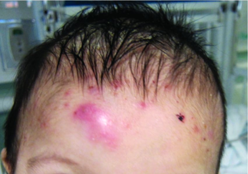
A 7-month-old male presents with pustules and inflamed papules on the scalp and extremities
The bacterial, fungal, and atypical mycobacterial cultures from the lesions performed at the emergency department were all negative.
Pediatric dermatology was consulted and a punch biopsy of one of the lesions was done. Histopathologic examination showed a mixed perifollicular infiltrate of predominantly eosinophils with some neutrophils and associated microabscesses. Periodic acid Schiff and Fite stains failed to reveal any organisms. CD1 immunostain was negative. Fresh tissue cultures for bacteria, fungi, and atypical mycobacteria were negative.
Given the clinical presentation of chronic recurrent sterile pustules on an infant with associated eosinophilia and the reported histopathologic findings, the patient was diagnosed with eosinophilic pustular folliculitis of infancy (EPFI).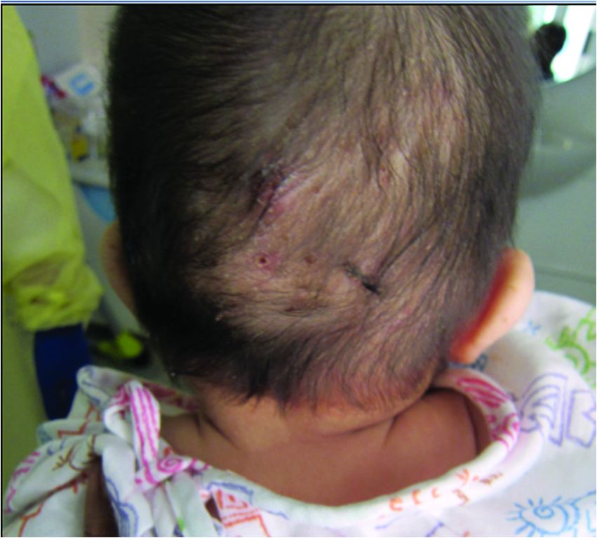
EPFI is a rare and idiopathic cutaneous disorder present in children. About 70% of the cases reported occur in the first 6 month of life and rarely present past 3 years of age. EPF encompasses a group of conditions including the classic adult form, or Ofuji disease. EPF is seen in immunosuppressed patients, mainly HIV positive, and EPF is also seen in infants and children.
In EPFI, males are most commonly affected. The condition presents, as it did in our patient, with recurrent crops of sterile papules and pustules mainly on the scalp, but they can occur in other parts of the body. The lesions go away within a few weeks to months without leaving any scars but it can take months to years to resolve. Histopathologic analysis of the lesions show an eosinophilic infiltrate which can be follicular, perifollicular, or periadnexal with associated flame figures in about 26% of cases.
Aggressive treatment is usually not needed as lesions are self-limited. Lesions can be treated with topical corticosteroids and oral antihistamine medications like cetirizine if symptomatic.
If the lesions start to present during the neonatal period, one may consider in the differential diagnosis, neonatal rashes like transient neonatal pustular melanosis and erythema toxicum neonatorum. Both of these neonatal conditions tend to resolve in the first month of life, compared with EPFI where lesions can come and go for months to years. EPFI lesions can be described as pustules and inflammatory papules, as well as furuncles and vesicles. All of the lesions may be seen in one patient at one time, which will not be typical for transient neonatal pustular melanosis or erythema toxicum. Eosinophils can be seen in erythema toxicum but folliculitis is not present. The inflammatory infiltrate seen in transient neonatal pustular melanosis is polymorphonuclear, not eosinophilic.
Early in the presentation, infectious conditions like staphylococcal or streptococcal folliculitis, cellulitis and furunculosis, tinea capitis, atypical mycobacterial infections, herpes simplex, and parasitic infections like scabies should be considered. In young infants, empiric antibiotic treatment may be started until cultures are finalized. If there is a family history of pruritic papules and pustules, scabies should be considered. A scabies prep can be done to rule out this entity.
Langerhans cell histiocytosis can also present with pustules and papules in early infancy and also has a predilection for the scalp. When this condition is in question, a skin biopsy should be performed which shows a CD1 positive histiocytic infiltrate.
In conclusion, EPFI is a benign rare condition that can present in infants as recurrent pustules and papules, mainly on the scalp, which are self-limited and if symptomatic can be treated with topical corticosteroids and antihistamines.
References
Alonso-Castro L et al. Dermatol Online J. 2012 Oct 15;18(10):6.
Frølunde AS et al. Clin Case Rep. 2021 May 11;9(5):e04167.
Hernández-Martín Á et al. J Am Acad Dermatol. 2013 Jan;68(1):150-5.
A 7-month-old male is brought to the emergency department for evaluation of pustules and inflamed papules on the scalp and extremities for several weeks of duration. The parents report the lesions started about a month prior and he has already been treated with cephalexin, clindamycin, and sulfamethoxazole without any improvement. Cultures sent prior by the child's pediatrician did not reveal any fungus or bacteria. The parents report a low-grade fever for about 3 days.
He was born via natural vaginal delivery with no instrumentation or external monitoring. Mom had prenatal care. Besides the skin lesions, the baby has been healthy and growing well. He has no history of eczema or severe infections. He has not been hospitalized before.
On physical examination the baby was not febrile. On the scalp and forehead, he had diffusely distributed pustules, erythematous papules, and nodules. He also presented with scattered, fine, small, crusted 1-2-mm pink papules on the trunk and extremities. He had no adenopathy or hepatosplenomegaly.
At the emergency department, samples from one of the pustules were sent for bacterial, fungal, and atypical mycobacteria cultures. Laboratory test showed a normal blood count with associated eosinophilia (2.8 x 10⁹ L), and normal liver and kidney function. A head ultrasound showed three ill-defined hypoechoic foci within the scalp.
The patient was admitted for treatment with broad-spectrum antibiotics and dermatology was consulted.
USPSTF releases updated recommendations on skin cancer screening
.
This final recommendation applies to the general public and is not meant for those at higher risk, such as people with a family history of skin cancer or who have any signs or symptoms, such as irregular moles.
“The new recommendations are consistent with those from 2016, and we are unable to balance benefits and harms,” said Task Force member Katrina Donahue, MD, MPH, professor and vice chair of research in the department of family medicine at the University of North Carolina, Chapel Hill. “Unfortunately, there is not enough evidence to recommend for or against screening, and health care professionals should use their judgment when deciding whether or not to screen.”
Dr. Donahue told this news organization that this is a call for more research: “Our recommendations are for patients who present to primary care without symptoms, and after a careful assessment of benefit and harms, we didn’t have evidence to push us towards screening as a benefit. We did look at data from two large screening programs, but they were from Europe and not representative of the U.S. population. They also did not show a benefit for reducing melanoma-related mortality.”
The USPSTF final recommendation statement and corresponding evidence summary have been published online in JAMA, as well as on the USPSTF website.
Skin cancer is the most commonly diagnosed cancer in the United States, but there are different types that vary in their incidence and severity. Basal and squamous cell carcinomas are the most common types of skin cancer, but they infrequently lead to death or substantial morbidity, notes the USPTSF. Melanomas represent about 1% of skin cancer and cause the most skin cancer deaths. An estimated 8,000 individuals in the United States will die of melanoma in 2023.
There are racial differences in melanoma incidence; it is about 30 times more common in White versus Black persons, but disease in persons with darker skin color tends to be diagnosed at a later stage. These disparities may be due to differences in risk factors, access to care, and clinical presentation.
In an accompanying editorial, Maryam M. Asgari, MD, MPH, of the department of dermatology, Massachusetts General Hospital, Boston, and Lori A. Crane, PhD, MPH, of the Colorado School of Public Health, University of Colorado, Aurora, point out that people with darker skin phenotypes also tend to be affected by skin cancers that are not associated with UV radiation, such as acral melanoma, which arises on the palms and soles, and skin cancers that arise in areas of chronic inflammation, such as wounds.

Thus, differences in anatomical distribution of skin cancers in in the various subpopulations needs to be considered when performing skin screening, they write. “Furthermore, while skin cancer risk is lower among people with darker skin pigmentation, survival is often worse for cancers like melanoma, highlighting the potential need for screening.”
“More data are needed, particularly regarding genetic and environmental risk factors for skin cancer in people with darker pigmentation, to help inform guidelines that can be broadly applied to the U.S. population,” add Dr. Asgari and Dr. Crane. “The diversity of the U.S. population extends also to geography, culture, and socioeconomic status, all of which affect skin cancer risk.”
Review of evidence
The USPSTF commissioned a systematic review to evaluate the benefits and harms of screening for skin cancer in asymptomatic adolescents and adults, including evidence for both keratinocyte carcinoma (basal cell carcinoma and squamous cell carcinoma) and cutaneous melanoma.
Foundational evidence showed that the sensitivity of visual skin examination by a clinician to detect melanoma ranged from 40% to 70% and specificity ranged from 86% to 98%. Evidence that evaluated the diagnostic accuracy of visual skin examination to detect keratinocyte carcinoma was limited and inconsistent. There were no new studies reporting on diagnostic accuracy for an asymptomatic screening population.
The USPSTF also reviewed 20 studies in 29 articles (n = 6,053,411). This included three nonrandomized studies evaluating two skin cancer screening programs in Germany, but results were inconsistent. In addition, the ecological and nonrandomized design of the studies limited the conclusions that could be drawn and the applicability to a U.S. population was difficult to assess because of differences in population diversity and health care delivery in the United States.
Other nonrandomized studies that looked at various outcomes, such as harms and stage at diagnosis and melanoma or all-cause mortality, also did not provide sufficient evidence to support screening.
Research is needed
In a second accompanying editorial published in JAMA Dermatology, Adewole S. Adamson, MD, MPP, of the division of dermatology and dermatologic surgery at the University of Texas, Austin, pointed out that unlike other cancer screening programs, such as those for breast, colon, and prostate cancer, skin cancer screening programs are somewhat less organized.

The other programs focus on defined groups of the population, generally with easily identifiable characteristics such as age, sex, and family history, and importantly, there are always defined ages for initiation and halting of screening and intervals for screening frequency. None of these basic screening parameters have been widely adopted among dermatologists in the United States, he wrote. “One important reason why skin cancer screening has remained inconsistent is that it is not covered by Medicare or by many commercial insurance companies,” Dr. Adamson told this news organization. “The test, in this case the skin exam, is often performed as part of a routine dermatology visit.”
Dermatologists should take the lead on this, he said. “Dermatologists should push for a high quality prospective clinical trial of skin cancer screening, preferably in a high-risk population.”
Dr. Donahue agrees that research is needed, as noted in the recommendation. For example, studies are needed demonstrating consistent data of the effects of screening on morbidity and mortality or early detection of skin cancer, and clearer descriptions of skin color and inclusion of a full spectrum of skin colors in study participants. Clinical research is also needed on outcomes in participants that reflect the diversity of the U.S. population.
“I hope funding agencies will be interested in this area of study,” she said. “We put out the whole systematic review and point out the gaps. We need consistent evidence in detecting cancer early and reducing complications from skin cancer.”
The U.S. Congress mandates that the Agency for Healthcare Research and Quality support the operations of the USPSTF.
None of the USPSTF authors report any disclosures. Dr. Asgari reported receiving royalties from UpToDate. Dr. Crane did not make any disclosures. Dr. Adamson reported serving as an expert reviewer for the U.S. Preventive Services Task Force skin cancer screening report, as well as support from the Robert Wood Johnson Foundation, the Dermatology Foundation Public Health Career Development Award, the National Institutes of Health, the American Cancer Society, and Meredith’s Mission for Melanoma.
A version of this article originally appeared on Medscape.com.
.
This final recommendation applies to the general public and is not meant for those at higher risk, such as people with a family history of skin cancer or who have any signs or symptoms, such as irregular moles.
“The new recommendations are consistent with those from 2016, and we are unable to balance benefits and harms,” said Task Force member Katrina Donahue, MD, MPH, professor and vice chair of research in the department of family medicine at the University of North Carolina, Chapel Hill. “Unfortunately, there is not enough evidence to recommend for or against screening, and health care professionals should use their judgment when deciding whether or not to screen.”
Dr. Donahue told this news organization that this is a call for more research: “Our recommendations are for patients who present to primary care without symptoms, and after a careful assessment of benefit and harms, we didn’t have evidence to push us towards screening as a benefit. We did look at data from two large screening programs, but they were from Europe and not representative of the U.S. population. They also did not show a benefit for reducing melanoma-related mortality.”
The USPSTF final recommendation statement and corresponding evidence summary have been published online in JAMA, as well as on the USPSTF website.
Skin cancer is the most commonly diagnosed cancer in the United States, but there are different types that vary in their incidence and severity. Basal and squamous cell carcinomas are the most common types of skin cancer, but they infrequently lead to death or substantial morbidity, notes the USPTSF. Melanomas represent about 1% of skin cancer and cause the most skin cancer deaths. An estimated 8,000 individuals in the United States will die of melanoma in 2023.
There are racial differences in melanoma incidence; it is about 30 times more common in White versus Black persons, but disease in persons with darker skin color tends to be diagnosed at a later stage. These disparities may be due to differences in risk factors, access to care, and clinical presentation.
In an accompanying editorial, Maryam M. Asgari, MD, MPH, of the department of dermatology, Massachusetts General Hospital, Boston, and Lori A. Crane, PhD, MPH, of the Colorado School of Public Health, University of Colorado, Aurora, point out that people with darker skin phenotypes also tend to be affected by skin cancers that are not associated with UV radiation, such as acral melanoma, which arises on the palms and soles, and skin cancers that arise in areas of chronic inflammation, such as wounds.
Thus, differences in anatomical distribution of skin cancers in in the various subpopulations needs to be considered when performing skin screening, they write. “Furthermore, while skin cancer risk is lower among people with darker skin pigmentation, survival is often worse for cancers like melanoma, highlighting the potential need for screening.”
“More data are needed, particularly regarding genetic and environmental risk factors for skin cancer in people with darker pigmentation, to help inform guidelines that can be broadly applied to the U.S. population,” add Dr. Asgari and Dr. Crane. “The diversity of the U.S. population extends also to geography, culture, and socioeconomic status, all of which affect skin cancer risk.”
Review of evidence
The USPSTF commissioned a systematic review to evaluate the benefits and harms of screening for skin cancer in asymptomatic adolescents and adults, including evidence for both keratinocyte carcinoma (basal cell carcinoma and squamous cell carcinoma) and cutaneous melanoma.
Foundational evidence showed that the sensitivity of visual skin examination by a clinician to detect melanoma ranged from 40% to 70% and specificity ranged from 86% to 98%. Evidence that evaluated the diagnostic accuracy of visual skin examination to detect keratinocyte carcinoma was limited and inconsistent. There were no new studies reporting on diagnostic accuracy for an asymptomatic screening population.
The USPSTF also reviewed 20 studies in 29 articles (n = 6,053,411). This included three nonrandomized studies evaluating two skin cancer screening programs in Germany, but results were inconsistent. In addition, the ecological and nonrandomized design of the studies limited the conclusions that could be drawn and the applicability to a U.S. population was difficult to assess because of differences in population diversity and health care delivery in the United States.
Other nonrandomized studies that looked at various outcomes, such as harms and stage at diagnosis and melanoma or all-cause mortality, also did not provide sufficient evidence to support screening.
Research is needed
In a second accompanying editorial published in JAMA Dermatology, Adewole S. Adamson, MD, MPP, of the division of dermatology and dermatologic surgery at the University of Texas, Austin, pointed out that unlike other cancer screening programs, such as those for breast, colon, and prostate cancer, skin cancer screening programs are somewhat less organized.
The other programs focus on defined groups of the population, generally with easily identifiable characteristics such as age, sex, and family history, and importantly, there are always defined ages for initiation and halting of screening and intervals for screening frequency. None of these basic screening parameters have been widely adopted among dermatologists in the United States, he wrote. “One important reason why skin cancer screening has remained inconsistent is that it is not covered by Medicare or by many commercial insurance companies,” Dr. Adamson told this news organization. “The test, in this case the skin exam, is often performed as part of a routine dermatology visit.”
Dermatologists should take the lead on this, he said. “Dermatologists should push for a high quality prospective clinical trial of skin cancer screening, preferably in a high-risk population.”
Dr. Donahue agrees that research is needed, as noted in the recommendation. For example, studies are needed demonstrating consistent data of the effects of screening on morbidity and mortality or early detection of skin cancer, and clearer descriptions of skin color and inclusion of a full spectrum of skin colors in study participants. Clinical research is also needed on outcomes in participants that reflect the diversity of the U.S. population.
“I hope funding agencies will be interested in this area of study,” she said. “We put out the whole systematic review and point out the gaps. We need consistent evidence in detecting cancer early and reducing complications from skin cancer.”
The U.S. Congress mandates that the Agency for Healthcare Research and Quality support the operations of the USPSTF.
None of the USPSTF authors report any disclosures. Dr. Asgari reported receiving royalties from UpToDate. Dr. Crane did not make any disclosures. Dr. Adamson reported serving as an expert reviewer for the U.S. Preventive Services Task Force skin cancer screening report, as well as support from the Robert Wood Johnson Foundation, the Dermatology Foundation Public Health Career Development Award, the National Institutes of Health, the American Cancer Society, and Meredith’s Mission for Melanoma.
A version of this article originally appeared on Medscape.com.
Atopic dermatitis positively linked with the risk for acne
Key clinical point: The overall risk for hospital-diagnosed acne is significantly higher in patients with atopic dermatitis (AD), highlighting the need to address comorbid skin diseases simultaneously along with the management of AD.
Major finding: Patients with AD vs control individuals had a 3-fold higher overall risk for hospital-diagnosed acne at ≤18 years (adjusted odds ratio [aOR] 3.44; 95% CI 3.13-3.78) and ≤30 years (aOR 3.15; 95% CI 2.90-3.42) of age.
Study details: The data come from a retrospective registry study that included 70,584 patients with AD aged ≤18 years at the time of their first AD diagnosis and 270,783 matched control individuals without AD.
Disclosures: This study did not receive any funding. Some authors declared serving as investigators and receiving educational grants, consulting fees, or speaker honoraria from various organizations.
Source: Sinikumpu SP et al. The association between atopic dermatitis and acne: A retrospective Finnish nationwide registry study. Br J Dermatol. 2023 (Mar 22). Doi: 10.1093/bjd/ljad086
Moderate-to-severe atopic dermatitis patients most likely to develop NMSC vs other malignancies
Key clinical point: Among malignancies, including breast cancer, melanoma, lymphoma, and non-melanoma skin cancer (NMSC), patients with moderate-to-severe atopic dermatitis (AD) had the highest incidence rate (IR) for NMSC followed by breast cancer and melanoma; NMSC incidence increased with age among patients with moderate but not severe AD.
Major finding: In patients with moderate and severe AD, the IR per 1000 person-years were 4.6 (95% CI 3.9-5.5) and 5.9 (95% CI 3.8-9.2) for NMSC, 2.2 (95% CI 1.6-3.0) and 0.5 (95% CI 0.1-3.9) for breast cancer, and 0.4 (95% CI 0.2-0.7) and 0.6 (95% CI 0.1-2.3) for melanoma, respectively. The NMSC IR increased with increasing age in patients with moderate AD (18-39 vs ≥65 years: 0.1 [95% CI 0.0-0.7] vs 18.0 [95% CI 13.9-23.2]).
Study details: This retrospective study analyzed the data of 7050 adults with moderate-to-severe AD from the Kaiser Permanente Northern California database.
Disclosures: This study was sponsored by Pfizer Inc. Some authors reported ties with various organizations, including Pfizer.
Source: Hedderson MM et al. Rates of malignancies among patients with moderate to severe atopic dermatitis: A retrospective cohort study. BMJ Open. 2023;13(3):e071172 (Mar 10). Doi: 10.1136/bmjopen-2022-071172
Enhanced topical treatment of infant atopic dermatitis prevents food allergy
Key clinical point: Compared with conventional treatment, enhanced treatment with topical corticosteroids (TCS) significantly reduced the incidence of hen’s egg allergy among infants with atopic dermatitis (AD) but retarded their growth.
Major finding: Infants receiving enhanced vs conventional treatment had a significantly lower incidence of hen’s egg allergy (31.4% vs 41.9%; P = .0028), but demonstrated lower body weight (mean difference −422 g; 95% CI −553 to −292 g) and height (mean difference −0.8 cm; 95% CI −1.22 to −0.33 cm) at 28 weeks of age.
Study details: This randomized controlled trial included 640 infants aged 7-13 weeks with AD who were randomly assigned to receive enhanced therapy (alclometasone dipropionate for the whole face and betamethasone valerate for whole body except face and scalp) followed by maintenance therapy (n = 318) or conventional therapy (alclometasone dipropionate and betamethasone valerate for the affected skin; n = 322).
Disclosures: This study was supported by the Japan Agency for Medical Research and Development (AMED). Some authors reported ties with various organizations, including AMED.
Source: Yamamoto-Hanada K et al on behalf of PACI Study Collaborators. Enhanced early skin treatment for atopic dermatitis in infants reduces food allergy. J Allergy Clin Immunol. 2023 (Mar 22). Doi: 10.1016/j.jaci.2023.03.008
Atopic dermatitis risk in children associated with residential distance from highly trafficked segments
Key clinical point: Children living closer to highly trafficked segments (HTS) are at a greater risk of developing atopic dermatitis (AD).
Major finding: Children living at ≥1,000 vs <500 m from an HTS had 27% lower odds of developing AD (P = .0009). The odds of AD decreased by 21% (P = .0002) with each factor of 10 increase in the distance from an HTS.
Study details: The data come from a retrospective cross-sectional analysis of 7247 children aged 0-18 years with AD and 7247 age- and sex-matched control individuals without AD.
Disclosures: This study was supported by the Department of Pediatrics, Division of Allergy and Clinical Immunology, National Jewish Health, Denver, Colorado, and Eugene F and Easton M Crawford Charitable Lead Unitrust, Chicago, Illinois. D Leung reported ties with various organizations.
Source: Nevid MZ et al. The association of residential distance from highly trafficked roads with atopic dermatitis risk. J Allergy Clin Immunol Pract. 2023 (Mar 20). Doi: 10.1016/j.jaip.2023.03.021
Abrocitinib offers comparable efficacy between adults and adolescents with moderate-to-severe atopic dermatitis
Key clinical point: An induction treatment of 200 mg abrocitinib followed by dose reduction (100 mg) or continuous dosing (200 mg) is efficacious and safe in adults and adolescents with moderate-to-severe atopic dermatitis (AD).
Major finding: In the 200 mg abrocitinib, 100 mg abrocitinib, and placebo arms, similar proportions of adolescents and adults experienced disease flare (14.9% and 16.9%, 42.9% and 38.9%, and 75.5% and 78.0%, respectively) and the Eczema Area and Severity Index response was recaptured by 28.6%, 25.0%, and 52.9% of adolescents and 34.3%, 33.7%, and 58.0% of adults, respectively. The safety profile was consistent in adolescents and adults.
Study details: This post hoc analysis of the JADE REGIMEN study included 246 adolescents (12-17 years) and 987 adults with moderate-to-severe AD who received 200 mg abrocitinib induction treatment; responders were randomly assigned to receive 40-week abrocitinib (200/100 mg) or placebo maintenance treatment and rescue treatment (if disease flared).
Disclosures: Pfizer Inc funded the study. Some authors reported various ties, including employment and stock ownership, with Pfizer or others.
Source: Flohr C et al. Efficacy and safety of abrocitinib monotherapy in adolescents and adults: A post hoc analysis of the phase 3 JAK1 atopic dermatitis efficacy and safety (JADE) REGIMEN clinical trial. J Dermatolog Treat. 2023;1-13 (Apr 10). Doi: 10.1080/09546634.2023.2200866
Maternal atopic dermatitis linked with the risk for childhood- and adult-onset atopic dermatitis
Key clinical point: Maternal atopic dermatitis (AD) significantly increases the risk for both childhood- and adult-onset AD, and active smoking is the main lifestyle risk factor for adult-onset AD.
Major finding: Compared with non-atopic and atopic control individuals, individuals with a maternal AD family history had a significantly higher risk for childhood-onset (adjusted odds ratio [aOR] 4.36, 95% CI 1.18-16.17; and aOR 32.97, 95% CI 4.03-269.68, respectively) and adult-onset AD (aOR 15.79, 95% CI 1.81-137.74; and aOR 34.15, 95% CI 3.15-370.28, respectively) and active smokers had an increased risk for adult-onset AD (aOR 5.54, 95% CI 1.06-29.01; and aOR 4.03, 95% CI 1.20-13.45, respectively).
Study details: This study analyzed the cross-sectional data of 736 adult individuals with childhood-onset (<18 years) or adult-onset (≥18 years) AD and 76 non-atopic and 91 atopic control individuals without AD.
Disclosures: This study was funded by the Christine Kühne-Center for Allergy Research and Education (CK-CARE), Switzerland. Some authors declared various ties, including employment, with CK-CARE or others.
Source: Maintz L et al. Atopic dermatitis: Correlation of distinct risk factors with age of onset in adulthood compared to childhood. Allergy. 2023 (Mar 22). Doi: 10.1111/all.15721
Topical tacrolimus and corticosteroids show similar efficacy and impact on airways in childhood atopic dermatitis
Key clinical point: Long-term topical corticosteroid (TCS) and tacrolimus (TAC) treatments had similar efficacy and impact on airway hyperresponsiveness or inflammation in children with moderate-to-severe atopic dermatitis (AD).
Major finding: At month 36, children treated with TCS and TAC had no significant difference in the mean body surface area (P = .12), mean Eczema Area and Severity Index score (P = .2), mean Investigator’s Global Assessment Score (P = .12), mean transepidermal water loss at eczema site (P = .96) and control site (P = .19), median exhaled nitric oxide level (P = .71), or median bronchial hyperresponsiveness to methacholine (P = .7).
Study details: Findings are from a single-center 3-year follow-up study including 152 children aged 1-3 years with moderate-to-severe AD who were randomly assigned to receive TCS (n = 75) or TAC (n = 77).
Disclosures: This study was supported by the Foundation for Paediatric Research, Finland, Orion Research Foundation, Finland, Orion Pharma, Finland, Astellas Pharma, Japan, and others. Orion and Astellas are commercial manufacturers of the TCS used in this study.
Source: Perälä M et al. Topical tacrolimus versus corticosteroids in childhood moderate-to-severe atopic dermatitis with impact on airways: A long-term randomized open-label study. Clin Exp Dermatol. 2023 (Mar 14). Doi: 10.1093/ced/llad098
Baricitinib allows flexibility in dosing regimens up to 104 weeks in moderate-to-severe atopic dermatitis
Key clinical point: Baricitinib offers the flexibility of dose down-titration and improvement in atopic dermatitis (AD) signs and symptoms up to treatment week 104 in patients with moderate-to-severe AD.
Major finding: Among patients receiving 4 mg baricitinib, 51.2% and 47.6% achieved or maintained a validated Investigator’s Global Assessment for AD score of 0 or 1 and 82.1%, and 73.8% maintained an Eczema Area and Severity Index 75 response at weeks 52 and 104, respectively. The clinical response was maintained after down-titration to 2 mg baricitinib.
Study details: This sub-study of the BREEZE-AD3 trial included 168 patients with moderate-to-severe AD who were responders or partial responders to 4 mg baricitinib in originating studies and were re-assigned (1:1) to receive 4 or 2 mg baricitinib at week 52.
Disclosures: This study was funded by Eli Lilly and Company under license from Incyte Corporation. Some authors reported ties with various organizations, including Eli Lilly. Four authors declared being employees and stockholders of Eli Lilly.
Source: Thyssen JP et al. Maintained improvement in physician- and patient-reported outcomes with baricitinib in adults with moderate-to-severe atopic dermatitis who were treated for up to 104 weeks in a randomized trial. J Dermatolog Treat. 2023;34(1):2190430 (Apr 5). Doi: 10.1080/09546634.2023.2190430