User login
Bringing you the latest news, research and reviews, exclusive interviews, podcasts, quizzes, and more.
div[contains(@class, 'header__large-screen')]
div[contains(@class, 'read-next-article')]
div[contains(@class, 'nav-primary')]
nav[contains(@class, 'nav-primary')]
section[contains(@class, 'footer-nav-section-wrapper')]
footer[@id='footer']
div[contains(@class, 'main-prefix')]
section[contains(@class, 'nav-hidden')]
div[contains(@class, 'ce-card-content')]
nav[contains(@class, 'nav-ce-stack')]
Sea buckthorn: What is it and what is it good for?
To avoid jumping on the bandwagon of another ingredient trend, we sought to examine the scientific background and properties of sea buckthorn oil and it’s utility for the skin.
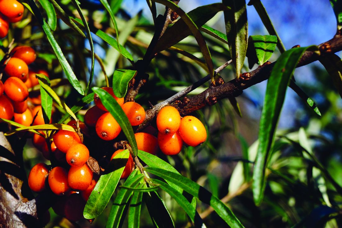
Sea buckthorn (Hippophae rhamnoides) – also known as a Siberian pineapple tree, and as sandthorn, sallowthorn, or seaberry – is a thorny, dioecious shrub (or tree) in the oleaster family. It can grow up to 23 feet high and is found in coastal sea cliff areas and on mountain slopes of Western Europe, and in dry sandy areas of Asia Minor and Central Asia, Siberia, China, and Tibet. Common sea buckthorn flowers in late April and early May, producing a large number of small, green and brown flowers, turning into edible, usually yellow or orange round berries. The berries have a bitter, sour taste and have a mild aroma, resembling that of a pineapple. The fruit contains a small stone that covers an oily seed.
The berries are a source of antioxidant vitamins, flavonoids, and organic acids, and when pressed, produce a juice that separates into three layers: a thick cream (upper layer), a combination of saturated and unsaturated fatty acids (middle layer), and juice that is a source of fat (lower layer). The berries contain mainly vitamin C, but also vitamin A (alpha- and beta-carotene) and a mixture of other carotenoids, as well as varying concentrations of tocopherols (vitamin E), folic acid, and vitamin B complex–group vitamins.

In addition to flavonoids, the berries contain catechins and procyanidins, cyclitols, phospholipids, tannins, sugars (galactose, fructose, xylose), organic acids (maleic acid, oxalic acid, malic acid, tartaric acid), phenolic acids (such as ferulic acid), and fatty oil. The amount of vitamin C content varies with the variety of the plant and where it is found. The oil of sea buckthorn may be extracted from two parts of the plant, with mechanical cold pressing of seeds (up to 12.5% weight as oil content) and fruit pulp (8%-12% oil content).
Among vegetable oils, sea buckthorn fruit oil has the highest content of palmitoleic acid (omega-7).
Fruit and seed oils contain tocotrienols and plant sterols. Pulp sea buckthorn oil has a high carotenoid content, as opposed to seed oil, and in Mongolia, Russia, and China, is used as a topical therapy for skin burns.
Other significant fatty acids found in sea buckthorn oil are saturated fatty acids (palmitic acid and stearic acid) and polyunsaturated fatty acids, which include alpha-linolenic acid (omega-3), gamma-linolenic acid (omega-6), linolic acid (omega-6), oleic acid (omega-9), and eicosanoic acid (omega-9). Gamma-linoleic acid in particular is reduced in dry skin conditions, such as aging and atopic dermatitis. The human body can produce some gamma-linolenic acid, oleic acid, and palmitoleic acid, but not linolic acid and alpha-linolenic acid. The addition of these substances to diet or skin care has been found to be beneficial in improving dryness and the skin barrier.

In addition, linolic acid, a natural component of human sebum, has been noted to be decreased in the sebum of people with acne-prone skin. Preliminary evidence indicates that dietary supplements containing fatty acids such as docosahexaenoic acid, sea buckthorn oil, and hemp seed oil may decrease the severity of atopic dermatitis.
Besides use in topical skin care and cosmetic preparations, sea buckthorn has also been used successfully in the treatment of chronic gastric ulcer disease, inflammation of the vagina and cervix, and cervical erosion. The bark and leaves of sea buckthorn used to be applied to treat diarrhea and dermatologic conditions, while berry oil has been applied topically or taken orally to soften the skin.
In traditional Indian, Chinese, and Tibetan medicines, sea buckthorn berries are used for medicinal purposes, as their ingredients were thought to have a beneficial effect on the function of the alimentary, respiratory, and circulatory systems. Current studies and uses are now confirming their utility experienced over hundreds of years.
Harvesting sea buckthorn fruit is difficult because of dense thorn arrangement among the berries. Therefore, sometimes the only way to obtain fruit is to remove the entire branch of the shrub, which reduces future crops. For this reason berries can only be harvested once every 2 years.
Sea buckthorn has interesting properties and could be of benefit in topical skin care, as long as it is not overharvested or harvested in a way that has a detrimental impact on the environment.
Dr. Wesley and Lily Talakoub, MD, are cocontributors to this column. Dr. Wesley practices dermatology in Beverly Hills, Calif. Dr. Talakoub is in private practice in McLean, Va. This month’s column is by Dr. Wesley. Write to them at [email protected]. They had no relevant disclosures.
References
United States Department of Agriculture. PLANTS Profile for Hippophae rhamnoides (seaberry). 2007.
Zielińska A and Nowak I. Lipids Health Dis. 2017 May 19;16(1):95.
Reynolds KA et al. Int J Dermatol. 2019 Dec;58(12):1371-6.
To avoid jumping on the bandwagon of another ingredient trend, we sought to examine the scientific background and properties of sea buckthorn oil and it’s utility for the skin.
Sea buckthorn (Hippophae rhamnoides) – also known as a Siberian pineapple tree, and as sandthorn, sallowthorn, or seaberry – is a thorny, dioecious shrub (or tree) in the oleaster family. It can grow up to 23 feet high and is found in coastal sea cliff areas and on mountain slopes of Western Europe, and in dry sandy areas of Asia Minor and Central Asia, Siberia, China, and Tibet. Common sea buckthorn flowers in late April and early May, producing a large number of small, green and brown flowers, turning into edible, usually yellow or orange round berries. The berries have a bitter, sour taste and have a mild aroma, resembling that of a pineapple. The fruit contains a small stone that covers an oily seed.
The berries are a source of antioxidant vitamins, flavonoids, and organic acids, and when pressed, produce a juice that separates into three layers: a thick cream (upper layer), a combination of saturated and unsaturated fatty acids (middle layer), and juice that is a source of fat (lower layer). The berries contain mainly vitamin C, but also vitamin A (alpha- and beta-carotene) and a mixture of other carotenoids, as well as varying concentrations of tocopherols (vitamin E), folic acid, and vitamin B complex–group vitamins.
In addition to flavonoids, the berries contain catechins and procyanidins, cyclitols, phospholipids, tannins, sugars (galactose, fructose, xylose), organic acids (maleic acid, oxalic acid, malic acid, tartaric acid), phenolic acids (such as ferulic acid), and fatty oil. The amount of vitamin C content varies with the variety of the plant and where it is found. The oil of sea buckthorn may be extracted from two parts of the plant, with mechanical cold pressing of seeds (up to 12.5% weight as oil content) and fruit pulp (8%-12% oil content).
Among vegetable oils, sea buckthorn fruit oil has the highest content of palmitoleic acid (omega-7).
Fruit and seed oils contain tocotrienols and plant sterols. Pulp sea buckthorn oil has a high carotenoid content, as opposed to seed oil, and in Mongolia, Russia, and China, is used as a topical therapy for skin burns.
Other significant fatty acids found in sea buckthorn oil are saturated fatty acids (palmitic acid and stearic acid) and polyunsaturated fatty acids, which include alpha-linolenic acid (omega-3), gamma-linolenic acid (omega-6), linolic acid (omega-6), oleic acid (omega-9), and eicosanoic acid (omega-9). Gamma-linoleic acid in particular is reduced in dry skin conditions, such as aging and atopic dermatitis. The human body can produce some gamma-linolenic acid, oleic acid, and palmitoleic acid, but not linolic acid and alpha-linolenic acid. The addition of these substances to diet or skin care has been found to be beneficial in improving dryness and the skin barrier.
In addition, linolic acid, a natural component of human sebum, has been noted to be decreased in the sebum of people with acne-prone skin. Preliminary evidence indicates that dietary supplements containing fatty acids such as docosahexaenoic acid, sea buckthorn oil, and hemp seed oil may decrease the severity of atopic dermatitis.
Besides use in topical skin care and cosmetic preparations, sea buckthorn has also been used successfully in the treatment of chronic gastric ulcer disease, inflammation of the vagina and cervix, and cervical erosion. The bark and leaves of sea buckthorn used to be applied to treat diarrhea and dermatologic conditions, while berry oil has been applied topically or taken orally to soften the skin.
In traditional Indian, Chinese, and Tibetan medicines, sea buckthorn berries are used for medicinal purposes, as their ingredients were thought to have a beneficial effect on the function of the alimentary, respiratory, and circulatory systems. Current studies and uses are now confirming their utility experienced over hundreds of years.
Harvesting sea buckthorn fruit is difficult because of dense thorn arrangement among the berries. Therefore, sometimes the only way to obtain fruit is to remove the entire branch of the shrub, which reduces future crops. For this reason berries can only be harvested once every 2 years.
Sea buckthorn has interesting properties and could be of benefit in topical skin care, as long as it is not overharvested or harvested in a way that has a detrimental impact on the environment.
Dr. Wesley and Lily Talakoub, MD, are cocontributors to this column. Dr. Wesley practices dermatology in Beverly Hills, Calif. Dr. Talakoub is in private practice in McLean, Va. This month’s column is by Dr. Wesley. Write to them at [email protected]. They had no relevant disclosures.
References
United States Department of Agriculture. PLANTS Profile for Hippophae rhamnoides (seaberry). 2007.
Zielińska A and Nowak I. Lipids Health Dis. 2017 May 19;16(1):95.
Reynolds KA et al. Int J Dermatol. 2019 Dec;58(12):1371-6.
Brodalumab or guselkumab: What to switch to in plaque psoriasis responding inadequately to ustekinumab?
Key clinical point: Patients with moderate-to-severe plaque psoriasis who respond inadequately to ustekinumab should preferably be switched to brodalumab rather than guselkumab.
Major finding: Patients with an inadequate response to ustekinumab who switched to brodalumab vs guselkumab had higher Psoriasis Area Severity Index (PASI) 100 response rate at week 36 (40.3% vs 20.0%; P < .001) and PASI 90 response rate at weeks 12 (62.7% vs 48.1%; P = .002) and 36 (63.7% vs 51.1%; P = .004).
Study details: This matching-adjusted indirect comparison study employed data for patients with moderate-to-severe plaque psoriasis who responded inadequately to ustekinumab and switched to receive brodalumab (n=121) in AMAGINE-2 and -3 and or to receive guselkumab (n=135) in NAVIGATE, phase 3, trials.
Disclosures: The study was supported by LEO Pharma A/S. P Hampton and M Augustin declared receiving research/educational grants, consultation/speaker honoraria, and/or travel expenses and serving as an advisory board member or clinical trial participant for various companies, including LEO Pharma. E Borg is a current employee and JB Hansen is a former employee of LEO Pharma.
Source: Hampton P et al. Psoriasis (Auckl). 2021 Nov 3. doi: 10.2147/PTT.S326121.
Psoriasis tied with impaired cortisol response to stress
Key clinical point: Patients with psoriasis exhibit significantly lower levels of salivary cortisol despite having higher psychological stress levels, suggesting the existence of an impaired cortisol response to stress.
Major finding: Although patients with psoriasis vs controls had a significantly higher Perceived Stress Scale score (17.2±0.6 vs 15.1±0.8; P = .0289), their salivary cortisol levels were significantly lower (9.6±0.5 vs 14.0±1.1 nmol/L; P < .001).
Study details: This was a cross-sectional study including 126 adult patients with plaque psoriasis who had not received systemic anti-psoriasis therapy since at least 6 months and 116 healthy adult controls.
Disclosures: The study was supported by Fondazione Cariplo. P Gisondi and G Girolomoni declared serving as consultants or speakers for various organizations.
Source: Gisondi P et al. J Pers Med. 2021 Oct 23. doi: 10.3390/jpm11111069.
Real-world persistence of biologics in psoriasis is low and may vary over time
Key clinical point: Given the chronic nature of the disease, antipsoriasis biologics are associated with low median persistence, which is relatively high for interleukin inhibitors vs tumor necrosis factor inhibitors and may change over time.
Major finding: Overall, the median persistence was 23.8 months (95% CI, 21.6-26.2), longest for ustekinumab (49.3 months [95% CI, 38.0-59.1]) and shortest for etanercept (16.3 months [95% CI, 14.5-19.0]). An approximately 50% decrease in the median persistence for ustekinumab, from 62.3 (95% CI, 45.6-∞) months to 32.7 (95% CI, 21.2-49.3) months, occurred between 2010-2011 and 2014-2016.
Study details: The data come from a retrospective cohort study consisting of 2,292 adult patients with psoriasis who had received at least 1 biologic between 2010 and 2018.
Disclosures: The study was sponsored by LEO Pharma. Levin L-Å declared receiving consulting fees from various sources including LEO Pharma, and some of the authors are employees of LEO Pharma.
Source: Schmitt-Egenolf M et al. Dermatol Ther (Heidelb). 2021 Oct 14. doi: 10.1007/s13555-021-00616-7.
AVT02 as effective as reference adalimumab in moderate-to-severe plaque psoriasis
Key clinical point: The biosimilar AVT02 is as efficacious, safe, and immunogenic as its originator, adalimumab, in moderate-to-severe chronic plaque psoriasis.
Major finding: At week 16, AVT02 and originator adalimumab induced comparable percentage improvement in Psoriasis Area Severity Index (91.6% and 89.6%, respectively), with the difference in percentage improvement within the predefined equivalence margins of ±10% (90% CI, −0.76 to 5.29), along with similar safety, tolerability, and immunogenicity profiles, which lasted through week 50.
Study details: This was a phase 3 study including 413 adult patients with moderate-to-severe chronic plaque psoriasis who were initially assigned to receive AVT02 or adalimumab; at week 16, the latter were randomly reassigned to continue receiving adalimumab or switch to AVT02 until week 48.
Disclosures: The study was sponsored by Alvotech Swiss AG. SR Feldman and R Kay disclosed receiving research grants, speaker honoraria, or consultancy fees from various sources, including Alvotech, and some of the authors declared being employees of Alvotech.
Source: Feldman SR et al. BioDrugs. 2021 Oct 16. doi: 10.1007/s40259-021-00502-w.
Ixekizumab bests secukinumab in the real-world race against psoriasis
Key clinical point: Compared with secukinumab, ixekizumab concorded with significantly increased adherence rates and decreased nonpersistence, discontinuation, and switching in biologic-experienced patients with psoriasis.
Major finding: After 18 months of follow-up, ixekizumab was associated with significantly higher rates of high treatment adherence (42% vs 35%; P = .019) and persistence (44.9% vs 36.9%; P = .007) and lower discontinuation (48.4% vs 56.0%; P = .018) and switching (26.6% vs 34.0%; P = .009) rates than secukinumab.
Study details: Findings are from a retrospective observational study consisting of prior biologic-experienced adult patients with psoriasis now receiving either secukinumab (n=780) or ixekizumab (n=411).
Disclosures: The study was supported by Eli Lilly and Company, USA. The lead author declared serving as a scientific advisor/clinical study investigator for various companies including Eli Lilly. Some of the authors are full-time employees or stakeholders of Eli Lilly, and a few others work for an alternative employer, which received compensation from Eli Lilly.
Source: Blauvelt A et al. Dermatol Ther (Heidelb). 2021 Oct 15. doi: 10.1007/s13555-021-00627-4.
Tapinarof cream 1% QD efficacious against extensive plaque psoriasis in phase 2a
Key clinical point: Tapinarof cream 1% applied once daily (QD) displayed significant efficacy after 4 weeks along with good tolerability and limited systemic exposure in patients with extensive plaque psoriasis.
Major finding: At day 29, the mean Psoriasis Area Severity Index score changed significantly with a mean percentage change from baseline of −59.56% (95% CI, −73.53% to −45.59%) and the tapinarof plasma concentration remained below the quantification level (< 50 pg/mL) in the majority (67.9%) of patients; 17 patients reported no irritation and 2 had mild irritation at the application site.
Study details: Findings are from a phase 2a trial involving 21 adult patients with extensive plaque psoriasis (20% or more body surface area involvement) who applied tapinarof cream 1% QD for 29 days.
Disclosures: The study was supported by Dermavant Sciences, Inc. Some of the authors, including the lead author, declared serving as an employee or investigator for Dermavant Sciences, Inc.
Source: Jett JE et al. Am J Clin Dermatol. 2021 Oct 28. doi: 10.1007/s40257-021-00641-4.
Is beta-blocker use in hypertension linked with psoriasis development?
Key clinical point: Clinical avoidance of beta-blockers (BBs) should not be considered a prerequisite for solely avoiding the onset of de novo psoriasis in patients with hypertension.
Major finding: Overall, 0.2% and 0.4% of patients developed de novo psoriasis in the first and second years after BB initiation, which was not significantly different from patients without exposure to BB (P = .77 and P = .96; respectively). The odds of de novo psoriasis were not significantly higher in patients with exposure to BB than those unexposed (odds ratio, 1.00; 95% CI, 0.60-1.67).
Study details: Findings are from a nationwide population-based retrospective cohort study including 105,529 patients aged 19 years or above with hypertension who had not been diagnosed with psoriasis.
Disclosures: The authors did not report any source of funding. No conflict of interests was reported.
Source: Kim YE et al. J Eur Acad Dermatol Venereol. 2021 Oct 9. doi: 10.1111/jdv.17733.
Phase 2a supports infrequent bimekizumab dosing for plaque psoriasis maintenance
Key clinical point: Bimekizumab may be dosed every 8 weeks during maintenance therapy for plaque psoriasis in contrast to the 4-week dosing regimen typically adopted for anti-interleukin-17A biologics.
Major finding: The absolute change in Psoriasis Area Severity Index (PASI) at week 28 in patients receiving an additional bimekizumab dose vs placebo was −19.7 (95% CI, −24.2 to −15.2) vs −10.8 (95% CI, −13.5 to −8.0). Patients receiving placebo vs bimekizumab at week 16 showed a higher reduction in PASI 100 (−34.4% vs −11.7%) and Investigator's Global Assessment 0/1 (−62.5% vs 0.0%) response rates between weeks 16 and 28.
Study details: This was a prospective phase 2a study including 49 patients with moderate-to-severe plaque psoriasis who received bimekizumab at weeks 0 and 4 and were subsequently randomly assigned to receive either placebo or bimekizumab third dose at week 16.
Disclosures: The study was sponsored by UCB Pharma. Most of the authors including the lead author declared serving as employees of UCB Pharma, and some received research grants or consultation fees from various sources including UCB Pharma.
Source: Oliver R et al. Br J Dermatol. 2021 Oct 23. doi: 10.1111/bjd.20827.
Novel CAL/BDP PAD-cream outshines the conventional counterpart
Key clinical point: The novel calcipotriol (CAL)/betamethasone dipropionate (BDP) PAD-cream offered greater benefits than the currently available topical suspension/gel (CAL/BDP TS) in terms of efficacy and patient quality of life along with favorable safety in plaque psoriasis.
Major finding: At 8 weeks, CAL/BDP PAD-cream vs CAL/BDP TS was associated with a significantly higher Physician's Global Assessment success rate (43.2% vs 31.9%; P < .0001), mean percent reduction in modified Psoriasis Area Severity Index (64.6% vs 56.4%; P < .0001), and Dermatology Life Quality Index 0/1 response rate (43.8% vs 34.2%; P = .0005) and no adverse drug reaction with a frequency greater than 1%.
Study details: This is a pooled analysis of 2 phase 3 trials consisting of a combined 1,271 patients with mild-to-moderate plaque psoriasis, treated with either CAL/BDP PAD-cream (n=551), CAL/BDP TS (n=542), or vehicle (n=178).
Disclosures: Both trials were sponsored by MC2 Therapeutics, Denmark. Some of the authors including the lead author received investigator honoraria for phase 3 trials from MC2, and the rest are employees of MC2.
Source: Pinter A et al. J Eur Acad Dermatol Venereol. 2021 Oct 10. doi: 10.1111/jdv.17734.