User login
Resident participation in surgery is safe for patients, study shows
Despite concerns that resident involvement in operations might pose a risk to the patient, the inclusion of general surgery residents in high-risk surgery cases does not negatively impact outcomes, according to a database study of more than 25,000 patients.
Adrienne N. Cobb, MD, of Loyola University Medical Center, Maywood, Ill., and her colleagues used the American College of Surgeons National Surgical Quality Improvement Program (ACS-NSQIP) database (2005-2012) to identify all patients undergoing any of six high-risk procedures on an elective basis.
Dr. Cobb and her colleagues compared outcomes for 25,363 patients who had procedures with and without resident participation: 4,018 and 21,345 patients, respectively. They also evaluated selected outcomes by postgraduate year (PGY). Junior residents were considered as PGY 1-2, senior residents were considered PGY 3, 4, and 5, and participants were considered fellows if they were PGY 6 or higher, according to their report published in the Journal of Surgical Research.
The six procedures assessed were esophagectomy (1,233 patients), open abdominal aortic aneurysm repair (162), laparoscopic paraesophageal resection with Nissen fundoplication (2,316), pancreaticoduodenectomy (Whipple, 10,309), abdominoperineal resection (2,003), and hepatectomy (9,329).
The primary outcome of the study was 30-day mortality with the exposure being resident involvement. Secondary outcomes that had complications included superficial and deep surgical site infection, wound disruption, bleeding requiring transfusion, return to the operating room, pneumonia, unplanned reintubation, pulmonary embolism, acute renal failure, stroke, MI, sepsis, urinary tract infection (UTI), and deep vein thrombosis, operative time, and length of stay.
In both univariate and multivariate analysis, there were no significant differences in mortality between patients who did or did not have procedures in which resident participation at any level of training was involved.
Overall, resident participation did increase the odds of a prolonged operative time (odds ratio, 1.5; 95% confidence interval, 1.1-2.1). Only abdominal perineal resection showed an increased odds risk of having a prolonged operation when residents were involved (OR, 4.3; 95% CI, 1.1-4.8). With regard to these results, the authors commented: “We assert that the additional time spent with trainees in the OR is integral to the production of confident and competent surgeons and does not lead to poorer outcomes for patients. It may, however, lead to increasing costs for the hospital and the patient.”
When risk-adjusted odds were calculated for all the other secondary outcomes and postoperative outcomes tested, UTI was the only one to show a negative impact when residents were involved (OR, 2.3; 95% CI, 1.1-4.8).
The study limitations include the age of the data and the limited number of procedures evaluated, and thus might not be generalizable to a more modern era and other procedures, according to the authors.
“Apart from UTI rates, resident participation did not significantly increase patient morbidity or mortality. Residents should continue to be given active and engaging roles in the OR, even in the most challenging cases,” Dr. Cobb and her colleagues concluded.
The study was funded by the National Institutes of Health. The authors reported that they had no disclosures.
SOURCE: Cobb AN et al. J Surg Res. 2018 Dec; 232:308-17.
Despite concerns that resident involvement in operations might pose a risk to the patient, the inclusion of general surgery residents in high-risk surgery cases does not negatively impact outcomes, according to a database study of more than 25,000 patients.
Adrienne N. Cobb, MD, of Loyola University Medical Center, Maywood, Ill., and her colleagues used the American College of Surgeons National Surgical Quality Improvement Program (ACS-NSQIP) database (2005-2012) to identify all patients undergoing any of six high-risk procedures on an elective basis.
Dr. Cobb and her colleagues compared outcomes for 25,363 patients who had procedures with and without resident participation: 4,018 and 21,345 patients, respectively. They also evaluated selected outcomes by postgraduate year (PGY). Junior residents were considered as PGY 1-2, senior residents were considered PGY 3, 4, and 5, and participants were considered fellows if they were PGY 6 or higher, according to their report published in the Journal of Surgical Research.
The six procedures assessed were esophagectomy (1,233 patients), open abdominal aortic aneurysm repair (162), laparoscopic paraesophageal resection with Nissen fundoplication (2,316), pancreaticoduodenectomy (Whipple, 10,309), abdominoperineal resection (2,003), and hepatectomy (9,329).
The primary outcome of the study was 30-day mortality with the exposure being resident involvement. Secondary outcomes that had complications included superficial and deep surgical site infection, wound disruption, bleeding requiring transfusion, return to the operating room, pneumonia, unplanned reintubation, pulmonary embolism, acute renal failure, stroke, MI, sepsis, urinary tract infection (UTI), and deep vein thrombosis, operative time, and length of stay.
In both univariate and multivariate analysis, there were no significant differences in mortality between patients who did or did not have procedures in which resident participation at any level of training was involved.
Overall, resident participation did increase the odds of a prolonged operative time (odds ratio, 1.5; 95% confidence interval, 1.1-2.1). Only abdominal perineal resection showed an increased odds risk of having a prolonged operation when residents were involved (OR, 4.3; 95% CI, 1.1-4.8). With regard to these results, the authors commented: “We assert that the additional time spent with trainees in the OR is integral to the production of confident and competent surgeons and does not lead to poorer outcomes for patients. It may, however, lead to increasing costs for the hospital and the patient.”
When risk-adjusted odds were calculated for all the other secondary outcomes and postoperative outcomes tested, UTI was the only one to show a negative impact when residents were involved (OR, 2.3; 95% CI, 1.1-4.8).
The study limitations include the age of the data and the limited number of procedures evaluated, and thus might not be generalizable to a more modern era and other procedures, according to the authors.
“Apart from UTI rates, resident participation did not significantly increase patient morbidity or mortality. Residents should continue to be given active and engaging roles in the OR, even in the most challenging cases,” Dr. Cobb and her colleagues concluded.
The study was funded by the National Institutes of Health. The authors reported that they had no disclosures.
SOURCE: Cobb AN et al. J Surg Res. 2018 Dec; 232:308-17.
Despite concerns that resident involvement in operations might pose a risk to the patient, the inclusion of general surgery residents in high-risk surgery cases does not negatively impact outcomes, according to a database study of more than 25,000 patients.
Adrienne N. Cobb, MD, of Loyola University Medical Center, Maywood, Ill., and her colleagues used the American College of Surgeons National Surgical Quality Improvement Program (ACS-NSQIP) database (2005-2012) to identify all patients undergoing any of six high-risk procedures on an elective basis.
Dr. Cobb and her colleagues compared outcomes for 25,363 patients who had procedures with and without resident participation: 4,018 and 21,345 patients, respectively. They also evaluated selected outcomes by postgraduate year (PGY). Junior residents were considered as PGY 1-2, senior residents were considered PGY 3, 4, and 5, and participants were considered fellows if they were PGY 6 or higher, according to their report published in the Journal of Surgical Research.
The six procedures assessed were esophagectomy (1,233 patients), open abdominal aortic aneurysm repair (162), laparoscopic paraesophageal resection with Nissen fundoplication (2,316), pancreaticoduodenectomy (Whipple, 10,309), abdominoperineal resection (2,003), and hepatectomy (9,329).
The primary outcome of the study was 30-day mortality with the exposure being resident involvement. Secondary outcomes that had complications included superficial and deep surgical site infection, wound disruption, bleeding requiring transfusion, return to the operating room, pneumonia, unplanned reintubation, pulmonary embolism, acute renal failure, stroke, MI, sepsis, urinary tract infection (UTI), and deep vein thrombosis, operative time, and length of stay.
In both univariate and multivariate analysis, there were no significant differences in mortality between patients who did or did not have procedures in which resident participation at any level of training was involved.
Overall, resident participation did increase the odds of a prolonged operative time (odds ratio, 1.5; 95% confidence interval, 1.1-2.1). Only abdominal perineal resection showed an increased odds risk of having a prolonged operation when residents were involved (OR, 4.3; 95% CI, 1.1-4.8). With regard to these results, the authors commented: “We assert that the additional time spent with trainees in the OR is integral to the production of confident and competent surgeons and does not lead to poorer outcomes for patients. It may, however, lead to increasing costs for the hospital and the patient.”
When risk-adjusted odds were calculated for all the other secondary outcomes and postoperative outcomes tested, UTI was the only one to show a negative impact when residents were involved (OR, 2.3; 95% CI, 1.1-4.8).
The study limitations include the age of the data and the limited number of procedures evaluated, and thus might not be generalizable to a more modern era and other procedures, according to the authors.
“Apart from UTI rates, resident participation did not significantly increase patient morbidity or mortality. Residents should continue to be given active and engaging roles in the OR, even in the most challenging cases,” Dr. Cobb and her colleagues concluded.
The study was funded by the National Institutes of Health. The authors reported that they had no disclosures.
SOURCE: Cobb AN et al. J Surg Res. 2018 Dec; 232:308-17.
FROM THE JOURNAL OF SURGICAL RESEARCH
Key clinical point: Resident participation in surgery during training was safe for the patients involved.
Major finding: UTI was the only secondary outcome to show a negative impact when residents were involved in surgery (OR, 2.3; 95% CI, 1.1-4.8).
Study details: A retrospective database analysis of six complex procedures involving more than 25,000 surgeries during 2005-2012.
Disclosures: The authors reported that they had no disclosures.
Source: Cobb AN et al. J Surg Res 2018 Dec;232:308-17.
Meeting preview: Mental health ED visits spiked between 2009 and 2015
Between 2009 and 2015, the number of emergency department visits related to mental health increased 56% among pediatric patients and 41% among adults, according to data that will be presented at the annual meeting of the American College of Emergency Physicians.

“The current mental health system is in crisis and the impact on emergency departments continues to increase,” lead researcher Genevieve Santillanes, MD, said in an interview in advance of the meeting. “We are seeing more patients with mental health disorders and when patients with mental health disorders require inpatient care, they board in the ED for long periods of time.”
Look to our onsite meeting coverage October 1-4 for additional details on this study and more news from ACEP18!
Between 2009 and 2015, the number of emergency department visits related to mental health increased 56% among pediatric patients and 41% among adults, according to data that will be presented at the annual meeting of the American College of Emergency Physicians.
“The current mental health system is in crisis and the impact on emergency departments continues to increase,” lead researcher Genevieve Santillanes, MD, said in an interview in advance of the meeting. “We are seeing more patients with mental health disorders and when patients with mental health disorders require inpatient care, they board in the ED for long periods of time.”
Look to our onsite meeting coverage October 1-4 for additional details on this study and more news from ACEP18!
Between 2009 and 2015, the number of emergency department visits related to mental health increased 56% among pediatric patients and 41% among adults, according to data that will be presented at the annual meeting of the American College of Emergency Physicians.
“The current mental health system is in crisis and the impact on emergency departments continues to increase,” lead researcher Genevieve Santillanes, MD, said in an interview in advance of the meeting. “We are seeing more patients with mental health disorders and when patients with mental health disorders require inpatient care, they board in the ED for long periods of time.”
Look to our onsite meeting coverage October 1-4 for additional details on this study and more news from ACEP18!
REPORTING FROM ACEP18
ACEP18: Toxicology updates & emerging trends
The dangers of synthetic drugs and heroin, and the opioid epidemic, are hitting emergency departments hard. A lack of guidelines and poor presentation to follow-up care make treatment decisions difficult. Pockets of outbreaks emerge when dangerous toxins are added to already dangerous drugs. One such regional outbreak occurred in March 2018 when synthetic cannabinoids laced with superwarfarin were led to 150 patients presenting to hospitals with severe coagulopathy in Illinois.
ACEP18 will feature a plethora of toxicology presentations and workshops, such as “Emergency Toxicology: Emerging Trends – Cases in Poisoning Management” on Tuesday, Oct. 2 at 8 am, led my Patrick M. Lank, MD, FACEP. Dr. Lark, an emergency medicine specialist in Chicago, will also be leading “Critical Update in Toxicology 2018" on Monday, Oct. 1, at 12:30 pm and “FAST FACTS: High-Yield Toxicology,” on Monday, at 4:30 pm.
The dangers of synthetic drugs and heroin, and the opioid epidemic, are hitting emergency departments hard. A lack of guidelines and poor presentation to follow-up care make treatment decisions difficult. Pockets of outbreaks emerge when dangerous toxins are added to already dangerous drugs. One such regional outbreak occurred in March 2018 when synthetic cannabinoids laced with superwarfarin were led to 150 patients presenting to hospitals with severe coagulopathy in Illinois.
ACEP18 will feature a plethora of toxicology presentations and workshops, such as “Emergency Toxicology: Emerging Trends – Cases in Poisoning Management” on Tuesday, Oct. 2 at 8 am, led my Patrick M. Lank, MD, FACEP. Dr. Lark, an emergency medicine specialist in Chicago, will also be leading “Critical Update in Toxicology 2018" on Monday, Oct. 1, at 12:30 pm and “FAST FACTS: High-Yield Toxicology,” on Monday, at 4:30 pm.
The dangers of synthetic drugs and heroin, and the opioid epidemic, are hitting emergency departments hard. A lack of guidelines and poor presentation to follow-up care make treatment decisions difficult. Pockets of outbreaks emerge when dangerous toxins are added to already dangerous drugs. One such regional outbreak occurred in March 2018 when synthetic cannabinoids laced with superwarfarin were led to 150 patients presenting to hospitals with severe coagulopathy in Illinois.
ACEP18 will feature a plethora of toxicology presentations and workshops, such as “Emergency Toxicology: Emerging Trends – Cases in Poisoning Management” on Tuesday, Oct. 2 at 8 am, led my Patrick M. Lank, MD, FACEP. Dr. Lark, an emergency medicine specialist in Chicago, will also be leading “Critical Update in Toxicology 2018" on Monday, Oct. 1, at 12:30 pm and “FAST FACTS: High-Yield Toxicology,” on Monday, at 4:30 pm.
REPORTING FROM ACEP18
Antithrombotic strategy 1 year after stenting in AF patients leans toward oral anticoagulant alone
SAN DIEGO – In patients with atrial fibrillation and stable coronary artery disease, a randomized trial of oral anticoagulation alone versus an anticoagulant plus a single antiplatelet agent failed to establish noninferiority of the single-agent approach. The trial could not demonstrate its primary endpoint of all-cause death, myocardial infarction, stroke, or systemic embolism.
But a secondary endpoint that included major bleeding did demonstrate equivalence, leading the researchers to suggest that oral anticoagulation (OAC) alone may be sufficient in most patients.
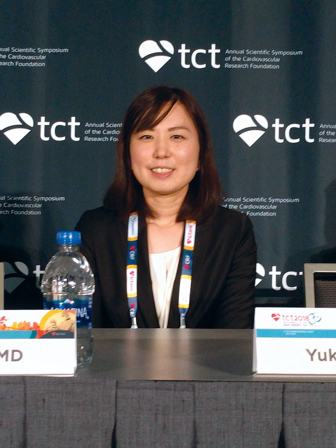
“Combined OAC and single antiplatelet therapy is unlikely to provide net clinical benefit over OAC alone. Thus, OAC alone might be reasonable for AF [atrial fibrillation] patients beyond 1 year after coronary stenting,” Yukiko Nakano, MD, of Kyoto (Japan) University Graduate School of Medicine, said during a press conference at the Transcatheter Cardiovascular Therapeutics annual meeting, sponsored by the Cardiovascular Research Foundation. The report was simultaneously published Sept. 24 in Circulation (doi: 10.1161/CIRCULATIONAHA.118.036768).
The results support the European Society of Cardiology practice guidelines, which recommend lifelong OAC without antiplatelet therapy. But physicians often continue to prescribe antiplatelet agents out of concern that stent thrombosis could occur if the therapy is stopped.
The study was stopped prematurely because of insufficient recruitment, which may have contributed to the failed primary endpoint. It’s a shortcoming that befalls many such studies, perhaps because cardiologists tend to be set in their ways when it comes to treatment of patients after a stent implant. “Cardiologists just think they know the answer, and they don’t want to expose their patients (to a clinical trial). They say, ‘I have my patients on whatever regimen. It seems to be working, and they’re not bleeding, so I don’t want to change it.’ This study suggests that we probably can stop one of the two (antiplatelet drugs) and get by with a single agent, and in this case they got by with no agent (in the monotherapy arm),” said C. Michael Gibson, MD, chief of clinical research in the division of cardiology at Beth Israel Deaconess Medical Center, Boston, who was a discussant at the press conference.
The study recruited 696 patients who were receiving OAC plus single antiplatelet therapy (SAPT) 1 year after receiving a stent. They were randomized 1:1 to continue combined therapy or to stop SAPT and then followed for a median of 2.5 years. A total of 74% of patients who received OAC alone were taking warfarin, while 26% were taking a direct oral anticoagulant. The SAPT group took aspirin or clopidogrel.
Overall, 15.7% of OAC patients experienced the primary endpoint, compared with 13.6% of the combined group (noninferiority P = .20). None of the individual components of the primary endpoint were statistically significantly different between the groups. International Society on Thrombosis and Haemostasis major bleeding and Thrombolysis in Myocardial Infarction major bleeding trended in favor of OAC alone. The secondary endpoint (primary endpoint plus major bleeding) achieved noninferiority, occurring in 19.5% of the OAC group and 19.4% of the combined therapy group (noninferiority P = .016; superiority P = .96).
Daiichi-Sankyo funded the trial. Dr. Nakano had no conflicts of interest. Dr. Gibson reported numerous financial ties to pharmaceutical companies, including Daiichi-Sankyo.
SAN DIEGO – In patients with atrial fibrillation and stable coronary artery disease, a randomized trial of oral anticoagulation alone versus an anticoagulant plus a single antiplatelet agent failed to establish noninferiority of the single-agent approach. The trial could not demonstrate its primary endpoint of all-cause death, myocardial infarction, stroke, or systemic embolism.
But a secondary endpoint that included major bleeding did demonstrate equivalence, leading the researchers to suggest that oral anticoagulation (OAC) alone may be sufficient in most patients.
“Combined OAC and single antiplatelet therapy is unlikely to provide net clinical benefit over OAC alone. Thus, OAC alone might be reasonable for AF [atrial fibrillation] patients beyond 1 year after coronary stenting,” Yukiko Nakano, MD, of Kyoto (Japan) University Graduate School of Medicine, said during a press conference at the Transcatheter Cardiovascular Therapeutics annual meeting, sponsored by the Cardiovascular Research Foundation. The report was simultaneously published Sept. 24 in Circulation (doi: 10.1161/CIRCULATIONAHA.118.036768).
The results support the European Society of Cardiology practice guidelines, which recommend lifelong OAC without antiplatelet therapy. But physicians often continue to prescribe antiplatelet agents out of concern that stent thrombosis could occur if the therapy is stopped.
The study was stopped prematurely because of insufficient recruitment, which may have contributed to the failed primary endpoint. It’s a shortcoming that befalls many such studies, perhaps because cardiologists tend to be set in their ways when it comes to treatment of patients after a stent implant. “Cardiologists just think they know the answer, and they don’t want to expose their patients (to a clinical trial). They say, ‘I have my patients on whatever regimen. It seems to be working, and they’re not bleeding, so I don’t want to change it.’ This study suggests that we probably can stop one of the two (antiplatelet drugs) and get by with a single agent, and in this case they got by with no agent (in the monotherapy arm),” said C. Michael Gibson, MD, chief of clinical research in the division of cardiology at Beth Israel Deaconess Medical Center, Boston, who was a discussant at the press conference.
The study recruited 696 patients who were receiving OAC plus single antiplatelet therapy (SAPT) 1 year after receiving a stent. They were randomized 1:1 to continue combined therapy or to stop SAPT and then followed for a median of 2.5 years. A total of 74% of patients who received OAC alone were taking warfarin, while 26% were taking a direct oral anticoagulant. The SAPT group took aspirin or clopidogrel.
Overall, 15.7% of OAC patients experienced the primary endpoint, compared with 13.6% of the combined group (noninferiority P = .20). None of the individual components of the primary endpoint were statistically significantly different between the groups. International Society on Thrombosis and Haemostasis major bleeding and Thrombolysis in Myocardial Infarction major bleeding trended in favor of OAC alone. The secondary endpoint (primary endpoint plus major bleeding) achieved noninferiority, occurring in 19.5% of the OAC group and 19.4% of the combined therapy group (noninferiority P = .016; superiority P = .96).
Daiichi-Sankyo funded the trial. Dr. Nakano had no conflicts of interest. Dr. Gibson reported numerous financial ties to pharmaceutical companies, including Daiichi-Sankyo.
SAN DIEGO – In patients with atrial fibrillation and stable coronary artery disease, a randomized trial of oral anticoagulation alone versus an anticoagulant plus a single antiplatelet agent failed to establish noninferiority of the single-agent approach. The trial could not demonstrate its primary endpoint of all-cause death, myocardial infarction, stroke, or systemic embolism.
But a secondary endpoint that included major bleeding did demonstrate equivalence, leading the researchers to suggest that oral anticoagulation (OAC) alone may be sufficient in most patients.
“Combined OAC and single antiplatelet therapy is unlikely to provide net clinical benefit over OAC alone. Thus, OAC alone might be reasonable for AF [atrial fibrillation] patients beyond 1 year after coronary stenting,” Yukiko Nakano, MD, of Kyoto (Japan) University Graduate School of Medicine, said during a press conference at the Transcatheter Cardiovascular Therapeutics annual meeting, sponsored by the Cardiovascular Research Foundation. The report was simultaneously published Sept. 24 in Circulation (doi: 10.1161/CIRCULATIONAHA.118.036768).
The results support the European Society of Cardiology practice guidelines, which recommend lifelong OAC without antiplatelet therapy. But physicians often continue to prescribe antiplatelet agents out of concern that stent thrombosis could occur if the therapy is stopped.
The study was stopped prematurely because of insufficient recruitment, which may have contributed to the failed primary endpoint. It’s a shortcoming that befalls many such studies, perhaps because cardiologists tend to be set in their ways when it comes to treatment of patients after a stent implant. “Cardiologists just think they know the answer, and they don’t want to expose their patients (to a clinical trial). They say, ‘I have my patients on whatever regimen. It seems to be working, and they’re not bleeding, so I don’t want to change it.’ This study suggests that we probably can stop one of the two (antiplatelet drugs) and get by with a single agent, and in this case they got by with no agent (in the monotherapy arm),” said C. Michael Gibson, MD, chief of clinical research in the division of cardiology at Beth Israel Deaconess Medical Center, Boston, who was a discussant at the press conference.
The study recruited 696 patients who were receiving OAC plus single antiplatelet therapy (SAPT) 1 year after receiving a stent. They were randomized 1:1 to continue combined therapy or to stop SAPT and then followed for a median of 2.5 years. A total of 74% of patients who received OAC alone were taking warfarin, while 26% were taking a direct oral anticoagulant. The SAPT group took aspirin or clopidogrel.
Overall, 15.7% of OAC patients experienced the primary endpoint, compared with 13.6% of the combined group (noninferiority P = .20). None of the individual components of the primary endpoint were statistically significantly different between the groups. International Society on Thrombosis and Haemostasis major bleeding and Thrombolysis in Myocardial Infarction major bleeding trended in favor of OAC alone. The secondary endpoint (primary endpoint plus major bleeding) achieved noninferiority, occurring in 19.5% of the OAC group and 19.4% of the combined therapy group (noninferiority P = .016; superiority P = .96).
Daiichi-Sankyo funded the trial. Dr. Nakano had no conflicts of interest. Dr. Gibson reported numerous financial ties to pharmaceutical companies, including Daiichi-Sankyo.
REPORTING FROM TCT 2018
Key clinical point:
Major finding: A measure that included cardiac events plus major bleeding showed an oral anticoagulant alone was noninferior to an OAC plus antiplatelet therapy.
Study details: Randomized, controlled trial of 696 patients.
Disclosures: Daiichi-Sankyo funded the trial. Dr. Nakano had no conflicts of interest. Dr. Gibson reported numerous financial ties to pharmaceutical companies, including Daiichi-Sankyo.
PCI bests medical therapy for FFR grey zone stable angina
PARIS – Patients with stable angina and a fractional flow reserve (FFR) value in the grey zone of 0.75-0.81 experienced a significant reduction in myocardial ischemia and substantially greater quality of life improvement if they were randomized to percutaneous coronary intervention (PCI) plus optimal medical therapy than to optimal medical therapy alone in the Scottish Grey-zone FFR Study.
The Grey-zone FFR Study was a single-center, prospective, unblinded, randomized trial that included 100 patients with stable angina, single-vessel disease, and a fractional flow reserve in the grey zone of 0.75-0.81. While broad consensus exists that an FFR below 0.75 constitutes evidence of a hemodynamically significant coronary lesion warranting revascularization and an FFR greater than 0.80 indicates a lesion isn’t functionally significant and therefore PCI can safely be deferred, there has been uncertainty on what to do about lesions in the grey zone, which are frequently encountered in the cardiac catheterization laboratory.
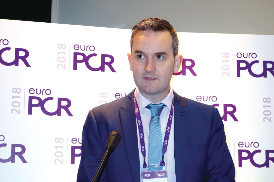
“In my clinical practice, I tend to go ahead with PCI for patients in the grey zone if I felt it was clinically feasible and safe to do so, particularly if I was worried about their lesion morphology,” Barry Hennigan, MD, said in response to questions after presenting the results at the annual meeting of the European Association of Percutaneous Cardiovascular Interventions. “If it’s a proximal LAD [left anterior descending artery] lesion and it’s a grey zone patient, particularly if it’s a lesion morphology that you’re not comfortable with, I think you need to be very careful before you defer a case.”
The twin purposes of the Scottish study were to define the prevalence of major ischemia by stress MRI and invasive flow assessment via a pressure wire in grey zone patients – something which hadn’t been done before – and to determine if PCI deferral in such patients is appropriate in terms of symptom control. The primary outcome was change in angina severity at 3 months follow-up using the Seattle Angina Questionnaire (SAQ).
Scores on two of the five domains of the SAQ – anginal frequency and quality of life – were significantly improved in the PCI group. Anginal frequency scores improved by a mean of 20.58 points in the PCI plus optimal medical therapy (OMT) group, compared with a 9.39-point improvement with OMT alone. Quality of life scores improved by 24.04 points in the PCI group versus 9.39 points in controls, said Dr. Hennigan, an interventional cardiologist at the University of Glasgow and Golden Jubilee National Hospital. Scores in the other three SAQ domains – physical limitations, anginal stability, and treatment satisfaction – didn’t differ significantly between the two study arms, although consistently greater improvements were seen in the PCI group.
Baseline stress perfusion MRI as assessed by two blinded observers demonstrated that 17.4% of patients with stable angina and a grey zone FFR had major ischemia, while any ischemia – major or minor – was present in 24.4%. Follow-up scans at 3 months showed a roughly 50% reduction in the prevalence of ischemia in the PCI group, with 7.3% of treated patients still having major ischemia and 12.2% having any ischemia.
Also, 28% of participants had evidence of ischemia at baseline based upon their coronary flow reserve measurements and 8% had a hyperemic stenosis resistance measurement indicative of ischemia. So the FFR grey zone encompasses a range of cardiovascular risks.
In the PCI plus OMT group, 89% of patients (eight of nine) with baseline ischemia on stress MRI had a greater than 10-point improvement in quality of life scores on the SAQ at follow-up in contrast to 53% of patients without ischemia, which made for a statistically significant difference. An improvement of that magnitude is generally considered clinically meaningful. In contrast, in the OMT-only group, 9 of 14 patients with baseline ischemia (64.2%) had a greater than 10-point quality of life improvement, which wasn’t significantly different from the 45.5% improvement rate in patients with no ischemia.
The lessons? Grey zone patients who benefit most from prompt revascularization are those with demonstrable ischemia. In addition, roughly half of grey zone patients with stable angina will improve their quality of life scores by more than 10 points with OMT alone regardless of the presence of myocardial ischemia or not.
Dr. Hennigan was repeatedly asked how he reconciles the results of the grey zone study with those of the much-discussed ORBITA trial, the first and only randomized trial of real versus sham PCI in patients with stable angina. ORBITA didn’t find a significant quality of life advantage for real PCI over sham PCI.
“It is quite possible that a lot of the effect that we saw in our PCI group was placebo related,” he conceded. “However, we do have objective evidence that we reduced ischemia on MRI. Also, 29% of ORBITA patients had an FFR above 0.8, whereas nearly all our patients were below that threshold. So we perhaps had more prevalent ischemia than the ORBITA cohort.”
Also informative is a comparison of SAQ scores at follow-up in the sham PCI ORBITA control group versus the grey zone Scottish PCI group, Dr. Hennigan continued. The Scottish PCI group had a mean 20.6-point improvement in anginal frequency scores while on an average of 1.3 antianginal medications, compared with a 9.6-point improvement in ORBITA patients on 2.9 drugs. The grey zone group who got PCI plus OMT also had a mean 16.1-point improvement in the SAQ physical limitations domain, versus a 5.0-point improvement in the ORBITA controls.
The Grey-zone FFR Study was supported by the British Heart Foundation. Dr. Hennigan reported having no financial conflicts of interest.
PARIS – Patients with stable angina and a fractional flow reserve (FFR) value in the grey zone of 0.75-0.81 experienced a significant reduction in myocardial ischemia and substantially greater quality of life improvement if they were randomized to percutaneous coronary intervention (PCI) plus optimal medical therapy than to optimal medical therapy alone in the Scottish Grey-zone FFR Study.
The Grey-zone FFR Study was a single-center, prospective, unblinded, randomized trial that included 100 patients with stable angina, single-vessel disease, and a fractional flow reserve in the grey zone of 0.75-0.81. While broad consensus exists that an FFR below 0.75 constitutes evidence of a hemodynamically significant coronary lesion warranting revascularization and an FFR greater than 0.80 indicates a lesion isn’t functionally significant and therefore PCI can safely be deferred, there has been uncertainty on what to do about lesions in the grey zone, which are frequently encountered in the cardiac catheterization laboratory.
“In my clinical practice, I tend to go ahead with PCI for patients in the grey zone if I felt it was clinically feasible and safe to do so, particularly if I was worried about their lesion morphology,” Barry Hennigan, MD, said in response to questions after presenting the results at the annual meeting of the European Association of Percutaneous Cardiovascular Interventions. “If it’s a proximal LAD [left anterior descending artery] lesion and it’s a grey zone patient, particularly if it’s a lesion morphology that you’re not comfortable with, I think you need to be very careful before you defer a case.”
The twin purposes of the Scottish study were to define the prevalence of major ischemia by stress MRI and invasive flow assessment via a pressure wire in grey zone patients – something which hadn’t been done before – and to determine if PCI deferral in such patients is appropriate in terms of symptom control. The primary outcome was change in angina severity at 3 months follow-up using the Seattle Angina Questionnaire (SAQ).
Scores on two of the five domains of the SAQ – anginal frequency and quality of life – were significantly improved in the PCI group. Anginal frequency scores improved by a mean of 20.58 points in the PCI plus optimal medical therapy (OMT) group, compared with a 9.39-point improvement with OMT alone. Quality of life scores improved by 24.04 points in the PCI group versus 9.39 points in controls, said Dr. Hennigan, an interventional cardiologist at the University of Glasgow and Golden Jubilee National Hospital. Scores in the other three SAQ domains – physical limitations, anginal stability, and treatment satisfaction – didn’t differ significantly between the two study arms, although consistently greater improvements were seen in the PCI group.
Baseline stress perfusion MRI as assessed by two blinded observers demonstrated that 17.4% of patients with stable angina and a grey zone FFR had major ischemia, while any ischemia – major or minor – was present in 24.4%. Follow-up scans at 3 months showed a roughly 50% reduction in the prevalence of ischemia in the PCI group, with 7.3% of treated patients still having major ischemia and 12.2% having any ischemia.
Also, 28% of participants had evidence of ischemia at baseline based upon their coronary flow reserve measurements and 8% had a hyperemic stenosis resistance measurement indicative of ischemia. So the FFR grey zone encompasses a range of cardiovascular risks.
In the PCI plus OMT group, 89% of patients (eight of nine) with baseline ischemia on stress MRI had a greater than 10-point improvement in quality of life scores on the SAQ at follow-up in contrast to 53% of patients without ischemia, which made for a statistically significant difference. An improvement of that magnitude is generally considered clinically meaningful. In contrast, in the OMT-only group, 9 of 14 patients with baseline ischemia (64.2%) had a greater than 10-point quality of life improvement, which wasn’t significantly different from the 45.5% improvement rate in patients with no ischemia.
The lessons? Grey zone patients who benefit most from prompt revascularization are those with demonstrable ischemia. In addition, roughly half of grey zone patients with stable angina will improve their quality of life scores by more than 10 points with OMT alone regardless of the presence of myocardial ischemia or not.
Dr. Hennigan was repeatedly asked how he reconciles the results of the grey zone study with those of the much-discussed ORBITA trial, the first and only randomized trial of real versus sham PCI in patients with stable angina. ORBITA didn’t find a significant quality of life advantage for real PCI over sham PCI.
“It is quite possible that a lot of the effect that we saw in our PCI group was placebo related,” he conceded. “However, we do have objective evidence that we reduced ischemia on MRI. Also, 29% of ORBITA patients had an FFR above 0.8, whereas nearly all our patients were below that threshold. So we perhaps had more prevalent ischemia than the ORBITA cohort.”
Also informative is a comparison of SAQ scores at follow-up in the sham PCI ORBITA control group versus the grey zone Scottish PCI group, Dr. Hennigan continued. The Scottish PCI group had a mean 20.6-point improvement in anginal frequency scores while on an average of 1.3 antianginal medications, compared with a 9.6-point improvement in ORBITA patients on 2.9 drugs. The grey zone group who got PCI plus OMT also had a mean 16.1-point improvement in the SAQ physical limitations domain, versus a 5.0-point improvement in the ORBITA controls.
The Grey-zone FFR Study was supported by the British Heart Foundation. Dr. Hennigan reported having no financial conflicts of interest.
PARIS – Patients with stable angina and a fractional flow reserve (FFR) value in the grey zone of 0.75-0.81 experienced a significant reduction in myocardial ischemia and substantially greater quality of life improvement if they were randomized to percutaneous coronary intervention (PCI) plus optimal medical therapy than to optimal medical therapy alone in the Scottish Grey-zone FFR Study.
The Grey-zone FFR Study was a single-center, prospective, unblinded, randomized trial that included 100 patients with stable angina, single-vessel disease, and a fractional flow reserve in the grey zone of 0.75-0.81. While broad consensus exists that an FFR below 0.75 constitutes evidence of a hemodynamically significant coronary lesion warranting revascularization and an FFR greater than 0.80 indicates a lesion isn’t functionally significant and therefore PCI can safely be deferred, there has been uncertainty on what to do about lesions in the grey zone, which are frequently encountered in the cardiac catheterization laboratory.
“In my clinical practice, I tend to go ahead with PCI for patients in the grey zone if I felt it was clinically feasible and safe to do so, particularly if I was worried about their lesion morphology,” Barry Hennigan, MD, said in response to questions after presenting the results at the annual meeting of the European Association of Percutaneous Cardiovascular Interventions. “If it’s a proximal LAD [left anterior descending artery] lesion and it’s a grey zone patient, particularly if it’s a lesion morphology that you’re not comfortable with, I think you need to be very careful before you defer a case.”
The twin purposes of the Scottish study were to define the prevalence of major ischemia by stress MRI and invasive flow assessment via a pressure wire in grey zone patients – something which hadn’t been done before – and to determine if PCI deferral in such patients is appropriate in terms of symptom control. The primary outcome was change in angina severity at 3 months follow-up using the Seattle Angina Questionnaire (SAQ).
Scores on two of the five domains of the SAQ – anginal frequency and quality of life – were significantly improved in the PCI group. Anginal frequency scores improved by a mean of 20.58 points in the PCI plus optimal medical therapy (OMT) group, compared with a 9.39-point improvement with OMT alone. Quality of life scores improved by 24.04 points in the PCI group versus 9.39 points in controls, said Dr. Hennigan, an interventional cardiologist at the University of Glasgow and Golden Jubilee National Hospital. Scores in the other three SAQ domains – physical limitations, anginal stability, and treatment satisfaction – didn’t differ significantly between the two study arms, although consistently greater improvements were seen in the PCI group.
Baseline stress perfusion MRI as assessed by two blinded observers demonstrated that 17.4% of patients with stable angina and a grey zone FFR had major ischemia, while any ischemia – major or minor – was present in 24.4%. Follow-up scans at 3 months showed a roughly 50% reduction in the prevalence of ischemia in the PCI group, with 7.3% of treated patients still having major ischemia and 12.2% having any ischemia.
Also, 28% of participants had evidence of ischemia at baseline based upon their coronary flow reserve measurements and 8% had a hyperemic stenosis resistance measurement indicative of ischemia. So the FFR grey zone encompasses a range of cardiovascular risks.
In the PCI plus OMT group, 89% of patients (eight of nine) with baseline ischemia on stress MRI had a greater than 10-point improvement in quality of life scores on the SAQ at follow-up in contrast to 53% of patients without ischemia, which made for a statistically significant difference. An improvement of that magnitude is generally considered clinically meaningful. In contrast, in the OMT-only group, 9 of 14 patients with baseline ischemia (64.2%) had a greater than 10-point quality of life improvement, which wasn’t significantly different from the 45.5% improvement rate in patients with no ischemia.
The lessons? Grey zone patients who benefit most from prompt revascularization are those with demonstrable ischemia. In addition, roughly half of grey zone patients with stable angina will improve their quality of life scores by more than 10 points with OMT alone regardless of the presence of myocardial ischemia or not.
Dr. Hennigan was repeatedly asked how he reconciles the results of the grey zone study with those of the much-discussed ORBITA trial, the first and only randomized trial of real versus sham PCI in patients with stable angina. ORBITA didn’t find a significant quality of life advantage for real PCI over sham PCI.
“It is quite possible that a lot of the effect that we saw in our PCI group was placebo related,” he conceded. “However, we do have objective evidence that we reduced ischemia on MRI. Also, 29% of ORBITA patients had an FFR above 0.8, whereas nearly all our patients were below that threshold. So we perhaps had more prevalent ischemia than the ORBITA cohort.”
Also informative is a comparison of SAQ scores at follow-up in the sham PCI ORBITA control group versus the grey zone Scottish PCI group, Dr. Hennigan continued. The Scottish PCI group had a mean 20.6-point improvement in anginal frequency scores while on an average of 1.3 antianginal medications, compared with a 9.6-point improvement in ORBITA patients on 2.9 drugs. The grey zone group who got PCI plus OMT also had a mean 16.1-point improvement in the SAQ physical limitations domain, versus a 5.0-point improvement in the ORBITA controls.
The Grey-zone FFR Study was supported by the British Heart Foundation. Dr. Hennigan reported having no financial conflicts of interest.
REPORTING FROM EUROPCR 2018
Key clinical point:
Major finding: Patients with stable angina and a fractional flow reserve in the grey zone of 0.75-0.81 experienced a 50% reduction in objectively defined myocardial ischemia if they received percutaneous coronary intervention plus medical therapy, compared with medical therapy alone.
Study details: This single-center, prospective, open-label trial randomized 100 stable angina patients with a grey zone fractional flow reserve of 0.75-0.81 to percutaneous coronary intervention plus optimal medical therapy or optimal medical therapy alone.
Disclosures: The study was supported by the British Heart Foundation. The presenter reported having no financial conflicts.
Mindfulness Training for Migraine Gathers Steam
Reductions in headache days were similar between those receiving mindfulness training and those receiving pharmacologic prophylaxis.
SAN FRANCISCO—A brief course of mindfulness training for patients with chronic migraine after their withdrawal from overuse of acute migraine medications proved as effective as prophylactic medication over the course of 12 months of follow-up, said Frank Andrasik, PhD, and colleagues at the 60th Annual Scientific Meeting of the American Headache Society.

“The effects of mindfulness by and large rivaled those of medication alone. Although [it was]not specifically assessed, patients commented that mindfulness did not have side effects and promoted greater involvement and adherence,” said Dr. Andrasik, Professor and Chair of the Department of Psychology and Director of the Center for Behavioral Medicine at the University of Memphis.
He noted that his study, which was published in the Journal of Headache Pain, is best considered exploratory because of its small size and nonrandomized design.
After five days of structured acute medication withdrawal in an outpatient day hospital setting, study participants were treated with either pharmacologic prophylaxis for migraine—most often using botulinum toxin—or a brief course in mindfulness training entailing six once-weekly 45-minute sessions plus home practice for seven to 10 minutes per day. “While this study design does not rise to level one randomized trial evidence, it does reflect real-world clinical practice, where patients often have a big say in choosing their treatment plan,” Dr. Andrasik observed.
At baseline, all 44 patients met diagnostic criteria for chronic migraine with associated acute medication overuse. They averaged 20.5 headache days per month, with 18.4 days of acute migraine medication use. At three, six, and 12 months of follow-up, the 22 patients in the mindfulness group averaged 8.3, 10.4, and 12.4 headache days per month, while the 22 on pharmacologic prophylaxis averaged 8.8, 11, and 8.6 headache days per month. Both groups averaged similar seven- to 10-day reductions in days of acute migraine medication use per month.
Using the widely accepted end point of at least a 50% reduction in headache days per month, 50% of the mindfulness-only group and 52.6% of the prophylactic medication-only group met that standard at 12 months of follow-up. Moreover, at 12 months, 65% of the mindfulness therapy group and 73.7% of the preventive medication group no longer met diagnostic criteria for chronic migraine.
The mindfulness protocol used in the study was based upon the popular mindfulness-based stress reduction program developed by Jon Kabat-Zinn, PhD, and colleagues in the mid 1980s.
Scores on the Migraine Disability Assessment (MIDAS) measure and the Beck Depression Inventory improved significantly and to a similar extent from baseline in both groups. In contrast, scores on the State-Trait Anxiety Inventory did not change significantly in either study arm.
Mindfulness for Migraine Is Still Emerging
Dr. Andrasik and session chair Elizabeth K. Seng, PhD, cautioned that despite solid evidence of efficacy for mindfulness training in the treatment of depression and several chronic pain disorders, mindfulness for treatment of migraine is still in its infancy. Large randomized, controlled clinical trials are ongoing or in the planning stages, and no results are available.

Dr. Seng, a Research Assistant Professor at Albert Einstein College of Medicine in New York, described mindfulness and acceptance as “third-wave” behavioral treatments for migraine. The first-wave therapies focused on fostering behavioral changes to reduce perceived stress to avoid triggering migraine attacks. Second-wave therapies involved interactions aimed at helping patients reframe maladaptive automatic thoughts to reduce stress stemming from the daily hassles of life.
“The focus in the first- and second-wave therapies is, ‘Change something and your life will be better. Change your behaviors, clean up your act, change your thoughts because your thoughts are not helping you, and thereby reduce stress and reduce migraine.’ These mindfulness therapies are incredibly different from that,” she explained.
Third-wave therapies are not directed toward changing daily stress or automatic thoughts; instead, they seek to change the patient’s relationship to them such that they no longer create barriers to engaging in life activities that the patient finds nourishing and meaningful. It is a matter of creating a willingness to experience pain to achieve worthwhile objectives, Dr. Seng explained.
Measuring Success
It is unclear that a reduction in migraine days—the traditional yardstick for therapeutic efficacy in migraine research—is the right primary outcome measure for third-wave therapies, according to the psychologist. “So far, our evidence would suggest that mindfulness-based therapies do not reduce migraine days as much as other behavioral treatments, but what they are doing is increasing migraine-related quality of life and reducing migraine-related disability to the same or possibly larger extent than our other behavioral treatments,” Dr. Seng said. “Maybe what these third-wave therapies are actually doing is impacting our cognitive and emotional functioning, and even if patients still experience similar levels of headache frequency, their reaction to those headache days no longer leads to suffering. That could be a clinically relevant outcome.”
Dr. Seng plans to formally study mindfulness therapies in a subgroup of migraine patients with high levels of depression. They might respond especially well, she hypothesized, since mindfulness was originally developed as a treatment for severe depression. “Patients who are depressed have a hard time overcoming barriers to engaging in nourishing life activities, and when they have a headache day, it is even worse. That is one of the things that is leading them to have migraine-related disability,” she said.
Dr. Andrasik, whose study was supported by the European Commission and an Italian research foundation, reported having no financial conflicts of interest regarding his presentation. Dr. Seng reported serving as a consultant to GlaxoSmithKline.
—Bruce Jancin
Suggested Reading
Grazzi L, Sansone E, Raggi A, et al. Mindfulness and pharmacological prophylaxis after withdrawal from medication overuse in patients with chronic migraine: an effectiveness trial with a one-year follow-up. J Headache Pain. 2017;18(1):15.
Kabat-Zinn J, Lipworth L, Burney R. The clinical use of mindfulness meditation for the self-regulation of chronic pain. J Behav Med. 1985;8(2):163-190.
Reductions in headache days were similar between those receiving mindfulness training and those receiving pharmacologic prophylaxis.
Reductions in headache days were similar between those receiving mindfulness training and those receiving pharmacologic prophylaxis.
SAN FRANCISCO—A brief course of mindfulness training for patients with chronic migraine after their withdrawal from overuse of acute migraine medications proved as effective as prophylactic medication over the course of 12 months of follow-up, said Frank Andrasik, PhD, and colleagues at the 60th Annual Scientific Meeting of the American Headache Society.
“The effects of mindfulness by and large rivaled those of medication alone. Although [it was]not specifically assessed, patients commented that mindfulness did not have side effects and promoted greater involvement and adherence,” said Dr. Andrasik, Professor and Chair of the Department of Psychology and Director of the Center for Behavioral Medicine at the University of Memphis.
He noted that his study, which was published in the Journal of Headache Pain, is best considered exploratory because of its small size and nonrandomized design.
After five days of structured acute medication withdrawal in an outpatient day hospital setting, study participants were treated with either pharmacologic prophylaxis for migraine—most often using botulinum toxin—or a brief course in mindfulness training entailing six once-weekly 45-minute sessions plus home practice for seven to 10 minutes per day. “While this study design does not rise to level one randomized trial evidence, it does reflect real-world clinical practice, where patients often have a big say in choosing their treatment plan,” Dr. Andrasik observed.
At baseline, all 44 patients met diagnostic criteria for chronic migraine with associated acute medication overuse. They averaged 20.5 headache days per month, with 18.4 days of acute migraine medication use. At three, six, and 12 months of follow-up, the 22 patients in the mindfulness group averaged 8.3, 10.4, and 12.4 headache days per month, while the 22 on pharmacologic prophylaxis averaged 8.8, 11, and 8.6 headache days per month. Both groups averaged similar seven- to 10-day reductions in days of acute migraine medication use per month.
Using the widely accepted end point of at least a 50% reduction in headache days per month, 50% of the mindfulness-only group and 52.6% of the prophylactic medication-only group met that standard at 12 months of follow-up. Moreover, at 12 months, 65% of the mindfulness therapy group and 73.7% of the preventive medication group no longer met diagnostic criteria for chronic migraine.
The mindfulness protocol used in the study was based upon the popular mindfulness-based stress reduction program developed by Jon Kabat-Zinn, PhD, and colleagues in the mid 1980s.
Scores on the Migraine Disability Assessment (MIDAS) measure and the Beck Depression Inventory improved significantly and to a similar extent from baseline in both groups. In contrast, scores on the State-Trait Anxiety Inventory did not change significantly in either study arm.
Mindfulness for Migraine Is Still Emerging
Dr. Andrasik and session chair Elizabeth K. Seng, PhD, cautioned that despite solid evidence of efficacy for mindfulness training in the treatment of depression and several chronic pain disorders, mindfulness for treatment of migraine is still in its infancy. Large randomized, controlled clinical trials are ongoing or in the planning stages, and no results are available.
Dr. Seng, a Research Assistant Professor at Albert Einstein College of Medicine in New York, described mindfulness and acceptance as “third-wave” behavioral treatments for migraine. The first-wave therapies focused on fostering behavioral changes to reduce perceived stress to avoid triggering migraine attacks. Second-wave therapies involved interactions aimed at helping patients reframe maladaptive automatic thoughts to reduce stress stemming from the daily hassles of life.
“The focus in the first- and second-wave therapies is, ‘Change something and your life will be better. Change your behaviors, clean up your act, change your thoughts because your thoughts are not helping you, and thereby reduce stress and reduce migraine.’ These mindfulness therapies are incredibly different from that,” she explained.
Third-wave therapies are not directed toward changing daily stress or automatic thoughts; instead, they seek to change the patient’s relationship to them such that they no longer create barriers to engaging in life activities that the patient finds nourishing and meaningful. It is a matter of creating a willingness to experience pain to achieve worthwhile objectives, Dr. Seng explained.
Measuring Success
It is unclear that a reduction in migraine days—the traditional yardstick for therapeutic efficacy in migraine research—is the right primary outcome measure for third-wave therapies, according to the psychologist. “So far, our evidence would suggest that mindfulness-based therapies do not reduce migraine days as much as other behavioral treatments, but what they are doing is increasing migraine-related quality of life and reducing migraine-related disability to the same or possibly larger extent than our other behavioral treatments,” Dr. Seng said. “Maybe what these third-wave therapies are actually doing is impacting our cognitive and emotional functioning, and even if patients still experience similar levels of headache frequency, their reaction to those headache days no longer leads to suffering. That could be a clinically relevant outcome.”
Dr. Seng plans to formally study mindfulness therapies in a subgroup of migraine patients with high levels of depression. They might respond especially well, she hypothesized, since mindfulness was originally developed as a treatment for severe depression. “Patients who are depressed have a hard time overcoming barriers to engaging in nourishing life activities, and when they have a headache day, it is even worse. That is one of the things that is leading them to have migraine-related disability,” she said.
Dr. Andrasik, whose study was supported by the European Commission and an Italian research foundation, reported having no financial conflicts of interest regarding his presentation. Dr. Seng reported serving as a consultant to GlaxoSmithKline.
—Bruce Jancin
Suggested Reading
Grazzi L, Sansone E, Raggi A, et al. Mindfulness and pharmacological prophylaxis after withdrawal from medication overuse in patients with chronic migraine: an effectiveness trial with a one-year follow-up. J Headache Pain. 2017;18(1):15.
Kabat-Zinn J, Lipworth L, Burney R. The clinical use of mindfulness meditation for the self-regulation of chronic pain. J Behav Med. 1985;8(2):163-190.
SAN FRANCISCO—A brief course of mindfulness training for patients with chronic migraine after their withdrawal from overuse of acute migraine medications proved as effective as prophylactic medication over the course of 12 months of follow-up, said Frank Andrasik, PhD, and colleagues at the 60th Annual Scientific Meeting of the American Headache Society.
“The effects of mindfulness by and large rivaled those of medication alone. Although [it was]not specifically assessed, patients commented that mindfulness did not have side effects and promoted greater involvement and adherence,” said Dr. Andrasik, Professor and Chair of the Department of Psychology and Director of the Center for Behavioral Medicine at the University of Memphis.
He noted that his study, which was published in the Journal of Headache Pain, is best considered exploratory because of its small size and nonrandomized design.
After five days of structured acute medication withdrawal in an outpatient day hospital setting, study participants were treated with either pharmacologic prophylaxis for migraine—most often using botulinum toxin—or a brief course in mindfulness training entailing six once-weekly 45-minute sessions plus home practice for seven to 10 minutes per day. “While this study design does not rise to level one randomized trial evidence, it does reflect real-world clinical practice, where patients often have a big say in choosing their treatment plan,” Dr. Andrasik observed.
At baseline, all 44 patients met diagnostic criteria for chronic migraine with associated acute medication overuse. They averaged 20.5 headache days per month, with 18.4 days of acute migraine medication use. At three, six, and 12 months of follow-up, the 22 patients in the mindfulness group averaged 8.3, 10.4, and 12.4 headache days per month, while the 22 on pharmacologic prophylaxis averaged 8.8, 11, and 8.6 headache days per month. Both groups averaged similar seven- to 10-day reductions in days of acute migraine medication use per month.
Using the widely accepted end point of at least a 50% reduction in headache days per month, 50% of the mindfulness-only group and 52.6% of the prophylactic medication-only group met that standard at 12 months of follow-up. Moreover, at 12 months, 65% of the mindfulness therapy group and 73.7% of the preventive medication group no longer met diagnostic criteria for chronic migraine.
The mindfulness protocol used in the study was based upon the popular mindfulness-based stress reduction program developed by Jon Kabat-Zinn, PhD, and colleagues in the mid 1980s.
Scores on the Migraine Disability Assessment (MIDAS) measure and the Beck Depression Inventory improved significantly and to a similar extent from baseline in both groups. In contrast, scores on the State-Trait Anxiety Inventory did not change significantly in either study arm.
Mindfulness for Migraine Is Still Emerging
Dr. Andrasik and session chair Elizabeth K. Seng, PhD, cautioned that despite solid evidence of efficacy for mindfulness training in the treatment of depression and several chronic pain disorders, mindfulness for treatment of migraine is still in its infancy. Large randomized, controlled clinical trials are ongoing or in the planning stages, and no results are available.
Dr. Seng, a Research Assistant Professor at Albert Einstein College of Medicine in New York, described mindfulness and acceptance as “third-wave” behavioral treatments for migraine. The first-wave therapies focused on fostering behavioral changes to reduce perceived stress to avoid triggering migraine attacks. Second-wave therapies involved interactions aimed at helping patients reframe maladaptive automatic thoughts to reduce stress stemming from the daily hassles of life.
“The focus in the first- and second-wave therapies is, ‘Change something and your life will be better. Change your behaviors, clean up your act, change your thoughts because your thoughts are not helping you, and thereby reduce stress and reduce migraine.’ These mindfulness therapies are incredibly different from that,” she explained.
Third-wave therapies are not directed toward changing daily stress or automatic thoughts; instead, they seek to change the patient’s relationship to them such that they no longer create barriers to engaging in life activities that the patient finds nourishing and meaningful. It is a matter of creating a willingness to experience pain to achieve worthwhile objectives, Dr. Seng explained.
Measuring Success
It is unclear that a reduction in migraine days—the traditional yardstick for therapeutic efficacy in migraine research—is the right primary outcome measure for third-wave therapies, according to the psychologist. “So far, our evidence would suggest that mindfulness-based therapies do not reduce migraine days as much as other behavioral treatments, but what they are doing is increasing migraine-related quality of life and reducing migraine-related disability to the same or possibly larger extent than our other behavioral treatments,” Dr. Seng said. “Maybe what these third-wave therapies are actually doing is impacting our cognitive and emotional functioning, and even if patients still experience similar levels of headache frequency, their reaction to those headache days no longer leads to suffering. That could be a clinically relevant outcome.”
Dr. Seng plans to formally study mindfulness therapies in a subgroup of migraine patients with high levels of depression. They might respond especially well, she hypothesized, since mindfulness was originally developed as a treatment for severe depression. “Patients who are depressed have a hard time overcoming barriers to engaging in nourishing life activities, and when they have a headache day, it is even worse. That is one of the things that is leading them to have migraine-related disability,” she said.
Dr. Andrasik, whose study was supported by the European Commission and an Italian research foundation, reported having no financial conflicts of interest regarding his presentation. Dr. Seng reported serving as a consultant to GlaxoSmithKline.
—Bruce Jancin
Suggested Reading
Grazzi L, Sansone E, Raggi A, et al. Mindfulness and pharmacological prophylaxis after withdrawal from medication overuse in patients with chronic migraine: an effectiveness trial with a one-year follow-up. J Headache Pain. 2017;18(1):15.
Kabat-Zinn J, Lipworth L, Burney R. The clinical use of mindfulness meditation for the self-regulation of chronic pain. J Behav Med. 1985;8(2):163-190.
Cardiovascular Health and Cognitive Decline: What Is the Connection?
Maintaining cardiovascular health may reduce white matter hyperintensities and decrease the risk of dementia.
Optimal measures of cardiovascular health are associated with brain health in adults, according to two studies published in the August 21 issue of JAMA.
In a French population-based cohort study, adults ages 65 and older with more cardiovascular health measures at ideal levels had a lower risk of dementia and lower rates of cognitive decline than did those with fewer optimal cardiovascular measures, such as blood pressure and physical activity.
In addition, a preliminary, cross-sectional study of younger adults found that the number of modifiable cardiovascular risk factors at recommended levels was associated with brain vessel structure and function and the number of white matter hyperintensities.

“These two studies convey an immediately actionable message to clinicians, policy makers, and patients,” said Jeffrey L. Saver, MD, and Mary Cushman, MD, in an accompanying editorial. “Available evidence indicates that to achieve a lifetime of robust brain health free of dementia, it is never too early or too late to strive for attainment of ideal cardiovascular health.” Dr. Saver is a Professor of Neurology and Director of the Stroke Center at the University of California, Los Angeles. Dr. Cushman is a Professor of Medicine and Pathology at Larner College of Medicine at the University of Vermont in Burlington.
Cardiovascular Health in Older Age
Vascular dementia is the second most common neuropathologic basis of dementia, after Alzheimer’s disease, and “most cases of dementia arise from a combination of [Alzheimer’s disease] and cerebrovascular pathology,” the editorialists noted. Studies have suggested a connection between cardiovascular health and dementia, but the evidence is limited.
Cécilia Samieri, PhD, a Senior Researcher at the Bordeaux Population Health Research Center at the Université de Bordeaux in France, and colleagues studied people age 65 and older from Bordeaux, Dijon, and Montpellier, France, to examine the association between cardiovascular health level and risk of dementia and cognitive decline in older adults.
The study included 6,626 people without a history of cardiovascular disease or dementia at baseline. Participants underwent in-person neuropsychologic testing between January 1999 and July 2016 and systematic detection of incident dementia. Participants had a mean age of 73.7, and 63.4% were women.
The investigators defined cardiovascular health using a seven-item tool from the American Heart Association (AHA). They determined the number of the AHA’s Life’s Simple Seven metrics that were at recommended levels (ie, nonsmoking, BMI < 25, regular physical activity, eating fish at least twice per week and fruits and vegetables at least three times per day, cholesterol < 200 mg/dL [untreated], fasting glucose < 100 mg/dL [untreated], and blood pressure < 120/80 mm Hg [untreated]).
Approximately 36.5% of the cohort had zero, one, or two optimal health metrics, 57.1% had three or four optimal health metrics, and 6.5% had five, six, or seven optimal health metrics.
During an average follow-up of 8.5 years, 745 participants developed dementia. Among participants with zero or one health metrics at optimal levels at baseline, the incidence rate of dementia was 1.76 per 100 person-years. Compared with this rate, the incidence rate per 100 person-years was 0.26 lower for participants with two optimal health metrics, 0.59 lower for participants with three optimal health metrics, 0.43 lower for participants with four optimal health metrics, 0.93 lower for participants with five optimal health metrics, and 0.96 lower for participants with six or seven optimal health metrics.
“In multivariable models, the hazard ratios for dementia were 0.90 per additional optimal metric,” the investigators said. “The study results support the recent recommendations of the AHA and the American Stroke Association for the promotion of the Life’s Simple Seven tool.”
Cerebrovascular Structure and Function in Young Adults
Wilby Williamson, BMBS
The researchers assessed patients’ cerebral vessel density, caliber, and tortuosity and brain white matter hyperintensity lesion count. In a subgroup of 52 participants, they assessed cerebral blood flow.
The researchers determined for each participant how many of eight modifiable risk factors were at recommended levels (ie, BMI < 25, highest tertile of cardiovascular fitness or physical activity, alcohol consumption < eight drinks per week, nonsmoker for more than six months, blood pressure on awake ambulatory monitoring < 130/80 mm Hg, a nonhypertensive diastolic response to exercise [ie, peak diastolic blood pressure < 90 mm Hg], total cholesterol < 200 mg/dL, and fasting glucose < 100 mg/dL).
On average, participants had six of the eight modifiable cardiovascular risk factors at recommended levels.
In multivariable models, cardiovascular risk factors were associated with cerebrovascular structure and the number of white matter hyperintensities. “For each additional modifiable risk factor categorized as healthy, vessel density was greater by 0.3 vessels/cm3, vessel caliber was greater by 8 μm, and white matter hyperintensity lesions were fewer by 1.6 lesions. Among the 52 participants with available data, cerebral blood flow varied with vessel density and was 2.5 mL/100 g/min higher for each healthier category of a modifiable risk factor,” Dr. Williamson and colleagues said.
The findings suggest that “some individuals may be starting to diverge to different risk trajectories for brain vascular health in early adulthood,” the researchers said. The study was exploratory, however, and follow-up studies are needed to determine the clinical significance of these findings, they said.
“The magnitude of changes was generally much less than would be expected to produce clinical symptoms such as cognitive impairment or gait difficulty,” said Drs. Saver and Cushman. The changes, however, “may portend more substantial abnormalities later in life,” they said. “Even during the late-life period, when septuagenarians become octogenarians, cardiovascular health is associated with substantial differences in cognitive trajectory and dementia onset.”
—Jake Remaly
Suggested Reading
Samieri C, Perier MC, Gaye B, et al. Association of cardiovascular health level in older age with cognitive decline and incident dementia. JAMA. 2018;320(7):657-664.
Saver JL, Cushman M. Striving for ideal cardiovascular and brain health: It is never too early or too late. JAMA. 2018; 320(7):645-647.
Williamson W, Lewandowski AJ, Forkert ND, et al. Association of cardiovascular risk factors with MRI indices of cerebrovascular structure and function and white matter hyperintensities in young adults. JAMA. 2018;320(7):665-673.
Maintaining cardiovascular health may reduce white matter hyperintensities and decrease the risk of dementia.
Maintaining cardiovascular health may reduce white matter hyperintensities and decrease the risk of dementia.
Optimal measures of cardiovascular health are associated with brain health in adults, according to two studies published in the August 21 issue of JAMA.
In a French population-based cohort study, adults ages 65 and older with more cardiovascular health measures at ideal levels had a lower risk of dementia and lower rates of cognitive decline than did those with fewer optimal cardiovascular measures, such as blood pressure and physical activity.
In addition, a preliminary, cross-sectional study of younger adults found that the number of modifiable cardiovascular risk factors at recommended levels was associated with brain vessel structure and function and the number of white matter hyperintensities.
“These two studies convey an immediately actionable message to clinicians, policy makers, and patients,” said Jeffrey L. Saver, MD, and Mary Cushman, MD, in an accompanying editorial. “Available evidence indicates that to achieve a lifetime of robust brain health free of dementia, it is never too early or too late to strive for attainment of ideal cardiovascular health.” Dr. Saver is a Professor of Neurology and Director of the Stroke Center at the University of California, Los Angeles. Dr. Cushman is a Professor of Medicine and Pathology at Larner College of Medicine at the University of Vermont in Burlington.
Cardiovascular Health in Older Age
Vascular dementia is the second most common neuropathologic basis of dementia, after Alzheimer’s disease, and “most cases of dementia arise from a combination of [Alzheimer’s disease] and cerebrovascular pathology,” the editorialists noted. Studies have suggested a connection between cardiovascular health and dementia, but the evidence is limited.
Cécilia Samieri, PhD, a Senior Researcher at the Bordeaux Population Health Research Center at the Université de Bordeaux in France, and colleagues studied people age 65 and older from Bordeaux, Dijon, and Montpellier, France, to examine the association between cardiovascular health level and risk of dementia and cognitive decline in older adults.
The study included 6,626 people without a history of cardiovascular disease or dementia at baseline. Participants underwent in-person neuropsychologic testing between January 1999 and July 2016 and systematic detection of incident dementia. Participants had a mean age of 73.7, and 63.4% were women.
The investigators defined cardiovascular health using a seven-item tool from the American Heart Association (AHA). They determined the number of the AHA’s Life’s Simple Seven metrics that were at recommended levels (ie, nonsmoking, BMI < 25, regular physical activity, eating fish at least twice per week and fruits and vegetables at least three times per day, cholesterol < 200 mg/dL [untreated], fasting glucose < 100 mg/dL [untreated], and blood pressure < 120/80 mm Hg [untreated]).
Approximately 36.5% of the cohort had zero, one, or two optimal health metrics, 57.1% had three or four optimal health metrics, and 6.5% had five, six, or seven optimal health metrics.
During an average follow-up of 8.5 years, 745 participants developed dementia. Among participants with zero or one health metrics at optimal levels at baseline, the incidence rate of dementia was 1.76 per 100 person-years. Compared with this rate, the incidence rate per 100 person-years was 0.26 lower for participants with two optimal health metrics, 0.59 lower for participants with three optimal health metrics, 0.43 lower for participants with four optimal health metrics, 0.93 lower for participants with five optimal health metrics, and 0.96 lower for participants with six or seven optimal health metrics.
“In multivariable models, the hazard ratios for dementia were 0.90 per additional optimal metric,” the investigators said. “The study results support the recent recommendations of the AHA and the American Stroke Association for the promotion of the Life’s Simple Seven tool.”
Cerebrovascular Structure and Function in Young Adults
Wilby Williamson, BMBS
The researchers assessed patients’ cerebral vessel density, caliber, and tortuosity and brain white matter hyperintensity lesion count. In a subgroup of 52 participants, they assessed cerebral blood flow.
The researchers determined for each participant how many of eight modifiable risk factors were at recommended levels (ie, BMI < 25, highest tertile of cardiovascular fitness or physical activity, alcohol consumption < eight drinks per week, nonsmoker for more than six months, blood pressure on awake ambulatory monitoring < 130/80 mm Hg, a nonhypertensive diastolic response to exercise [ie, peak diastolic blood pressure < 90 mm Hg], total cholesterol < 200 mg/dL, and fasting glucose < 100 mg/dL).
On average, participants had six of the eight modifiable cardiovascular risk factors at recommended levels.
In multivariable models, cardiovascular risk factors were associated with cerebrovascular structure and the number of white matter hyperintensities. “For each additional modifiable risk factor categorized as healthy, vessel density was greater by 0.3 vessels/cm3, vessel caliber was greater by 8 μm, and white matter hyperintensity lesions were fewer by 1.6 lesions. Among the 52 participants with available data, cerebral blood flow varied with vessel density and was 2.5 mL/100 g/min higher for each healthier category of a modifiable risk factor,” Dr. Williamson and colleagues said.
The findings suggest that “some individuals may be starting to diverge to different risk trajectories for brain vascular health in early adulthood,” the researchers said. The study was exploratory, however, and follow-up studies are needed to determine the clinical significance of these findings, they said.
“The magnitude of changes was generally much less than would be expected to produce clinical symptoms such as cognitive impairment or gait difficulty,” said Drs. Saver and Cushman. The changes, however, “may portend more substantial abnormalities later in life,” they said. “Even during the late-life period, when septuagenarians become octogenarians, cardiovascular health is associated with substantial differences in cognitive trajectory and dementia onset.”
—Jake Remaly
Suggested Reading
Samieri C, Perier MC, Gaye B, et al. Association of cardiovascular health level in older age with cognitive decline and incident dementia. JAMA. 2018;320(7):657-664.
Saver JL, Cushman M. Striving for ideal cardiovascular and brain health: It is never too early or too late. JAMA. 2018; 320(7):645-647.
Williamson W, Lewandowski AJ, Forkert ND, et al. Association of cardiovascular risk factors with MRI indices of cerebrovascular structure and function and white matter hyperintensities in young adults. JAMA. 2018;320(7):665-673.
Optimal measures of cardiovascular health are associated with brain health in adults, according to two studies published in the August 21 issue of JAMA.
In a French population-based cohort study, adults ages 65 and older with more cardiovascular health measures at ideal levels had a lower risk of dementia and lower rates of cognitive decline than did those with fewer optimal cardiovascular measures, such as blood pressure and physical activity.
In addition, a preliminary, cross-sectional study of younger adults found that the number of modifiable cardiovascular risk factors at recommended levels was associated with brain vessel structure and function and the number of white matter hyperintensities.
“These two studies convey an immediately actionable message to clinicians, policy makers, and patients,” said Jeffrey L. Saver, MD, and Mary Cushman, MD, in an accompanying editorial. “Available evidence indicates that to achieve a lifetime of robust brain health free of dementia, it is never too early or too late to strive for attainment of ideal cardiovascular health.” Dr. Saver is a Professor of Neurology and Director of the Stroke Center at the University of California, Los Angeles. Dr. Cushman is a Professor of Medicine and Pathology at Larner College of Medicine at the University of Vermont in Burlington.
Cardiovascular Health in Older Age
Vascular dementia is the second most common neuropathologic basis of dementia, after Alzheimer’s disease, and “most cases of dementia arise from a combination of [Alzheimer’s disease] and cerebrovascular pathology,” the editorialists noted. Studies have suggested a connection between cardiovascular health and dementia, but the evidence is limited.
Cécilia Samieri, PhD, a Senior Researcher at the Bordeaux Population Health Research Center at the Université de Bordeaux in France, and colleagues studied people age 65 and older from Bordeaux, Dijon, and Montpellier, France, to examine the association between cardiovascular health level and risk of dementia and cognitive decline in older adults.
The study included 6,626 people without a history of cardiovascular disease or dementia at baseline. Participants underwent in-person neuropsychologic testing between January 1999 and July 2016 and systematic detection of incident dementia. Participants had a mean age of 73.7, and 63.4% were women.
The investigators defined cardiovascular health using a seven-item tool from the American Heart Association (AHA). They determined the number of the AHA’s Life’s Simple Seven metrics that were at recommended levels (ie, nonsmoking, BMI < 25, regular physical activity, eating fish at least twice per week and fruits and vegetables at least three times per day, cholesterol < 200 mg/dL [untreated], fasting glucose < 100 mg/dL [untreated], and blood pressure < 120/80 mm Hg [untreated]).
Approximately 36.5% of the cohort had zero, one, or two optimal health metrics, 57.1% had three or four optimal health metrics, and 6.5% had five, six, or seven optimal health metrics.
During an average follow-up of 8.5 years, 745 participants developed dementia. Among participants with zero or one health metrics at optimal levels at baseline, the incidence rate of dementia was 1.76 per 100 person-years. Compared with this rate, the incidence rate per 100 person-years was 0.26 lower for participants with two optimal health metrics, 0.59 lower for participants with three optimal health metrics, 0.43 lower for participants with four optimal health metrics, 0.93 lower for participants with five optimal health metrics, and 0.96 lower for participants with six or seven optimal health metrics.
“In multivariable models, the hazard ratios for dementia were 0.90 per additional optimal metric,” the investigators said. “The study results support the recent recommendations of the AHA and the American Stroke Association for the promotion of the Life’s Simple Seven tool.”
Cerebrovascular Structure and Function in Young Adults
Wilby Williamson, BMBS
The researchers assessed patients’ cerebral vessel density, caliber, and tortuosity and brain white matter hyperintensity lesion count. In a subgroup of 52 participants, they assessed cerebral blood flow.
The researchers determined for each participant how many of eight modifiable risk factors were at recommended levels (ie, BMI < 25, highest tertile of cardiovascular fitness or physical activity, alcohol consumption < eight drinks per week, nonsmoker for more than six months, blood pressure on awake ambulatory monitoring < 130/80 mm Hg, a nonhypertensive diastolic response to exercise [ie, peak diastolic blood pressure < 90 mm Hg], total cholesterol < 200 mg/dL, and fasting glucose < 100 mg/dL).
On average, participants had six of the eight modifiable cardiovascular risk factors at recommended levels.
In multivariable models, cardiovascular risk factors were associated with cerebrovascular structure and the number of white matter hyperintensities. “For each additional modifiable risk factor categorized as healthy, vessel density was greater by 0.3 vessels/cm3, vessel caliber was greater by 8 μm, and white matter hyperintensity lesions were fewer by 1.6 lesions. Among the 52 participants with available data, cerebral blood flow varied with vessel density and was 2.5 mL/100 g/min higher for each healthier category of a modifiable risk factor,” Dr. Williamson and colleagues said.
The findings suggest that “some individuals may be starting to diverge to different risk trajectories for brain vascular health in early adulthood,” the researchers said. The study was exploratory, however, and follow-up studies are needed to determine the clinical significance of these findings, they said.
“The magnitude of changes was generally much less than would be expected to produce clinical symptoms such as cognitive impairment or gait difficulty,” said Drs. Saver and Cushman. The changes, however, “may portend more substantial abnormalities later in life,” they said. “Even during the late-life period, when septuagenarians become octogenarians, cardiovascular health is associated with substantial differences in cognitive trajectory and dementia onset.”
—Jake Remaly
Suggested Reading
Samieri C, Perier MC, Gaye B, et al. Association of cardiovascular health level in older age with cognitive decline and incident dementia. JAMA. 2018;320(7):657-664.
Saver JL, Cushman M. Striving for ideal cardiovascular and brain health: It is never too early or too late. JAMA. 2018; 320(7):645-647.
Williamson W, Lewandowski AJ, Forkert ND, et al. Association of cardiovascular risk factors with MRI indices of cerebrovascular structure and function and white matter hyperintensities in young adults. JAMA. 2018;320(7):665-673.
FDA approves oral dacomitinib for some metastatic NSCLC
The Food and Drug Administration has approved dacomitinib tablets (VIZIMPRO) for the first-line treatment of metastatic non-small cell lung cancer in patients with epidermal growth factor receptor (EGFR) exon 19 deletion or exon 21 L858R substitution mutations.

The second-generation, irreversible EGFR tyrosine kinase inhibitor (TKI) has a recommended dose of 45 mg once daily with or without food.
The approval is based on the randomized, active-controlled 452-patient ARCHER 1050 trial, in which the drug showed improvement in progression-free survival (PFS) versus gefitinib. The median PFS was 14.7 in the dacomitinib arm, compared with 9.2 months in the gefitinib arm (hazard ratio 0.59).
ARCHER 1050 trial participants had good performance status, no prior therapy for metastatic disease or recurrent disease and were disease-free for at least 12 months following systemic non-EGFR TKI-containing therapy. Dacomitinib and gefitinib were given once-daily at 45 mg and 250 mg oral doses, respectively, until disease progression or unacceptable toxicity.
No improvement was seen in overall response rate or overall survival with dacomitinib vs. gefitinib.
Of 394 patients who received dacomitinib, 27% experienced serious adverse reactions – most often diarrhea, rash, paronychia, stomatitis, decreased appetite, dry skin, decreased weight, alopecia, cough, and pruritus, according to the FDA. Those that most often lead to discontinuation were diarrhea and interstitial lung disease. Prescribing information for dacomitinib contains warnings and precautions for interstitial lung disease, diarrhea, and dermatologic adverse reactions.
The Food and Drug Administration has approved dacomitinib tablets (VIZIMPRO) for the first-line treatment of metastatic non-small cell lung cancer in patients with epidermal growth factor receptor (EGFR) exon 19 deletion or exon 21 L858R substitution mutations.
The second-generation, irreversible EGFR tyrosine kinase inhibitor (TKI) has a recommended dose of 45 mg once daily with or without food.
The approval is based on the randomized, active-controlled 452-patient ARCHER 1050 trial, in which the drug showed improvement in progression-free survival (PFS) versus gefitinib. The median PFS was 14.7 in the dacomitinib arm, compared with 9.2 months in the gefitinib arm (hazard ratio 0.59).
ARCHER 1050 trial participants had good performance status, no prior therapy for metastatic disease or recurrent disease and were disease-free for at least 12 months following systemic non-EGFR TKI-containing therapy. Dacomitinib and gefitinib were given once-daily at 45 mg and 250 mg oral doses, respectively, until disease progression or unacceptable toxicity.
No improvement was seen in overall response rate or overall survival with dacomitinib vs. gefitinib.
Of 394 patients who received dacomitinib, 27% experienced serious adverse reactions – most often diarrhea, rash, paronychia, stomatitis, decreased appetite, dry skin, decreased weight, alopecia, cough, and pruritus, according to the FDA. Those that most often lead to discontinuation were diarrhea and interstitial lung disease. Prescribing information for dacomitinib contains warnings and precautions for interstitial lung disease, diarrhea, and dermatologic adverse reactions.
The Food and Drug Administration has approved dacomitinib tablets (VIZIMPRO) for the first-line treatment of metastatic non-small cell lung cancer in patients with epidermal growth factor receptor (EGFR) exon 19 deletion or exon 21 L858R substitution mutations.
The second-generation, irreversible EGFR tyrosine kinase inhibitor (TKI) has a recommended dose of 45 mg once daily with or without food.
The approval is based on the randomized, active-controlled 452-patient ARCHER 1050 trial, in which the drug showed improvement in progression-free survival (PFS) versus gefitinib. The median PFS was 14.7 in the dacomitinib arm, compared with 9.2 months in the gefitinib arm (hazard ratio 0.59).
ARCHER 1050 trial participants had good performance status, no prior therapy for metastatic disease or recurrent disease and were disease-free for at least 12 months following systemic non-EGFR TKI-containing therapy. Dacomitinib and gefitinib were given once-daily at 45 mg and 250 mg oral doses, respectively, until disease progression or unacceptable toxicity.
No improvement was seen in overall response rate or overall survival with dacomitinib vs. gefitinib.
Of 394 patients who received dacomitinib, 27% experienced serious adverse reactions – most often diarrhea, rash, paronychia, stomatitis, decreased appetite, dry skin, decreased weight, alopecia, cough, and pruritus, according to the FDA. Those that most often lead to discontinuation were diarrhea and interstitial lung disease. Prescribing information for dacomitinib contains warnings and precautions for interstitial lung disease, diarrhea, and dermatologic adverse reactions.
PACIFIC trial: Durvalumab improves both OS and PFS in stage III NSCLC
TORONTO – The programmed death-ligand 1 (PD-L1) inhibitor durvalumab significantly improves overall survival in patients with stage III unresectable non–small-cell lung cancer without progression after chemoradiotherapy, according to updated results from the global phase 3 PACIFIC study.

The findings, presented at the World Conference on Lung Cancer, follow a prior report from the study showing improved progression-free survival (PFS) in durvalumab-treated patients (stratified hazard ratio, 0.52), and together these survival benefits mark the first major advance in this disease setting in decades,” Scott J. Antonia, MD, reported at the conference, sponsored by the International Association for the Study of Lung Cancer (IASLC).
“The fact is this is a new standard of care treatment for the patients with this disease,” he said, adding that “in all likelihood we are improving the cure rate for the patients with this disease.”
The findings were published simultaneously in the New England Journal of Medicine.
Overall survival at a median follow-up of 25.2 months in 473 patients randomized to receive durvalumab was significantly greater than among 236 who received placebo (stratified HR, 0.68; median survival not reached vs. 28.7 months in the groups, respectively), said Dr. Antonia of the H. Lee Moffitt Cancer Center and Research Institute, and professor of oncologic sciences at the University of South Florida, Tampa.
Durvalumab also improved overall survival in all prespecified subgroups, and PFS was similar to that in previous reports (stratified HR 0.51; median of 17.2 vs. 5.6 months with durvalumab and placebo, respectively), he said, noting that “interestingly, patients who were nonsmokers did benefit from durvalumab.”
This is notable because prior research suggests that never-smokers with advanced stage cancer have less of a chance of responding to immunotherapy (although they should still be offered immunotherapy), he explained.
“Also interestingly, it appears as if cisplatin was the better drug to use in the conventional therapy portion of the treatment,” he said.
Durvalumab also provided continued improvement vs. placebo in time to death or distant metastasis (stratified HR, 0.53), time to second progression (stratified HR, 0.58), time to first subsequent therapy or death (stratified HR, 0.58), and time to second subsequent therapy or death (stratified HR, 0.63).
Study subjects were patients with World Health Organization Performance Status scores of 0 or 1 with any PD-L1 tumor status, who received at least two cycles of conventional standard-of-care platinum-based chemoradiotherapy (CRT). They were randomized between May 2014 and April 2016 – at 1-42 days after CRT – to receive intravenous durvalumab at a dose of 10 mg/kg given intravenously every 2 weeks or placebo, and were stratified by age, gender, and smoking history.
Durvalumab was well tolerated; 30.5% and 26.1% of treatment and placebo patients, respectively, had grade 3/4 adverse events, and 15.4% and 9.8%, respectively, discontinued because of adverse events.
“There were no new safety signals with this longer follow-up,” Dr. Antonia said.
After study treatment ended, 41% and 54% in the groups, respectively, received additional anticancer therapy, and 8% and 22.4%, respectively, received additional immunotherapy, he noted.
The results are not only statistically significant, but clinically meaningful, and they offer new hope for patients with a disease that, in those who receive chemoradiotherapy, has a 3-year survival rate of only about 27%, he said.
During a press briefing at the conference, moderator Frances Shepherd, MD, a medical oncologist at Princess Margaret Cancer Centre in Toronto and a past president of the International Association for the Study of Lung Cancer, called the results “very exciting” given that this type of cancer represents about a third of all lung cancers and therefore affects an “enormous number of patients in Canada and globally.”
The PACIFIC trial was sponsored by AstraZeneca. Dr. Antonia reported being a speaker or advisory board member or receiving funding support from AstraZeneca and numerous other companies. Dr. Shepherd reported receiving honoraria and support from AstraZeneca and others. She is a speaker or advisory board member for Eli Lilly, AstraZeneca, and Merck, and she has ownership Interest in Eli Lilly and AstraZeneca.
SOURCE: Antonia S et al., WCLC 2018 Abstract PL02.01.
TORONTO – The programmed death-ligand 1 (PD-L1) inhibitor durvalumab significantly improves overall survival in patients with stage III unresectable non–small-cell lung cancer without progression after chemoradiotherapy, according to updated results from the global phase 3 PACIFIC study.
The findings, presented at the World Conference on Lung Cancer, follow a prior report from the study showing improved progression-free survival (PFS) in durvalumab-treated patients (stratified hazard ratio, 0.52), and together these survival benefits mark the first major advance in this disease setting in decades,” Scott J. Antonia, MD, reported at the conference, sponsored by the International Association for the Study of Lung Cancer (IASLC).
“The fact is this is a new standard of care treatment for the patients with this disease,” he said, adding that “in all likelihood we are improving the cure rate for the patients with this disease.”
The findings were published simultaneously in the New England Journal of Medicine.
Overall survival at a median follow-up of 25.2 months in 473 patients randomized to receive durvalumab was significantly greater than among 236 who received placebo (stratified HR, 0.68; median survival not reached vs. 28.7 months in the groups, respectively), said Dr. Antonia of the H. Lee Moffitt Cancer Center and Research Institute, and professor of oncologic sciences at the University of South Florida, Tampa.
Durvalumab also improved overall survival in all prespecified subgroups, and PFS was similar to that in previous reports (stratified HR 0.51; median of 17.2 vs. 5.6 months with durvalumab and placebo, respectively), he said, noting that “interestingly, patients who were nonsmokers did benefit from durvalumab.”
This is notable because prior research suggests that never-smokers with advanced stage cancer have less of a chance of responding to immunotherapy (although they should still be offered immunotherapy), he explained.
“Also interestingly, it appears as if cisplatin was the better drug to use in the conventional therapy portion of the treatment,” he said.
Durvalumab also provided continued improvement vs. placebo in time to death or distant metastasis (stratified HR, 0.53), time to second progression (stratified HR, 0.58), time to first subsequent therapy or death (stratified HR, 0.58), and time to second subsequent therapy or death (stratified HR, 0.63).
Study subjects were patients with World Health Organization Performance Status scores of 0 or 1 with any PD-L1 tumor status, who received at least two cycles of conventional standard-of-care platinum-based chemoradiotherapy (CRT). They were randomized between May 2014 and April 2016 – at 1-42 days after CRT – to receive intravenous durvalumab at a dose of 10 mg/kg given intravenously every 2 weeks or placebo, and were stratified by age, gender, and smoking history.
Durvalumab was well tolerated; 30.5% and 26.1% of treatment and placebo patients, respectively, had grade 3/4 adverse events, and 15.4% and 9.8%, respectively, discontinued because of adverse events.
“There were no new safety signals with this longer follow-up,” Dr. Antonia said.
After study treatment ended, 41% and 54% in the groups, respectively, received additional anticancer therapy, and 8% and 22.4%, respectively, received additional immunotherapy, he noted.
The results are not only statistically significant, but clinically meaningful, and they offer new hope for patients with a disease that, in those who receive chemoradiotherapy, has a 3-year survival rate of only about 27%, he said.
During a press briefing at the conference, moderator Frances Shepherd, MD, a medical oncologist at Princess Margaret Cancer Centre in Toronto and a past president of the International Association for the Study of Lung Cancer, called the results “very exciting” given that this type of cancer represents about a third of all lung cancers and therefore affects an “enormous number of patients in Canada and globally.”
The PACIFIC trial was sponsored by AstraZeneca. Dr. Antonia reported being a speaker or advisory board member or receiving funding support from AstraZeneca and numerous other companies. Dr. Shepherd reported receiving honoraria and support from AstraZeneca and others. She is a speaker or advisory board member for Eli Lilly, AstraZeneca, and Merck, and she has ownership Interest in Eli Lilly and AstraZeneca.
SOURCE: Antonia S et al., WCLC 2018 Abstract PL02.01.
TORONTO – The programmed death-ligand 1 (PD-L1) inhibitor durvalumab significantly improves overall survival in patients with stage III unresectable non–small-cell lung cancer without progression after chemoradiotherapy, according to updated results from the global phase 3 PACIFIC study.
The findings, presented at the World Conference on Lung Cancer, follow a prior report from the study showing improved progression-free survival (PFS) in durvalumab-treated patients (stratified hazard ratio, 0.52), and together these survival benefits mark the first major advance in this disease setting in decades,” Scott J. Antonia, MD, reported at the conference, sponsored by the International Association for the Study of Lung Cancer (IASLC).
“The fact is this is a new standard of care treatment for the patients with this disease,” he said, adding that “in all likelihood we are improving the cure rate for the patients with this disease.”
The findings were published simultaneously in the New England Journal of Medicine.
Overall survival at a median follow-up of 25.2 months in 473 patients randomized to receive durvalumab was significantly greater than among 236 who received placebo (stratified HR, 0.68; median survival not reached vs. 28.7 months in the groups, respectively), said Dr. Antonia of the H. Lee Moffitt Cancer Center and Research Institute, and professor of oncologic sciences at the University of South Florida, Tampa.
Durvalumab also improved overall survival in all prespecified subgroups, and PFS was similar to that in previous reports (stratified HR 0.51; median of 17.2 vs. 5.6 months with durvalumab and placebo, respectively), he said, noting that “interestingly, patients who were nonsmokers did benefit from durvalumab.”
This is notable because prior research suggests that never-smokers with advanced stage cancer have less of a chance of responding to immunotherapy (although they should still be offered immunotherapy), he explained.
“Also interestingly, it appears as if cisplatin was the better drug to use in the conventional therapy portion of the treatment,” he said.
Durvalumab also provided continued improvement vs. placebo in time to death or distant metastasis (stratified HR, 0.53), time to second progression (stratified HR, 0.58), time to first subsequent therapy or death (stratified HR, 0.58), and time to second subsequent therapy or death (stratified HR, 0.63).
Study subjects were patients with World Health Organization Performance Status scores of 0 or 1 with any PD-L1 tumor status, who received at least two cycles of conventional standard-of-care platinum-based chemoradiotherapy (CRT). They were randomized between May 2014 and April 2016 – at 1-42 days after CRT – to receive intravenous durvalumab at a dose of 10 mg/kg given intravenously every 2 weeks or placebo, and were stratified by age, gender, and smoking history.
Durvalumab was well tolerated; 30.5% and 26.1% of treatment and placebo patients, respectively, had grade 3/4 adverse events, and 15.4% and 9.8%, respectively, discontinued because of adverse events.
“There were no new safety signals with this longer follow-up,” Dr. Antonia said.
After study treatment ended, 41% and 54% in the groups, respectively, received additional anticancer therapy, and 8% and 22.4%, respectively, received additional immunotherapy, he noted.
The results are not only statistically significant, but clinically meaningful, and they offer new hope for patients with a disease that, in those who receive chemoradiotherapy, has a 3-year survival rate of only about 27%, he said.
During a press briefing at the conference, moderator Frances Shepherd, MD, a medical oncologist at Princess Margaret Cancer Centre in Toronto and a past president of the International Association for the Study of Lung Cancer, called the results “very exciting” given that this type of cancer represents about a third of all lung cancers and therefore affects an “enormous number of patients in Canada and globally.”
The PACIFIC trial was sponsored by AstraZeneca. Dr. Antonia reported being a speaker or advisory board member or receiving funding support from AstraZeneca and numerous other companies. Dr. Shepherd reported receiving honoraria and support from AstraZeneca and others. She is a speaker or advisory board member for Eli Lilly, AstraZeneca, and Merck, and she has ownership Interest in Eli Lilly and AstraZeneca.
SOURCE: Antonia S et al., WCLC 2018 Abstract PL02.01.
REPORTING FROM WCLC 2018
Key clinical point: Durvalumab improves survival in stage III unresectable NSCLC patients without progression after CRT.
Major finding: Median overall survival not reached vs. 28.7 months for durvalumab compared with placebo (stratified HR, 0.58).
Study details: The global, randomized, placebo-controlled phase 3 PACIFIC trial of 709 patients.
Disclosures: The PACIFIC trial was sponsored by AstraZeneca. Dr. Antonia reported being a speaker or advisory board member or receiving funding support from AstraZeneca and numerous other companies. Dr. Shepherd reported receiving honoraria and support from AstraZeneca and others. She is a speaker or advisory board member for Eli Lilly, AstraZeneca, and Merck, and she has ownership Interest in Eli Lilly and AstraZeneca.
Source: Antonia S et al. WCLC 2018 Abstract PL02.01.
Erythematous Verrucous Plaque on the Hand
The Diagnosis: Chromomycosis
Skin scrapings revealed brownish sclerotic bodies. A review of the skin biopsy performed 4 years prior showed florid pseudoepitheliomatous hyperplasia overlying dense mixed inflammatory infiltrates of predominantly granulomatous microabscesses in the dermis. Numerous sclerotic bodies were evident within multinucleated giant cells and scattered among epidermal and dermal microabscesses (Figure). Few atypical basal keratinocytes were noted, but frank pleomorphism and aberrant mitosis was absent.
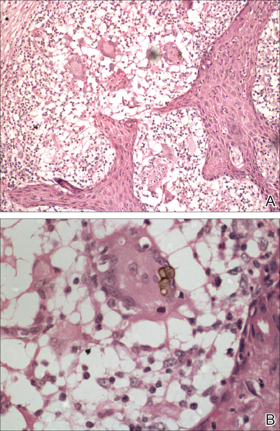
Chromomycosis is a chronic subcutaneous fungal infection caused by pigmented (dematiaceous) fungi growing in soil, decaying vegetables, and rotting wood. Infection usually occurs via traumatic inoculation from splinters and thorns. Some of the agents responsible include Fonsecaea pedrosoi, Cladophialophora carrionii, and Phialophora verrucosa.1
Diverse cutaneous manifestations have been observed with 5 different clinical forms: nodules, verrucous hyperkeratotic plaques, cicatricial lesions with central sparing, scaly plaques, and tumoral (cauliflowerlike) lesions.2 Of these clinical presentations, verrucous hyperkeratotic plaques are the most common, as seen in our patient. However, this presentation is not exclusive to chromomycosis because many conditions appear similarly, including sporotrichosis, nontuberculous mycobacterial infection, tuberculosis verrucosa cutis, and squamous cell carcinoma (SCC). The presence of small ulcerations may appear as the black dots seen on the plaques of chromomycosis, distinguishing chromomycosis from other conditions. Although this feature may be a fundamental clue for diagnosis, it should be emphasized that in many occasions, clinical differences between chromomycosis and its differentials are subtle. A study involving 9 patients with chromomycosis reported that only 1 was given the initial diagnosis of mycosis. Six patients initially were diagnosed with cutaneous malignancies, 1 patient with viral warts, and another patient with ganglion.3 Therefore, unless there is a high index of suspicion, these conditions may easily be mistaken for others by clinicians who are unfamiliar with their presentations, particularly in the setting of a busy clinic.
Chromomycosis routinely is diagnosed based on histologic examination and culture. Apart from sclerotic bodies, other histopathologic features include an inflammatory infiltrate characterized by neutrophilic microabscesses, multinucleated cells, fibrosis, acanthosis, papillomatosis, hyperkeratosis, and pseudoepitheliomatous hyperplasia (PEH).2 Pseudoepitheliomatous hyperplasia is an exaggerated proliferation of the epidermis, usually secondary to chronic inflammatory skin conditions.4 Because most verrucous lesions are thought to be neoplastic and carcinomas more commonly are seen and expected in dermatopathology, PEH can sometimes be mistaken for SCC. At times, the squamous epithelium of PEH can appear infiltrative, giving the illusion of well-differentiated SCC.5 However, absence of marked cellular atypia and abnormal mitotic activity should suggest otherwise. Thorough scrutiny for a concomitant infective process is necessary to avoid the overdiagnosis of SCC. Special stains for infectious agents such as periodic acid-Schiff and Grocott-Gomori methenamine-silver for fungal spores and Ziehl-Neelsen for acid-fast bacilli may reveal infectious organisms. Multilevel sections of deeper levels also may be essential to uncover sparse organisms.6
There is no standard treatment of chromomycosis. Some treatment options are available based on few open clinical studies and expert opinions. Systemic antifungals such as itraconazole or terbinafine most commonly are used with 15% to 80% cure rates.7 In invasive refractory cases, a combination of itraconazole and terbinafine has been employed as salvage therapy. Recently, the use of newer azoles such as posaconazole is favored due to its expanded-spectrum profile along with better pharmacodynamics and pharmacokinetic profile versus itraconazole. Physical methods such as cryotherapy, heat therapy, laser therapy, and photodynamic therapy frequently are practiced in conjunction with systemic antifungal therapy.8 Surgical procedures such as photocoagulation, Mohs micrographic surgery, and curettage sometimes are recommended for smaller well-defined lesions. Amputation, however, is rarely ever indicated, as there rarely is deep tissue involvement.2
Our case highlights the importance of clinicopathologic correlation in diagnosing squamous epithelial lesions. A high index of clinical suspicion and a wider list of differential diagnoses of verrucous plaques are necessary to minimize pitfalls in diagnosing lesions with squamous proliferation and therefore reduces the need for unnecessary interventions.
- Queiroz-Telles F, Esterre P, Perez-Blanco M, et al. Chromoblastomycosis: an overview of clinical manifestations, diagnosis and treatment. Med Mycol. 2009;47:3-15.
- Krzyściak PM, Pindycka-Piaszczyńska M, Piaszczyński M. Chromoblastomycosis. Postepy Dermatol Allergol. 2014;31:310-321.
- Jayalakshmi P, Looi LM, Soo-Hoo TS. Chromoblastomycosis in Malaysia. Mycopathologica. 1990;109:27-31.
- Zayour M, Lazova R. Pseudoepitheliomatous hyperplasia: a review. Am J Dermatopathol. 2011;33:112-126.
- El-Khoury J, Kibbi AG, Abbas O. Mucocutaneous pseudoepitheliomatous hyperplasia: a review. Am J Dermatopathol. 2012;34:165-175.
- Tan KB, Tan SH, Aw DC, et al. Simulators of squamous cell carcinoma of the skin: diagnostic challenges on small biopsies and clinicopathological correlation [published online June 25, 2013]. J Skin Cancer. 2013;2013:752864.
- Queiroz-Telles F, Santos DW. Challenges in the therapy of chromoblastomycosis. Mycopathologia. 2013;175:477-488.
- Queiroz-Telles F, de Hoog S, Santos DW, et al. Chromoblastomycosis. Clin Microbiol Rev. 2017;30:233-276.
The Diagnosis: Chromomycosis
Skin scrapings revealed brownish sclerotic bodies. A review of the skin biopsy performed 4 years prior showed florid pseudoepitheliomatous hyperplasia overlying dense mixed inflammatory infiltrates of predominantly granulomatous microabscesses in the dermis. Numerous sclerotic bodies were evident within multinucleated giant cells and scattered among epidermal and dermal microabscesses (Figure). Few atypical basal keratinocytes were noted, but frank pleomorphism and aberrant mitosis was absent.
Chromomycosis is a chronic subcutaneous fungal infection caused by pigmented (dematiaceous) fungi growing in soil, decaying vegetables, and rotting wood. Infection usually occurs via traumatic inoculation from splinters and thorns. Some of the agents responsible include Fonsecaea pedrosoi, Cladophialophora carrionii, and Phialophora verrucosa.1
Diverse cutaneous manifestations have been observed with 5 different clinical forms: nodules, verrucous hyperkeratotic plaques, cicatricial lesions with central sparing, scaly plaques, and tumoral (cauliflowerlike) lesions.2 Of these clinical presentations, verrucous hyperkeratotic plaques are the most common, as seen in our patient. However, this presentation is not exclusive to chromomycosis because many conditions appear similarly, including sporotrichosis, nontuberculous mycobacterial infection, tuberculosis verrucosa cutis, and squamous cell carcinoma (SCC). The presence of small ulcerations may appear as the black dots seen on the plaques of chromomycosis, distinguishing chromomycosis from other conditions. Although this feature may be a fundamental clue for diagnosis, it should be emphasized that in many occasions, clinical differences between chromomycosis and its differentials are subtle. A study involving 9 patients with chromomycosis reported that only 1 was given the initial diagnosis of mycosis. Six patients initially were diagnosed with cutaneous malignancies, 1 patient with viral warts, and another patient with ganglion.3 Therefore, unless there is a high index of suspicion, these conditions may easily be mistaken for others by clinicians who are unfamiliar with their presentations, particularly in the setting of a busy clinic.
Chromomycosis routinely is diagnosed based on histologic examination and culture. Apart from sclerotic bodies, other histopathologic features include an inflammatory infiltrate characterized by neutrophilic microabscesses, multinucleated cells, fibrosis, acanthosis, papillomatosis, hyperkeratosis, and pseudoepitheliomatous hyperplasia (PEH).2 Pseudoepitheliomatous hyperplasia is an exaggerated proliferation of the epidermis, usually secondary to chronic inflammatory skin conditions.4 Because most verrucous lesions are thought to be neoplastic and carcinomas more commonly are seen and expected in dermatopathology, PEH can sometimes be mistaken for SCC. At times, the squamous epithelium of PEH can appear infiltrative, giving the illusion of well-differentiated SCC.5 However, absence of marked cellular atypia and abnormal mitotic activity should suggest otherwise. Thorough scrutiny for a concomitant infective process is necessary to avoid the overdiagnosis of SCC. Special stains for infectious agents such as periodic acid-Schiff and Grocott-Gomori methenamine-silver for fungal spores and Ziehl-Neelsen for acid-fast bacilli may reveal infectious organisms. Multilevel sections of deeper levels also may be essential to uncover sparse organisms.6
There is no standard treatment of chromomycosis. Some treatment options are available based on few open clinical studies and expert opinions. Systemic antifungals such as itraconazole or terbinafine most commonly are used with 15% to 80% cure rates.7 In invasive refractory cases, a combination of itraconazole and terbinafine has been employed as salvage therapy. Recently, the use of newer azoles such as posaconazole is favored due to its expanded-spectrum profile along with better pharmacodynamics and pharmacokinetic profile versus itraconazole. Physical methods such as cryotherapy, heat therapy, laser therapy, and photodynamic therapy frequently are practiced in conjunction with systemic antifungal therapy.8 Surgical procedures such as photocoagulation, Mohs micrographic surgery, and curettage sometimes are recommended for smaller well-defined lesions. Amputation, however, is rarely ever indicated, as there rarely is deep tissue involvement.2
Our case highlights the importance of clinicopathologic correlation in diagnosing squamous epithelial lesions. A high index of clinical suspicion and a wider list of differential diagnoses of verrucous plaques are necessary to minimize pitfalls in diagnosing lesions with squamous proliferation and therefore reduces the need for unnecessary interventions.
The Diagnosis: Chromomycosis
Skin scrapings revealed brownish sclerotic bodies. A review of the skin biopsy performed 4 years prior showed florid pseudoepitheliomatous hyperplasia overlying dense mixed inflammatory infiltrates of predominantly granulomatous microabscesses in the dermis. Numerous sclerotic bodies were evident within multinucleated giant cells and scattered among epidermal and dermal microabscesses (Figure). Few atypical basal keratinocytes were noted, but frank pleomorphism and aberrant mitosis was absent.
Chromomycosis is a chronic subcutaneous fungal infection caused by pigmented (dematiaceous) fungi growing in soil, decaying vegetables, and rotting wood. Infection usually occurs via traumatic inoculation from splinters and thorns. Some of the agents responsible include Fonsecaea pedrosoi, Cladophialophora carrionii, and Phialophora verrucosa.1
Diverse cutaneous manifestations have been observed with 5 different clinical forms: nodules, verrucous hyperkeratotic plaques, cicatricial lesions with central sparing, scaly plaques, and tumoral (cauliflowerlike) lesions.2 Of these clinical presentations, verrucous hyperkeratotic plaques are the most common, as seen in our patient. However, this presentation is not exclusive to chromomycosis because many conditions appear similarly, including sporotrichosis, nontuberculous mycobacterial infection, tuberculosis verrucosa cutis, and squamous cell carcinoma (SCC). The presence of small ulcerations may appear as the black dots seen on the plaques of chromomycosis, distinguishing chromomycosis from other conditions. Although this feature may be a fundamental clue for diagnosis, it should be emphasized that in many occasions, clinical differences between chromomycosis and its differentials are subtle. A study involving 9 patients with chromomycosis reported that only 1 was given the initial diagnosis of mycosis. Six patients initially were diagnosed with cutaneous malignancies, 1 patient with viral warts, and another patient with ganglion.3 Therefore, unless there is a high index of suspicion, these conditions may easily be mistaken for others by clinicians who are unfamiliar with their presentations, particularly in the setting of a busy clinic.
Chromomycosis routinely is diagnosed based on histologic examination and culture. Apart from sclerotic bodies, other histopathologic features include an inflammatory infiltrate characterized by neutrophilic microabscesses, multinucleated cells, fibrosis, acanthosis, papillomatosis, hyperkeratosis, and pseudoepitheliomatous hyperplasia (PEH).2 Pseudoepitheliomatous hyperplasia is an exaggerated proliferation of the epidermis, usually secondary to chronic inflammatory skin conditions.4 Because most verrucous lesions are thought to be neoplastic and carcinomas more commonly are seen and expected in dermatopathology, PEH can sometimes be mistaken for SCC. At times, the squamous epithelium of PEH can appear infiltrative, giving the illusion of well-differentiated SCC.5 However, absence of marked cellular atypia and abnormal mitotic activity should suggest otherwise. Thorough scrutiny for a concomitant infective process is necessary to avoid the overdiagnosis of SCC. Special stains for infectious agents such as periodic acid-Schiff and Grocott-Gomori methenamine-silver for fungal spores and Ziehl-Neelsen for acid-fast bacilli may reveal infectious organisms. Multilevel sections of deeper levels also may be essential to uncover sparse organisms.6
There is no standard treatment of chromomycosis. Some treatment options are available based on few open clinical studies and expert opinions. Systemic antifungals such as itraconazole or terbinafine most commonly are used with 15% to 80% cure rates.7 In invasive refractory cases, a combination of itraconazole and terbinafine has been employed as salvage therapy. Recently, the use of newer azoles such as posaconazole is favored due to its expanded-spectrum profile along with better pharmacodynamics and pharmacokinetic profile versus itraconazole. Physical methods such as cryotherapy, heat therapy, laser therapy, and photodynamic therapy frequently are practiced in conjunction with systemic antifungal therapy.8 Surgical procedures such as photocoagulation, Mohs micrographic surgery, and curettage sometimes are recommended for smaller well-defined lesions. Amputation, however, is rarely ever indicated, as there rarely is deep tissue involvement.2
Our case highlights the importance of clinicopathologic correlation in diagnosing squamous epithelial lesions. A high index of clinical suspicion and a wider list of differential diagnoses of verrucous plaques are necessary to minimize pitfalls in diagnosing lesions with squamous proliferation and therefore reduces the need for unnecessary interventions.
- Queiroz-Telles F, Esterre P, Perez-Blanco M, et al. Chromoblastomycosis: an overview of clinical manifestations, diagnosis and treatment. Med Mycol. 2009;47:3-15.
- Krzyściak PM, Pindycka-Piaszczyńska M, Piaszczyński M. Chromoblastomycosis. Postepy Dermatol Allergol. 2014;31:310-321.
- Jayalakshmi P, Looi LM, Soo-Hoo TS. Chromoblastomycosis in Malaysia. Mycopathologica. 1990;109:27-31.
- Zayour M, Lazova R. Pseudoepitheliomatous hyperplasia: a review. Am J Dermatopathol. 2011;33:112-126.
- El-Khoury J, Kibbi AG, Abbas O. Mucocutaneous pseudoepitheliomatous hyperplasia: a review. Am J Dermatopathol. 2012;34:165-175.
- Tan KB, Tan SH, Aw DC, et al. Simulators of squamous cell carcinoma of the skin: diagnostic challenges on small biopsies and clinicopathological correlation [published online June 25, 2013]. J Skin Cancer. 2013;2013:752864.
- Queiroz-Telles F, Santos DW. Challenges in the therapy of chromoblastomycosis. Mycopathologia. 2013;175:477-488.
- Queiroz-Telles F, de Hoog S, Santos DW, et al. Chromoblastomycosis. Clin Microbiol Rev. 2017;30:233-276.
- Queiroz-Telles F, Esterre P, Perez-Blanco M, et al. Chromoblastomycosis: an overview of clinical manifestations, diagnosis and treatment. Med Mycol. 2009;47:3-15.
- Krzyściak PM, Pindycka-Piaszczyńska M, Piaszczyński M. Chromoblastomycosis. Postepy Dermatol Allergol. 2014;31:310-321.
- Jayalakshmi P, Looi LM, Soo-Hoo TS. Chromoblastomycosis in Malaysia. Mycopathologica. 1990;109:27-31.
- Zayour M, Lazova R. Pseudoepitheliomatous hyperplasia: a review. Am J Dermatopathol. 2011;33:112-126.
- El-Khoury J, Kibbi AG, Abbas O. Mucocutaneous pseudoepitheliomatous hyperplasia: a review. Am J Dermatopathol. 2012;34:165-175.
- Tan KB, Tan SH, Aw DC, et al. Simulators of squamous cell carcinoma of the skin: diagnostic challenges on small biopsies and clinicopathological correlation [published online June 25, 2013]. J Skin Cancer. 2013;2013:752864.
- Queiroz-Telles F, Santos DW. Challenges in the therapy of chromoblastomycosis. Mycopathologia. 2013;175:477-488.
- Queiroz-Telles F, de Hoog S, Santos DW, et al. Chromoblastomycosis. Clin Microbiol Rev. 2017;30:233-276.

A 75-year-old retired farmer presented with an erythematous verrucous plaque on the dorsal aspect of the left hand of 4 years' duration. Superficial biopsies from the lesion 4 years prior to presentation revealed pseudoepitheliomatous hyperplasia suggestive of squamous cell carcinoma, which led to the excision of the lesion along with 2 digits of the left hand. Despite surgery, the lesions promptly recurred and continued to progress. Physical examination revealed a verrucous plaque with crusting and small ulcerations (black dots) over the extensor aspect of the left hand and forearm.