User login
Durvalumab approved for advanced urothelial carcinoma
for patients with locally advanced or metastatic urothelial carcinoma who have disease progression after prior treatment with a platinum-containing chemotherapy.
The agency also approved a complementary diagnostic for the assessment of the PD-L1 protein the tumor tissue.
The most common adverse reactions were fatigue, musculoskeletal pain, constipation, decreased appetite, nausea, peripheral edema, and urinary tract infection. Pneumonitis, hepatitis, colitis, thyroid disease, adrenal insufficiency, and diabetes also occurred in patients taking durvalumab.
The recommended dose of durvalumab is 10 mg/kg IV over a period of 60 minutes, every 2 weeks, until disease progression or unacceptable toxicity occurs. Full prescribing information is available here.
Durvalumab is marketed as Imfinzi by AstraZeneca. The complementary diagnostic is the Ventana PD-L1 (SP263) Assay from Ventana Medical Systems.
Spontaneous coronary artery dissection: New insights
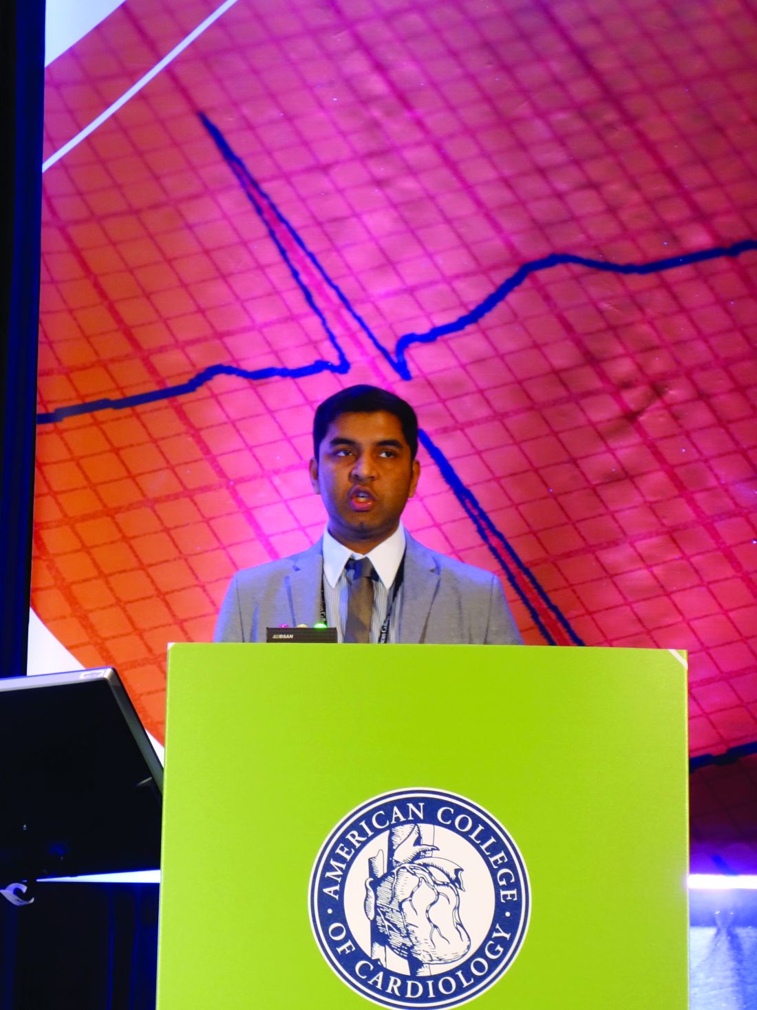
Washington – Patients with spontaneous coronary artery dissection as their presentation of acute coronary syndrome have a markedly better long-term survival rate than do patients with ACS in its various other forms, Rahul Potluri, MD, reported at the annual meeting of the American College of Cardiology.
In his retrospective cohort study of 182 U.K. patients diagnosed with spontaneous coronary artery dissection (SCAD) as the cause of their ACS and 32,981 controls with ACS without SCAD, the 15-year all-cause mortality rate was 10.4% in the SCAD group vs. 32.1% in the controls.
Although SCAD was first described in 1931, half of the roughly 1,500 cases reported in the medical literature have been published within the past 5 years. That makes this series of 182 SCAD patients with 15-year outcome data unique in providing the longest follow-up to date reported in a sizable patient series, said Dr. Potluri of the University of Alberta, Edmonton, and Aston University in Birmingham, England.
SCAD is a rupture in a coronary artery wall not related to atherosclerotic heart disease, trauma, or iatrogenic causes. The dissection is thought to result from an intimal tear or medial hemorrhage from the vasa vasorum, with subsequent accumulation of blood in a false lumen.
It’s not entirely unexpected that patients with SCAD have a better long-term prognosis than do those with ACS without SCAD, Dr. Potluri said. After all, patients with SCAD tend to be considerably younger: In Dr. Potluri’s series, the average age was younger than 52 years, vs. 66 years in controls. Plus they had lower levels of cardiovascular risk factors, and by definition they were free of atherosclerotic heart disease. But SCAD often occurs in conjunction with fibromuscular dysplasia, and the long-term health implications of that combination have not been well studied.
In a multivariate logistic regression analysis adjusted for age, sex, ethnicity, and comorbid conditions, the likelihood of dying within 5 years was 89% greater in the non-SCAD group. During follow-up, the non-SCAD ACS group was 4.1-fold more likely to have another ACS and 32% more likely to be diagnosed with heart failure.
SCAD was typically managed conservatively in Dr. Potluri’s series: In the SCAD group, 11% underwent PCI and 2.7% had CABG, compared with 51% and 10.7%, respectively, of controls.
“Conservative treatment appears to be safe in the SCAD patient population,” he said.
The prevalence of SCAD in his series of more than 33,000 patients admitted for ACS to U.K. hospitals in 2000-2014 was 0.54%. Dr. Potluri said that low figure likely reflects considerable underreporting due to the fact that some data were from the early 2000s, when SCAD was still largely below physicians’ radar.
Dr. Potluri is an authority on big data analytics in medical research. He formed his 182-patient SCAD series using ACALM (Algorithm for Comorbidity, Associations, Length of Stay, and Mortality), an analytic tool he developed years ago as a medical student. At the time of his SCAD study, the ACALM registry included anonymous, deidentified data on 1.8 million U.K. patients. The ACALM methodology utilizes a form of artificial intelligence known as novel field derivation to analyze huge amalgamated data sets. This is made possible by the fact that all U.K. hospitals report patient data in an identical standardized way, which is not the case in the United States.
Dr. Potluri has previously applied the ACALM methodology to a variety of other health care issues, including studies of an association between hyperlipidemia and breast cancer, the relationship between cardiovascular disease and mental health, and the so-called weekend effect, whereby U.K. patients hospitalized for cardiovascular reasons on the weekend have higher mortality than do those admitted on weekdays.
THE ACALM registry now exceeds 4 million patients. Dr. Potluri said he plans to analyze this full group to develop a large, comprehensive SCAD patient registry. This will enable investigators to evaluate potential psychosocial risk factors for SCAD, the role of fibromuscular dysplasia and connective tissue diseases, and other practical questions.
He reported having no financial conflicts regarding his study.
At ACC 17
Key clinical point:
Major finding: The 15-year all-cause mortality rate was 10% in patients with spontaneous coronary artery dissection, compared with 32% in those with ACS without spontaneous coronary artery dissection.
Data source: A retrospective cohort study including 15-year outcomes for 182 patients with spontaneous coronary artery dissection and more than 32,000 controls with ACS without dissection.
Disclosures: The study presenter reported having no financial conflicts.
Student-Resident Luncheon offers relaxed networking
The ritual of networking at medical conferences often involves waiting to chat with a speaker after a session or finessing an introduction in a busy hallway. That’s why, at the Student-Resident Luncheon, organized by SHM’s Physicians-in-Training Committee, Tuesday, May 2, at noon, trainees will have a chance to interact with experts in the field in a much more relaxed setting.
The free luncheon will include several tables, each dedicated to a specific topic, such as pediatric hospital medicine, with an experienced hospitalist at each one. The residents and students who attend can choose their table and will have a chance to sit at two different tables. There will also be an “open forum” segment at the end, when the trainees can seek out other experts, said Darlene Tad-y, MD, committee chair and assistant professor of medicine at the University of Colorado at Denver, Aurora.
“The purpose behind it was to bring our trainees together so that they can meet each other and to bring some of the SHM leaders to them so that they can learn about what [the leaders] do in hospital medicine and see the breadth of work that hospitalists are doing around the country,” she said.
Pediatric hospital medicine, medical education, and global health are three of the confirmed topics that will be covered at the luncheon, Dr. Tad-y said. There will be a maximum of 10 people at each table, including the expert.
“We wanted it to be very immediate for the students and residents who are going to be there,” she said. “It’s a pretty small group setting.”
The event has been getting more popular each year and is now in its third year. SHM said 500 students and residents participated last year. Residents and students who register for the annual meeting receive an invitation to attend the luncheon. Those who decide on-site that they want to attend will have the ability to do so, Dr. Tad-y said.
Such close interaction with people whose literature they may have read can be very helpful for trainees, she said. They can “sit at a table with them and hear their story and learn how they got to where they were. I think it’s quite impactful.
Knee osteoarthritis linked to premature mortality
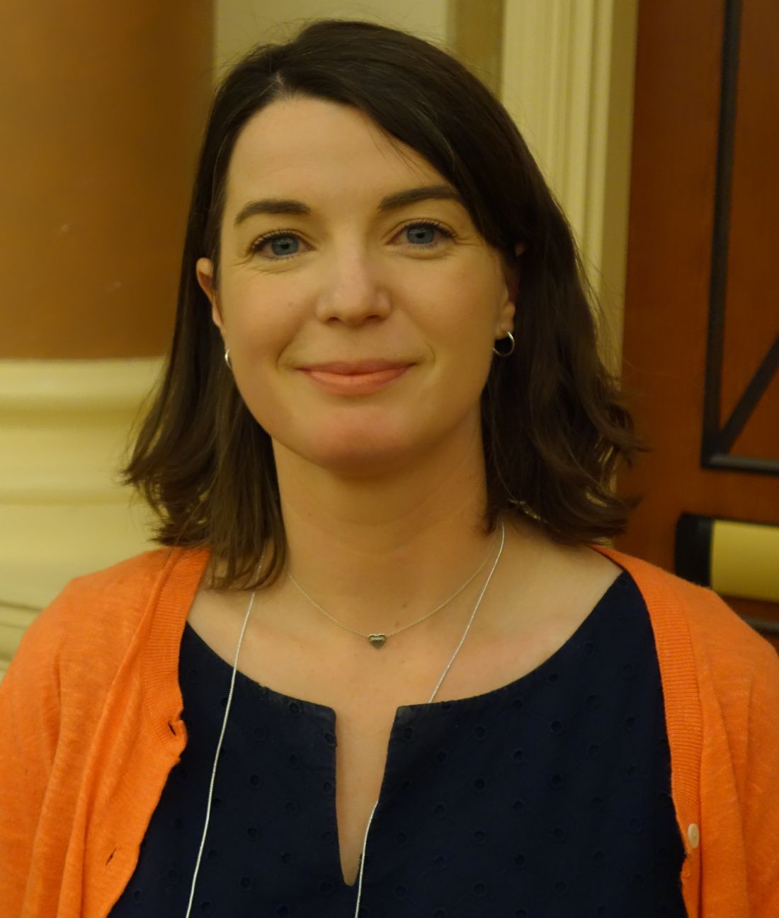
LAS VEGAS – Knee osteoarthritis was independently associated with increased risk of premature mortality in the largest-ever meta-analysis to examine the issue, Kirsten M. Leyland, PhD, reported at the World Congress on Osteoarthritis.
The meta-analysis used individual patient-level data on 11,954 participants in six prospective observational cohort studies conducted in four countries. The key finding: Subjects with symptomatic and radiographically confirmed knee OA were at 17% greater risk of premature mortality, compared with pain-free, radiographically negative participants, independent of age, sex, and race, according to Dr. Leyland, a musculoskeletal epidemiologist at the University of Oxford (England).
The six prospective cohort studies included in the new analysis are well known in the field of osteoarthritis research: the Framingham (Mass.) Osteoarthritis Study, the Rotterdam (the Netherlands) Study, the Multicenter Osteoarthritis Study (MOST), the Johnston County (N.C.) Osteoarthritis Project, the Tasmanian Older Adult Cohort Study, and the Chingford (U.K.) 1000 Women Study. These studies feature 7.4-23.7 years of prospective follow-up.
In an interview, Dr. Leyland said that the current analysis uses all-cause mortality as the outcome because causes of death are recorded differently in the countries where the studies are taking place. It will take several more years for statisticians to harmonize the cause-of-death data and draw definitive conclusions; however, preliminary analysis suggests the causes of premature mortality in the knee OA patients are disproportionately cardiovascular, she added.
Dr. Leyland reported having no financial conflicts regarding the study, which was supported by Arthritis Research UK.
AT OARSI 2017
Key clinical point:
Major finding: Knee osteoarthritis is independently associated with a 17% increased likelihood of premature mortality.
Data source: This meta-analysis harmonized individual patient-level data drawn from six major prospective, population-based cohort studies worldwide.
Disclosures: The presenter reported having no financial conflicts regarding the study, which was supported by Arthritis Research UK.
Optimism in the face of change
The first HM17 plenary is focused on health policy at a time when a dynamically evolving health care delivery system may seem daunting, opaque, and labyrinthine.
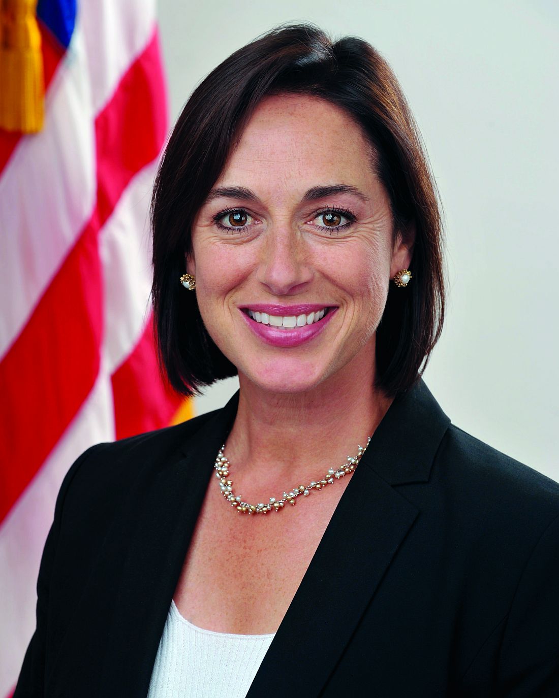
“Though it feels uncertain in some ways, the health care world is pretty united that we need to continue the progress we’ve made on moving away from the old fee-for-service model, toward one that lets people practice medicine the way they want and focus on patients and outcomes,” said HM17 keynote speaker Karen DeSalvo, MD, MPH, MSc, former acting assistant secretary for health in the U.S. Department of Health and Human Services (HHS).

“That’s just the way that we finance or pay for care,” she said. “There’s this entire care system that everybody’s working and innovating in every day, trying to find more efficient, effective ways to get better outcomes. Hospitalists, quite frankly, have been in the lead for 20 years and really understand in granular detail what it takes.”
Dr. DeSalvo believes that the progress of the past 5 years has already laid the path that must now be followed. The public sector’s move away from fee for service, toward payment for episodic care, has combined with emerging technology platforms to create a new age in which physicians and insurers can judge, in real-time, how well care is working.
Add to this the growth of accountable care organizations, other alternative payment models, value-based purchasing, and the implementation of the Medicare Access and CHIP Reauthorization Act of 2015 (MACRA), and it is clear the direction in which the industry is headed.
Of course, Dr. DeSalvo understands the fears of those wondering where the next wave of change will carry them. She’s heard the political debate over the past 8 years and the rancorous discussions in just the first few months of 2017. But she believes the path for health care delivery continues to be “toward the natural place as opposed to the unnatural place.”
“That gives me a lot of optimism,” she said. “One of the most important things for hospitalists to be doing right now is to keep standing up and speaking on behalf of patients and saying that the right thing is for us not to regress but to continue moving forward so that we can all have the kind of system we want for our patients.
The first HM17 plenary is focused on health policy at a time when a dynamically evolving health care delivery system may seem daunting, opaque, and labyrinthine.
“Though it feels uncertain in some ways, the health care world is pretty united that we need to continue the progress we’ve made on moving away from the old fee-for-service model, toward one that lets people practice medicine the way they want and focus on patients and outcomes,” said HM17 keynote speaker Karen DeSalvo, MD, MPH, MSc, former acting assistant secretary for health in the U.S. Department of Health and Human Services (HHS).
“That’s just the way that we finance or pay for care,” she said. “There’s this entire care system that everybody’s working and innovating in every day, trying to find more efficient, effective ways to get better outcomes. Hospitalists, quite frankly, have been in the lead for 20 years and really understand in granular detail what it takes.”
Dr. DeSalvo believes that the progress of the past 5 years has already laid the path that must now be followed. The public sector’s move away from fee for service, toward payment for episodic care, has combined with emerging technology platforms to create a new age in which physicians and insurers can judge, in real-time, how well care is working.
Add to this the growth of accountable care organizations, other alternative payment models, value-based purchasing, and the implementation of the Medicare Access and CHIP Reauthorization Act of 2015 (MACRA), and it is clear the direction in which the industry is headed.
Of course, Dr. DeSalvo understands the fears of those wondering where the next wave of change will carry them. She’s heard the political debate over the past 8 years and the rancorous discussions in just the first few months of 2017. But she believes the path for health care delivery continues to be “toward the natural place as opposed to the unnatural place.”
“That gives me a lot of optimism,” she said. “One of the most important things for hospitalists to be doing right now is to keep standing up and speaking on behalf of patients and saying that the right thing is for us not to regress but to continue moving forward so that we can all have the kind of system we want for our patients.
Elders and Falls Lead TBI-Related ED Visits
Nearly 3 million emergency department (ED) visits, hospitalizations, and deaths were related to traumatic brain injury (TBI) in 2013, according to researchers from the National Center for Injury Prevention and Control. The age-adjusted rate of ED visits was higher in 2013 than in 2007 (787.1 vs 534.4), a change driven largely by people aged ≥ 75 years, who accounted for 18% of the increase in the number of TBI-related ED visits.
Related: Ideas for Helping TBI Patients
The most common mechanisms of injury in the study were falls, being struck by or against an object, and motor-vehicle crashes. Particular age groups were disproportionately affected by specific mechanisms. The researchers say about half of all fall-related TBI visits/hospitalizations/deaths were among babies, toddlers, and adults aged > 75 years.
Those data suggest an urgent need for more and stronger fall-prevention efforts, say the researchers. In older adults, TBIs are more likely to lead to hospitalizations that can be complicated by comorbidities. Moreover, the researchers say, older adults are more likely to use anticoagulants, which can increase the likelihood of intracranial hemorrhage.
Related: Making Fall Prevention “Routine”
Prevention strategies that have proved effective in randomized controlled trials include multicomponent exercise programs, tai chi, vitamin D supplements, cataract surgery, and making the home environment safer. The CDC also has developed the Stopping Elderly Accidents Deaths and Injuries (STEADI) program, which incorporates empirically supported clinical guidelines and scientifically tested interventions to help primary care providers address fall risk and use effective interventions.
Conway: HM well positioned for ‘system transformation’
Patrick Conway, MD, MSc, MHM, may have had a lot of job titles recently, but his work address hasn’t changed in 6 years.
Dr. Conway, deputy administrator for innovation and quality for the Centers for Medicare & Medicaid Services and director of its Center for Medicare and Medicaid Innovation, has been at the intersection of policy and practice in Washington, since joining CMS in 2011. The still-practicing hospitalist was acting CMS administrator for several months earlier this year, holding the top post while President Donald Trump’s nominee to lead the agency, Seema Verma, awaited U.S. Senate confirmation. His title before that was principal deputy administrator and CMS chief medical officer.
Dr. Conway said he believes that at a “macro legislation” level, Washington is committed to health care reform that improves patient care and incentivizes physicians to join alternative payment models (APMs). He said hospitalists should be encouraged by how well the field has already adapted to the proliferation of accountable-care organizations, value-based purchasing, and the Medicare Access and CHIP Reauthorization Act of 2015, or MACRA. He said that as innovations lead to better patient care and more coordinated care, that’s good for hospitalists, patients, and the hospitals that bring them together.
“I want to leave people with the idea that value-based payment innovation and delivery system reform will continue to be critical aspects of improving our health system,” he said. “I also want hospitalists to continue to stay engaged in these new payment models and help lead them and provide better patient care as part of them.”
Dr. Conway said that focusing on innovations is essential to hospitalists to ensure the specialty remains at the forefront of improved patient care, decreased readmissions, decreased infections and other quality improvements. He notes that by still practicing on an academic medical service with faculty, residents and medical students, he’s encouraged and excited that “people get it.”
“Every doctor wants better care for their patients,” Dr. Conway said. “The reality of … new payment systems and new fields of innovation we’re entering into is it unleashes hospitalists and aligns incentives with what they want for their patients.”
Take bundled payments around episodic care. With innovations in payment models, hospitalists are increasingly being paid for how good their care is, not how much care there is.
“That’s exciting,” Dr. Conway said. “Very different than 10 years ago where you had a lot of groups where it was about how much volume can we churn through. Now I think hospitals and HM groups [realize] value is going to continue to increasingly be based on the quality of care and the efficiency of care delivery.”
High-value care abstracts in spotlight
If the annual meeting of the Society of Hospital Medicine could be boiled down to a single goal, it might be to give hospitalists the information they need to be the best doctors they can be.
It shouldn’t come as a surprise, then, that research performed by hospitalists on high-value care (HVC) will be featured in an abstract session at this year’s conference.
The “Best of HVC Abstract Submissions” session, scheduled for Tuesday, May 2, 3:05–3:45 p.m., will be part of a day-long track devoted entirely to HVC, a new feature at this year’s annual meeting.
“The panel will give them feedback and ask questions and sort of engage in conversation about it,” Dr. Feldman said. He thinks that featuring these abstracts in a high-profile setting is a good way for hospitalists to learn from one another. “There are so many hospitalists interested in this topic right now and people working in this area and coming up with quality improvement projects around high-value care. It’s nice to know what other folks are doing.”
Dr. Feldman hopes that, if hospitalists find out what is happening at other centers, it will spur even more quality projects.
“We don’t often have the opportunity to share the work that we’ve done and to motivate each other,” he said. “When you hear about the great work that other people are doing, it is invigorating; it’s exciting; it gets you jazzed up about doing the work in your own institution. A lot of this isn’t incredibly sophisticated. It is stuff that can be relatively easily disseminated from one institution to another without having a ton of support. To be able to see the type of work that people do is thrilling for the hospitalists who attend.”
“With the growing recognition of the costs and harms from medical overuse, high-value care has become a national imperative,” he said. “Hospitalists are natural leaders for high-value care, much like we have been for patient safety and quality improvement. Hospitalists have already led the way on developing programs that target areas of overuse in hospitals. Our ‘Best of HVC’ session will highlight many of these promising examples. There will be plenty of opportunities to learn from innovators and take back the best ideas to our own hospitals.”
Organizers said that the HVC track “will guide attendees on how to avoid diagnostic and therapeutic overuse and how to move toward the right care for every hospital medicine patient.” Other sessions in the track will cover “things we do for no reason,” how to use imaging wisely, and tips on overcoming cultures fraught with overuse.
Dr. Feldman said the track is intended to meet the demand of meeting attendees.
“There are lots of hospitalists who are engaging in this type of work in their institutions and systems,” he said. “So, it’s just timely. People are engaged in it and they’re excited about it, so it makes sense that adding a track right now would fulfill the needs of many of the hospitalists who are going to be attending.”
Best of HVC Abstract Submissions
Tuesday, May 2, 3:05–3:45 p.m.
Speed-mentoring event targets junior hospitalists
When senior physicians and researchers – including hospitalists – talk about their careers, it seems they never fail to mention the mentors who helped shape their professional lives. Mentoring matters – a lot.
Junior hospitalists have a chance to receive mentoring in an area of their choice at HM17.
“This is their opportunity to get ‘rapid-fire’ advice from senior hospitalists from a variety of perspectives,” said event organizer Joanna Bonsall, MD, PhD, SFHM, assistant professor of medicine at Emory University, Atlanta.
The advisers are chosen because of their commitment to supporting career development and because of their reputation and expertise. Many of the speed mentors are returning to the event for the fourth time. It has been held at the SHM annual meeting since 2013.
“This event doesn’t replace traditional mentorship but does give junior hospitalists an opportunity to discuss an aspect of their career with multiple senior hospitalists from across the country,” Dr. Bonsall said.
Advances in Hematology and Oncology (May 2017)
Table of Contents
- Thirteen Years and Still Growing: An AVAHO History
- Criteria for Use Updates for Enzalutamide, Daratumumab, Elotuzumab, Carfilzomib, and Ixazomib
- Lung Cancer Screening: Translating Research Into Practice
- Open Clinical Trials for Patients With Colorectal Cancer
- Treatment and Management of Patients With Prostate Cancer
- Cancer Care Collaborative Approach to Optimize Clinical Care
- Patient Knowledge of and Barriers to Breast, Colon, and Cervical Cancer Screenings: A Cross-Sectional Survey of TRICARE Beneficiaries
- Novel Neuroendocrine Tumor in Multiple Endocrine Neoplasia Type 1