User login
Monday’s Lillehei Forum highlighted basic research
The Lillehei Forum is a series of presentations on basic research by residents in the field of cardiothoracic surgery.
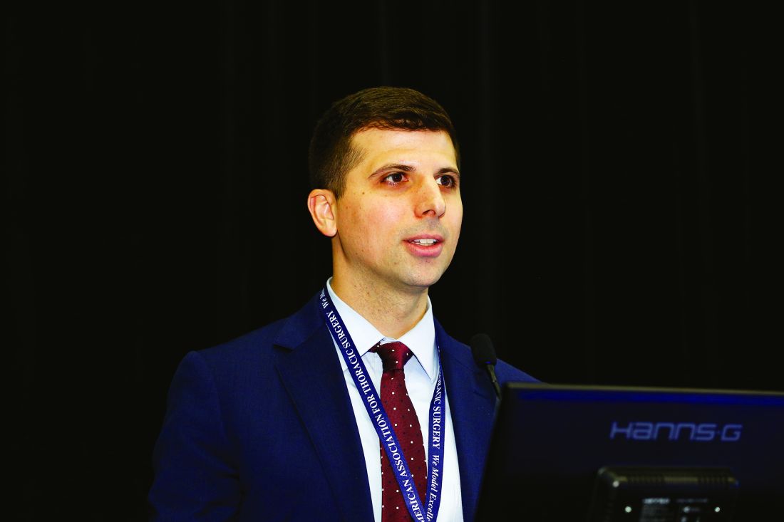
A targeted near-infrared contrast agent specific for lung adenocarcinomas was assessed for effectiveness during intraoperative molecular imaging (IMI) in animal models. Use of the contrast agent proved significantly more effective at detecting positive margins than did surgery alone, according to a study presented by Jarrod D. Predina, MD.
Initial human experiences with IMI for NSCLC using a fluorescent contrast agent have been limited by technical hurdles including high background noise in inflammatory tissues and low signal output, according to Dr. Predina. As an alternative, he and his colleagues at the University of Pennsylvania hypothesized that a targeted near-infrared contrast agent specific for lung adenocarcinomas would improve sensitivity and specificity during surgery.
Using a mouse surgical model of non–small cell lung cancer (NSCLC) that recapitulates local and systemic post-operative recurrences, Dr. Predina and his colleagues at the University of Pennsylvania injected 140 mice intravenously prior to resection with a near-infrared imaging agent (OTL0038). This agent is specific for pulmonary adenocarcinomas due to its high affinity binding of the folate receptor alpha. Tumor-bearing mice were randomized to surgery with or without IMI. Suspicious residual disease was resected and analyzed by immunohistochemistry, flow cytometry, and immunofluorescence. Based on this data, OTL0038 was tested in a pilot study of five canines with spontaneously occurring lung cancer.
In a local recurrence model system, of 80 mice assessed, surgeons identified 10 positive margins in those randomized to imaging with IMI vs. three positive margins in mice undergoing surgery alone, a significant difference. In systemic recurrence models (60 mice), the mean number of pulmonary nodules located with IMI was 7.2 vs. 3.4 in controls, also a significant difference, according to Dr. Predina.
In five canines with a presumed diagnosis of NSCLC, no toxicity was observed. Four of five canines had fluorescent tumors; the non-fluorescing tumor was discovered to be a metastatic mammary tumor on final pathologic analysis. In one canine, an otherwise undetectable 8-mm pulmonary adenocarcinoma was discovered with IMI.
“Our data suggest that a targeted near-infrared contrast agent may improve IMI technology. Ultimately, this will enable accurate identification of residual disease that may otherwise be overlooked. These results are the basis of an ongoing phase I human trial,” concluded Dr. Predina.
The complexity of the lung has limited the prospects of bioengineering strategies utilizing stem cells and fully decellularized or bioartificial scaffolds, according to Brandon A. Guenthart, MD, of Columbia University.
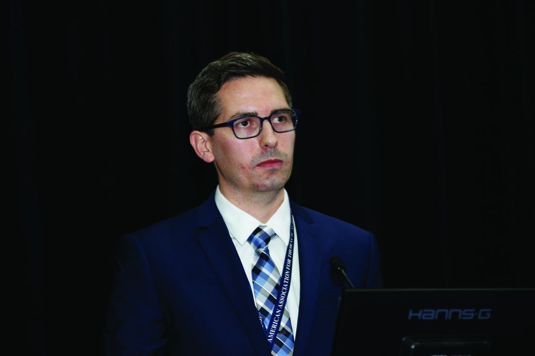
Using standard protocols, Dr. Guenthart and his colleagues procured human lungs rejected for transplantation on the basis of standard clinical criteria. Lungs were placed on a custom-built EVLP system, ventilated and perfused. Utilizing video bronchoscopy, real-time transpleural imaging, and a custom designed micro-catheter delivery and occlusion system, the team delivered a decellularization solution into targeted lung regions that resulted in the selective removal of airway epithelium.
Human airway epithelial cells or human-derived alveolar progenitor cells were labeled (with a quantum dot or a near-infrared dye) and delivered into decellularized lung regions. Following delivery, EVLP was continued for 4-6 hours to allow for cell engraftment. Lung wedge samples were collected at each time point for histologic analysis.
Following decellularization, hematoxylin and eosin staining confirmed removal of pseudostratified and columnar epithelium in proximal airways and removal of type I and II pneumocytes in the distal lung. Delivered cells were retained in the lung after EVLP and fixation, and cellular morphology and distribution within the alveoli indicated early engraftment.
“Bioengineering human lungs utilizing advanced therapeutic interventions such as cell replacement may help combat the critical shortage of transplantable lungs,” said Dr. Guenthart. “Additionally, in the future, targeted intervention and patient-specific cell replacement strategies may have translational applications in vivo and eliminate the need for transplantation in select patients,” he concluded.
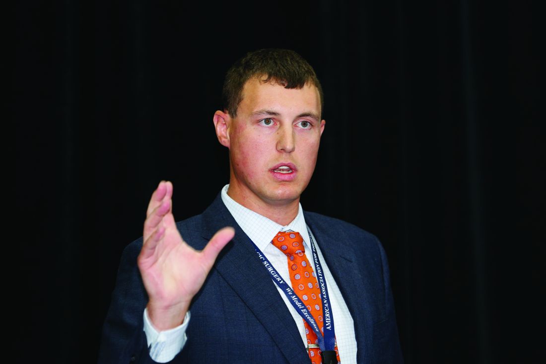
They used a previously validated porcine lung injury model of intravenous lipopolysaccharide (LPS) to induce a systemic inflammatory response and subsequent severe ARDS requiring ECMO support. A total of eight mature adult swine were administered LPS via the external jugular vein followed by sternotomy and central ECMO cannulation (right atrium to ascending aorta).
The left pulmonary artery (inflow) and left superior and inferior pulmonary veins (outflow) were dissected out and cannulated to isolate the left lung, which then underwent 4 hours normorthermic IVLP with Steen Solution followed by 4 hours of lung reperfusion after IVLP decannulation.
Dr. Mehaffey and his colleagues then compared the right (LPS control) and left lungs (LPS+IVLP) of the same animal.
All animals demonstrated a significant reduction in PaO2/FiO2 ratio and total lung compliance 2 hours after the start of LPS infusion. During IVLP, the left (treated) pulmonary vein oxygenation was superior to right (control) pulmonary vein oxygenation. After reperfusion and IVLP decannulation, six (75%) animals had improved lung function allowing for ECMO decannulation.
The left lung showed significant improvement of lung-specific oxygenation compared to the right control at 4 hours of reperfusion. Similarly, total lung compliance improved after targeted rehabilitation of the left lung, according to Dr. Mehaffey. In addition, the wet-to-dry ratio of lung tissue demonstrated significantly reduced edema in the rehabilitated left lungs compared to right controls.
Dr. Mehaffey and his colleagues concluded that IVLP may reduce the duration of ECMO and ventilator support resulting in major reduction in morbidity, mortality, and health care–related costs in patients with ARDS.
The Lillehei Forum is a series of presentations on basic research by residents in the field of cardiothoracic surgery.
A targeted near-infrared contrast agent specific for lung adenocarcinomas was assessed for effectiveness during intraoperative molecular imaging (IMI) in animal models. Use of the contrast agent proved significantly more effective at detecting positive margins than did surgery alone, according to a study presented by Jarrod D. Predina, MD.
Initial human experiences with IMI for NSCLC using a fluorescent contrast agent have been limited by technical hurdles including high background noise in inflammatory tissues and low signal output, according to Dr. Predina. As an alternative, he and his colleagues at the University of Pennsylvania hypothesized that a targeted near-infrared contrast agent specific for lung adenocarcinomas would improve sensitivity and specificity during surgery.
Using a mouse surgical model of non–small cell lung cancer (NSCLC) that recapitulates local and systemic post-operative recurrences, Dr. Predina and his colleagues at the University of Pennsylvania injected 140 mice intravenously prior to resection with a near-infrared imaging agent (OTL0038). This agent is specific for pulmonary adenocarcinomas due to its high affinity binding of the folate receptor alpha. Tumor-bearing mice were randomized to surgery with or without IMI. Suspicious residual disease was resected and analyzed by immunohistochemistry, flow cytometry, and immunofluorescence. Based on this data, OTL0038 was tested in a pilot study of five canines with spontaneously occurring lung cancer.
In a local recurrence model system, of 80 mice assessed, surgeons identified 10 positive margins in those randomized to imaging with IMI vs. three positive margins in mice undergoing surgery alone, a significant difference. In systemic recurrence models (60 mice), the mean number of pulmonary nodules located with IMI was 7.2 vs. 3.4 in controls, also a significant difference, according to Dr. Predina.
In five canines with a presumed diagnosis of NSCLC, no toxicity was observed. Four of five canines had fluorescent tumors; the non-fluorescing tumor was discovered to be a metastatic mammary tumor on final pathologic analysis. In one canine, an otherwise undetectable 8-mm pulmonary adenocarcinoma was discovered with IMI.
“Our data suggest that a targeted near-infrared contrast agent may improve IMI technology. Ultimately, this will enable accurate identification of residual disease that may otherwise be overlooked. These results are the basis of an ongoing phase I human trial,” concluded Dr. Predina.
The complexity of the lung has limited the prospects of bioengineering strategies utilizing stem cells and fully decellularized or bioartificial scaffolds, according to Brandon A. Guenthart, MD, of Columbia University.
Using standard protocols, Dr. Guenthart and his colleagues procured human lungs rejected for transplantation on the basis of standard clinical criteria. Lungs were placed on a custom-built EVLP system, ventilated and perfused. Utilizing video bronchoscopy, real-time transpleural imaging, and a custom designed micro-catheter delivery and occlusion system, the team delivered a decellularization solution into targeted lung regions that resulted in the selective removal of airway epithelium.
Human airway epithelial cells or human-derived alveolar progenitor cells were labeled (with a quantum dot or a near-infrared dye) and delivered into decellularized lung regions. Following delivery, EVLP was continued for 4-6 hours to allow for cell engraftment. Lung wedge samples were collected at each time point for histologic analysis.
Following decellularization, hematoxylin and eosin staining confirmed removal of pseudostratified and columnar epithelium in proximal airways and removal of type I and II pneumocytes in the distal lung. Delivered cells were retained in the lung after EVLP and fixation, and cellular morphology and distribution within the alveoli indicated early engraftment.
“Bioengineering human lungs utilizing advanced therapeutic interventions such as cell replacement may help combat the critical shortage of transplantable lungs,” said Dr. Guenthart. “Additionally, in the future, targeted intervention and patient-specific cell replacement strategies may have translational applications in vivo and eliminate the need for transplantation in select patients,” he concluded.
They used a previously validated porcine lung injury model of intravenous lipopolysaccharide (LPS) to induce a systemic inflammatory response and subsequent severe ARDS requiring ECMO support. A total of eight mature adult swine were administered LPS via the external jugular vein followed by sternotomy and central ECMO cannulation (right atrium to ascending aorta).
The left pulmonary artery (inflow) and left superior and inferior pulmonary veins (outflow) were dissected out and cannulated to isolate the left lung, which then underwent 4 hours normorthermic IVLP with Steen Solution followed by 4 hours of lung reperfusion after IVLP decannulation.
Dr. Mehaffey and his colleagues then compared the right (LPS control) and left lungs (LPS+IVLP) of the same animal.
All animals demonstrated a significant reduction in PaO2/FiO2 ratio and total lung compliance 2 hours after the start of LPS infusion. During IVLP, the left (treated) pulmonary vein oxygenation was superior to right (control) pulmonary vein oxygenation. After reperfusion and IVLP decannulation, six (75%) animals had improved lung function allowing for ECMO decannulation.
The left lung showed significant improvement of lung-specific oxygenation compared to the right control at 4 hours of reperfusion. Similarly, total lung compliance improved after targeted rehabilitation of the left lung, according to Dr. Mehaffey. In addition, the wet-to-dry ratio of lung tissue demonstrated significantly reduced edema in the rehabilitated left lungs compared to right controls.
Dr. Mehaffey and his colleagues concluded that IVLP may reduce the duration of ECMO and ventilator support resulting in major reduction in morbidity, mortality, and health care–related costs in patients with ARDS.
Monday’s Plenary tackled key issues
Presentations of the latest research in cardiothoracic surgery are the hallmark of the AATS Plenary session and this year’s session did not disappoint.
When repairing large paraesophageal hernias, transthoracic Belsey Mark IV fundoplication led to lower rates of leaks and reoperations than did the widely used laparoscopic Nissen fundoplication, according to a study presented by Danuel Laan, MD.
Dr. Laan, a resident at the Mayo Clinic in Rochester, Minn., and his colleagues compared outcomes after Belsey Mark IV fundoplication with those after laparoscopic Nissen fundoplication.
The researchers performed a retrospective review of prospectively collected data on all Mayo patients from 2002 to 2011 who underwent repair of a large paraesophageal hernia, defined as having more than 50% of the stomach within the chest. The analysis excluded patients who had already undergone a fundoplication.
Matching 118 Belsey fundoplication patients 1:1 with 118 Nissen fundoplication patients, the investigators compared the two groups for recurrence, need for reoperation, and perioperative outcomes. Nearly 30% of patients in the Belsey and Nissen groups were male. Mean age was 68.7 years and 69.8 years, respectively. Body mass index was 30.3 kg/m2 and 28.8 kg/m2, respectively.
Ten Belsey patients (8.4%) had a hernia recurrence, as did 19 Nissen patients (16.1%). Rates of leak and reoperation were greater in Nissen fundoplication patients than in Belsey patients. There were no leaks in the Belsey patients, compared with eight leaks (6.8%) in the Nissen patients, a statistically significant difference. Reoperation was necessary in 3 Belsey patients (2.5%), significantly fewer than the 11 Nissen patients (9.3%) who underwent reoperation.
Among 77 patients in each group followed for 5 years or less, there was no difference in symptoms. However, in 41 patients in each group followed for more than 5 years, symptoms were excellent or good in 85% of Belsey patients, compared with 56% of Nissen patients.
AATS: Surgical intervention relatively safe for AAOCA in youth
It’s relatively safe to surgically treat anomalous aortic origin of a coronary artery in children and adolescents, with essentially all patients cleared for full activity after 3 months, according to a study presented by Carlos M. Mery, MD.
Anomalous aortic origin of a coronary artery (AAOCA) is the second leading cause of sudden cardiac death (SCD) in young people, noted Dr. Mery of Texas Children’s Hospital.
He and his colleagues prospectively analyzed outcomes for 44 AAOCA patients who underwent surgical intervention as part of a standardized management program. The researchers included all AAOCA patients aged 2-18 years who underwent surgical intervention between December 2012 and April 2017 as part of the multidisciplinary Coronary Anomalies Program at Texas Children’s Hospital.
Surgical indications included an anomalous left coronary artery, ischemic symptoms, a positive nuclear perfusion test (NPT), or high-risk anatomy such as long intramural segment or ostial stenosis identified via CT angiography. Median patient age was 14 years.
Nine of the patients (20%) underwent surgery for an anomalous left coronary artery, with 32 patients (80%) receiving surgical intervention for an anomalous right coronary artery. A total of 34 surgical procedures (77%) were unroofing of intramural segments, 7 were coronary translocations (16%), and 2 were ostioplasties (4%).
There were no operative deaths. One patient required coronary artery bypass grafting after developing ischemia following a coronary translocation. Minor complications were seen in eight other patients (18%). One patient presented with a second episode of aborted sudden cardiac death 1 year after unroofing of an anomalous left coronary artery with a short intramural segment, and underwent successful coronary translocation and unroofing of a previously identified myocardial bridge.
At follow-up (median 2 years), 40 patients were asymptomatic (91%), while 4 patients had nonspecific chest pain (9%). Forty-one patients (93%) had returned to full activity, while 3 patients were waiting for their 3-month clearance to return to full activity.
Lobectomy can be done safely after concurrent chemotherapy and high-dose radiation in patients with resectable N2-positive stage IIIA non–small-cell lung cancer, according to study findings presented by Jessica S. Donington, MD.

Dr. Donington, of New York University, and her colleagues analyzed two prospective trials conducted by NRG Oncology, RTOG 0229 and RTOG 0839. Both trials’ primary endpoint was mediastinal node sterilization after concurrent chemotherapy and full-dose radiation and has been previously reported.
Dr. Donington and her fellow investigators specifically examined short-term surgical outcomes, given the significant controversy regarding the safety of resection after full-dose thoracic radiation.
In both trials, patients received weekly carboplatin and paclitaxel. Those in the 0229 trial underwent 61.2 Gy of radiation in 34 fractions, while patients in the 0839 trial underwent 60 Gy in 30 fractions. In addition, patients in the 0839 trial were randomized 2:1 to receive weekly panitumumab, an EGFR monoclonal antibody, with their induction therapy.
Surgical expertise was considered essential to this treatment strategy. Therefore, all surgeons were certified by RTOG prior to enrolling patients, and all patients were surgically evaluated before beginning induction therapy to determine resectability and appropriateness for trimodality therapy.
Of 125 eligible patients enrolled in the two trials, 93 patients (74%) underwent anatomic resection. A total of 77 patients underwent lobectomy, 8 underwent pneumonectomy, 6 underwent bilobectomy, and 2 patients had sleeve lobectomy. Medical contraindication and persistent nodal disease found during post-induction invasive staging were the most common reasons patients didn’t undergo resection.
Eighty-five of the 93 surgical patients had R0 resections (91%). Surgeons attempted 14 minimally invasive resections (15%), 2 of which uneventfully converted to open resection.
Just over one-quarter (28%) of patients suffered greater than Grade 3 adverse events (AEs) related to surgery, the majority of which were pulmonary in nature. The 30-day mortality rate was 4%, and all four deaths were linked to pulmonary AEs, including acute respiratory distress syndrome, bronchopleural fistula, pulmonary artery hemorrhage, and respiratory failure. Multivariable analysis for mortality identified the addition of panitumumab and use of an extended resection to be associated with an increased risk for operative mortality.
In the patients undergoing lobectomy, rates for greater than Grade 3 AEs and 30-day mortality were 26%, and 1.3%, respectively. Those rates are similar to rates reported for lobectomy without induction therapy in the National Inpatient Sample (NIS) and the STS General Thoracic Surgery Database over the same time period.
These two RTOG trials are the first to prospectively demonstrate that trimodality therapy with full-dose neoadjuvant radiation therapy is safe in a multi-institutional setting, Dr. Donington said.
Presentations of the latest research in cardiothoracic surgery are the hallmark of the AATS Plenary session and this year’s session did not disappoint.
When repairing large paraesophageal hernias, transthoracic Belsey Mark IV fundoplication led to lower rates of leaks and reoperations than did the widely used laparoscopic Nissen fundoplication, according to a study presented by Danuel Laan, MD.
Dr. Laan, a resident at the Mayo Clinic in Rochester, Minn., and his colleagues compared outcomes after Belsey Mark IV fundoplication with those after laparoscopic Nissen fundoplication.
The researchers performed a retrospective review of prospectively collected data on all Mayo patients from 2002 to 2011 who underwent repair of a large paraesophageal hernia, defined as having more than 50% of the stomach within the chest. The analysis excluded patients who had already undergone a fundoplication.
Matching 118 Belsey fundoplication patients 1:1 with 118 Nissen fundoplication patients, the investigators compared the two groups for recurrence, need for reoperation, and perioperative outcomes. Nearly 30% of patients in the Belsey and Nissen groups were male. Mean age was 68.7 years and 69.8 years, respectively. Body mass index was 30.3 kg/m2 and 28.8 kg/m2, respectively.
Ten Belsey patients (8.4%) had a hernia recurrence, as did 19 Nissen patients (16.1%). Rates of leak and reoperation were greater in Nissen fundoplication patients than in Belsey patients. There were no leaks in the Belsey patients, compared with eight leaks (6.8%) in the Nissen patients, a statistically significant difference. Reoperation was necessary in 3 Belsey patients (2.5%), significantly fewer than the 11 Nissen patients (9.3%) who underwent reoperation.
Among 77 patients in each group followed for 5 years or less, there was no difference in symptoms. However, in 41 patients in each group followed for more than 5 years, symptoms were excellent or good in 85% of Belsey patients, compared with 56% of Nissen patients.
AATS: Surgical intervention relatively safe for AAOCA in youth
It’s relatively safe to surgically treat anomalous aortic origin of a coronary artery in children and adolescents, with essentially all patients cleared for full activity after 3 months, according to a study presented by Carlos M. Mery, MD.
Anomalous aortic origin of a coronary artery (AAOCA) is the second leading cause of sudden cardiac death (SCD) in young people, noted Dr. Mery of Texas Children’s Hospital.
He and his colleagues prospectively analyzed outcomes for 44 AAOCA patients who underwent surgical intervention as part of a standardized management program. The researchers included all AAOCA patients aged 2-18 years who underwent surgical intervention between December 2012 and April 2017 as part of the multidisciplinary Coronary Anomalies Program at Texas Children’s Hospital.
Surgical indications included an anomalous left coronary artery, ischemic symptoms, a positive nuclear perfusion test (NPT), or high-risk anatomy such as long intramural segment or ostial stenosis identified via CT angiography. Median patient age was 14 years.
Nine of the patients (20%) underwent surgery for an anomalous left coronary artery, with 32 patients (80%) receiving surgical intervention for an anomalous right coronary artery. A total of 34 surgical procedures (77%) were unroofing of intramural segments, 7 were coronary translocations (16%), and 2 were ostioplasties (4%).
There were no operative deaths. One patient required coronary artery bypass grafting after developing ischemia following a coronary translocation. Minor complications were seen in eight other patients (18%). One patient presented with a second episode of aborted sudden cardiac death 1 year after unroofing of an anomalous left coronary artery with a short intramural segment, and underwent successful coronary translocation and unroofing of a previously identified myocardial bridge.
At follow-up (median 2 years), 40 patients were asymptomatic (91%), while 4 patients had nonspecific chest pain (9%). Forty-one patients (93%) had returned to full activity, while 3 patients were waiting for their 3-month clearance to return to full activity.
Lobectomy can be done safely after concurrent chemotherapy and high-dose radiation in patients with resectable N2-positive stage IIIA non–small-cell lung cancer, according to study findings presented by Jessica S. Donington, MD.
Dr. Donington, of New York University, and her colleagues analyzed two prospective trials conducted by NRG Oncology, RTOG 0229 and RTOG 0839. Both trials’ primary endpoint was mediastinal node sterilization after concurrent chemotherapy and full-dose radiation and has been previously reported.
Dr. Donington and her fellow investigators specifically examined short-term surgical outcomes, given the significant controversy regarding the safety of resection after full-dose thoracic radiation.
In both trials, patients received weekly carboplatin and paclitaxel. Those in the 0229 trial underwent 61.2 Gy of radiation in 34 fractions, while patients in the 0839 trial underwent 60 Gy in 30 fractions. In addition, patients in the 0839 trial were randomized 2:1 to receive weekly panitumumab, an EGFR monoclonal antibody, with their induction therapy.
Surgical expertise was considered essential to this treatment strategy. Therefore, all surgeons were certified by RTOG prior to enrolling patients, and all patients were surgically evaluated before beginning induction therapy to determine resectability and appropriateness for trimodality therapy.
Of 125 eligible patients enrolled in the two trials, 93 patients (74%) underwent anatomic resection. A total of 77 patients underwent lobectomy, 8 underwent pneumonectomy, 6 underwent bilobectomy, and 2 patients had sleeve lobectomy. Medical contraindication and persistent nodal disease found during post-induction invasive staging were the most common reasons patients didn’t undergo resection.
Eighty-five of the 93 surgical patients had R0 resections (91%). Surgeons attempted 14 minimally invasive resections (15%), 2 of which uneventfully converted to open resection.
Just over one-quarter (28%) of patients suffered greater than Grade 3 adverse events (AEs) related to surgery, the majority of which were pulmonary in nature. The 30-day mortality rate was 4%, and all four deaths were linked to pulmonary AEs, including acute respiratory distress syndrome, bronchopleural fistula, pulmonary artery hemorrhage, and respiratory failure. Multivariable analysis for mortality identified the addition of panitumumab and use of an extended resection to be associated with an increased risk for operative mortality.
In the patients undergoing lobectomy, rates for greater than Grade 3 AEs and 30-day mortality were 26%, and 1.3%, respectively. Those rates are similar to rates reported for lobectomy without induction therapy in the National Inpatient Sample (NIS) and the STS General Thoracic Surgery Database over the same time period.
These two RTOG trials are the first to prospectively demonstrate that trimodality therapy with full-dose neoadjuvant radiation therapy is safe in a multi-institutional setting, Dr. Donington said.
Novel med-rec program keys on timely, accurate patient histories
A novel medication reconciliation program that hospitalists might adopt in their own centers will be featured Wednesday during a 4:15 p.m. session, “Medication Reconciliation Implementation: A Hospital Case.”
Dr. Shabbir’s hospital was one of six that participated in the nationwide Multi-Center Medication Reconciliation Quality Improvement Study (MARQUIS), an effort to develop a more accurate and safe med-rec process when patients enter and leave the hospital. Among other interventions, the hospitals employed pharmacy technicians in the newly created role of “medication reconciliation assistants,” responsible for taking a best possible medication history. These technicians had expertise in recognizing frequently prescribed pills and verified patient medication lists with at least two sources (i.e., local pharmacies, nursing homes, doctor’s offices). They entered the information in patients’ electronic health records and, if they noticed discrepancies, alerted the hospitalist attending.
The service “gave me, as a physician, a sigh of relief and a confidence in that history,” Dr. Shabbir said, noting that it also saved time. “In the age of polypharmacy and medication complexity, this is really new work that we didn’t have anybody to do until we had this program.”
A med-rec assistant program is worth considering, Dr. Shabbir said, as it has “the potential to impact the quadruple aim – patient outcomes, patient experience, joy in work, and lower costs.”
“Hospitalists may not appreciate the extent to which quality is often lacking in medication reconciliation,” he said. “We will teach hospitalists how to make a case for a good medication reconciliation program at their own medical centers. Not having a good history as a hospitalist – and trying to obtain it when a patient is moved up to the floor [and] the family has left with the medications, if they even were brought in – and having out-of-date medication lists is really troubling. This is something that affects anyone who’s admitting, following, or discharging a patient.
“It’s a big deal. This program perhaps will give some ideas and guidance. Hopefully, if it’s replicated, it can prevent harm to patients. There’s a very compelling case to be made for these programs from a business point of view.”
Medication Reconciliation Implementation: A Hospital Case
Wednesday, 4:15–5:20 p.m.
Exploring issues at academic centers affiliated with HM
How hospitalists at community hospitals can leverage resources from their larger, affiliated academic centers will be discussed Wednesday at 4:20 p.m. at “Hospitalists Affiliated with Academic Centers: Challenges & How to Address Them.”
“In a lot of community hospitals, pediatric units tend to be fairly small,” said copresenter James O’Callaghan, MD, FAAP, SFHM, lead pediatric hospitalist at a community hospital affiliated with Seattle Children’s Hospital. “You may feel you’re a little bit isolated from the main center and a very small piece of the pie. There are sometimes difficulties getting resources or quality work done, partly because, at a community site, you can’t justify spending money for QI [quality improvement] resources or helping the pediatric unit do chart review when they’ve got much bigger fish to fry in the adult world.”
During a bed crunch at the main hospital over the winter, he and his colleagues worked to accept some patients from the main hospital because the community site had some open beds.
“It was a win-win arrangement,” he said. “The community site got more patients and filled more beds, and the main hospital was able to free up beds for more patients who were postsurgical or acutely ill.”

The talk also will explore benefits such as resources, academic research experience, and the potential to recruit skilled professionals. Dr. Alvarez said, “Affiliated community-based pediatric hospitalist programs have a significant resource advantage for quality and safety initiatives, stemming from the direct affiliation with their tertiary institution that they can use to make significant care improvement within their community.”
“If you are at a single community-based hospital without any larger affiliation, the challenges are bigger because you don’t have extra resources to draw on,” he said.
Community Hospitalists Affiliated with Academic Centers: Challenges & How to Address Them
Wednesday, 4:20–5:20 p.m.
Available via HM17 On Demand
How hospitalists at community hospitals can leverage resources from their larger, affiliated academic centers will be discussed Wednesday at 4:20 p.m. at “Hospitalists Affiliated with Academic Centers: Challenges & How to Address Them.”
“In a lot of community hospitals, pediatric units tend to be fairly small,” said copresenter James O’Callaghan, MD, FAAP, SFHM, lead pediatric hospitalist at a community hospital affiliated with Seattle Children’s Hospital. “You may feel you’re a little bit isolated from the main center and a very small piece of the pie. There are sometimes difficulties getting resources or quality work done, partly because, at a community site, you can’t justify spending money for QI [quality improvement] resources or helping the pediatric unit do chart review when they’ve got much bigger fish to fry in the adult world.”
During a bed crunch at the main hospital over the winter, he and his colleagues worked to accept some patients from the main hospital because the community site had some open beds.
“It was a win-win arrangement,” he said. “The community site got more patients and filled more beds, and the main hospital was able to free up beds for more patients who were postsurgical or acutely ill.”
The talk also will explore benefits such as resources, academic research experience, and the potential to recruit skilled professionals. Dr. Alvarez said, “Affiliated community-based pediatric hospitalist programs have a significant resource advantage for quality and safety initiatives, stemming from the direct affiliation with their tertiary institution that they can use to make significant care improvement within their community.”
“If you are at a single community-based hospital without any larger affiliation, the challenges are bigger because you don’t have extra resources to draw on,” he said.
Community Hospitalists Affiliated with Academic Centers: Challenges & How to Address Them
Wednesday, 4:20–5:20 p.m.
Available via HM17 On Demand
Trends in value-based care favor hospitalists, expert says
Value-based care’s ascendancy equals increased opportunities for hospitalists who know how to wield informatics, speak the language of systems improvement, and have an ability to function as a commodity within a health system, according to the dean of hospital medicine.
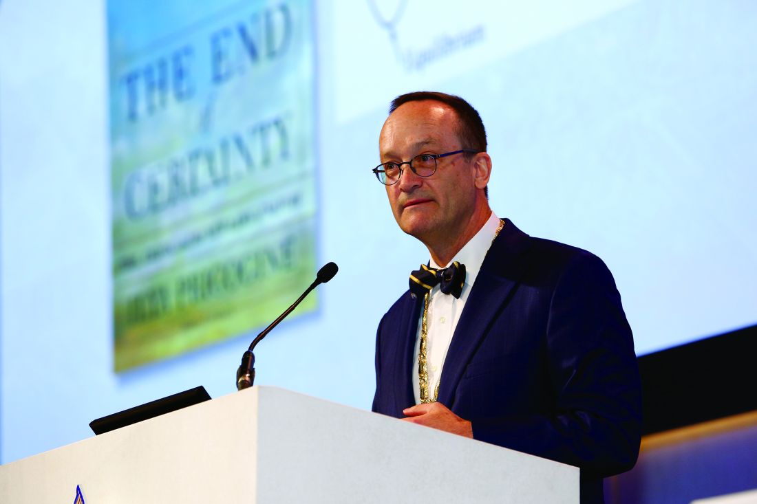
“If I were a betting man, I’d say there is no way value-based care can happen without hospitalists,” Robert Wachter, MD, MHM, said during a leadership summit at this year’s annual meeting of the Society of Hospital Medicine. Dr. Wachter is the New York Times bestselling author of “The Digital Doctor: Hope, Hype, and Harm at the Dawn of Medicine’s Computer Age.” He also is a professor and chair of the department of medicine at the University of California, San Francisco.

“There will be talk about closing hospitals and building competencies in ambulatory care. That’s the first 5 minutes, but then there is the realization people are still going to get sick, representing a massive amount of cost, and as far as I can tell, there is no alternative to having hospitals for this,” Dr. Wachter said. “Hospitalists will come out just fine.”
This will be true particularly for hospitalists who understand that electronic health records companies have helped health systems collect massive amounts of data but do not offer help for how those data can be applied to drive better outcomes and improve performance ratings, according to Dr. Wachter.
“These companies have no competencies in data analytics or data visualization,” Dr. Wachter said. “I am hopeful because Silicon Valley has woken up to the possibilities of this and are looking for ways to partner with health care. … The world of improving value, safety, and patient experience is going to be a world enabled by thoughtful use of informatics. So, if you don’t have those competencies, I think it’s important to grow them over time.”
Speaking the language of “lean” or that of other systematic strategies for improvement will also increase the value a hospitalist can bring to an organization, according to Dr. Wachter, who said that as hospital senior leadership and frontline personnel work together to implement system improvement, new leadership roles for hospitalists, ranging from chief patient experience officer to chief quality officers, and everything in between, are being created.
Another “hopeful trend” for hospitalists is the growing focus on the provider experience, said Dr. Wachter, since improving systems is not possible if staff are not engaged. “It’s impossible to believe you’re going to accomplish your goals of improving care and improving patient experience, and efficiencies, with a cadre of burned-out doctors,” he said.
Dr. Wachter said that to be indispensable, hospitalists need to understand they often are commoditized, offered by their senior leadership as bargaining chips when deals are made with surrounding health systems seeking to outsource some of their hospitalist functions.
“Managing a large community-affiliated network was not a core competency in the past,” Dr Wachter said. “But it will become increasingly important.”
Value-based care’s ascendancy equals increased opportunities for hospitalists who know how to wield informatics, speak the language of systems improvement, and have an ability to function as a commodity within a health system, according to the dean of hospital medicine.
“If I were a betting man, I’d say there is no way value-based care can happen without hospitalists,” Robert Wachter, MD, MHM, said during a leadership summit at this year’s annual meeting of the Society of Hospital Medicine. Dr. Wachter is the New York Times bestselling author of “The Digital Doctor: Hope, Hype, and Harm at the Dawn of Medicine’s Computer Age.” He also is a professor and chair of the department of medicine at the University of California, San Francisco.
“There will be talk about closing hospitals and building competencies in ambulatory care. That’s the first 5 minutes, but then there is the realization people are still going to get sick, representing a massive amount of cost, and as far as I can tell, there is no alternative to having hospitals for this,” Dr. Wachter said. “Hospitalists will come out just fine.”
This will be true particularly for hospitalists who understand that electronic health records companies have helped health systems collect massive amounts of data but do not offer help for how those data can be applied to drive better outcomes and improve performance ratings, according to Dr. Wachter.
“These companies have no competencies in data analytics or data visualization,” Dr. Wachter said. “I am hopeful because Silicon Valley has woken up to the possibilities of this and are looking for ways to partner with health care. … The world of improving value, safety, and patient experience is going to be a world enabled by thoughtful use of informatics. So, if you don’t have those competencies, I think it’s important to grow them over time.”
Speaking the language of “lean” or that of other systematic strategies for improvement will also increase the value a hospitalist can bring to an organization, according to Dr. Wachter, who said that as hospital senior leadership and frontline personnel work together to implement system improvement, new leadership roles for hospitalists, ranging from chief patient experience officer to chief quality officers, and everything in between, are being created.
Another “hopeful trend” for hospitalists is the growing focus on the provider experience, said Dr. Wachter, since improving systems is not possible if staff are not engaged. “It’s impossible to believe you’re going to accomplish your goals of improving care and improving patient experience, and efficiencies, with a cadre of burned-out doctors,” he said.
Dr. Wachter said that to be indispensable, hospitalists need to understand they often are commoditized, offered by their senior leadership as bargaining chips when deals are made with surrounding health systems seeking to outsource some of their hospitalist functions.
“Managing a large community-affiliated network was not a core competency in the past,” Dr Wachter said. “But it will become increasingly important.”
VIDEO: Dr. Lisa Newman on triple negative breast cancer in African American women
LAS VEGAS – The heavy burden of triple negative and other aggressive breast cancers among African American women cannot be simplified to socioeconomic factors alone.
International investigations by Lisa Newman, MD, director of the Breast Oncology Program at the Henry Ford Health System, Detroit, and other researchers are making it clear that genetic factors play a significant role.
She explained the latest findings and what they mean for screening, genetic referral, and treatment in an interview at the American Society of Breast Surgeons annual meeting.
The video associated with this article is no longer available on this site. Please view all of our videos on the MDedge YouTube channel
AT ASBS 2017

Attendees drill down on infections at ID boot camp
No hands went up when Glenn Wortmann, MD, chief of infectious diseases at MedStar Washington Hospital Center, asked whether any of the hospitalists in front of him had handled any cases of Candida auris, a kind of yeast that is highly resistant to several potent antifungals and, in some cases, has been found to be resistant to every antifungal thrown at it.
They might not have seen this dreadful bug, first identified in Japan in 2009, yet. But they will soon, Dr. Wortmann said.
James Pile, MD, SFHM, a pre-course director and associate professor of internal medicine at Case Western Reserve University, Cleveland, said he expected that most of those who signed up already knew quite a bit about I.D. issues, but the point of the course was to go deeper.
“Our goal was to find the sweet spot in what they don’t know or need a refresher on,” he said.
In his talk, he focused on infectious disease emergencies – such as infective endocarditis – which hospitalists may not see as often but that “we just have to get right when we do see them because the stakes are very high.”
He covered spinal epidural abscess, bacterial meningitis, and soft tissue necrotizing infections, saying that a crucial element in managing these cases is to seriously consider them a possibility in the first place. He also recommended having a low threshold for obtaining a surgical consultation, a CT scan, or both in patients with what appears to be severe cellulitis.
John Sanders, MD, MPH, head of infectious diseases at Wake Forest Baptist Health, Winston-Salem, N.C., dug into the nitty-gritty of C. difficile infections, touching on the possible role of acid suppressants, especially proton pump inhibitors, in the increasing incidence of these infections. The news wasn’t all bad: He also discussed emerging therapies, such as CRS3123 – a narrow-spectrum antibiotic that inhibits protein synthesis, toxin production, and sporulation – and monoclonal antibodies, which have been shown to lower recurrence rates when used with antibiotics.
The keys to controlling C. diff infections, he said, are hand-washing, remembering that the spores are resistant to ethanol, limiting fluoroquinolone use, and isolating patients with active infections.
Ultraviolet lighting for C. diff control is a gray area, he said.
“The data [are] mixed,” he said. “I think it’s a good idea to do it, and it’s certainly being pushed. But it’s somewhat controversial as to whether it’s cost effective.”

Presidential address focused on learning and teams
Thoralf M. Sundt III, MD, titled his AATS Presidential Address, “Ancora Imparo: Always Learning,” to emphasize the fact that learning is at the core of team development and of dealing with a complex world.
The world once seemed simple, deterministic – a Newtonian world of individual performance, he said. But in fact, it is a complex, unpredictable world, where the solutions of a previous generation of leaders that relied on individual knowledge and authority no longer apply.
“Complex is not the same as complicated,” said Dr. Sundt. “A watch is complicated, but it is entirely predictable. Complexity is different.” Complex systems are composed of many diverse autonomous parts. They are independent, they are linked as a system, and most important – they adapt. They change in response to the environment and to their own component parts.
How they change depends critically on the nature of the interactions among those components, allowing them to demonstrate emergent properties. And they are unpredictable too, said Dr. Sundt.
This unpredictability poses problems, he added, pointing to the inevitability of systems failures in truly complex endeavors from nuclear power to petrochemical plants.
“The implication of this is that we must think beyond error prevention because we simply cannot prevent them all,” said Dr. Sundt. “That is why checklists alone will not solve our problems.” The focus, instead, he suggested, needs to be on “error management” in order to include detection and recovery.
This is all relevant to surgeons because “our world, our patients, the procedures we perform, and the institutions in which we performed them – are increasingly complex.” Therefore, “no matter how much we care, and no matter how much we try, accidents, mistakes and mishaps will occur – individual effort is not enough.”
But complexity is only half the problem, he said. “The other half is us.”
Dr. Sundt discussed two ways of thinking – fast and slow – with fast thinking grabbing on to patterns and making quick, seemingly intuitive decisions, such as those made by “experts,” and the slow following a deliberative and logical path.
Both types of thinking have their benefits and lacks, which points to how important it is to have access to each type in order to maximize success. Thus, he pointed out, there must be a focus on teams and on a diversity of views, because diversity increases the chances of finding the best good solution.
So diversity is necessary, but is it sufficient? Two other things are necessary, according to Dr. Sundt.
The first is synergy, which is the difference between another useless administrative meeting and an exciting, generative one during which everyone shares opinions, changes their views a bit, and collectively comes up with an entirely original idea.
“But the key to transforming a workgroup to a high-performing team is learning,” he said. “It is an active process that requires both cognitive, and - unfortunately for us task-oriented personalities – affective skills. The aim is to create a ‘learning organization’ – a culture that encourages learning through open interactions in a psychologically safe space.
“It is entirely within our power to change the nature of the interactions we have within our teams, and we can do it today. No hospital committee or departmental approval is required. You cannot solve all of the problems, but you do have an enormous impact on those individuals around you.”
As the leader of a team, “everyone is watching you.” he added.
Dr. Sundt concluded by pointing out that it is also extremely important to confront inevitable failure as a learning experience. “You must wring every drop of learning out of it,” he counseled. And ultimately, “trust your team - you need them. The patient needs them,” he concluded.
Routine surveillance adds cost but little benefit following a pediatric tumor
MONTREAL – It can be a tough sell to worried patients or their caregivers, but surveillance imaging in children with a history of some treated solid tumors may not add anything to care except costs, a team of investigators suggested.
A study of the relationship between surveillance imaging and outcomes in patients who were followed after therapy for solid tumors found that “off therapy surveillance imaging did not affect outcomes in our relapsed solid tumor patients,” wrote Jacob Zimmerli and his coinvestigators at the University of Utah Huntsman Cancer Institute, Salt Lake City.
“In the case of rhabdomyosarcoma, we had a 31% relapse rate of our 39 unique cancer diagnoses that we looked at over a 10-year period. We had 12 relapses, and, of those relapses, 10 were found through nonroutine imaging,” Mr. Zimmerli said in an interview.
Survival rates for patients with rhabdomyosarcoma were identical whether their relapsed disease was detected via surveillance imaging or imaging performed when relapse was directly suspected because of clinical or physical findings, he said.
Surveillance imaging also came at a significant financial cost. Of the $345,000 total expenditure for imaging of rhabdomyosarcoma, $223,000 was spent on imaging for patients who never experienced a relapse. Additionally, $95,000 was spent on imaging for patients who had a relapse but whose relapses were not detected by routine surveillance imaging. Therefore, the investigators calculated, 92% of costs associated with surveillance did not lead to a relapse diagnosis.
“This finding in itself will lead us to look at a few more specific cancer subgroups and maybe do more cost analysis to see if there is a better way to treat these patients,” Mr. Zimmerli said.
To see whether surveillance imaging was associated with outcomes, the investigators performed a retrospective review of data on 292 patients treated at Primary Children’s Hospital in Salt Lake City for osteosarcoma, Wilms’ tumor, neuroblastoma, hepatoblastoma, Ewing sarcoma , or rhabdomyosarcoma.
All patients had either completed a minimum of 2 years of posttherapy follow-up or had a disease relapse within 2 years of completing therapy.
The investigators found that 8 of 10 osteosarcoma relapses, five of seven Wilms’ tumor relapses, and 9 of 15 neuroblastoma relapses were detected on routine imaging.
In contrast, only two of four hepatoblastoma relapses, three of nine Ewings sarcoma relapses, and 2 of 12 rhabdomyosarcoma relapses were found on routine imaging.
The overall survival rate for patients with relapses diagnosed on routine imaging was 51.72%, compared with 50% for patients whose relapses were diagnosed on imaging following the appearance of symptoms. The difference represented only one patient and was not statistically significant.
Mr. Zimmerli acknowledged that many routine posttherapy imaging studies are performed to assuage patient concerns and that it may be difficult to convince parents of children with a history of cancer that routine imaging of some tumors may not offer the assurances of relapse-free survival that they seek.
The study was supported by Intermountain Healthcare. The investigators reported no conflicts of interest.
FROM ASPHO
Key clinical point: Routine surveillance was less effective at detecting relapse of some pediatric solid tumors than imaging performed to confirm a clinical finding.
Major finding: Using imaging performed after symptoms appeared, 10 of 12 rhabdomyosarcoma relapses were detected.
Data source: A retrospective review of outcomes following routine and nonroutine imaging among children followed after treatment for one of six solid tumor types.
Disclosures: The study was supported by Intermountain Healthcare. The investigators reported no conflicts of interest.
5-Fluorouracil failed four separate measures of photoaging
PORTLAND – A standard course of topical 5-fluorouracil (5-FU) does not noticeably improve visual signs of facial photoaging, such as forehead lines and crow’s feet, according to the results of a blinded, controlled study of 281 elderly white men.
Four validated photonumeric measures revealed no statistically significant differences between the intervention and vehicle control arms at 6, 12, or 18 months’ follow-up, Kaveri Korgavkar, MD, said at the annual meeting of the Society for Investigative Dermatology. “This might be a true lack of impact, or current scales may not be sensitive enough to capture aspects of aging that are improved by 5-fluorouracil,” commented Dr. Korgavkar, who presented the findings on behalf of the VAKCCT (the Veterans Affairs Keratinocyte Carcinoma Chemoprevention Trial) work group.
The treatment and control groups resembled each other demographically and clinically at baseline. Participants averaged 71.5 years of age (standard deviation, 0.57 years), 97% were male, 99% were white, and all had clinically meaningful histories of sun damage with at least two keratinocyte carcinomas in the previous 2 years, including at least one lesion on the face or ears. Previously, the VAKCCT investigators reported positive results for 5-FU as a chemopreventive – for example, it was associated with about a 60% reduction in actinic keratoses, compared with placebo, and the effects persisted for up to 3 years.
However, none of the four photonumeric scales of photoaging uncovered significant differences between the treatment and control groups at 6, 12, or 18 months’ follow-up, Dr. Korgavkar reported. That finding belies the results of two other previous studies, but they were small and uncontrolled, she added. One study of 19 patients reported statistically significant improvements over time in wrinkling, hyperpigmentation, lentigines, and sallowness based on the Griffith’s scale, while a second prospective study of 32 patients reported significant improvements in visual signs of photoaging on the forearms, with a corresponding rise in levels of procollagen 1 and a decrease in dermal elastosis at 1 month.
Existing scales might more effectively capture some aspects of photoaging – such as wrinkles or crow’s feet – than others, Dr. Korgavkar said in an interview. Therefore, she and her associates are working to construct more sensitive and comprehensive visual scales of photoaging, she said.
The VAKCCT was sponsored by the VA Office of Research and Development. Dr. Korgavkar had no conflicts of interest.
AT SID 2017
Key clinical point: A standard topical course of 5-fluorouracil did not noticeably improve visual signs of photoaging, such as forehead lines and crow’s feet.
Major finding: Four validated photonumeric measures of photoaging revealed no statistically significant differences between the intervention and the vehicle control at 6, 12, or 18 months’ follow-up.
Data source: An analysis of data from 281 participants in the Veterans Affairs Keratinocyte Carcinoma Chemoprevention trial (VAKCCT).
Disclosures: The VAKCCT was sponsored by the VA Office of Research and Development. Dr. Korgavkar had no conflicts of interest.