User login
Pembrolizumab proves promising for treating advanced SCLC
VIENNA – One third of patients with extensive-stage small-cell lung cancer (SCLC) treated with the checkpoint inhibitor pembrolizumab achieved an objective response, according to updated data from the KEYNOTE-28 trial.
Of the 24 patients with advanced SCLC who received pembrolizumab in the multicohort phase Ib study, one had a complete response, seven had a partial response, and one further patient had stable disease for 6 or more months, giving a clinical benefit rate of 33.3% (95% CI, 15.6-53.3%).
“Pembrolizumab demonstrated meaningful antitumor activity in previously treated patients with PD-L1-positive small-cell lung cancer,” said presenting study author Patrick A. Ott, MD, PhD, of the Dana-Faber Cancer Institute in Boston.
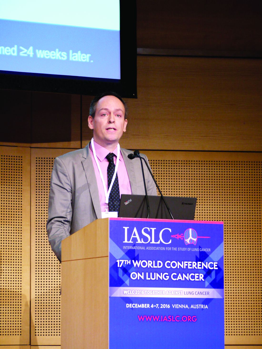
“Responses were durable, with a median duration of response of 19.4 months,” Dr. Ott said at the World Conference on Cancer, which is sponsored by the International Association for the Study of Lung Cancer. About 40% of patients exhibited a decrease in tumor size, he added.
Treatment with pembrolizumab was associated with a progression-free survival (PFS) of 1.2 months, with a range of 1.7-5.9 months. The 6- and 12-month PFS rates were 28.6% and 23.8%, respectively.
The median overall survival (OS) was 9.7 months, ranging from 4.1 months to an upper limit that has not yet been reached. The respective 6- and 12-month OS rates were 66.0% and 37.7%.
“The safety experience was consistent with previous experience for pembrolizumab in other tumor types, and was identical with longer follow-up” Dr. Ott said.
Over a median follow-up duration of 9.8 months, treatment-related adverse events occurred in two-thirds of patients, with arthralgia, asthenia, and rash reported by four (16.7%) patients each, and diarrhea and fatigue by three (12.5%) patients each. There were no cases of pneumonitis.
The mean age of patients recruited into the KEYNOTE-28 trial was 60.5 years; more than half of the patients (58%) were male. Five (20.8%) had stable brain metastases, and 95.8% had small cell histology.
All recruited patients had received prior standard-of-care chemotherapy with cisplatin or carboplatin plus etoposide, 45.8% had received irinotecan or topotecan, and 29.2% had been treated with a taxane. In addition, one patient had received prior radiotherapy, one had been given an investigational tyrosine kinase inhibitor therapy, and another had received another, unspecified, investigational treatment.
After enrollment, all patients were treated with pembrolizumab at an intravenous dose of 10 mg/kg every 2 weeks. Response was assessed every 8 weeks for the first 6 months and then annually, with patients continuing to respond continuing to be treated for up to 2 years or until progression or unacceptable toxicity.
There are two ongoing, phase II trials with pembrolizumab in patients with advanced SCLC. One is looking at maintenance therapy with pembrolizumab after combination chemotherapy and is sponsored by the Barbara Ann Karmanos Cancer Institute in collaboration with the National Cancer Institute (NCT02359019). The other, the REACTION trial sponsored by the European Organisation for Research and Treatment of Cancer, is looking at the use of pembrolizumab together with etoposide and platinum chemotherapy in previously untreated patients (NCT02580994).
Merck funded the KEYNOTE-28 study. Dr. Ott disclosed ties with Bristol-Myers Squibb, Merck, ArmoBiosciences, AstraZeneca/MedImmune, Celldex, Genentech, Alexion, and CytomX.
VIENNA – One third of patients with extensive-stage small-cell lung cancer (SCLC) treated with the checkpoint inhibitor pembrolizumab achieved an objective response, according to updated data from the KEYNOTE-28 trial.
Of the 24 patients with advanced SCLC who received pembrolizumab in the multicohort phase Ib study, one had a complete response, seven had a partial response, and one further patient had stable disease for 6 or more months, giving a clinical benefit rate of 33.3% (95% CI, 15.6-53.3%).
“Pembrolizumab demonstrated meaningful antitumor activity in previously treated patients with PD-L1-positive small-cell lung cancer,” said presenting study author Patrick A. Ott, MD, PhD, of the Dana-Faber Cancer Institute in Boston.
“Responses were durable, with a median duration of response of 19.4 months,” Dr. Ott said at the World Conference on Cancer, which is sponsored by the International Association for the Study of Lung Cancer. About 40% of patients exhibited a decrease in tumor size, he added.
Treatment with pembrolizumab was associated with a progression-free survival (PFS) of 1.2 months, with a range of 1.7-5.9 months. The 6- and 12-month PFS rates were 28.6% and 23.8%, respectively.
The median overall survival (OS) was 9.7 months, ranging from 4.1 months to an upper limit that has not yet been reached. The respective 6- and 12-month OS rates were 66.0% and 37.7%.
“The safety experience was consistent with previous experience for pembrolizumab in other tumor types, and was identical with longer follow-up” Dr. Ott said.
Over a median follow-up duration of 9.8 months, treatment-related adverse events occurred in two-thirds of patients, with arthralgia, asthenia, and rash reported by four (16.7%) patients each, and diarrhea and fatigue by three (12.5%) patients each. There were no cases of pneumonitis.
The mean age of patients recruited into the KEYNOTE-28 trial was 60.5 years; more than half of the patients (58%) were male. Five (20.8%) had stable brain metastases, and 95.8% had small cell histology.
All recruited patients had received prior standard-of-care chemotherapy with cisplatin or carboplatin plus etoposide, 45.8% had received irinotecan or topotecan, and 29.2% had been treated with a taxane. In addition, one patient had received prior radiotherapy, one had been given an investigational tyrosine kinase inhibitor therapy, and another had received another, unspecified, investigational treatment.
After enrollment, all patients were treated with pembrolizumab at an intravenous dose of 10 mg/kg every 2 weeks. Response was assessed every 8 weeks for the first 6 months and then annually, with patients continuing to respond continuing to be treated for up to 2 years or until progression or unacceptable toxicity.
There are two ongoing, phase II trials with pembrolizumab in patients with advanced SCLC. One is looking at maintenance therapy with pembrolizumab after combination chemotherapy and is sponsored by the Barbara Ann Karmanos Cancer Institute in collaboration with the National Cancer Institute (NCT02359019). The other, the REACTION trial sponsored by the European Organisation for Research and Treatment of Cancer, is looking at the use of pembrolizumab together with etoposide and platinum chemotherapy in previously untreated patients (NCT02580994).
Merck funded the KEYNOTE-28 study. Dr. Ott disclosed ties with Bristol-Myers Squibb, Merck, ArmoBiosciences, AstraZeneca/MedImmune, Celldex, Genentech, Alexion, and CytomX.
AT WCLC 2016
Key clinical point: Pembrolizumab has antitumor activity in patients with advanced small-cell lung cancer.
Major finding: The objective response rate was 33.3% (95% CI 15.6-55.3%), including one complete and seven partial responses.
Data source: Phase Ib, nonrandomized, multicohort trial of 24 heavily pretreated patients with extensive-disease small-cell lung cancer.
Disclosures: Merck funded the study. Dr. Ott disclosed ties with Bristol-Myers Squibb, Merck, ArmoBiosciences, AstraZeneca/MedImmune, Celldex, Genentech, Alexion, and CytomX.
Skin cancer a concern in pediatric solid organ transplant recipients
As survival rates among pediatric organ transplant recipients increase, so do the rates of cutaneous malignancies later in life for this population, who are at a greater risk for skin cancers that include nonmelanoma skin cancers (NMSCs), melanoma, Kaposi sarcoma, and anogenital carcinoma, according to the authors of a literature review.
In studies, skin cancers account for 13%-55% of all cancers in pediatric organ transplant recipients (POTRs), according to Alexander L. Fogel of Stanford (Calif.) University and his coauthors. The review article provides an update on this topic, as well as information on the prevention and management of skin cancers in this population, and the differences between this group and adult organ transplant recipients (AOTRs).

NMSC is the most common type of skin cancer in the pediatric group – and the second most common type of malignancy (NMSCs are the most common type of cancer affecting adult organ transplant recipients). NMSCs typically appear an average of 12-18 years post transplantation in this population (at an average age of 26-34 years). Length of posttransplantation follow-up, sunlight exposure, fair skin, and Northern European ancestry are among the factors associated with increased risk. This type of cancer involves the lip nearly twice as often as in adult recipients: 23% vs. 12%. The pediatric cohort also experiences more nonmelanoma cancer spreading to the lymph nodes than do adults: 9% vs. 6%.
Among pediatric transplant recipients, squamous cell carcinomas appear 2.8 times more often than basal cell carcinomas, “a trend that is opposite that observed in the nontransplant population,” the authors wrote.
In one study, anogenital carcinomas accounted for 4% of posttransplant cancers in this cohort, at an average of 12 years after the transplant, at a mean age of 27 years.
Some data indicate that in adult transplant recipients, there is an association between the human papillomavirus, and anal and genital warts and posttransplant anogenital cancer, but there are little data looking at this association in the pediatric group, the authors noted.
Although melanoma and Kaposi sarcoma are also found in this cohort at rates greater than in the general population, and are associated with high mortality rates, the data are too few to draw conclusions, the authors wrote.
In 2014, 1,795 pediatric solid organ transplants were performed, accounting for 6% of all such transplants. The absolute number of pediatric transplants has remained fairly stable over 5 years, yet very little pediatric-specific literature exists for prevention and management of skin cancers post transplantation, the authors pointed out.
Changing immunosuppressive medications used in transplantation may be effective in reducing skin cancer risk, they said, noting that including rapamycin inhibitors in combination therapy has been shown to reduce the risk of developing skin cancers in some transplant patients by more than half.
The authors emphasized that regular sunscreen use and dermatologic checkups are also essential in this population, and that “the importance of regular dermatologic evaluation should be stressed to patients and their families.”
Mr. Fogel’s coauthors were Mari Miyar, MD, of the department of dermatology, Kaiser Permanente, San Jose, Calif., and Joyce Teng, MD, of the departments of dermatology and pediatrics, Stanford. The authors had no disclosures listed, and no funding source for the review was listed.
This article was updated 12/8/16.
[email protected]
On Twitter @whitneymcknight
As survival rates among pediatric organ transplant recipients increase, so do the rates of cutaneous malignancies later in life for this population, who are at a greater risk for skin cancers that include nonmelanoma skin cancers (NMSCs), melanoma, Kaposi sarcoma, and anogenital carcinoma, according to the authors of a literature review.
In studies, skin cancers account for 13%-55% of all cancers in pediatric organ transplant recipients (POTRs), according to Alexander L. Fogel of Stanford (Calif.) University and his coauthors. The review article provides an update on this topic, as well as information on the prevention and management of skin cancers in this population, and the differences between this group and adult organ transplant recipients (AOTRs).
NMSC is the most common type of skin cancer in the pediatric group – and the second most common type of malignancy (NMSCs are the most common type of cancer affecting adult organ transplant recipients). NMSCs typically appear an average of 12-18 years post transplantation in this population (at an average age of 26-34 years). Length of posttransplantation follow-up, sunlight exposure, fair skin, and Northern European ancestry are among the factors associated with increased risk. This type of cancer involves the lip nearly twice as often as in adult recipients: 23% vs. 12%. The pediatric cohort also experiences more nonmelanoma cancer spreading to the lymph nodes than do adults: 9% vs. 6%.
Among pediatric transplant recipients, squamous cell carcinomas appear 2.8 times more often than basal cell carcinomas, “a trend that is opposite that observed in the nontransplant population,” the authors wrote.
In one study, anogenital carcinomas accounted for 4% of posttransplant cancers in this cohort, at an average of 12 years after the transplant, at a mean age of 27 years.
Some data indicate that in adult transplant recipients, there is an association between the human papillomavirus, and anal and genital warts and posttransplant anogenital cancer, but there are little data looking at this association in the pediatric group, the authors noted.
Although melanoma and Kaposi sarcoma are also found in this cohort at rates greater than in the general population, and are associated with high mortality rates, the data are too few to draw conclusions, the authors wrote.
In 2014, 1,795 pediatric solid organ transplants were performed, accounting for 6% of all such transplants. The absolute number of pediatric transplants has remained fairly stable over 5 years, yet very little pediatric-specific literature exists for prevention and management of skin cancers post transplantation, the authors pointed out.
Changing immunosuppressive medications used in transplantation may be effective in reducing skin cancer risk, they said, noting that including rapamycin inhibitors in combination therapy has been shown to reduce the risk of developing skin cancers in some transplant patients by more than half.
The authors emphasized that regular sunscreen use and dermatologic checkups are also essential in this population, and that “the importance of regular dermatologic evaluation should be stressed to patients and their families.”
Mr. Fogel’s coauthors were Mari Miyar, MD, of the department of dermatology, Kaiser Permanente, San Jose, Calif., and Joyce Teng, MD, of the departments of dermatology and pediatrics, Stanford. The authors had no disclosures listed, and no funding source for the review was listed.
This article was updated 12/8/16.
[email protected]
On Twitter @whitneymcknight
FROM PEDIATRIC DERMATOLOGY
Key clinical point:
Major finding: Pediatric solid organ transplant recipients experience skin cancer rates between 13% and 55%.
Data source: A literature review of malignancies among pediatric organ transplant recipients.
Disclosures: The authors listed no disclosures, and no funding source for the review was listed.
New-onset pain rare after Essure placement
ORLANDO – While some women report new-onset pelvic pain after placement of an Essure sterilization device, results of a retrospective study suggest this pain is actually associated with placement of the device in about 1% of cases.
Among 1,430 women who had an Essure micro-insert (Bayer) placed at a tertiary care hospital in Canada from June 2002 to June 2013, 62 secondary surgeries were performed, including some for removal of fallopian tubes and removal of the device.
In total, 27 patients reported new-onset pelvic pain after Essure placement and another 11 reported worsening of previous pain. Upon further workup, 15 of the 27 women in the new-onset pain group had another possible explanation for their pain, including surgical or pathology findings of endometriosis or adenomyosis. The investigators concluded there was a link between the pain and the device in just 12 (0.8%) of the women.
Among these dozen patients, the investigators linked the pain in eight women to perforation or migration of the Essure device. Investigators found no other obvious cause for the new-onset pain in the remaining four patients and attributed it to the Essure device.

Set realistic expectations, take a comprehensive pain history, and reassure women when they report post-Essure placement pain, James Robinson, MD, a minimally invasive gynecologic surgeon at Medstar Washington Hospital in Washington, D.C., advised at the meeting, which was sponsored by AAGL.
Dr. Robinson pointed out that there is no standardized approach to managing women with complaints of pain or guidelines on how best to remove the device. Imaging to confirm proper placement and to rule out other sources of pelvic pain, followed by medical or surgical management as warranted, can be effective strategies.
“There is less science here – it’s more the art of medicine, I think,” he said.
Dr. Robinson filled in as a presenter for one of the study coauthors, John A. Thiel, MD, of the University of Saskatchewan, Saskatoon, who was unable to attend the conference.
The study by Dr. Thiel and his colleagues suggests a thorough examination will typically reveal other reasons for pelvic pain and rules out the Essure device as the cause, Dr. Robinson said. The full findings of the study are published in the Journal of Minimally Invasive Gynecology (2016 Nov-Dec;23[7]:1158-1162).
Does removal help?
In a recently published case series of 29 women who had their Essure device removed laparoscopically because of pain, 23 reported relief following excision (Contraception. 2016 Aug;94[2]:190-2).
“Again, a subset had misplaced inserts or had another condition such as endometriosis,” Dr. Robinson said.
The majority of women whose pain resolved with removal of the devices reported their pain early on, so there is an important takeaway from this,” Dr. Robinson said. “We need to listen to our patients when they report pain shortly after device placement … and respond to that.”
Dr. Robinson advised physicians to be ready to surgically remove the device if that is warranted. “I think a lot of people doing these procedures are not comfortable taking their patient back to the operating room or don’t know who to send them to,” he said. “If you are going to place the device, you should be able to take it out or know someone who can.”
The bigger picture
Even though the Essure device is not frequently the cause of pelvic pain, physicians needs to be aware that some patients are likely to assume that it is.
Dr. Robinson pointed to a case in which a woman who had the Essure micro-insert placed 7 years earlier presented with a complaint of a bilateral tingling sensation over the course of 6 months. Online research led her to suspect Essure as the cause of her symptoms. However, on further investigation, it turned out she had relatively high levels of lead in her system from leaky pipes in her home. “It wasn’t an Essure issue,” he said. “But because it’s out there, people will jump to the conclusion that the foreign body is likely the cause of their problem.”
Ob.gyns. should become familiar with websites such as essureproblems.webs.com, which chronicle problems patients have reported with the device, he said.
When talking to patients, start with informed consent and listen to their concerns, Dr. Robinson advised. As part of the counseling about Essure permanent birth control, discuss the risks and benefits of alternatives, such as laparoscopic tubal ligation, long-acting reversible contraception, and vasectomy.
“I took the time to listen to the FDA hearing in Sept 2015 and … it moved me to listen to those patients, and I’ve been a huge advocate of Essure sterilization. I felt for a while I would never do another tubal ligation,” Dr. Robinson said. “But when you listen to patients who have real complaints, what sticks out in your mind is so many of these people are upset because no one took them seriously and listened to them.”
ORLANDO – While some women report new-onset pelvic pain after placement of an Essure sterilization device, results of a retrospective study suggest this pain is actually associated with placement of the device in about 1% of cases.
Among 1,430 women who had an Essure micro-insert (Bayer) placed at a tertiary care hospital in Canada from June 2002 to June 2013, 62 secondary surgeries were performed, including some for removal of fallopian tubes and removal of the device.
In total, 27 patients reported new-onset pelvic pain after Essure placement and another 11 reported worsening of previous pain. Upon further workup, 15 of the 27 women in the new-onset pain group had another possible explanation for their pain, including surgical or pathology findings of endometriosis or adenomyosis. The investigators concluded there was a link between the pain and the device in just 12 (0.8%) of the women.
Among these dozen patients, the investigators linked the pain in eight women to perforation or migration of the Essure device. Investigators found no other obvious cause for the new-onset pain in the remaining four patients and attributed it to the Essure device.
Set realistic expectations, take a comprehensive pain history, and reassure women when they report post-Essure placement pain, James Robinson, MD, a minimally invasive gynecologic surgeon at Medstar Washington Hospital in Washington, D.C., advised at the meeting, which was sponsored by AAGL.
Dr. Robinson pointed out that there is no standardized approach to managing women with complaints of pain or guidelines on how best to remove the device. Imaging to confirm proper placement and to rule out other sources of pelvic pain, followed by medical or surgical management as warranted, can be effective strategies.
“There is less science here – it’s more the art of medicine, I think,” he said.
Dr. Robinson filled in as a presenter for one of the study coauthors, John A. Thiel, MD, of the University of Saskatchewan, Saskatoon, who was unable to attend the conference.
The study by Dr. Thiel and his colleagues suggests a thorough examination will typically reveal other reasons for pelvic pain and rules out the Essure device as the cause, Dr. Robinson said. The full findings of the study are published in the Journal of Minimally Invasive Gynecology (2016 Nov-Dec;23[7]:1158-1162).
Does removal help?
In a recently published case series of 29 women who had their Essure device removed laparoscopically because of pain, 23 reported relief following excision (Contraception. 2016 Aug;94[2]:190-2).
“Again, a subset had misplaced inserts or had another condition such as endometriosis,” Dr. Robinson said.
The majority of women whose pain resolved with removal of the devices reported their pain early on, so there is an important takeaway from this,” Dr. Robinson said. “We need to listen to our patients when they report pain shortly after device placement … and respond to that.”
Dr. Robinson advised physicians to be ready to surgically remove the device if that is warranted. “I think a lot of people doing these procedures are not comfortable taking their patient back to the operating room or don’t know who to send them to,” he said. “If you are going to place the device, you should be able to take it out or know someone who can.”
The bigger picture
Even though the Essure device is not frequently the cause of pelvic pain, physicians needs to be aware that some patients are likely to assume that it is.
Dr. Robinson pointed to a case in which a woman who had the Essure micro-insert placed 7 years earlier presented with a complaint of a bilateral tingling sensation over the course of 6 months. Online research led her to suspect Essure as the cause of her symptoms. However, on further investigation, it turned out she had relatively high levels of lead in her system from leaky pipes in her home. “It wasn’t an Essure issue,” he said. “But because it’s out there, people will jump to the conclusion that the foreign body is likely the cause of their problem.”
Ob.gyns. should become familiar with websites such as essureproblems.webs.com, which chronicle problems patients have reported with the device, he said.
When talking to patients, start with informed consent and listen to their concerns, Dr. Robinson advised. As part of the counseling about Essure permanent birth control, discuss the risks and benefits of alternatives, such as laparoscopic tubal ligation, long-acting reversible contraception, and vasectomy.
“I took the time to listen to the FDA hearing in Sept 2015 and … it moved me to listen to those patients, and I’ve been a huge advocate of Essure sterilization. I felt for a while I would never do another tubal ligation,” Dr. Robinson said. “But when you listen to patients who have real complaints, what sticks out in your mind is so many of these people are upset because no one took them seriously and listened to them.”
Key clinical point:
Major finding: Of 1,430 women who had the Essure inserts placed, just 12 had new-onset pain that was found to be related to the device.
Data source: Retrospective cohort study of 1,430 women treated at a tertiary care hospital in Canada.
Disclosures: Dr. Robinson reported having no financial disclosures.
Prescribing the landmark hemangioma drug: The challenges and the benefits
For Beth Drolet, MD, a pediatric dermatologist in Wisconsin, the tremendous impact oral propranolol has had on the treatment of severe infantile hemangioma is written on the faces of children diagnosed with the condition in recent years.
“You can tell which drugs the kids were on by their age,” said Dr. Drolet, professor of dermatology and pediatrics at the Medical College of Wisconsin, Milwaukee. “If they were born before 2008, before we used this medication, those kids have had multiple surgeries and are still not looking that good. But we rarely see that in the kids born after.”
Still, it is possible for dermatologists to successfully treat their smallest patients with oral propranolol, according to Dr. Drolet and Ilona J. Frieden, MD, professor of dermatology and pediatrics at the University of California, San Francisco.
In interviews, the two pediatric dermatologists spoke about the challenges and benefits of treating hemangioma patients with oral propranolol solution, which was approved by the Food and Drug Administration in 2014 for “proliferating infantile hemangioma requiring systemic therapy.” It is the only FDA-approved systemic treatment for this indication.
The oral form of the drug was used off label to treat patients with hemangioma after a French dermatologist discovered in 2007 that it could effectively treat the condition. A topical form of propranolol is also used for hemangiomas that do not require systemic treatment.
Prior to about a decade ago, Dr. Drolet said, steroids were used to treat severe hemangiomas with limited success.
In general, infantile hemangiomas “have a natural course of gradually involuting even without treatment,” Dr. Frieden noted. But the most severe cases can produce functional impairment, scarring, and anatomic distortion.
Dr. Drolet said she considers treatment if hemangioma threatens a vital function (hearing, sight, breathing) or can lead to pain, infection, or scarring.
One challenge for dermatologists is that standard of care treatment with oral propranolol requires in-office cardiac monitoring, especially as the dose is increased over the first week or two of treatment.
“I don’t think most dermatologists are comfortable taking a heart rate and blood pressure in an infant,” said Dr. Drolet, who is director of the birthmarks and vascular anomalies section at Children’s Hospital of Wisconsin, Milwaukee. Instead, they tend to refer patients to a pediatrician or pediatric cardiologist.
Her clinic hired a cardiac nurse to train the staff in how to take heart rate and blood pressure in babies. “Partnering with cardiology was really important for us,” she commented. “We worked really closely with our pediatric cardiology team to gain that expertise for our staff to assess that. You have to be pretty comfortable with it. If you’re not, you’re going to have to find someone else.”
Another option for dermatologists, Dr. Frieden said, is to focus on heart rate alone since blood pressure in infants is difficult to measure. “It’s not FDA sanctioned, but many people seem to do that and it’s OK,” she said.
Dr. Frieden and Dr. Drolet provided the following recommendations about treating babies with oral propranolol:
• Caution parents about side effects. Cardiac side effects have been “extraordinarily rare,” Dr. Drolet said. “We have seen problems with wheezing and, very rarely, severe hypoglycemia,” which can be prevented by educating the family. While it’s uncommon for the medication alone to produce wheezing, this may occur when a respiratory infection and propranolol combine to stress the body, she noted.
In some cases, physicians prescribe albuterol for wheezing without realizing that it will interact with propranolol, she added. “One is a beta-blocker, and the other is a beta-antagonist. They completely cancel each other out.”
To prevent hypoglycemia, Dr. Frieden said she recommends that children be fed every 6 hours if they’re under 6 months old or every 8 hours if they’re over 6 months of age. And Dr. Drolet said she advises parents to stop propranolol when their infants are sick.
A major focus of an educational video provided by Dr. Drolet’s clinic is advising parents “to stop the medication if the infant is not eating regularly, vomiting, or has diarrhea. It interferes with how you respond to low blood sugar if you’re not eating,” she said. “That surprised us. Now that we’ve been teaching parents about when to call us, that’s been pretty preventable.”
Minor side effects include cold hands and feet and sleep disturbances such as sleepiness and apparent nightmares, Dr. Frieden pointed out.
• Monitor guidelines regarding safety and protocols. “Over time, we’re getting more and more expertise,” Dr. Drolet said. For example, her clinic no longer performs ECGs on babies who take the medication because research has suggested they are not needed.
• Spend time developing an education program for parents. Dr. Drolet’s clinic provides the educational video to teach parents about how oral propranolol is used. “We haven’t done that for any other drugs,” she said. “But we want to make sure we aren’t overdosing it. We’ve been very careful about our parent education to prevent that.”
Guidelines on the diagnosis and management of infantile hemangioma were published in 2015 in Pediatrics (2015 Oct;136[4]:e1060-104).
Dr. Frieden has consulted for Pierre Fabre Dermatologie, the manufacturer of the oral propranolol product, marketed as Hemangeol. Dr. Drolet has received an investigator-initiated grant from the company.
Study: Pretreatment ECG not always needed in babies with hemangiomas
Routine ECG screening in infants before they receive propranolol for hemangiomas is “not likely to be an effective screening tool in patients with otherwise normal physical examination and family history” and may even cause harmful delays in treatment, study authors concluded.
“As previously published guidelines suggest, it is likely that an indication-driven ECG strategy is a better approach, because there is a low incidence of ECG abnormalities that would limit propranolol use in children,” wrote Kevin B. Yarbrough, MD, a dermatologist at Phoenix Children’s Hospital, and his associates. The results “support published guidelines for propranolol initiation and are congruent with findings from other investigators” (Pediatr Dermatol. 2016 Nov;33[6]:615-20).
In the retrospective study, Dr. Yarbrough and his associates tracked 162 patients (median age, 5.2 months) who underwent routine ECG screening at several clinics before propranolol treatment for hemangiomas from 2008 to 2013. The ECGs were read as abnormal in 69 cases (43%); the most common abnormality was left ventricular hypertrophy (16 patients), followed by right ventricular hypertrophy (8), sinus bradycardia (6), and sinus tachycardia (5).
Cardiologists cleared all 69 patients for propranolol treatment, which they received. “No patients in our cohort experienced an adverse effect during treatment that could have been predicted or prevented by ECG before initiation of the propranolol,” the authors wrote.
“Routine ECG adds to the cost of treating hemangiomas and leads to unnecessary consultations and testing. Even more importantly, abnormalities detected on ECG can lead to delays in treatment initiation, which in turn can lead to greater patient morbidity, as seen in the case of our patient whose hemangioma ulcerated while awaiting cardiology consultation,” they added.
Still, they noted that ECG tests should still be performed on “infants with bradycardia or cardiac arrhythmia found during initial physical examination, a family history of congenital heart disease or arrhythmias, and a maternal history of connective tissue disease.”
Study funding information was not provided. One of the study authors reported that he was a clinical investigator for Pierre Fabre Dermatologie, the manufacturer of the oral propranolol product Hemangeol.
FROM PEDIATRIC DERMATOLOGY
Key clinical point: While it’s appropriate in some cases, routine ECG screening appears to be unnecessary before administering propranolol to infants to treat hemangiomas.
Major finding: All 69 infants whose screening ECGs turned up abnormalities were subsequently cleared by cardiologists.
Data source: A retrospective analysis of 162 patients with infantile hemangiomas seen at various clinics from 2008 to 2013.
Disclosures: Study funding information was not provided. One of the study authors, Alfons L. Krol, MD, reported being a clinical investigator for Pierre Fabre Dermatologie, the manufacturer of the oral propranolol product Hemangeol.
Stem cell therapy for STEMI falls short at 2 years
NEW ORLEANS – Longer follow-up has not altered initial negative findings of the randomized phase II TIME trial, which tested the efficacy of early intracoronary stem cell therapy, given on day 3 or 7 after an ST-segment acute MI (STEMI). However, loss of the sickest patients from follow-up may have influenced the findings.
“When TIME (Transplantation in Myocardial Infarction Evaluation) was developed several years ago, the optimal timing for cell delivery following myocardial infarction was not known and had not been directly tested in a prospective clinical trial,” explained lead investigator Jay H. Traverse, MD, director of research at the Minneapolis Heart Institute Foundation and a senior consulting cardiologist at the Abbott Northwestern Hospital, Minneapolis.

With the new, additional follow-up out to 2 years in 85 patients, both groups saw further gains in LV ejection fraction and further reductions in LV infarct size, as well as stable LV end-diastolic volume index, but still with no significant differences between them.
Of note, however, 10 of the patients lost to follow-up were lost because they received implantable cardioverter defibrillators (ICDs) and, given technologic limitations at the time, could no longer undergo cardiac MRI, Dr. Traverse noted. “These 10 patients are important as we look at the long-term follow-up of TIME because these patients were more likely to have the most severe LV dysfunction and most remodeled left ventricles. This [subset] represents a limitation to long-term follow-up studies in this population.”
Challenges to research
Two challenging issues seen in many trials of cell therapy for cardiovascular disease are their underpowered nature and the changing natural history of the disease, according to session comoderator Timothy D. Henry, MD, head of cardiology at Cedars-Sinai in Los Angeles.
“One of the things with the TIME trial that’s striking, really, is that even though these are high-risk patients, there was almost no mortality in 2 years,” he commented. “Second of all... there were no differences in ejection fraction, but that’s because you are missing 30% of people. And your missing 30% of people are the sick people, so of course if you are just going to follow normal people for 2 years, you’re going to have normal results.”
“What you are seeing is a little bit illusory because all the sick patients got ICDs and we could no longer image them at that time,” Dr. Traverse agreed. “That will be less of an issue in some of our future trials that we have started now, like CONCERT and SENECA, where we are now able to do MRI analysis of people with devices. So that will certainly help.”
Nonetheless, all trials in similar patient populations are plagued by substantial rates of dropout over time and faced with improving outcomes generally, because of better medical therapy, he acknowledged. “You can see as far as the natural history, even taking into account the ICD changes that would have lowered volumes, people are pretty stable on medical therapy. Their ejection fractions and volumes out to 2 years were really quite stable. We have certainly impacted that.”
That issue raises the question of whether investigators should be using other endpoints going forward, according to session panelist Doris A. Taylor, PhD, director of Regenerative Medicine Research at the Texas Heart Institute in Houston.
“One of the things I’ve seen over and over in this field is constantly evolving questions about which endpoints we should follow and what constitutes positive effects,” she commented. “So given the natural history, what are the endpoints we should be using?”
“Patients don’t care what their ejection fraction is per se. But they want to know if they have to get an ICD or if they will be hospitalized for heart failure, and their family is affected if they die,” Dr. Traverse replied. “We definitely need these hard endpoints. The problem is that you need so many patients with these hard endpoints that [the trials] are just financially not very doable. So that’s a big issue.”
Trial details
Patients enrolled in TIME had a first anterior STEMI, underwent reperfusion by angioplasty and stenting, and had LV dysfunction with an ejection fraction of 45% or lower. They all received either autologous bone marrow mononuclear cells or placebo on day 3 or day 7 after their MI by intracoronary infusion.
Of the initial 120 randomized patients, 10 patients were lost to follow-up because of receipt of an ICD, 3 had died (1 each from cardiovascular causes, pancreatitis, and hemorrhagic stroke), 7 were lost to follow-up for other reasons, and 15 had acquired other contraindications to MRI, according to Dr. Traverse.
At 2 years, the remaining patients in both the cell therapy and placebo groups had roughly 5% absolute increases in LV ejection fraction from baseline and roughly 45% reductions in infarct size from baseline, with no significant differences between groups.
When all patients were combined, about half were determined to have had microvascular obstruction at baseline. This finding was an adverse prognostic factor, associated with poorer recovery of LV function over time, greater adverse LV remodeling, and a higher likelihood of receiving an ICD, Dr. Traverse reported.
He has received a research grant from the National Heart, Lung, and Blood Institute, which sponsored the TIME trial.
NEW ORLEANS – Longer follow-up has not altered initial negative findings of the randomized phase II TIME trial, which tested the efficacy of early intracoronary stem cell therapy, given on day 3 or 7 after an ST-segment acute MI (STEMI). However, loss of the sickest patients from follow-up may have influenced the findings.
“When TIME (Transplantation in Myocardial Infarction Evaluation) was developed several years ago, the optimal timing for cell delivery following myocardial infarction was not known and had not been directly tested in a prospective clinical trial,” explained lead investigator Jay H. Traverse, MD, director of research at the Minneapolis Heart Institute Foundation and a senior consulting cardiologist at the Abbott Northwestern Hospital, Minneapolis.
With the new, additional follow-up out to 2 years in 85 patients, both groups saw further gains in LV ejection fraction and further reductions in LV infarct size, as well as stable LV end-diastolic volume index, but still with no significant differences between them.
Of note, however, 10 of the patients lost to follow-up were lost because they received implantable cardioverter defibrillators (ICDs) and, given technologic limitations at the time, could no longer undergo cardiac MRI, Dr. Traverse noted. “These 10 patients are important as we look at the long-term follow-up of TIME because these patients were more likely to have the most severe LV dysfunction and most remodeled left ventricles. This [subset] represents a limitation to long-term follow-up studies in this population.”
Challenges to research
Two challenging issues seen in many trials of cell therapy for cardiovascular disease are their underpowered nature and the changing natural history of the disease, according to session comoderator Timothy D. Henry, MD, head of cardiology at Cedars-Sinai in Los Angeles.
“One of the things with the TIME trial that’s striking, really, is that even though these are high-risk patients, there was almost no mortality in 2 years,” he commented. “Second of all... there were no differences in ejection fraction, but that’s because you are missing 30% of people. And your missing 30% of people are the sick people, so of course if you are just going to follow normal people for 2 years, you’re going to have normal results.”
“What you are seeing is a little bit illusory because all the sick patients got ICDs and we could no longer image them at that time,” Dr. Traverse agreed. “That will be less of an issue in some of our future trials that we have started now, like CONCERT and SENECA, where we are now able to do MRI analysis of people with devices. So that will certainly help.”
Nonetheless, all trials in similar patient populations are plagued by substantial rates of dropout over time and faced with improving outcomes generally, because of better medical therapy, he acknowledged. “You can see as far as the natural history, even taking into account the ICD changes that would have lowered volumes, people are pretty stable on medical therapy. Their ejection fractions and volumes out to 2 years were really quite stable. We have certainly impacted that.”
That issue raises the question of whether investigators should be using other endpoints going forward, according to session panelist Doris A. Taylor, PhD, director of Regenerative Medicine Research at the Texas Heart Institute in Houston.
“One of the things I’ve seen over and over in this field is constantly evolving questions about which endpoints we should follow and what constitutes positive effects,” she commented. “So given the natural history, what are the endpoints we should be using?”
“Patients don’t care what their ejection fraction is per se. But they want to know if they have to get an ICD or if they will be hospitalized for heart failure, and their family is affected if they die,” Dr. Traverse replied. “We definitely need these hard endpoints. The problem is that you need so many patients with these hard endpoints that [the trials] are just financially not very doable. So that’s a big issue.”
Trial details
Patients enrolled in TIME had a first anterior STEMI, underwent reperfusion by angioplasty and stenting, and had LV dysfunction with an ejection fraction of 45% or lower. They all received either autologous bone marrow mononuclear cells or placebo on day 3 or day 7 after their MI by intracoronary infusion.
Of the initial 120 randomized patients, 10 patients were lost to follow-up because of receipt of an ICD, 3 had died (1 each from cardiovascular causes, pancreatitis, and hemorrhagic stroke), 7 were lost to follow-up for other reasons, and 15 had acquired other contraindications to MRI, according to Dr. Traverse.
At 2 years, the remaining patients in both the cell therapy and placebo groups had roughly 5% absolute increases in LV ejection fraction from baseline and roughly 45% reductions in infarct size from baseline, with no significant differences between groups.
When all patients were combined, about half were determined to have had microvascular obstruction at baseline. This finding was an adverse prognostic factor, associated with poorer recovery of LV function over time, greater adverse LV remodeling, and a higher likelihood of receiving an ICD, Dr. Traverse reported.
He has received a research grant from the National Heart, Lung, and Blood Institute, which sponsored the TIME trial.
AT THE AHA SCIENTIFIC SESSIONS
Key clinical point:
Major finding: At 2 years, differences in LV ejection fraction, infarct size, and end-diastolic volume index between the cell therapy and placebo groups remained nonsignificant.
Data source: TIME, a randomized, phase II trial of intracoronary delivery of autologous bone marrow mononuclear cells in 120 patients with ST-segment acute MI and LV dysfunction.
Disclosures: Dr. Traverse has received a research grant from the National Heart, Lung, and Blood Institute.
Lupus patients may be shortchanged on lipid management
WASHINGTON – Many patients with systemic lupus erythematosus may not be getting lipid testing or statin prescriptions despite having risk factors and even frank cardiovascular conditions, according to a large Medicaid database study presented at the annual meeting of the American College of Rheumatology.
The study found that systemic lupus erythematosus (SLE) patients were half as likely to get a lipid screening and 77% less likely to get a statin prescription, compared with patients with diabetes, a group that has a similarly elevated baseline CV disease risk.
This is concerning because it has been shown in several studies that SLE patients have higher rates of CV events than do age- and sex-matched patients with diabetes, and hyperlipidemia has been associated with CV disease in SLE patients. However, no studies have directly demonstrated reduced CV disease risk with statin use in SLE patients, unlike the general population and in those with diabetes, and so no clear guidelines exist, said lead study author Sarah Chen, MD, a rheumatology fellow at Brigham and Women’s Hospital, Boston.

Analyses of 1 year of billing records for each patient showed that SLE patients visited their doctor significantly more often than did the diabetes patients during their index year (mean of 4.7 vs. 3.2 visits). They were more likely to have a renal manifestation of their disease (13% vs. 4%). Although hypertension was similar among lupus and diabetes patients (35% vs. 40%), lupus patients were less likely to have hyperlipidemia (11% vs. 24%) but more likely to have concurrent cardiovascular disease, defined as angina, heart attack, atherosclerosis, percutaneous coronary intervention, coronary artery bypass grafting, stroke, or peripheral artery disease (13% vs. 10%).
Overall, 27% of lupus patients and 42% of diabetes patients had at least one lipid screen during their study year. This proportion stayed steady among diabetes patients as they aged, but rose significantly among lupus patients from 23% in the youngest group of 18-39 years to 32% of the oldest group of 50-65 years.
“This suggests that diabetes patients are getting consistent cardiovascular assessment as they age, while lupus patients are not,” Dr. Chen said at the meeting.
Statin prescriptions were also significantly less common among lupus patients overall (12% vs. 33%). The proportion of lupus patients who got at least one statin prescription increased with age, from 7% in the youngest group to 21% in the oldest group. But it increased more among diabetes patients, from 23% among the youngest to 42% among the oldest, Dr. Chen noted.
She conducted a multivariate analysis that controlled for age, gender, race, ethnicity, region of residence, socioeconomic status, Charlson Comorbidity Index, and the presence of cardiovascular disease. This determined that lupus patients were 54% less likely to have lipid testing and 77% less likely to get a statin.
A subanalysis found some significant predictors of SLE patients’ receipt of lipid screening. Increasing age increased the odds of screening by 2% per year. Lipid screening was 6% more likely for each additional medication they took. Lupus patients with nephritis were 36% more likely to have a screening than were those without nephritis. Those with existing cardiovascular disease were 11% more likely to have a test than were those without it, although this odds ratio (OR) had a nonsignificant 95% confidence interval of 1.00-1.24.
Hispanic and Asian patients were significantly more likely to receive lipid testing than were whites.
A second subanalysis found significant predictors of lupus patients receiving a statin prescription among lupus patients. With every passing year, the chance of getting a statin increased by 5%; it also increased by 7% for every medication that a patient was taking. Being a male increased the likelihood of receiving a statin prescription by 23%. Lupus nephritis more than doubled the odds of getting a statin (OR, 2.34), and the presence of cardiovascular disease was a similarly strong predictor (OR, 1.95). Hispanics and Asians were more likely to get the medication than were whites.
One significant limitation of the study is a lack of results on lipid testing, particularly in light of the potential effect the results might have had on decisions to prescribe statins.
Dr. Chen had no financial disclosures. The study was supported by a grant from the National Institutes of Health.
[email protected]
On Twitter @alz_gal
WASHINGTON – Many patients with systemic lupus erythematosus may not be getting lipid testing or statin prescriptions despite having risk factors and even frank cardiovascular conditions, according to a large Medicaid database study presented at the annual meeting of the American College of Rheumatology.
The study found that systemic lupus erythematosus (SLE) patients were half as likely to get a lipid screening and 77% less likely to get a statin prescription, compared with patients with diabetes, a group that has a similarly elevated baseline CV disease risk.
This is concerning because it has been shown in several studies that SLE patients have higher rates of CV events than do age- and sex-matched patients with diabetes, and hyperlipidemia has been associated with CV disease in SLE patients. However, no studies have directly demonstrated reduced CV disease risk with statin use in SLE patients, unlike the general population and in those with diabetes, and so no clear guidelines exist, said lead study author Sarah Chen, MD, a rheumatology fellow at Brigham and Women’s Hospital, Boston.
Analyses of 1 year of billing records for each patient showed that SLE patients visited their doctor significantly more often than did the diabetes patients during their index year (mean of 4.7 vs. 3.2 visits). They were more likely to have a renal manifestation of their disease (13% vs. 4%). Although hypertension was similar among lupus and diabetes patients (35% vs. 40%), lupus patients were less likely to have hyperlipidemia (11% vs. 24%) but more likely to have concurrent cardiovascular disease, defined as angina, heart attack, atherosclerosis, percutaneous coronary intervention, coronary artery bypass grafting, stroke, or peripheral artery disease (13% vs. 10%).
Overall, 27% of lupus patients and 42% of diabetes patients had at least one lipid screen during their study year. This proportion stayed steady among diabetes patients as they aged, but rose significantly among lupus patients from 23% in the youngest group of 18-39 years to 32% of the oldest group of 50-65 years.
“This suggests that diabetes patients are getting consistent cardiovascular assessment as they age, while lupus patients are not,” Dr. Chen said at the meeting.
Statin prescriptions were also significantly less common among lupus patients overall (12% vs. 33%). The proportion of lupus patients who got at least one statin prescription increased with age, from 7% in the youngest group to 21% in the oldest group. But it increased more among diabetes patients, from 23% among the youngest to 42% among the oldest, Dr. Chen noted.
She conducted a multivariate analysis that controlled for age, gender, race, ethnicity, region of residence, socioeconomic status, Charlson Comorbidity Index, and the presence of cardiovascular disease. This determined that lupus patients were 54% less likely to have lipid testing and 77% less likely to get a statin.
A subanalysis found some significant predictors of SLE patients’ receipt of lipid screening. Increasing age increased the odds of screening by 2% per year. Lipid screening was 6% more likely for each additional medication they took. Lupus patients with nephritis were 36% more likely to have a screening than were those without nephritis. Those with existing cardiovascular disease were 11% more likely to have a test than were those without it, although this odds ratio (OR) had a nonsignificant 95% confidence interval of 1.00-1.24.
Hispanic and Asian patients were significantly more likely to receive lipid testing than were whites.
A second subanalysis found significant predictors of lupus patients receiving a statin prescription among lupus patients. With every passing year, the chance of getting a statin increased by 5%; it also increased by 7% for every medication that a patient was taking. Being a male increased the likelihood of receiving a statin prescription by 23%. Lupus nephritis more than doubled the odds of getting a statin (OR, 2.34), and the presence of cardiovascular disease was a similarly strong predictor (OR, 1.95). Hispanics and Asians were more likely to get the medication than were whites.
One significant limitation of the study is a lack of results on lipid testing, particularly in light of the potential effect the results might have had on decisions to prescribe statins.
Dr. Chen had no financial disclosures. The study was supported by a grant from the National Institutes of Health.
[email protected]
On Twitter @alz_gal
AT THE ACR ANNUAL MEETING
Key clinical point:
Major finding: Lupus patients were 54% less likely to have lipid level testing and 77% less likely to have a statin prescribed than were diabetes patients.
Data source: A Medicaid database review comprising nearly 20,000 lupus patients and 40,000 diabetes patients during 2007-2010.
Disclosures: Dr. Chen had no financial disclosures. The study was supported by a grant from the National Institutes of Health.
Tocilizumab raises cholesterol, but not cardiovascular events
WASHINGTON – The lipid changes induced by tocilizumab do not translate into an increased risk of heart attack or stroke in patients with rheumatoid arthritis, a 5-year postmarketing study has determined.
Patients taking the drug experienced quick and dramatic increases in low-density lipoprotein cholesterol: 12% in just 4 weeks. But when compared against patients taking etanercept, they showed no significant increases in stroke, heart attack, heart failure hospitalization, or cardiac death, Jon Giles, MD, said at the annual meeting of the American College of Rheumatology.

“This has been a lingering question,” he said. “These data are very reassuring. Practitioners can feel comfortable that they’re not inducing harm with this drug in patients with high cardiovascular risk. Now when we’re evaluating RA patients for the best drug, we can make a choice based on other parameters ... how the patient wants to take the drug, for example, or other safety indicators. This study takes away one element of uncertainty.”
Tocilizumab’s profound effects on lipids have been confirmed in all of its pivotal trials, with average across-the-board increases of 17%-25% in total cholesterol, low-and high-density cholesterol, and triglycerides. ENTRACTE is the first trial, however, to compare its cardiovascular safety profile to that of another RA medication.
The study comprised 3,080 patients with active, seropositive RA from 353 centers across 31 countries. The mean age of the patients was 61 years. All patients had at least one cardiovascular risk factor, including current smoking (30%), hypertension (71%), or diabetes (18%). About a quarter were already taking a statin, and about 5% had a history of heart attack, stroke, or coronary revascularization.
They were randomized to open-label tocilizumab 8 mg/kg infused every 4 weeks or etanercept 50 mg injected weekly. The mean follow-up reported was 3.5 years, but some patients had full 5-year data. “This was plenty of time to see early atherothrombotic complications if they were going to develop,” Dr. Giles said.
The primary endpoint was a composite of major cardiovascular outcomes including cardiovascular death, nonfatal myocardial infarction, or nonfatal stroke. Secondary outcomes included individual components of the primary endpoint, as well as all-cause mortality, heart failure hospitalization, fatal and nonfatal myocardial infarction and stroke, and lipid levels.
By week 4, LDL-cholesterol levels had risen by almost 12% in the tocilizumab group. This moderated somewhat, but stayed elevated, reaching 9% above baseline by week 144. In contrast, LDL-cholesterol in the etanercept group was virtually unchanged throughout the study.
In the intent-to-treat analysis, there were 161 occurrences of the composite outcome: 83 in the tocilizumab group and 78 in the etanercept group (hazard ratio, 1.05). This was not a significant difference. The event rate was 1.70 per 100 person-years in the etanercept group and 1.82 in the tocilizumab group – also not significantly different.
The individual secondary endpoints were almost identical in each group: cardiovascular death, 35 with etanercept vs. 36 with tocilizumab; nonfatal MI, 31 vs. 28; nonfatal stroke, 15 vs. 24; and all-cause mortality, 64 in each group. A multivariate regression analysis showed no significantly increased risks associated with tocilizumab in any of the secondary endpoints. The rates and types of adverse events were also similar.
Because this was a safety study, the investigators were interested in the uncertainty in their estimate of risk, Dr. Giles said in an interview.
“The study was designed to exclude at most an 80% increase in cardiovascular events for the patients treated with tocilizumab, compared with etanercept. What we determined was that we could exclude with 95% certainty that if a much larger sample of similar patients was studied, the risk of such events in the tocilizumab group would not exceed a 43% higher rate at the extreme. Notably, the degree of uncertainty included the possibility that the event rate could be lower in the tocilizumab group, compared with those treated with etanercept.”
Dr. Giles reported receiving consulting fees from Genentech, which markets tocilizumab. All other authors also disclosed financial relationships with Genentech or its parent company, Roche. Four authors were employees of Roche/Genentech.
[email protected]
On Twitter @alz_gal
WASHINGTON – The lipid changes induced by tocilizumab do not translate into an increased risk of heart attack or stroke in patients with rheumatoid arthritis, a 5-year postmarketing study has determined.
Patients taking the drug experienced quick and dramatic increases in low-density lipoprotein cholesterol: 12% in just 4 weeks. But when compared against patients taking etanercept, they showed no significant increases in stroke, heart attack, heart failure hospitalization, or cardiac death, Jon Giles, MD, said at the annual meeting of the American College of Rheumatology.
“This has been a lingering question,” he said. “These data are very reassuring. Practitioners can feel comfortable that they’re not inducing harm with this drug in patients with high cardiovascular risk. Now when we’re evaluating RA patients for the best drug, we can make a choice based on other parameters ... how the patient wants to take the drug, for example, or other safety indicators. This study takes away one element of uncertainty.”
Tocilizumab’s profound effects on lipids have been confirmed in all of its pivotal trials, with average across-the-board increases of 17%-25% in total cholesterol, low-and high-density cholesterol, and triglycerides. ENTRACTE is the first trial, however, to compare its cardiovascular safety profile to that of another RA medication.
The study comprised 3,080 patients with active, seropositive RA from 353 centers across 31 countries. The mean age of the patients was 61 years. All patients had at least one cardiovascular risk factor, including current smoking (30%), hypertension (71%), or diabetes (18%). About a quarter were already taking a statin, and about 5% had a history of heart attack, stroke, or coronary revascularization.
They were randomized to open-label tocilizumab 8 mg/kg infused every 4 weeks or etanercept 50 mg injected weekly. The mean follow-up reported was 3.5 years, but some patients had full 5-year data. “This was plenty of time to see early atherothrombotic complications if they were going to develop,” Dr. Giles said.
The primary endpoint was a composite of major cardiovascular outcomes including cardiovascular death, nonfatal myocardial infarction, or nonfatal stroke. Secondary outcomes included individual components of the primary endpoint, as well as all-cause mortality, heart failure hospitalization, fatal and nonfatal myocardial infarction and stroke, and lipid levels.
By week 4, LDL-cholesterol levels had risen by almost 12% in the tocilizumab group. This moderated somewhat, but stayed elevated, reaching 9% above baseline by week 144. In contrast, LDL-cholesterol in the etanercept group was virtually unchanged throughout the study.
In the intent-to-treat analysis, there were 161 occurrences of the composite outcome: 83 in the tocilizumab group and 78 in the etanercept group (hazard ratio, 1.05). This was not a significant difference. The event rate was 1.70 per 100 person-years in the etanercept group and 1.82 in the tocilizumab group – also not significantly different.
The individual secondary endpoints were almost identical in each group: cardiovascular death, 35 with etanercept vs. 36 with tocilizumab; nonfatal MI, 31 vs. 28; nonfatal stroke, 15 vs. 24; and all-cause mortality, 64 in each group. A multivariate regression analysis showed no significantly increased risks associated with tocilizumab in any of the secondary endpoints. The rates and types of adverse events were also similar.
Because this was a safety study, the investigators were interested in the uncertainty in their estimate of risk, Dr. Giles said in an interview.
“The study was designed to exclude at most an 80% increase in cardiovascular events for the patients treated with tocilizumab, compared with etanercept. What we determined was that we could exclude with 95% certainty that if a much larger sample of similar patients was studied, the risk of such events in the tocilizumab group would not exceed a 43% higher rate at the extreme. Notably, the degree of uncertainty included the possibility that the event rate could be lower in the tocilizumab group, compared with those treated with etanercept.”
Dr. Giles reported receiving consulting fees from Genentech, which markets tocilizumab. All other authors also disclosed financial relationships with Genentech or its parent company, Roche. Four authors were employees of Roche/Genentech.
[email protected]
On Twitter @alz_gal
AT THE ACR ANNUAL MEETING
Key clinical point:
Major finding: LDL-cholesterol levels rose by almost 12% in the tocilizumab group, but there was no corresponding increase in cardiovascular events.
Data source: ENTRACTE, which randomized 3,080 patients with RA to either tocilizumab or etanercept.
Disclosures: Dr. Giles reported receiving consulting fees from Genentech, which markets tocilizumab. All other authors also disclosed financial relationships with Genentech or its parent company, Roche. Four authors were employees of Roche/Genentech.
Hospitalists See Benefit from Working with ‘Surgicalists’

Time was critical. He needed surgery right away to remove his gallbladder. But for that, he needed a surgeon.
“There was a surgeon on call, but the surgeon was not picking up the phone,” Dr. Singh says. “I’m scratching my head. Why is the surgeon not calling back? Where is the surgeon? Did the pager get lost? What if the patient has a bad outcome?”
Eventually, Dr. Singh had to give up on the on-call surgeon, and the patient was flown to a hospital 45 miles away in downtown Sacramento. His surgery had been delayed for almost 12 hours.
The man lived largely due to good luck, Dr. Singh says. The unresponsive surgeon had disciplinary proceedings started against his license but retired rather than face the consequences.
Today, hospitalists at Sutter Amador no longer have to anxiously wait for those responses to emergency pages. It’s one of many hospitals that have turned to a “surgicalist” model, with a surgeon always on hand at the hospital. Surgicalists perform both emergency procedures and procedures that are tied to a hospital admission, without which a patient can’t be discharged. Although it is growing in popularity, the model is still only seen in a small fraction of hospitals.
The model is widely supported by hospitalists because it brings several advantages, mainly a greater availability of the surgeon for consult.
“We don’t have to hunt them down, trying to call their office, trying to see if they’re available to call back,” says Dr. Singh, who is now also the chair of medical staff performance at Sutter Amador and adds that the change has helped with his job satisfaction.
A Clear Delineation
Arrangements between hospitalists and surgicalists vary depending on the hospital, but there typically are clearly delineated criteria on who cares for whom, with the more urgent surgical cases tending to fall under the surgicalists’ care and those with less urgent problems, even though surgery might be involved, tending to go to hospitalists.
When a surgery-related question or the need for actual surgery arises, the model calls for a quick response time from the surgicalist. Hospitalists and surgicalists collaborate on ways to reduce length of stay and prevent readmissions since they share the same institutional goals. Hospitalists are also more in tune with the needs of the surgeons, for instance, not feeding a patient who is going to need quick surgery and not administering blood thinners when a surgery is imminent unless there’s an overriding reason not to do so.
One advantage of this collaboration is that a hospitalist working alongside a surgicalist can get extra surgery-related guidance even when surgery probably isn’t needed, says John Nelson, MD, MHM, a hospitalist at Overlake Medical Center in Bellevue, Wash., a hospitalist management consultant, and a past president of SHM.
“Maybe the opinion of a general surgeon could be useful, but maybe I can get along without it because the general surgeons are busy. It’s going to be hard for them to find time to see this patient, and they’re not going to be very interested in it,” he says. “But if instead I have a surgical hospitalist who’s there all day, it’s much less of a bother for them to come by and take a look at my patient.”
Remaining Challenges
The model is not without its hurdles. When surgicalists are on a 24-hour shift, the patients will see a new one each day, sometimes prompting them to ask, “Who’s my doctor?” Also, complex cases can pose a challenge as they move from one surgicalist to another day to day.
John Maa, MD, who wrote a seminal paper on surgicalists in 2007 based on an early surgicalist model he started at San Mateo Medical Center in California,1 says he is now concerned that the principles he helped make popular—the absorption of surgeons into a system as they work hand in hand with other hospital staff all the time—might be eroding. Some small staffing companies are calling themselves surgicalists, promising fast response times, but are actually locum tenens surgeons under a surgicalist guise, he says.
Properly rolled out, surgicalist programs mean a much better working relationship between hospitalists and surgeons, says Lynette Scherer, MD, FACS, chief medical officer at Surgical Affiliates Management Group in Sacramento. The company, founded in 1996, employs about 200 surgeons, twice as many as three years ago, Dr. Scherer says, but the company declined to share what that amounts to in full-time equivalent positions.
“The hospitalists know all of our algorithms, and they know when to call us,” Dr. Scherer says. “We share the patients on the inpatient side as we need to. We keep the ones that are appropriate for us, and they keep the ones that are appropriate for them.”
The details depend on the hospital, she says.
“Whenever we go to a new site, we sit down with the hospitalist team and say, ‘What do you need here?’ And our admitting grids are different based on what the different needs of the hospitals are.”
To stay on top of complex cases with very sick patients, the medical director rounds with the team nearly every day to help guide that care, Dr. Scherer says.
At Sutter Amador, the arrival of the surgicalist model has helped shorten the length of stay by almost one day for surgery admissions, Dr. Singh says.
Reported outcomes, however, seem to be mixed.
In 2008, Sutter Medical Center in Sacramento switched from a nine-surgeon call panel to four surgeons who covered the acute-care surgery service in 24-hour shifts. Researchers looked at outcomes from 2007, before the new model was adopted, and from the four subsequent years. The results were published in 2014 in the Journal of the American College of Surgeons.2
The total number of operations rose significantly, with 497 performed in 2007 and 640 in 2011. The percentage of cases with complications also fell significantly, from 21% in 2007 to 12% in 2011, with a low of 11% in 2010.
But the mortality rate rose significantly, from 1.4% in 2007 to 2.2% in 2011, with a high of 4.1% in 2008. The study authors note that the mortality rate ultimately fell back to levels not statistically significantly higher than the rate before the service. They suggested the spike could have been due to a greater willingness by the service to treat severely ill patients and due to the “immaturity” of the service in its earlier years. The percentage of cases with a readmission fell from 6.4% in 2007 to 4.7% in 2011, with a low of 3% in 2009, but that change wasn’t quite statistically significant.
“The data’s really bearing out that emergency patients are different in terms of the care they demand,” Dr. Scherer says. “So the patient with alcoholic cirrhosis who presents with a hole in his colon is very different than somebody who presents for an elective colon resection. And you can really reduce complications when you have a team of educated people taking care of these patients.”
Dr. Nelson says adopting the model “just means you’re a smoother operator and you can provide better service to people.” He adds that for any hospital that is getting poor surgical coverage and is paying for it, “it might make sense to consider it.”
Thomas R. Collins is a freelance medical writer based in Florida.
References
- Maa J, Carter JT, Gosnell JE, Wachter R, Harris HW. The surgical hospitalist: a new model for emergency surgical care. J Am Coll Surg. 2007;205(5):704-711.
- O’Mara MS, Scherer L, Wisner D, Owens LJ. Sustainability and success of the acute care surgery model in the nontrauma setting. J Am Coll Surg. 2014;219(1):90-98.
For a Model That Many Say Makes Sense, Why Not More Growth?

So why hasn’t the surgicalist field exploded? About 10 years ago, that’s what was predicted. In 2007, John Nelson, MD, MHM, a hospitalist at Overlake Medical Center in Bellevue, Wash., a hospitalist management consultant, and a past president of SHM, wrote an article saying the field could be close to a “surge” similar to the medical hospitalist explosion.1
It’s not known how many of the nation’s roughly 5,600 hospitals use a surgicalist model. There is no association for surgicalists, who are also sometimes called “surgical hospitalists.”
But according to Dr. Nelson’s anecdotal impressions, the number could range from 300 to 800 hospitals, he says. That would mean it is in place only in roughly 10% of U.S. hospitals.
Expansion of the field has been slowed by cost and politics.
Most hospitals don’t have enough surgeons to pull off a surgicalist program, and adding the right number of surgeons costs money, Dr. Nelson says.
And at smaller community hospitals that have used on-call surgeons for years, it’s a thorny issue. Those surgeons often get a call stipend for being on call. If surgeons don’t have a full slate of elective surgeries, they could rely on that on-call pay and resist the adoption of the surgicalist model, which would mean losing that pay, Dr. Nelson says.
“Some of the surgeons in the community might say, ‘I don’t really like ED call, but I have to keep doing it because of the stipend. I depend on it for my income,’” he says.
Even those who wouldn’t mind losing that on-call pay might not be enthusiastic about a move to a surgicalist model because it would bring more general surgeons into the region.
Some, Dr. Nelson says, might say, “How do I know that in two years they aren’t stealing my referrals? The new surgical hospitalist is also a potential competitor for referrals I depend on.”
And other surgeons might resist simply because they like the professional gratification of emergency surgery work, he says.
According to John Maa, MD, who wrote a seminal paper on surgicalists in 2007 based on an early surgicalist model he started at San Mateo Medical Center in California,2 “In a lot of academic centers that didn’t pay call stipend, this was just very simple for them to implement some model of this. … Once you try to send someone to an established group where there was already someone taking call, that became very controversial, and I’ve seen litigation result from that.”
Trauma surgeons resisted the model, at least in part, because of semantics: The term “surgical hospitalist” was too similar to “medical hospitalist,” and they worried it might imply a lack of surgical training, says Dr. Maa.
For now, the typical assessment of the model that Dr. Nelson hears is: “They’ve thought about it, they may in the future, but no, they don’t have one.”
“It’s not growing as fast as the medical hospitalist idea did,” he says. “But it is growing. It’s not going to go away, but it’s still relatively small.”
—Thomas R. Collins
References
- Nelson J. The surgical surge. The Hospitalist website. Accessed October 25, 2016.
- Maa J, Carter JT, Gosnell JE, Wachter R, Harris HW. The surgical hospitalist: a new model for emergency surgical care. J Am Coll Surg. 2007 ;205(5):704-711.
Time was critical. He needed surgery right away to remove his gallbladder. But for that, he needed a surgeon.
“There was a surgeon on call, but the surgeon was not picking up the phone,” Dr. Singh says. “I’m scratching my head. Why is the surgeon not calling back? Where is the surgeon? Did the pager get lost? What if the patient has a bad outcome?”
Eventually, Dr. Singh had to give up on the on-call surgeon, and the patient was flown to a hospital 45 miles away in downtown Sacramento. His surgery had been delayed for almost 12 hours.
The man lived largely due to good luck, Dr. Singh says. The unresponsive surgeon had disciplinary proceedings started against his license but retired rather than face the consequences.
Today, hospitalists at Sutter Amador no longer have to anxiously wait for those responses to emergency pages. It’s one of many hospitals that have turned to a “surgicalist” model, with a surgeon always on hand at the hospital. Surgicalists perform both emergency procedures and procedures that are tied to a hospital admission, without which a patient can’t be discharged. Although it is growing in popularity, the model is still only seen in a small fraction of hospitals.
The model is widely supported by hospitalists because it brings several advantages, mainly a greater availability of the surgeon for consult.
“We don’t have to hunt them down, trying to call their office, trying to see if they’re available to call back,” says Dr. Singh, who is now also the chair of medical staff performance at Sutter Amador and adds that the change has helped with his job satisfaction.
A Clear Delineation
Arrangements between hospitalists and surgicalists vary depending on the hospital, but there typically are clearly delineated criteria on who cares for whom, with the more urgent surgical cases tending to fall under the surgicalists’ care and those with less urgent problems, even though surgery might be involved, tending to go to hospitalists.
When a surgery-related question or the need for actual surgery arises, the model calls for a quick response time from the surgicalist. Hospitalists and surgicalists collaborate on ways to reduce length of stay and prevent readmissions since they share the same institutional goals. Hospitalists are also more in tune with the needs of the surgeons, for instance, not feeding a patient who is going to need quick surgery and not administering blood thinners when a surgery is imminent unless there’s an overriding reason not to do so.
One advantage of this collaboration is that a hospitalist working alongside a surgicalist can get extra surgery-related guidance even when surgery probably isn’t needed, says John Nelson, MD, MHM, a hospitalist at Overlake Medical Center in Bellevue, Wash., a hospitalist management consultant, and a past president of SHM.
“Maybe the opinion of a general surgeon could be useful, but maybe I can get along without it because the general surgeons are busy. It’s going to be hard for them to find time to see this patient, and they’re not going to be very interested in it,” he says. “But if instead I have a surgical hospitalist who’s there all day, it’s much less of a bother for them to come by and take a look at my patient.”
Remaining Challenges
The model is not without its hurdles. When surgicalists are on a 24-hour shift, the patients will see a new one each day, sometimes prompting them to ask, “Who’s my doctor?” Also, complex cases can pose a challenge as they move from one surgicalist to another day to day.
John Maa, MD, who wrote a seminal paper on surgicalists in 2007 based on an early surgicalist model he started at San Mateo Medical Center in California,1 says he is now concerned that the principles he helped make popular—the absorption of surgeons into a system as they work hand in hand with other hospital staff all the time—might be eroding. Some small staffing companies are calling themselves surgicalists, promising fast response times, but are actually locum tenens surgeons under a surgicalist guise, he says.
Properly rolled out, surgicalist programs mean a much better working relationship between hospitalists and surgeons, says Lynette Scherer, MD, FACS, chief medical officer at Surgical Affiliates Management Group in Sacramento. The company, founded in 1996, employs about 200 surgeons, twice as many as three years ago, Dr. Scherer says, but the company declined to share what that amounts to in full-time equivalent positions.
“The hospitalists know all of our algorithms, and they know when to call us,” Dr. Scherer says. “We share the patients on the inpatient side as we need to. We keep the ones that are appropriate for us, and they keep the ones that are appropriate for them.”
The details depend on the hospital, she says.
“Whenever we go to a new site, we sit down with the hospitalist team and say, ‘What do you need here?’ And our admitting grids are different based on what the different needs of the hospitals are.”
To stay on top of complex cases with very sick patients, the medical director rounds with the team nearly every day to help guide that care, Dr. Scherer says.
At Sutter Amador, the arrival of the surgicalist model has helped shorten the length of stay by almost one day for surgery admissions, Dr. Singh says.
Reported outcomes, however, seem to be mixed.
In 2008, Sutter Medical Center in Sacramento switched from a nine-surgeon call panel to four surgeons who covered the acute-care surgery service in 24-hour shifts. Researchers looked at outcomes from 2007, before the new model was adopted, and from the four subsequent years. The results were published in 2014 in the Journal of the American College of Surgeons.2
The total number of operations rose significantly, with 497 performed in 2007 and 640 in 2011. The percentage of cases with complications also fell significantly, from 21% in 2007 to 12% in 2011, with a low of 11% in 2010.
But the mortality rate rose significantly, from 1.4% in 2007 to 2.2% in 2011, with a high of 4.1% in 2008. The study authors note that the mortality rate ultimately fell back to levels not statistically significantly higher than the rate before the service. They suggested the spike could have been due to a greater willingness by the service to treat severely ill patients and due to the “immaturity” of the service in its earlier years. The percentage of cases with a readmission fell from 6.4% in 2007 to 4.7% in 2011, with a low of 3% in 2009, but that change wasn’t quite statistically significant.
“The data’s really bearing out that emergency patients are different in terms of the care they demand,” Dr. Scherer says. “So the patient with alcoholic cirrhosis who presents with a hole in his colon is very different than somebody who presents for an elective colon resection. And you can really reduce complications when you have a team of educated people taking care of these patients.”
Dr. Nelson says adopting the model “just means you’re a smoother operator and you can provide better service to people.” He adds that for any hospital that is getting poor surgical coverage and is paying for it, “it might make sense to consider it.”
Thomas R. Collins is a freelance medical writer based in Florida.
References
- Maa J, Carter JT, Gosnell JE, Wachter R, Harris HW. The surgical hospitalist: a new model for emergency surgical care. J Am Coll Surg. 2007;205(5):704-711.
- O’Mara MS, Scherer L, Wisner D, Owens LJ. Sustainability and success of the acute care surgery model in the nontrauma setting. J Am Coll Surg. 2014;219(1):90-98.
For a Model That Many Say Makes Sense, Why Not More Growth?
So why hasn’t the surgicalist field exploded? About 10 years ago, that’s what was predicted. In 2007, John Nelson, MD, MHM, a hospitalist at Overlake Medical Center in Bellevue, Wash., a hospitalist management consultant, and a past president of SHM, wrote an article saying the field could be close to a “surge” similar to the medical hospitalist explosion.1
It’s not known how many of the nation’s roughly 5,600 hospitals use a surgicalist model. There is no association for surgicalists, who are also sometimes called “surgical hospitalists.”
But according to Dr. Nelson’s anecdotal impressions, the number could range from 300 to 800 hospitals, he says. That would mean it is in place only in roughly 10% of U.S. hospitals.
Expansion of the field has been slowed by cost and politics.
Most hospitals don’t have enough surgeons to pull off a surgicalist program, and adding the right number of surgeons costs money, Dr. Nelson says.
And at smaller community hospitals that have used on-call surgeons for years, it’s a thorny issue. Those surgeons often get a call stipend for being on call. If surgeons don’t have a full slate of elective surgeries, they could rely on that on-call pay and resist the adoption of the surgicalist model, which would mean losing that pay, Dr. Nelson says.
“Some of the surgeons in the community might say, ‘I don’t really like ED call, but I have to keep doing it because of the stipend. I depend on it for my income,’” he says.
Even those who wouldn’t mind losing that on-call pay might not be enthusiastic about a move to a surgicalist model because it would bring more general surgeons into the region.
Some, Dr. Nelson says, might say, “How do I know that in two years they aren’t stealing my referrals? The new surgical hospitalist is also a potential competitor for referrals I depend on.”
And other surgeons might resist simply because they like the professional gratification of emergency surgery work, he says.
According to John Maa, MD, who wrote a seminal paper on surgicalists in 2007 based on an early surgicalist model he started at San Mateo Medical Center in California,2 “In a lot of academic centers that didn’t pay call stipend, this was just very simple for them to implement some model of this. … Once you try to send someone to an established group where there was already someone taking call, that became very controversial, and I’ve seen litigation result from that.”
Trauma surgeons resisted the model, at least in part, because of semantics: The term “surgical hospitalist” was too similar to “medical hospitalist,” and they worried it might imply a lack of surgical training, says Dr. Maa.
For now, the typical assessment of the model that Dr. Nelson hears is: “They’ve thought about it, they may in the future, but no, they don’t have one.”
“It’s not growing as fast as the medical hospitalist idea did,” he says. “But it is growing. It’s not going to go away, but it’s still relatively small.”
—Thomas R. Collins
References
- Nelson J. The surgical surge. The Hospitalist website. Accessed October 25, 2016.
- Maa J, Carter JT, Gosnell JE, Wachter R, Harris HW. The surgical hospitalist: a new model for emergency surgical care. J Am Coll Surg. 2007 ;205(5):704-711.

Prenatal exposure to TNF inhibitors does not increase infections in newborns
WASHINGTON – Prenatal exposure to tumor necrosis factor–inhibiting drugs does not significantly increase the risk of a serious antenatal infection in infants born to women taking the drugs for rheumatoid arthritis, according to a large database study.
Researchers from McGill University, Montreal, and the University of Alabama at Birmingham who conducted the study did find a higher rate of serious infections among infants born to users of a tumor necrosis factor inhibitor (TNFi), especially among those exposed to infliximab, but after adjustment for maternal age and other antirheumatic drugs, the risk was not statistically significant.
Infliximab is unique among the TNFi drugs in that it concentrates in cord blood, reaching levels that can exceed 150% of the maternal blood level, Dr. Vinet noted. Adalimumab concentrates similarly, although the current study did not find any significantly increased infection risk associated with that medication.
Dr. Vinet and her colleagues analyzed drug exposure in 2,455 infants born to mothers with rheumatoid arthritis (RA), who were included in the PregnAncies in RA mothers and Outcomes in offspring in the United States cohort (PAROUS) registry. This cohort is drawn from data in the national MarketScan commercial database. The infants were age- and gender-matched with more than 11,000 matched controls born to women without RA, and with no prenatal TNFi exposure. Among these drugs, she looked for exposure to adalimumab, certolizumab pegol, etanercept, golimumab, and infliximab, as well as corticosteroids and other biologic and nonbiologic disease-modifying antirheumatic drugs (DMARDs).
Two exposures were considered: drugs taken during pregnancy and drugs taken before conception but not during pregnancy. These were compared with infants of mothers with RA who didn’t take TNFi drugs, and to the control infants. Serious infections were those that required a hospitalization during the first 12 months of life; only the index incident was counted.
Among the RA cohort, 290 (12%) were exposed to a TNFi during pregnancy and 109 (4%) were born to women who had taken a TNFi before conception. The remainder of the cohort was unexposed to those medications.
The mean maternal age was 32 years and similar in all RA categories and controls.
Corticosteroid use was common in women with RA, whether they took a TNFi during pregnancy (55%), before pregnancy (44%), or not at all (26%). Nonbiologic DMARDs were given to 19% of the TNFi cohort during pregnancy and 16% before pregnancy, as well as to 15% of those who didn’t take a TNFi.
The rate of serious neonatal infection was 2% among both infants born to RA mothers who didn’t take a TNFi and those born to RA mothers who took a TNFi before conception. Control infants born to women without RA had a serious infection rate of 0.2%.
Among infants exposed to a TNFi during pregnancy, the serious infection rate was 3%; it was also 3% among those exposed only in the third trimester.
A multivariate analysis that controlled for maternal age, prepregnancy diabetes, gestational diabetes, preterm birth, and exposure to the other drug categories determined that TNFi drugs did not significantly increase the risk of a serious infection in neonates with gestational exposure (odds ratio, 1.4) or whose mothers took the drugs before conception (OR, 0.9), compared with controls. The findings were similar when the analysis was restricted to TNFi exposure in the third trimester only.
When Dr. Vinet examined each drug independently, she found numerical differences in infection rates: golimumab and certolizumab pegol, 0%; adalimumab, 2.4%; etanercept 2.7%; and infliximab, 8.3%.
Because of the relatively small number of events, this portion of the regression analysis could not control for preterm birth and gestational diabetes. But after adjusting for maternal age and in utero corticosteroid exposure, Dr. Vinet found no significant associations with serious neonatal infection and any of the TNFi drugs, including infliximab (OR, 3.5; 95% CI, 0.8-15.0).
She and her colleagues had no financial disclosures.
[email protected]
On Twitter @alz_gal
AT THE ACR ANNUAL MEETING
Key clinical point:
Major finding: The rates of serious neonatal infection were 2% among infants born to RA mothers without exposure to TNFi drugs and 3% among those exposed to the drugs during gestation.
Data source: The case-control study comprised 2,455 cases and more than 11,000 controls.
Disclosures: Dr. Vinet and her colleagues had no financial disclosures.