User login
LDL cholesterol lowering tied to less risk of first CVD events in patients older than 70
TOPLINE:
, which is similar to the benefit seen among younger patients in primary prevention, new research shows.
METHODOLOGY:
- Using various cross-linked Danish registries, researchers analyzed 65,190 participants aged 50 years and older (49,155 aged 50-69 and 16,035 aged 70+) without a history of atherosclerotic cardiovascular disease (ASCVD) who initiated new lipid-lowering treatment and had a baseline LDL cholesterol measurement and a subsequent measurement within a year.
- The primary outcome was hospitalization for a major vascular event, defined as a composite of acute coronary syndrome, nonhemorrhagic stroke, and coronary revascularization. Secondary outcomes included individual cardiovascular components of the primary outcome and all-cause mortality.
TAKEAWAY:
- During a median follow-up of 2.5 years, 626 older (70 years and over) and 1,123 younger (aged 50-69) participants had a major vascular event, with crude incidence rates of 13.4 and 7.1 per 1000 person-years, respectively.
- After adjustment for potential confounders, each 1-mmol/L reduction in LDL cholesterol in people aged 70 and older was associated with a significant 23% lower risk for major vascular events (hazard ratio [HR] 0.77; 95% confidence interval [CI], 0.71-0.83), similar to results for those younger than 70 (HR, 0.76; 95% CI, 0.71-0.80; P value for the difference between the age groups, 0.79).
- Results across all cardiovascular secondary analyses supported the main findings, and there was no significant difference between older and younger participants across all subgroup analyses, including using 75 years as the age cutoff.
- There was no association with all-cause mortality for either the older (HR, 1.03; 95% CI, 0.98-1.09) or younger (HR, 1.00; 95% CI, 0.95-1.06) groups.
IN PRACTICE:
“Our results, based on a substantial sample size representative of a contemporary general population, may contribute to informing future guideline recommendations,” and to discussions with older patients about the benefits of LDL lowering therapy, the authors wrote. They stressed that any potential benefits should be balanced against potential harms in this population, as these individuals may have comorbidities and may be taking multiple medications.
In an accompanying editorial, Safi U. Khan, MD, from the department of cardiology at Houston Methodist DeBakey Heart and Vascular Center, said the study “contributes valuable insights regarding the effects of LDL-C-lowering therapy, especially as the burgeoning aging population faces escalating burden of ASCVD,” and added future research “should focus on corroborating these findings and addressing the safety of lipid-lowering treatments in older individuals.”
SOURCE:
The study was conducted by Niklas Worm Andersson, MD, department of epidemiology research, Statens Serum Institut, Copenhagen, and colleagues. It was published online Journal of the American College of Cardiology.
LIMITATIONS:
The results may not apply to individuals without LDL monitoring when receiving lipid-lowering treatment. Outcomes relied on the validity of recorded diagnostic codes in the registries, and medical record review of cases was not done. Residual confounding can’t be ruled out, in part because data on potentially important risk factors such as smoking, blood pressure, and body mass index weren’t available. The results may not generalize to clinical scenarios or subpopulations not directly studied.
DISCLOSURES:
Dr. Andersson has no relevant conflicts of interest. Author Tine Lovsø Dohlmann, PhD, was employed by Statens Serum Institut during the study, but has been employed by Novo Nordisk since January 2023. All other study authors and the editorialist Dr. Khan have no relevant conflicts of interest.
A version of this article first appeared on Medscape.com.
FROM THE JOURNAL OF THE AMERICAN COLLEGE OF CARDIOLOGY
It’s safe to skip SLNB for small, ultrasound-negative breast cancer
Sentinel lymph node biopsy (SLNB) is the standard of care for axillary node staging in early breast cancer, but previous studies have shown that removal of axillary lymph nodes has no therapeutic effect.
The finding raises a question: Is SLNB even necessary when preoperative axillary imaging is negative?
A new randomized trial addresses the question and brings much “welcome clarity” to the issue, Seema Khan, MD, a surgical oncologist and breast cancer researcher at Northwestern University, Chicago, said in an editorial to the trial, both of which were published in JAMA Oncology.
In short, European
At a median of follow-up of 5.7 years, distant disease-free survival and other outcomes were essentially equivalent between 708 women randomized to SLNB followed by full axillary dissection if nodes are positive and 697 other women randomized to observation without SLNB.
Adjuvant therapies were not significantly different between the two groups largely because adjuvant decisions were driven by tumor characteristics, not axillary studies.
The results support “the argument that sentinel node positivity is not, in and of itself, a critical parameter that determines therapeutic plans and outcomes in patients with stage I breast cancer. This is a major accomplishment of the SOUND [Sentinel Node vs. Observation After Axillary Ultra-Sound] trial, which will be strengthened when longer-term data become available” at 10-year follow up, Dr. Khan said.
Investigators led by Oreste Davide Gentilini, MD, a breast cancer surgeon at San Raffaele Scientific and Research Hospital, Milan, estimated that, of the 2.3 million breast cancers diagnosed globally each year, the study suggests “that approximately 500,000 patients might be able to take advantage of the total omission of axillary surgery,” sparing women lymphedema and other serious side effects and saving health care systems substantial dollars.
A case-by-case decision
The study included women of all ages, with a median age of 60 years.
Subjects had a single tumor 0.8-1.5 cm across with negative preoperative axillary ultrasonography. Almost 90% had estrogen receptor ERBB2 (formerly HER2) positive tumors. Almost all of the subjects had radiotherapy, and about 20% of women in each arm also had chemotherapy.
The trial was conducted in 18 sites, most in Italy with other sites in Switzerland, Spain, and Chile.
Overall, 13.7% of women in the SLNB group turned out to have positive axillary nodes, with 0.6% having four or more positive nodes.
However, it didn’t seem to make a difference in the overall study results.
Five-year distant disease-free survival was 97.7% in the SLNB group and 98% in the observation arm (P for noninferiority = .02).
Secondary outcomes were also essentially identical, including local/regional relapse (1.7% with SLNB versus 1.6% without); distant metastases (1.8% vs. 2%), and death from breast cancer (1% vs. 0.6%). The cumulative incidence of axillary lymph node recurrences in the observation arm was just 0.4%.
“These findings suggest that patients with BC of a diameter equal to or smaller than 2 cm and a negative result on preoperative axillary lymph node ultrasonography can be safely spared any axillary surgery whenever the lack of pathological information does not affect the postoperative treatment plan,” Dr. Gentilini and associates concluded.
The team cautioned that decision to forgo SLNB must be made on a case-by-case basis in a multidisciplinary setting because there are still situations where nodal pathology is relevant, for instance when deciding to add chemotherapy to endocrine treatments in premenopausal women with endocrine-responsive disease.
Only about 20% of the subjects were under 50 years old and the team didn’t break down their results by age, which makes it difficult to apply their results to the situation.
The work was funded by the European Institute of Oncology. Dr. Khan didn’t have any disclosures. Dr. Gentilini reported personal fees from AstraZeneca, Bayer, BD, Eli Lilly, and MSD. Two other investigators also reported personal fees from those and/or other companies.
FROM JAMA ONCOLOGY
Commentary: DMARD types, guselkumab, and interleukin inhibitors in PsA, October 2023

To address this gap in knowledge, Möller and colleagues compared the effectiveness of the first bDMARD in patients with PsA with low vs high joint counts (LJC and HJC, respectively). Using the Swiss Clinical Quality Management registry for rheumatic diseases, they obtained data on 387 patients with PsA who had either LJC (n = 197) or HJC (n = 190) and received bDMARD. As expected, patients with HJC had a higher burden of disease. Despite the higher burden, patients in both groups showed similar treatment efficacy in terms of drug retention. Consistent with previous reports, female sex was associated with lower treatment persistence, whereas concomitant treatment with conventional synthetic DMARD (csDMARD) was associated with longer bDMARD persistence. Thus, baseline joint counts may not be a good criterion for choosing who should be treated with bDMARD. The presence of active disease and lack of response to prior csDMARD is sufficient.
Persistence with therapy is an important indicator of drug effectiveness in the real world. A recent report from the CorEvitas registry by Mease and colleagues demonstrated that nearly 80% of patients with PsA persisted with guselkumab (an interleukin [IL]–23 inhibitor) treatment for 6 months and showed improvements in peripheral joint and skin symptoms. This study evaluated 114 patients with active PsA, > 90% of whom were previously treated with b- and tsDMARD. The mean scores for clinical Disease Activity Index in PsA, overall joint and skin activity, patient-reported pain, and body surface area with psoriasis improved significantly.
Choosing the next therapy after lack of success with treatment with a tumour necrosis factor (TNF) inhibitor and an IL-17A inhibitor is difficult. One question is whether one should try another IL-17A inhibitor or move to another class of therapy. Hansen and colleagues tried to address this question by analyses of data from the Danish Rheumatology Registry. Patients with PsA who underwent prior treatment with one or more TNF inhibitor and switched to either first-line (n = 534) or second-line (n = 102) IL-17A inhibitors (ixekizumab or secukinumab) were included. Similar persistence with therapy was observed between first-line and second-line IL-17A inhibitor switchers and between second-line secukinumab and second-line ixekizumab switchers. Withdrawal reasons were similar for both first-line and second-line switchers when considering adverse events; however, withdrawal due to lack of successful therapy was higher for the first-line vs second-line switchers (34% vs 18%). An important piece of information missing in the report was whether the lack of successful treatment with first-line therapy with an IL-17A inhibitor was primary (no response at all) or secondary (initial response and later failure). One presumes that patients with primary failures are less likely to respond to another IL-17A inhibitor compared with patients with secondary failures. Nevertheless, this large population-based study suggests that the failure of first-line IL-17A inhibitor therapy should not deter treatment with second-line IL-17A inhibitors.
Finally, Schett and colleagues looked at serum cytokine changes after treatment with guselkumab in patients with PsA with inadequate response to TNF inhibitor (TNFI-IR). Using clinical data and biosamples from patients enrolled in the COSMOS study, which included patients with active PsA and TNFI-IR who were randomly assigned to receive either guselkumab (n = 189) or placebo (n = 96), they showed that the serum levels of IL-17A, IL-17F, IL-22, and serum amyloid A were reduced significantly by week 4 and were sustained through week 48 in the guselkumab group vs the placebo group. Patients who achieved a clinical response to guselkumab at week 24 showed higher baseline IL-22 and interferon-γ levels as well as a significant reduction in IL-6 levels at week 4 compared with nonresponders. These markers are candidates for predictors for response to guselkumab in this population.
Pediatrician with SCD gives her young patients hope
These days, thanks to transformative advances in treating SCD that have substantially improved survival, Dr. Fasipe’s mission for a new generation of patients and their families is to replace their pain and fear with relief and hope.
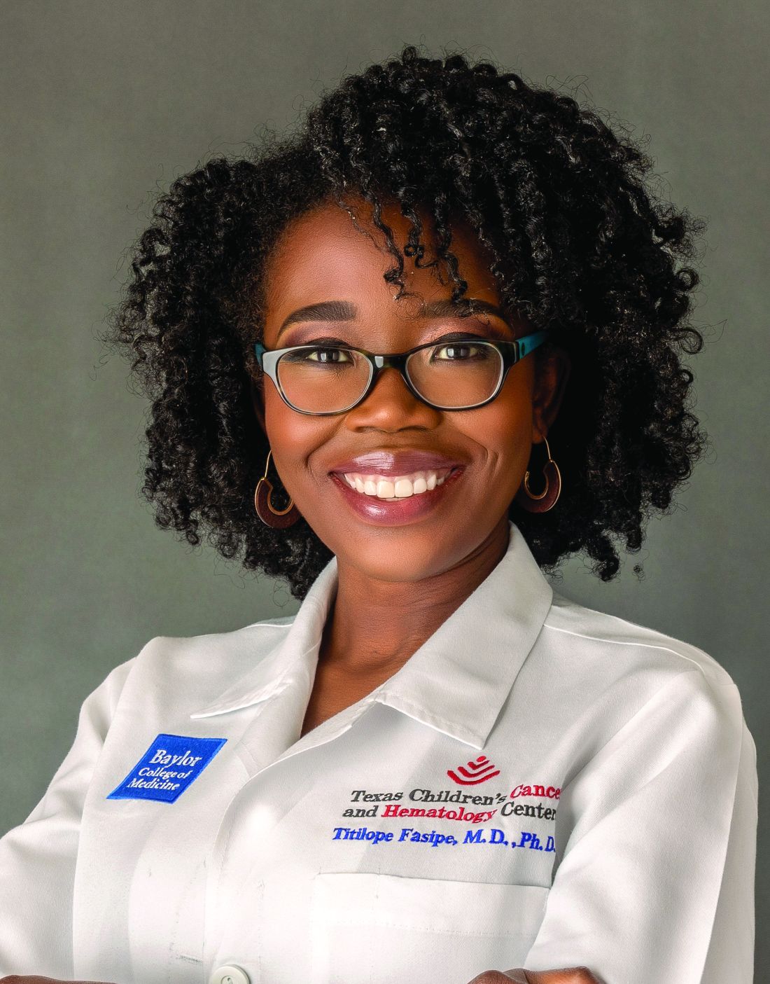
“If you grow up thinking that you’re going to die when you’re 18, it changes your world and your viewpoints, and it impacts your mental health,” she told this news organization.
“We are trying to make sure our children and their families know that there is a new story for sickle cell disease, and you don’t have to use any age as your prediction marker for your lifespan,” Dr. Fasipe said.
SCD, which affects about 100,000 people nationwide, is an inherited blood disorder, with the majority of patients – but not all – being of African descent. This condition is characterized by pain crises, or vaso-occlusive episodes, triggered when cells that are sickled get stuck and impede blood flow. These crises can come on suddenly and range from mild to severe.
Dr. Fasipe was born in Nigeria, where rates of SCD are among the world’s highest. She attended elementary school in the United States, where her father was studying theology, before returning to Nigeria with her family at age 11.
Back in those days, in both nations only about 50% of children with SCD lived beyond their 18th birthday. The survival rates in Nigeria and sub-Saharan Africa countries continue to be poor. In some more developed regions elsewhere, advances such as universal newborn screening, penicillin prophylaxis, pneumococcal vaccination, stroke screening, and hydroxyurea therapy have yielded substantial improvements, with 95% or more patients with SCD reaching their 18th birthday.
“With measures such as newborn screening, we can immediately start prevention measures in sickle cell disease, such as prevention of infection, which was the number one reason why children were dying,” Dr. Fasipe explained. “With global initiatives, we want that story to be the same in sub-Saharan Africa as well.”
Cousin’s early death inspires medical studies
In an essay published by Texas Medical Center that describes her childhood experiences, Dr. Fasipe recounts a pivotal event in her life: The heartbreaking death of her beloved cousin at the age of just 17, from a complication of SCD. This bereavement fueled Dr. Fasipe’s determination to pursue a medical career, to do all that she could to prevent such losses.
“Having sickle cell disease myself wasn’t the trigger that made me become a doctor. But when Femi [her cousin] died, I thought: ‘This shouldn’t happen,’ ” Dr. Fasipe wrote.
When she applied to medical school back in the United States, she declared in her application essay: “I want to cure sickle cell.”
By the time Dr. Fasipe was ready to undertake residency and fellowship applications, her essay had shifted to focus on pediatrics “specifically because I want to reach sickle cell patients before they’ve defined how their lives are going to be,” she said. “I want to give them hope.”
Hope for a cure
Fast-forwarding to this point in Dr. Fasipe’s career, she noted that her dream of a cure for SCD is no longer a distant aspiration, thanks to the advent of stem cell transplantation and more recently, gene therapy. These advancements have elevated her hope for a cure to an entirely new level.
Each new treatment comes with caveats. Stem cell transplantation requires a matching donor, leaving the majority of patients ineligible. And while gene therapy eliminates the need for a donor, treatment can reportedly cost nearly $3 million. Nevertheless, Dr. Fasipe emphasized the promise that these new advancements represent.
“The scientists that work in these spaces do appreciate these [accessibility barriers], and the expectation is these therapies will be more accessible with time and effort,” she said. “We’ve got to start somewhere, and it’s exciting that they’re making these early successes.”
Advice for clinicians
With firsthand knowledge of how it feels to be the patient, as well as on the clinician side of SCD treatment, Dr. Fasipe advises colleagues on some ways that they can improve care while boosting their patients’ hope:
Speak with empathy
Acknowledge the ‘elephant in the room’; the pain that patients with SCD can experience is real.
“When I’m managing any patient with pain, I first acknowledge the suffering because while we may not understand what that person is going through, acknowledgment is part of showing empathy,” she explains.
Seek out resources
Patients with SCD may typically seek treatment in primary care, where expertise in the disease may be lacking, and general practitioners may feel frustrated that there are limited treatment options.
“If you do find yourself treating a sickle cell disease patient, you may not have all of the answers, but there are good resources, whether it’s a nearby sickle cell disease centers or national guidelines,” Dr. Fasipe said.
Access to treatment
With research, including a recent study, showing that only about 25% of patients with SCD are prescribed hydroxyurea and even fewer – only about 5% – receive more recently approved SCD treatments, clinicians should be proactive by making sure that patients receive needed treatments.
“Clearly medicines like hydroxyurea are not as optimized in this community space as they should be, and then there are newer therapies that families, patients, and even providers may not be aware of, so it is important to be informed of the guidelines and provide all patients with comprehensive, high-quality care,” Dr. Fasipe said.
In the ED, patients with SCD are ‘care-seeking,’ not drug-seeking
Due to the sometimes rapid onset of severe pain symptoms, patients with SCD commonly wind up in the emergency department. In this time of an opioid epidemic, patients too often are suspected of merely seeking drugs.
“Sickle cell disease tends to get lumped into a category of a disease of pain, but pain is subjective and it is difficult to quantify, so unfortunately, patients can be labeled as potentially drug-seeking,” Dr. Fasipe explained, citing an article that detailed this problem.
Consequently, patients may have particularly negative experiences in the emergency department, but the use of resources such as a sickle cell disease point-of-care tool developed by the American College of Emergency Physicians and the American Society of Hematology can help improve care for those patients.
“One of the [point-of-care recommendations] before even managing the pain is that physicians show compassion by acknowledging the patient’s pain and that they understand why pain with sickle cell disease might look different than other types of pain,” Dr. Fasipe said.
Building trust
Encounters such as negative emergency department experiences can perpetuate a deeper issue of distrust between those with SCD and the medical community, which originated in long-held, well-documented racial disparities in health care.
“We know historically and even today that there are difficulties facing our families who are impacted by sickle cell disease, and they are related to structural racism and socioeconomic barriers,” Dr. Fasipe explained.
With these issues in mind, she said, “I refer to sickle cell disease as the medical representation of the Black experience in America.” However, she added, the good news is “we are now doing our best now to improve that.”
Among key efforts in building trust is the inclusion of patients with SCD and their families in as many aspects of research and clinical care as possible.
“In the global health care community, it is imperative to invite people with sickle cell disease and from the community to the decision-making table,” she noted.
“Now, when we’re talking about research for therapies, their expectation is that research trials and other initiatives for sickle cell disease must have input from the community; there are no initiatives for sickle cell disease that do not have input from the community.
“The patients and community members may not be experts on the science of sickle cell, but they’re experts on the lived experience and that’s very important when you’re thinking about new bringing in a new therapy.”
Forward momentum
Meanwhile, Dr. Fasipe observed, with the collective, advocacy-driven, forward momentum of the SCD community as a whole, things should only continue to improve.
“Because of the various barriers, some progress may not be immediately around the corner, but I do have confidence that this current generation of children with sickle cell will have improved health equity by the time they reach adulthood,” she said.
“I believe in this future, so I’m doing the work now, and it’s a promise I tell parents: I want your future adult child to live their best life, and we’re working hard to ensure that that becomes their future reality.”
Sickle cell disease awareness
September is National Sickle Cell Disease Awareness Month, and the National Heart, Lung, and Blood Institute offers a comprehensive website that clinicians can pass along to their patients, with information ranging from fact sheets on the disease and treatments to social media resources and inspiring stories of people with the disease.
In a comment, Lewis Hsu, MD, PhD, chief medical officer of the Sickle Cell Disease Association of America, underscored the uniquely important contributions of people like Dr. Fasipe, in providing inspiration to patients and clinicians alike.
“I have worked with several physicians, nurses, psychologists, and public health specialists who have sickle cell disease,” said Dr. Hsu, who is a pediatric hematologist who also serves as director of the Sickle Cell Center and professor of pediatrics for the University of Illinois at Chicago.
“They are ambassadors who have the trust of both patients and healthcare providers,” Dr. Hsu said.
In addition to providing inspiration of resilience, such care providers can serve as “communication bridges,” he explained.
“When they are conference speakers, everybody wants to hear them; when they sit on advisory committees or focus groups, they can help find the compromise or set the priorities.”
“Their impact on the whole sickle cell community is very large,” Dr. Hsu said.
These days, thanks to transformative advances in treating SCD that have substantially improved survival, Dr. Fasipe’s mission for a new generation of patients and their families is to replace their pain and fear with relief and hope.
“If you grow up thinking that you’re going to die when you’re 18, it changes your world and your viewpoints, and it impacts your mental health,” she told this news organization.
“We are trying to make sure our children and their families know that there is a new story for sickle cell disease, and you don’t have to use any age as your prediction marker for your lifespan,” Dr. Fasipe said.
SCD, which affects about 100,000 people nationwide, is an inherited blood disorder, with the majority of patients – but not all – being of African descent. This condition is characterized by pain crises, or vaso-occlusive episodes, triggered when cells that are sickled get stuck and impede blood flow. These crises can come on suddenly and range from mild to severe.
Dr. Fasipe was born in Nigeria, where rates of SCD are among the world’s highest. She attended elementary school in the United States, where her father was studying theology, before returning to Nigeria with her family at age 11.
Back in those days, in both nations only about 50% of children with SCD lived beyond their 18th birthday. The survival rates in Nigeria and sub-Saharan Africa countries continue to be poor. In some more developed regions elsewhere, advances such as universal newborn screening, penicillin prophylaxis, pneumococcal vaccination, stroke screening, and hydroxyurea therapy have yielded substantial improvements, with 95% or more patients with SCD reaching their 18th birthday.
“With measures such as newborn screening, we can immediately start prevention measures in sickle cell disease, such as prevention of infection, which was the number one reason why children were dying,” Dr. Fasipe explained. “With global initiatives, we want that story to be the same in sub-Saharan Africa as well.”
Cousin’s early death inspires medical studies
In an essay published by Texas Medical Center that describes her childhood experiences, Dr. Fasipe recounts a pivotal event in her life: The heartbreaking death of her beloved cousin at the age of just 17, from a complication of SCD. This bereavement fueled Dr. Fasipe’s determination to pursue a medical career, to do all that she could to prevent such losses.
“Having sickle cell disease myself wasn’t the trigger that made me become a doctor. But when Femi [her cousin] died, I thought: ‘This shouldn’t happen,’ ” Dr. Fasipe wrote.
When she applied to medical school back in the United States, she declared in her application essay: “I want to cure sickle cell.”
By the time Dr. Fasipe was ready to undertake residency and fellowship applications, her essay had shifted to focus on pediatrics “specifically because I want to reach sickle cell patients before they’ve defined how their lives are going to be,” she said. “I want to give them hope.”
Hope for a cure
Fast-forwarding to this point in Dr. Fasipe’s career, she noted that her dream of a cure for SCD is no longer a distant aspiration, thanks to the advent of stem cell transplantation and more recently, gene therapy. These advancements have elevated her hope for a cure to an entirely new level.
Each new treatment comes with caveats. Stem cell transplantation requires a matching donor, leaving the majority of patients ineligible. And while gene therapy eliminates the need for a donor, treatment can reportedly cost nearly $3 million. Nevertheless, Dr. Fasipe emphasized the promise that these new advancements represent.
“The scientists that work in these spaces do appreciate these [accessibility barriers], and the expectation is these therapies will be more accessible with time and effort,” she said. “We’ve got to start somewhere, and it’s exciting that they’re making these early successes.”
Advice for clinicians
With firsthand knowledge of how it feels to be the patient, as well as on the clinician side of SCD treatment, Dr. Fasipe advises colleagues on some ways that they can improve care while boosting their patients’ hope:
Speak with empathy
Acknowledge the ‘elephant in the room’; the pain that patients with SCD can experience is real.
“When I’m managing any patient with pain, I first acknowledge the suffering because while we may not understand what that person is going through, acknowledgment is part of showing empathy,” she explains.
Seek out resources
Patients with SCD may typically seek treatment in primary care, where expertise in the disease may be lacking, and general practitioners may feel frustrated that there are limited treatment options.
“If you do find yourself treating a sickle cell disease patient, you may not have all of the answers, but there are good resources, whether it’s a nearby sickle cell disease centers or national guidelines,” Dr. Fasipe said.
Access to treatment
With research, including a recent study, showing that only about 25% of patients with SCD are prescribed hydroxyurea and even fewer – only about 5% – receive more recently approved SCD treatments, clinicians should be proactive by making sure that patients receive needed treatments.
“Clearly medicines like hydroxyurea are not as optimized in this community space as they should be, and then there are newer therapies that families, patients, and even providers may not be aware of, so it is important to be informed of the guidelines and provide all patients with comprehensive, high-quality care,” Dr. Fasipe said.
In the ED, patients with SCD are ‘care-seeking,’ not drug-seeking
Due to the sometimes rapid onset of severe pain symptoms, patients with SCD commonly wind up in the emergency department. In this time of an opioid epidemic, patients too often are suspected of merely seeking drugs.
“Sickle cell disease tends to get lumped into a category of a disease of pain, but pain is subjective and it is difficult to quantify, so unfortunately, patients can be labeled as potentially drug-seeking,” Dr. Fasipe explained, citing an article that detailed this problem.
Consequently, patients may have particularly negative experiences in the emergency department, but the use of resources such as a sickle cell disease point-of-care tool developed by the American College of Emergency Physicians and the American Society of Hematology can help improve care for those patients.
“One of the [point-of-care recommendations] before even managing the pain is that physicians show compassion by acknowledging the patient’s pain and that they understand why pain with sickle cell disease might look different than other types of pain,” Dr. Fasipe said.
Building trust
Encounters such as negative emergency department experiences can perpetuate a deeper issue of distrust between those with SCD and the medical community, which originated in long-held, well-documented racial disparities in health care.
“We know historically and even today that there are difficulties facing our families who are impacted by sickle cell disease, and they are related to structural racism and socioeconomic barriers,” Dr. Fasipe explained.
With these issues in mind, she said, “I refer to sickle cell disease as the medical representation of the Black experience in America.” However, she added, the good news is “we are now doing our best now to improve that.”
Among key efforts in building trust is the inclusion of patients with SCD and their families in as many aspects of research and clinical care as possible.
“In the global health care community, it is imperative to invite people with sickle cell disease and from the community to the decision-making table,” she noted.
“Now, when we’re talking about research for therapies, their expectation is that research trials and other initiatives for sickle cell disease must have input from the community; there are no initiatives for sickle cell disease that do not have input from the community.
“The patients and community members may not be experts on the science of sickle cell, but they’re experts on the lived experience and that’s very important when you’re thinking about new bringing in a new therapy.”
Forward momentum
Meanwhile, Dr. Fasipe observed, with the collective, advocacy-driven, forward momentum of the SCD community as a whole, things should only continue to improve.
“Because of the various barriers, some progress may not be immediately around the corner, but I do have confidence that this current generation of children with sickle cell will have improved health equity by the time they reach adulthood,” she said.
“I believe in this future, so I’m doing the work now, and it’s a promise I tell parents: I want your future adult child to live their best life, and we’re working hard to ensure that that becomes their future reality.”
Sickle cell disease awareness
September is National Sickle Cell Disease Awareness Month, and the National Heart, Lung, and Blood Institute offers a comprehensive website that clinicians can pass along to their patients, with information ranging from fact sheets on the disease and treatments to social media resources and inspiring stories of people with the disease.
In a comment, Lewis Hsu, MD, PhD, chief medical officer of the Sickle Cell Disease Association of America, underscored the uniquely important contributions of people like Dr. Fasipe, in providing inspiration to patients and clinicians alike.
“I have worked with several physicians, nurses, psychologists, and public health specialists who have sickle cell disease,” said Dr. Hsu, who is a pediatric hematologist who also serves as director of the Sickle Cell Center and professor of pediatrics for the University of Illinois at Chicago.
“They are ambassadors who have the trust of both patients and healthcare providers,” Dr. Hsu said.
In addition to providing inspiration of resilience, such care providers can serve as “communication bridges,” he explained.
“When they are conference speakers, everybody wants to hear them; when they sit on advisory committees or focus groups, they can help find the compromise or set the priorities.”
“Their impact on the whole sickle cell community is very large,” Dr. Hsu said.
Overburdened: Health care workers more likely to die by suicide
This transcript has been edited for clarity.
Welcome to Impact Factor, your weekly dose of commentary on a new medical study.
If you run into a health care provider these days and ask, “How are you doing?” you’re likely to get a response like this one: “You know, hanging in there.” You smile and move on. But it may be time to go a step further. If you ask that next question – “No, really, how are you doing?” Well, you might need to carve out some time.
It’s been a rough few years for those of us in the health care professions. Our lives, dominated by COVID-related concerns at home, were equally dominated by COVID concerns at work. On the job, there were fewer and fewer of us around as exploitation and COVID-related stressors led doctors, nurses, and others to leave the profession entirely or take early retirement. Even now, I’m not sure we’ve recovered. Staffing in the hospitals is still a huge problem, and the persistence of impersonal meetings via teleconference – which not only prevent any sort of human connection but, audaciously, run from one into another without a break – robs us of even the subtle joy of walking from one hallway to another for 5 minutes of reflection before sitting down to view the next hastily cobbled together PowerPoint.
I’m speaking in generalities, of course.
I’m talking about how bad things are now because, in truth, they’ve never been great. And that may be why health care workers – people with jobs focused on serving others – are nevertheless at substantially increased risk for suicide.
Analyses through the years have shown that physicians tend to have higher rates of death from suicide than the general population. There are reasons for this that may not entirely be because of work-related stress. Doctors’ suicide attempts are more often lethal – we know what is likely to work, after all.
And, according to this paper in JAMA, it is those people who may be suffering most of all.
The study is a nationally representative sample based on the 2008 American Community Survey. Records were linked to the National Death Index through 2019.
Survey respondents were classified into five categories of health care worker, as you can see here. And 1,666,000 non–health care workers served as the control group.
Let’s take a look at the numbers.
I’m showing you age- and sex-standardized rates of death from suicide, starting with non–health care workers. In this study, physicians have similar rates of death from suicide to the general population. Nurses have higher rates, but health care support workers – nurses’ aides, home health aides – have rates nearly twice that of the general population.
Only social and behavioral health workers had rates lower than those in the general population, perhaps because they know how to access life-saving resources.
Of course, these groups differ in a lot of ways – education and income, for example. But even after adjustment for these factors as well as for sex, race, and marital status, the results persist. The only group with even a trend toward lower suicide rates are social and behavioral health workers.
There has been much hand-wringing about rates of physician suicide in the past. It is still a very real problem. But this paper finally highlights that there is a lot more to the health care profession than physicians. It’s time we acknowledge and support the people in our profession who seem to be suffering more than any of us: the aides, the techs, the support staff – the overworked and underpaid who have to deal with all the stresses that physicians like me face and then some.
There’s more to suicide risk than just your job; I know that. Family matters. Relationships matter. Medical and psychiatric illnesses matter. But to ignore this problem when it is right here, in our own house so to speak, can’t continue.
Might I suggest we start by asking someone in our profession – whether doctor, nurse, aide, or tech – how they are doing. How they are really doing. And when we are done listening, we use what we hear to advocate for real change.
Dr. Wilson is associate professor of medicine and public health and director of the Clinical and Translational Research Accelerator at Yale University, New Haven, Conn. He has disclosed no relevant financial relationships.
A version of this article appeared on Medscape.com.
The unappreciated healing power of awe
I’m standing atop the Klein Matterhorn, staring out at the Alps, their moonscape peaks forming a jagged, terrifying, glorious white horizon.
I am small. But the emotions are huge. The joy: I get to be a part of all this today. The fear: It could kill me. More than kill me, it could consume me.
That’s what I always used to feel when training in Zermatt, Switzerland.
I was lucky. As a former U.S. Ski Team athlete, I was regularly able to experience such magnificent scenescapes – and feel the tactile insanity of it, too, the rise and fall of helicopters or trams taking us up the mountains, the slicing, frigid air at the summit, and the lurking on-edge feeling that you, tiny human, really aren’t meant to be standing where you are standing.
“Awe puts things in perspective,” said Craig Anderson, PhD, postdoctoral scholar at Washington University at St. Louis, and researcher of emotions and behavior. “It’s about feeling connected with people and part of the larger collective – and that makes it okay to feel small.”
Our modern world is at odds with awe. We tend to shrink into our daily lives, our problems, our devices, and the real-time emotional reactions to those things, especially anger.
It doesn’t have to be that way.
‘In the upper reaches of pleasure and on the boundary of fear’
That’s how New York University ethical leadership professor Jonathan Haidt, PhD, and psychology professor Dacher Keltner, PhD, of the University of California, Berkeley, defined awe in a seminal report from 2003.
The feeling is composed of two elements: perceived vastness (sensing something larger than ourselves) and accommodation (our need to process and understand that vastness). The researchers also wrote that awe could “change the course of life in profound and permanent ways.”
“There’s a correlation between people who are happier and those who report more feelings of awe,” said David Yaden, PhD, assistant professor in the department of psychiatry and behavioral sciences at Johns Hopkins University, Baltimore, and coauthor of “The Varieties of Spiritual Experience.” “It’s unclear, though, which way the causality runs. Is it that having more awe experiences makes people happier? Or that happy people have more awe. But there is a correlation.”
One aspect about awe that’s clear: When people experience it, they report feeling more connected. And that sense of connection can lead to prosocial behavior – such as serving others and engaging with one’s community.
“Feelings of isolation are quite difficult, and we’re social creatures, so when we feel connected, we can benefit from it,” Dr. Yaden said.
A 2022 study published in the Journal of Personality and Social Psychology revealed that awe “awakens self-transcendence, which in turn invigorates pursuit of the authentic self.”
While these effects can be seen as one individual’s benefits, the researchers posited that they also lead to prosocial behaviors. Another study conducted by the same scientists showed that awe led to greater-good behavior during the pandemic, to the tune of an increased willingness to donate blood. In this study, researchers also cited a correlation between feelings of awe and increased empathy.
The awe experience
Dr. Yaden joined Dr. Keltner and other researchers in creating a scale for the “awe experience,” and found six related factors: a feeling that time momentarily slows; a sense of self-diminishment (your sense of self becomes smaller); a sense of connectedness; feeling in the presence of something grand; the need to mentally process the experience; and physical changes, like goosebumps or feeling your jaw slightly drop.
“Any of these factors can be large or small,” Dr. Yaden noted, adding that awe can also feel positive or negative. A hurricane can instill awe, for example, and the experience might not be pleasant.
However, “it’s more common for the awe experience to be positive,” Dr. Yaden said.
How your brain processes awe
Functional MRI, by which brain activity is measured through blood flow, allows researchers to see what’s happening in the brain after an awe experience.
One study that was conducted in the Netherlands and was published in the journal Human Brain Mapping suggested that certain parts of the brain that are responsible for self-reflection were less “activated” when participants watched awe-inspiring videos.
The researchers posit that the “captivating nature of awe stimuli” could be responsible for such reductions, meaning participants’ brains were geared more toward feelings of connection with others or something greater – and a smaller sense of self.
Another study published in the journal Emotion revealed a link between awe and lower levels of inflammatory cytokines, so awe could have positive and potentially protective health benefits, as well.
And of course there are the physical and emotional benefits of nature, as dozens of studies reveal. Dr. Anderson’s research in the journal Emotion showed that nature “experiences” led to more feelings of awe and that the effects of nature also reduced stress and increased well-being.
Why we turn away from awe
The world we inhabit day to day isn’t conducive to experiencing awe – indoors, seated, reacting negatively to work or social media. The mentalities we forge because of this sometimes work against experiencing any form of awe.
Example: Some people don’t like to feel small. That requires a capacity for humility.
“That [feeling] can be threatening,” noted Dr. Anderson, who earned his doctorate studying as part of Dr. Keltner’s “Project Awe” research team at UC Berkeley.
The pandemic and politics and rise in angry Internet culture also contribute. And if you didn’t know, humans have a “negativity bias.”
“Our responses to stress tend to be stronger in magnitude than responses to positive things,” Dr. Anderson said. “Browsing the Internet and seeing negative things can hijack our responses. Anger really narrows our attention on what makes us angry.”
In that sense, anger is the antithesis of awe. As Dr. Anderson puts it: Awe broadens our attention to the world and “opens us up to other people and possibilities,” he said. “When we’re faced with daily hassles, when we experience something vast and awe-inspiring, those other problems aren’t as big of a deal.”
We crave awe in spite of ourselves
An awful lot of us are out there seeking awe, knowingly or not.
People have been stopping at scenic overlooks and climbing local peaks since forever, but let’s start with record-setting attendance at the most basic and accessible source of natural awe we have in the United States: national parks.
In 2022, 68% of the 312 million visitors sought out nature-based or recreational park activities (as opposed to historical or cultural activities). Even though a rise in national park visits in 2021 and 2022 could be attributed to pandemic-related behavior (the need for social distancing and/or the desire to get outside), people were flocking to parks prior to COVID-19. In fact, 33 parks set visitation records in 2019; 12 did so in 2022.
We also seek awe in man-made spectacle. Consider annual visitor numbers for the following:
- Golden Gate Bridge: 10 million
- : 4 million
- : 1.62 million
And what about the most awe-inducing experience ever manufactured: Space tourism. While catering to the wealthy for now, flying to space allows untrained people to enjoy something only a chosen few astronauts have been able to feel: the “overview effect,” a term coined by author Frank White for the shift in perspective that occurs in people who see Earth from space.
Upon his return from his Blue Origin flight, actor William Shatner was candid about his emotional experience. “I was crying,” he told NPR. “I didn’t know what I was crying about. It was the death that I saw in space and the lifeforce that I saw coming from the planet – the blue, the beige, and the white. And I realized one was death and the other was life.”
We want awe. We want to feel this way.
Adding everyday awe to your life
It may seem counterintuitive: Most awe-inspiring places are special occasion destinations, but in truth it’s possible to find awe each day. Outdoors and indoors.
Park Rx America, led by Robert Zarr, MD, MPH, boasts a network of nearly 1500 healthcare providers ready to “prescribe” walks or time in nature as part of healing. “Our growing community of ‘nature prescribers’ incorporate nature as a treatment option for their willing clients and patients,” Dr. Zarr said.
He also noted that awe is all about where you look, including in small places.
“Something as simple as going for a walk and stopping to notice the complexity of fractal patterns in the leaves, for example, leaves me with a sense of awe,” he said. “Although difficult to measure, there is no doubt that an important part of our health is intricately linked to these daily awe-filled moments.”
Nature is not the only way. Dr. Yaden suggested that going to a museum to see art or sporting events is also a way to experience the feeling.
An unexpected source of man-made awe: Screens. A study published in Nature showed that immersive video experiences (in this case, one achieved by virtual reality) were effective in eliciting an awe response in participants.
While virtual reality isn’t ubiquitous, immersive film experiences are. IMAX screens were created for just this purpose (as anyone who saw the Avatar films in this format can attest).
Is it perfect? No. But whether you’re witnessing a birth, hiking an autumn trail bathed in orange, or letting off a little gasp when you see Oppenheimer’s nuclear explosion in 70 mm, it all counts.
Because it’s not about the thing. It’s about your openness to be awed by the thing.
I’m a little like Dr. Zarr in that I can find wonder in the crystalline structures of a snowflake. And I also love to hike and inhale expansive views. If you can get to Switzerland, and specifically Zermatt, take the old red tram to the top. I highly recommend it.
A version of this article appeared on Medscape.com.
Are ketogenic supplements the key to healthy aging?
A century ago, pediatricians began prescribing for children with intractable seizures the “keto diet,” which they also used to treat diabetes in children and adults. The low-carbohydrate, high-fat meals were designed to induce a near hypoglycemic state, forcing the body to use ketones for fuel instead of glucose.
The strategy fell out of favor after the discovery of insulin in the 1920s and the development of better antiseizure medications. The global market for the ketogenic diet topped $11 billion in 2022.
Is it just a fad, or has the public – and science – caught up with the 100-year-old approach?
Although scientists still don’t know why the ketogenic diet was effective for controlling seizures, they have documented the effectiveness of ketogenic diets for the treatment of diabetes and metabolic syndrome. An extensive body of literature has documented their use in athletes, but less is known regarding conditions such as heart disease and dementia.
Although the data are promising, much of the research has been conducted with mice or has come from trials of short-term use in humans. But recently, the National Institutes of Health awarded a $3.5 million federal grant for a double-blind, randomized, placebo-controlled clinical trial to understand the effects of the long-term use of ketone ester supplementation on frailty. Developed 20 years ago, ketone esters are precursor molecules that the body quickly breaks down into ketone bodies when carbohydrates aren’t available.
“We’ve learned so much recently about how ketone bodies interact with aging biology,” John Newman, MD, PhD, of the Buck Institute for Research on Aging in Novato, Calif., and the study’s principal investigator, said in an interview. “And we’re only just starting to translate that out of the laboratory and into human studies to see how we can take advantage of ketone bodies to improve people’s health.”
Researchers from the Ohio State University and the University of Connecticut will also participate in the TAKEOFF (Targeting Aging With Ketone Ester in Older Adults for Function in Frailty) trial, which seeks to recruit a total of 180 people across the three sites.
Dr. Newman, assistant professor at the Buck Institute and associate professor in the division of geriatrics at the University of California, San Francisco, said
One of the common things that happen during aging is that tissues – such as of the heart, brain, and muscle – lose the ability to metabolize glucose effectively. Over time, resistance to insulin can develop.
Researchers can map out areas of the brain affected by Alzheimer’s disease, for example, by assessing where patients’ glucose uptake drops. In heart failure, the heart has difficulty obtaining enough energy from glucose and instead burns fats and ketone bodies.
How might ketones affect frailty in the elderly?
As a practicing geriatrician, Dr. Newman measures frailty by evaluating patients’ strength, endurance, and how they react to stresses. He and his colleagues believe certain molecular and cellular changes may make patients more likely to fall, to recover more slowly from surgery, or to lose mobility.
The main hypothesis of the TAKEOFF study is “that if you target these fundamental mechanisms of aging, you would be able to impact many different diseases of aging across different organ systems.”
Dr. Newman and Brianna Stubbs, DPhil, lead translational scientist at the Buck Institute, are still finishing up the BIKE (Buck Institute Ketone Ester) pilot study, which was the first double-blind, randomized, placebo-controlled study to evaluate the use of ketone ester supplements in adults older than 65 years. “The BIKE study is 12 weeks long. That’s actually the longest that anyone has studied ketone ester supplements in humans,” Dr. Stubbs said. The results will help them firm up the protocol for the TAKEOFF trial, which will likely treat patients for up to 24 weeks.
The primary outcome measure at all three study sites will be leg press strength. Researchers will also assess a variety of secondary outcomes that cover geriatric and cognitive function – measures such as gait speed and walking endurance, cognitive tests, and quality of life. And at the Buck, Dr. Newman and Dr. Stubbs will be evaluating the use of biomarkers that are often available in clinical labs – insulin, C-reactive protein, cystatin, and natriuretic peptide tests – for use as outcome measures that are responsive to treatment interventions and that can be used to track outcomes in future research on aging.
To achieve the goal of looking broadly at different organ systems likely to be affected by ketogenic supplements, they have assembled a team of coinvestigators with wide-ranging expertise in ketone and aging research.
Jeff Volek, PhD, professor in the department of human sciences at the Ohio State University, in Columbus, has contributed extensively to the literature on the use of ketogenic diets and supplements in a variety of populations, such as endurance athletes and patients with insulin resistance or diabetes.
Dr. Volek has demonstrated that ketones can have an anticatabolic effect on muscle tissue. “They could help offset some of the muscle loss with aging, which would in turn improve their physical functioning and ability to do daily activities,” he said.
The anti-inflammatory property of ketones may provide another benefit to older people. They can reduce oxidative stress, which is considered one of the chief pathologic mechanisms responsible for conditions such as heart disease, Alzheimer’s disease, asthma, and arthritis.
In addition to the main study outcomes, Dr. Volek’s lab will study muscle physiology by performing biopsies at baseline and after consumption of ketogenic supplements to assess metabolic changes in muscle cells as they consume energy. Study participants will also undergo MRIs to detect subtle changes in muscle size before and after treatment.
From elite athletes to everyday agers
As a graduate student in Dr. Volek’s lab, Jenna Bartley, PhD, studied the effects of a ketogenic diet on elite athletes. But her work has taken a turn. Now an assistant professor in the department of immunology and the center on aging at the University of Connecticut in Farmington, she focuses on how immune responses and physical function decline with age.
“Ketogenic diets and the main ketone bodies – mainly beta-hydroxybutyrate – have been shown to have really powerful influences on a lot of things that go wrong with aging,” Dr. Bartley said. The decline in immune function in the elderly is not isolated to one cell type or even one arm of the immune system. There is reason to believe ketone supplementation could improve immune function.
“T cells really love ketones for energy,” Dr. Bartley said. Some data show that production of ketone bodies is impaired in individuals with severe SARS-CoV-2 infection. Mouse models of SARS-CoV-2 infection have found that ketogenic diets led to improvement in the response to antiviral therapy.
In her lab, she’ll assess serum markers of inflammation in patients, as well as cytokine secretion following stimulation of T cells. T cells in culture from older people produce more inflammatory cytokines than those from younger people, leading to a dysfunctional immune response. Dr. Bartley is curious to see whether ketones can fix that. Additional work will include single-cell RNA sequencing of different classes of immune cells to investigate how ketones might change metabolic pathways.
Why use ketogenic supplements instead of having people consume ketogenic diets? “There are no cheat days in the keto diet,” Dr. Bartley said. Administering the diet requires intense supervision of research participants to enforce adherence. Use of supplements will improve compliance and likely make any findings translatable to more of the population, she said.
Drawbacks of the initial formulations of ketone esters, first developed 20 years ago, included high cost and terrible taste. Dr, Stubbs, a former world class rowing champion who competed in the Ironman World Championship last year, has firsthand experience with them as a research participant.
“It tasted like drinking nail polish,” she said. Recent advances in manufacturing have made them cheaper – roughly $5 per day – and more palatable, enabling research studies such as TAKEOFF.
For Dr. Newman, the studies are early building blocks in the emerging field of geroscience, which aims to translate fundamental mechanisms of aging into therapies to treat disease.
“We’re hoping that this will be an example of a proof-of-concept geroscience study that will really help to translate ketone body biology out of the laboratory and hopefully into a diversity of clinical applications,” he said. “There’s a lot we don’t understand still about the molecular mechanisms of frailty.”
Dr. Newman and Dr. Stubbs own stock in BHB Therapeutics Ltd, the company providing the product being studied, and are inventors on patents that relate to the product being studied. The Buck Institute has an ownership interest in BHB Therapeutics. Dr. Bartley and Dr. Volek report no relevant financial relationships.
A version of this article appeared on Medscape.com .
How to get paid if your patient passes on
The death of a patient comes with many challenges for physicians, including a range of emotional and professional issues. Beyond those concerns,
“When a patient passes away, obviously there is, unfortunately, a lot of paperwork and stress for families, and it’s a very difficult situation,” says Shikha Jain, MD, an oncologist and associate professor of medicine at the University of Illinois at Chicago. “Talking about finances in that moment can be difficult and uncomfortable, and one thing I’d recommend is that the physicians themselves not get involved.”
Instead, Dr. Jain said, someone in the billing department in the practice or the hospital should take a lead on dealing with any outstanding debts.
“That doctor-patient relationship is a very precious relationship, so you don’t want to mix that financial aspect of providing care with the doctor-patient relationship,” Dr. Jain said. “That’s one thing that’s really important.”
The best approach in such situations is for practices to have a standing policy in place that dictates how to handle bills once a patient has died.
In most cases, the executor of the patient’s will must inform all creditors, including doctors, that the decedent has died, but sometimes there’s a delay.
Hoping the doctor’s office writes it off
“Even though the person in charge of the estate is supposed to contact the doctor’s office and let them know when a patient has passed, that doesn’t always happen,” says Hope Wen, head of billing at practice management platform Soundry Health. “It can be very challenging to track down that information, and sometimes they’re just crossing their fingers hoping that the doctor’s office will just write off the balance, which they often do.”
Some offices use a service that compares accounts receivable lists to Social Security death files and state records to identify deaths more quickly. Some physicians might also use a debt collection agency or an attorney who has experience collecting decedent debts and dealing with executors and probate courts.
Once the practice becomes aware that a patient has died, it can no longer send communications to the name and address on file, although it can continue to go through the billing process with the insurer for any bills incurred up to the date of the death.
At that point, the estate becomes responsible for the debt, and all communication must go to the executor of the estate (in some states, this might be called a personal representative). The office can reach out to any contacts on file to see if they are able to identify the executor.
“You want to do that in a compassionate way,” says Jack Brown III, JD, MBA, president of Gulf Coast Collection Bureau. “You’ll tell them you’re sorry for their loss, but you’re wondering who is responsible for the estate. Once you’ve identified that person and gotten their letter of administration from the probate court or a power of attorney, then you can speak with that person as if they were the patient.”
The names of executors are also public record and are available through the probate court (sometimes called the surrogate court) in the county where the decedent lived.
“Even if there’s no will or no executive named, the court will appoint an administrator for the estate, which is usually a family member,” said Robert Bernstein, an estate lawyer in Parsippany, N.J. “Their information will be on file in the court.”
Insurance coverage
Typically, insurance will pay for treatment (after deductibles and copays) up until the date of the patient’s death. But, of course, it can take months for some insurance companies to make their final payments, allowing physicians to know exactly how much they’re owed by that estate. In such cases, it’s important for physicians to know the rules in the decedent’s state for how long they have to file a claim.
Most states require that claims occur within 6-9 months of the person’s death. However, in some states, claimants can continue to file for much longer if the estate has not yet paid out all of its assets.
“Sometimes there is real estate to sell or a business to wind down, and it can take years for the estate to distribute all of the assets,” Mr. Bernstein says. “If it’s a year later and they still haven’t distributed the assets, the physician can still file the claim and should be paid.”
In some cases, especially if the decedent received compassionate, quality care, their family will want to make good on any outstanding debts to the health care providers who took care of their loved ones in their final days. In other cases, especially when a family member has had a long illness, their assets have been depleted over time or were transferred to other family members so that there is little left in the estate itself when the patient dies.
Regardless of other circumstances, the estate alone is responsible for such payments, and family members, including spouses and children, typically have no liability. (Though rarely enforced, some states do have filial responsibility laws that could hold children responsible for their parents’ debts, including unpaid medical bills. In addition, states with community property laws might require a surviving spouse to cover their partner’s debt, even after death.)
The probate process varies from state to state, but in general, the probate system and the executor will gather all existing assets and then notify all creditors about how to submit a claim. Typically, the claim will need to include information about how much is owed and documentation, such as bills and an explanation of benefits to back up the claim. It should be borne in mind that even those who’ve passed away have privacy protections under the Health Insurance Portability and Accountability Act, so practices must be careful as to how much information they’re sharing through their claim.
Once the estate has received all the claims, the executor will follow a priority of claims, starting with secured creditors. Typically, medical bills, especially those incurred in the last 90 days of the decedent’s life, have priority in the probate process, Mr. Brown says.
How to minimize losses
In that case, the practice would write off the unpaid debt as a business loss. If there are not enough assets in the estate to pay all claims, the executor will follow a state schedule that apportions those assets that are available.
There are some steps that practices can take to protect themselves from incurring such losses. For example, before beginning treatment, practices might consider asking patients to name a guarantor, who will essentially promise to cover any outstanding debts that the patient incurs.
To be binding, the office will need a signature from both the patient and the guarantor. Some offices may also keep a patient credit card number on file with written authorization that they can use to pay bills that are past due, although this payment method would no longer be valid after a patient dies.
While it’s important for all physicians to document and verify the financial information for their patients, oncologists often must consider an additional layer of fiduciary responsibility when it comes to their patients. Ms. Wen suggests that oncology offices check in with insurance companies to determine whether a patient has exhausted their benefits.
“That can happen with cancer patients, depending on how long they’ve been receiving treatment and what type of treatment they’ve been getting,” she said. “Some of the clinical trials, insurance will pay for them, but they’re really expensive and can get toward that max. So knowing where they are with their insurance coverage is big.”
When time is of the essence, some patients will choose to go forward with a treatment before receiving insurance approval. In those cases, the office must have an additional conversation in which the costs of the treatment are discussed. The office should obtain written confirmation of who will pay if the insurer does not, Ms. Wen said. While it’s the patient’s responsibility to keep track of their insurance benefits, oncology practices and hospitals must also exercise due diligence in monitoring the benefits that are available.
“That’s part of their contract with insurance companies if they’re in network, helping patients understand their benefits,” Ms. Wen saids.
It’s also important for practices to keep clear, consistent records to make it easier to identify outstanding bills and the correct contact information for them. If bills had gone unpaid prior to a patient’s death and the office started legal action and received a judgment, that claim would typically go ahead of other creditors’ claims.
Dr. Jain says that some practices might also consider keeping a financial adviser or social worker on staff who can assist patients and their families with understanding their out-of-pocket costs for treatment.
“Financial toxicity in oncology and medical care is a very real problem,” she says. “At the beginning of the relationship, I recommend that my patients get set up with a financial specialist that can help them navigate that aspect, not only when a patient passes away but during the process of receiving treatment, so they’re not shocked by the bills.”
A version of this article first appeared on Medscape.com.
People with long COVID have specific blood biomarkers, study says
The findings may be a step toward creating blood tests to positively identify people with long COVID so specialized treatments can be employed, researchers said.
“This is a decisive step forward in the development of valid and reliable blood testing protocols for long COVID,” said David Putrino, PhD., lead author and professor of rehabilitation and human performance and director of the Abilities Research Center at Icahn Mount Sinai Health System, New York.
Researchers from the Icahn School of Medicine at Mount Sinai and Yale School of Medicine looked at blood samples from about 270 people between January 2021 and June 2022. The people had never been infected with COVID, had fully recovered from an infection, or still showed symptoms at least four months after infection.
Using machine learning, the research teams were able to differentiate between people with and without long COVID with 96% accuracy based on distinctive features in the blood samples, according to a news release from Mount Sinai.
People with long COVID had abnormal T-cell activity and low levels of the hormone cortisol. Cortisol helps people feel alert and awake, which would explain why people with long COVID often report fatigue, NBC News said in a report on the study.
“It was one of the findings that most definitively separated the folks with long Covid from the people without long Covid,” Dr. Putrino told NBC News.
The study also found that long COVID appears to reactivate latent viruses including Epstein-Barr and mononucleosis, the study said.
The blood tests could allow doctors to come up with specialized treatments in people who report a wide variety of long COVID symptoms, Dr. Putrino said.
“There is no ‘silver bullet’ for treating long COVID, because it is an illness that infiltrates complex systems such as the immune and hormonal regulation,” he said.
The Centers for Disease Control and Prevention says about one in five Americans who had COVID still have long COVID. Symptoms include fatigue, brain fog, dizziness, digestive problems, and loss of smell and taste.
A version of this article appeared on WebMD.com.
Hypotrichosis and Hair Loss on the Occipital Scalp
The Diagnosis: Monilethrix
A diagnosis of monilethrix was rendered based on the clinical and trichoscopic findings. Simple surveillance of the patient’s condition and prevention of further hair trauma were proposed as management options.
Monilethrix is a hair shaft disorder that is inherited in a predominantly autosomal-dominant pattern with variable expressiveness and penetrance resulting from heterozygous mutations in hair keratin genes KRT81, KRT83, and KRT86 in a region of chromosome 12q13.13.1,2 An autosomalrecessive form has been described with mutation in desmoglein 4, but it differs from the classical form by the variable periodicity of the region between the nodules.3,4
The morphologic alteration consists of the formation of fusiform nodules of normal structure alternated with narrow and dystrophic constrictions (Figure). These internodes are fragile areas that cause breakage at constricted points.5 Clinically, monilethrix presents as areas of focal or diffuse alopecia with frequent involvement of the terminal follicles, mainly in areas of friction. The hair is normal at birth due to the predominance of lanugo in the neonatal period, but it subsequently is replaced by abnormal hairs in the first months of life.6 Initial clinical signs begin to appear when the terminal hairs begin to form.7 Although rarer, the eyebrows and eyelashes, as well as the axillary, pubic, and body hair, may be involved.5
.")
Other hair shaft anomalies merit consideration in the differential diagnosis of monilethrix, including pseudomonilethrix, pressure alopecia, trichorrhexis invaginata, ectodermal dysplasia, tinea capitis, and trichothiodystrophy.6 The diagnosis is reached by clinical history and physical examination. Trichoscopy and light microscopy are used to confirm the diagnosis. Trichoscopic examination shows markedly higher rates of anagen hair. The shafts examined in our patient revealed 0.7- to 1-mm intervals between nodes. Hair can be better visualized under a polarized microscope, and the condition can be distinguished from pseudomonilethrix using this approach.5,6 In our patient, the diagnosis was made based on light microscopy and trichoscopic findings with no genetic testing; however, genetic testing for the classic mutations of the keratin genes would be desirable to confirm the diagnosis but was not done in our patient.6 The prognosis of monilethrix is variable; most cases persist into adulthood, though spontaneous improvement may occur with advancing age, during summer, and during pregnancy.8
There is no definitive therapy for monilethrix. Although there have been reports of cases treated with systemic corticosteroids, oral retinoids, topical minoxidil, vitamins, and peeling ointments (desquamative oil), the cornerstone of management is protecting the hair against traumatic procedures such as excessive combing, brushing, and friction, as well as parent and patient education about the benign nature of the condition.9 Additionally, some cases have shown improvement with minoxidil solution at 2% and 5% concentrations, oral minoxidil, or acitretin.7-9
- Fontenelle de Oliveira E, Cotta de Alencar Araripe AL. Monilethrix: a typical case report with microscopic and dermatoscopic findings. An Bras Dermatol. 2015;90:126-127.
- de Cruz R, Horev L, Green J, et al. A novel monilethrix mutation in coil 2A of KRT86 causing autosomal dominant monilethrix with incomplete penetrance. Br J Dermatol. 2012;166(suppl 2):20-26.
- Baltazard T, Dhaille F, Chaby G, et al. Value of dermoscopy for the diagnosis of monilethrix. Dermatol Online J. 2017;23:13030 /qt9hf1p3xm.
- Kato M, Shimizu A, Yokoyama Y, et al. An autosomal recessive mutation of DSG4 causes monilethrix through the ER stress response. J Invest Dermatol. 2015;135:1253-1260.
- Gummer CL, Dawber RP, Swift JA. Monilethrix: an electron microscopic and electron histochemical study. Br J Dermatol. 1981;105:529-541.
- Sharma VK, Chiramel MJ, Rao A. Dermoscopy: a rapid bedside tool to assess monilethrix. Indian J Dermatol Venereol Leprol. 2016;82:73-74.
- Sinclair R. Treatment of monilethrix with oral minoxidil. JAAD Case Rep. 2016;2:212-215.
- Rakowska A, Slowinska M, Czuwara J, et al. Dermoscopy as a tool for rapid diagnosis of monilethrix. J Drugs Dermatol. 2007;6:222-224.
- Karincaoglu Y, Coskun BK, Seyhan ME, et al. Monilethrix. Am J Clin Dermatol. 2005;6:407-410.
The Diagnosis: Monilethrix
A diagnosis of monilethrix was rendered based on the clinical and trichoscopic findings. Simple surveillance of the patient’s condition and prevention of further hair trauma were proposed as management options.
Monilethrix is a hair shaft disorder that is inherited in a predominantly autosomal-dominant pattern with variable expressiveness and penetrance resulting from heterozygous mutations in hair keratin genes KRT81, KRT83, and KRT86 in a region of chromosome 12q13.13.1,2 An autosomalrecessive form has been described with mutation in desmoglein 4, but it differs from the classical form by the variable periodicity of the region between the nodules.3,4
The morphologic alteration consists of the formation of fusiform nodules of normal structure alternated with narrow and dystrophic constrictions (Figure). These internodes are fragile areas that cause breakage at constricted points.5 Clinically, monilethrix presents as areas of focal or diffuse alopecia with frequent involvement of the terminal follicles, mainly in areas of friction. The hair is normal at birth due to the predominance of lanugo in the neonatal period, but it subsequently is replaced by abnormal hairs in the first months of life.6 Initial clinical signs begin to appear when the terminal hairs begin to form.7 Although rarer, the eyebrows and eyelashes, as well as the axillary, pubic, and body hair, may be involved.5
Other hair shaft anomalies merit consideration in the differential diagnosis of monilethrix, including pseudomonilethrix, pressure alopecia, trichorrhexis invaginata, ectodermal dysplasia, tinea capitis, and trichothiodystrophy.6 The diagnosis is reached by clinical history and physical examination. Trichoscopy and light microscopy are used to confirm the diagnosis. Trichoscopic examination shows markedly higher rates of anagen hair. The shafts examined in our patient revealed 0.7- to 1-mm intervals between nodes. Hair can be better visualized under a polarized microscope, and the condition can be distinguished from pseudomonilethrix using this approach.5,6 In our patient, the diagnosis was made based on light microscopy and trichoscopic findings with no genetic testing; however, genetic testing for the classic mutations of the keratin genes would be desirable to confirm the diagnosis but was not done in our patient.6 The prognosis of monilethrix is variable; most cases persist into adulthood, though spontaneous improvement may occur with advancing age, during summer, and during pregnancy.8
There is no definitive therapy for monilethrix. Although there have been reports of cases treated with systemic corticosteroids, oral retinoids, topical minoxidil, vitamins, and peeling ointments (desquamative oil), the cornerstone of management is protecting the hair against traumatic procedures such as excessive combing, brushing, and friction, as well as parent and patient education about the benign nature of the condition.9 Additionally, some cases have shown improvement with minoxidil solution at 2% and 5% concentrations, oral minoxidil, or acitretin.7-9
- Fontenelle de Oliveira E, Cotta de Alencar Araripe AL. Monilethrix: a typical case report with microscopic and dermatoscopic findings. An Bras Dermatol. 2015;90:126-127.
- de Cruz R, Horev L, Green J, et al. A novel monilethrix mutation in coil 2A of KRT86 causing autosomal dominant monilethrix with incomplete penetrance. Br J Dermatol. 2012;166(suppl 2):20-26.
- Baltazard T, Dhaille F, Chaby G, et al. Value of dermoscopy for the diagnosis of monilethrix. Dermatol Online J. 2017;23:13030 /qt9hf1p3xm.
- Kato M, Shimizu A, Yokoyama Y, et al. An autosomal recessive mutation of DSG4 causes monilethrix through the ER stress response. J Invest Dermatol. 2015;135:1253-1260.
- Gummer CL, Dawber RP, Swift JA. Monilethrix: an electron microscopic and electron histochemical study. Br J Dermatol. 1981;105:529-541.
- Sharma VK, Chiramel MJ, Rao A. Dermoscopy: a rapid bedside tool to assess monilethrix. Indian J Dermatol Venereol Leprol. 2016;82:73-74.
- Sinclair R. Treatment of monilethrix with oral minoxidil. JAAD Case Rep. 2016;2:212-215.
- Rakowska A, Slowinska M, Czuwara J, et al. Dermoscopy as a tool for rapid diagnosis of monilethrix. J Drugs Dermatol. 2007;6:222-224.
- Karincaoglu Y, Coskun BK, Seyhan ME, et al. Monilethrix. Am J Clin Dermatol. 2005;6:407-410.
A 6-month-old infant girl was referred to the dermatology service with hypotrichosis and hair loss on the occipital region of the scalp of 4 months’ duration (top). The patient was born at full term by cesarean delivery without complications. There were no comorbidities or family history of alopecia. Clinical examination revealed an alopecic plaque in the occipital region with broken hairs and some dystrophic hairs associated with follicular papules and perifollicular hyperkeratosis. A hair pull test was positive for telogen hairs. Trichoscopy revealed black dots and broken hairs resembling Morse code (bottom). Hair microscopy showed regular alternation of constriction zones separated by intervals of normal thickness.

