User login
ESC’s new lipid guidelines keep LDL-cholesterol targets
ROME – LDL cholesterol treatment targets remain alive and well in non-U.S. lipid management guidelines.
New dyslipidemia management guidelines from the European Society of Cardiology, issued in late August, retain the same LDL cholesterol targets as the prior, 2011 guidelines, a sharp and purposeful departure from the “risk-based” U.S. guidelines introduced in 2013 that eliminated treating patients to specific LDL cholesterol targets.

The new ESC guidelines also incorporated the new class of lipid-lowering drugs, PCSK9 inhibitors (evolocumab [Repatha] and alirocumab [Praluent]), into the treatment algorithm, and carved out a role for ezetimibe (Zetia) following its proven success as an add-on agent to statins (Eur Heart J. 2016. doi: 10.1093/eurheartj/ehw272).
But it’s retention of LDL cholesterol targets as a cornerstone of dyslipidemia management in the new ESC report, written jointly with the European Atherosclerosis Society, that especially distinguishes the new guidelines.
“It seemed to us logical that if you have drugs [statins] that lower LDL cholesterol, then you target LDL cholesterol,” explained Ian M. Graham, MD, professor of cardiovascular medicine at Trinity College in Dublin and cochair of the guidelines panel. “If a patient’s risk is high, they still need to lower LDL cholesterol. It’s not really contradictory to the U.S. approach,” Dr. Graham said in an interview.
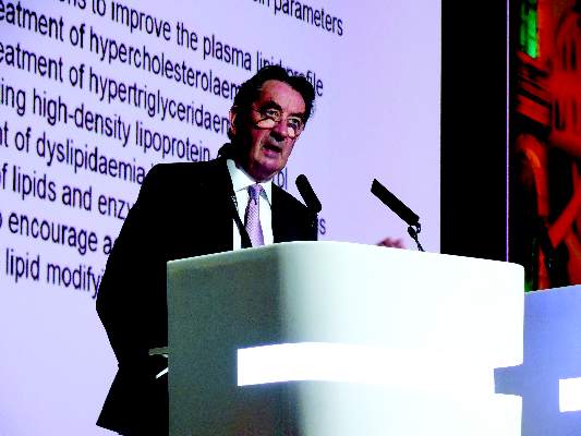
The ESC panel’s discussions about the LDL cholesterol targets were “difficult and long,” said Guy De Backer, MD, a member of the guidelines committee and professor of cardiology at the University of Ghent, Belgium, in a session devoted to the new guidelines during the annual congress of the ESC.
The ESC’s decision to retain the 2011 LDL cholesterol targets won praise from U.S. cardiologist Eugene Braunwald, MD. “I think that not measuring and following LDL cholesterol is silly. I don’t agree” with current U.S. guidelines, he said in a talk during the congress. “If you don’t follow LDL cholesterol then you don’t know a patient’s compliance.
Regularly measuring LDL cholesterol is important,” said Dr. Braunwald, professor of medicine at Harvard Medical School in Boston. But he questioned the LDL cholesterol targets set by the ESC panel, specifically the LDL cholesterol goal of less than 70 mg/dL for very-high-risk patients.

“I don’t think the ESC went low enough; the goal should be less than 50 mg/dL,” declared Dr. Braunwald, who added “anything above 50 mg/dL is toxic.”
The new guidelines also made the definition of “very-high-risk” patients “stricter and more precise,” said Alberico L. Catapano, MD, cochair of the panel and professor of pharmacology at the University of Milan.
The guideline’s detailed list defining very-high-risk patients includes those with either clinical or “unequivocal” imaging evidence for cardiovascular disease, as well as patients with diabetes and target-organ damage, severe chronic kidney disease, or a 10% or greater 10-year risk for fatal cardiovascular disease calculated by the European Score risk formula.
The potent lipid-lowering PCSK9 inhibitors came onto the U.S. and European markets in 2015, labeled in the United States specifically for patients with familial hypercholesterolemia.

In July 2016, an American College of Cardiology Task Force issued a model clinical-decision pathway for lowering LDL cholesterol levels that for the first time included the PCSK9 inhibitors, specified as a “may be considered” option for selected patients (J Am Coll Cardiol. 2016 Jul;68[1]:92-125). This consensus decision pathway was not a set of actual guidelines, which means the ESC revision is the first guideline to include the PCSK9 inhibitors. The panel took the same stance as the U.S. decision pathway and designated the PCSK9 inhibitors as a “may be considered” option.
Dr. Graham explained that this designation was driven by the current absence of evidence for clinical benefit from the large lipid-lowering effect of the PCSK9 antibodies, although trial results that address this are expected to appear very soon. Experts not on the guidelines panel agreed with this decision.
“We know that it’s crucial to await results from the clinical endpoint studies” before the guidelines committee makes a more forceful recommendation, commented Erik S.G. Stroes, MD, professor of vascular medicine at the Academic Medical Center in Amsterdam. He added, however, that many clinicians, himself included, have been pleased to offer PCSK9-inhibitor treatment to very-high-risk patients with no good alternatives for effectively lowering their LDL cholesterol to target levels, such as statin-intolerant patients or those with LDL cholesterol levels that remain high despite maximal therapy.

The current ESC guideline’s statement on when to use a PCSK9 inhibitor “is vague” said Dr. Braunwald. “Within the next year, we’ll have results from two huge trials that will show clinical outcomes. We know that PCSK9 inhibitors are extremely powerful at lowering LDL cholesterol, but I would like to see the loop closed” with proven effects on clinical outcomes. “I would bet 100 to 1 that they will be effective, but LDL cholesterol is just a surrogate marker, and you need to look in large populations before you make a guideline recommendation to use these drugs, so we’ll wait.”
Once the clinical value of treatment with PCSK9 inhibitors is settled, the next issue will be the cost of these drugs, something that will become a big consideration once they become more widely used, Dr. Braunwald added.
Dr. De Backer and Dr. Graham had no disclosures. Dr. Catapano has been a consultant to Aegerion, Amgen, AstraZeneca, Merck, Pfizer, and Sigma-Tau. Dr. Braunwald has been a consultant to Bayer, Daiichi Sankyo, the Medicines Company, Merck, Novartis, and Sanofi. Dr. Stroes has been a consultant to Amgen, Bristol-Myers Squibb, Merck, and Sanofi.
On Twitter @mitchelzoler
ROME – LDL cholesterol treatment targets remain alive and well in non-U.S. lipid management guidelines.
New dyslipidemia management guidelines from the European Society of Cardiology, issued in late August, retain the same LDL cholesterol targets as the prior, 2011 guidelines, a sharp and purposeful departure from the “risk-based” U.S. guidelines introduced in 2013 that eliminated treating patients to specific LDL cholesterol targets.
The new ESC guidelines also incorporated the new class of lipid-lowering drugs, PCSK9 inhibitors (evolocumab [Repatha] and alirocumab [Praluent]), into the treatment algorithm, and carved out a role for ezetimibe (Zetia) following its proven success as an add-on agent to statins (Eur Heart J. 2016. doi: 10.1093/eurheartj/ehw272).
But it’s retention of LDL cholesterol targets as a cornerstone of dyslipidemia management in the new ESC report, written jointly with the European Atherosclerosis Society, that especially distinguishes the new guidelines.
“It seemed to us logical that if you have drugs [statins] that lower LDL cholesterol, then you target LDL cholesterol,” explained Ian M. Graham, MD, professor of cardiovascular medicine at Trinity College in Dublin and cochair of the guidelines panel. “If a patient’s risk is high, they still need to lower LDL cholesterol. It’s not really contradictory to the U.S. approach,” Dr. Graham said in an interview.
The ESC panel’s discussions about the LDL cholesterol targets were “difficult and long,” said Guy De Backer, MD, a member of the guidelines committee and professor of cardiology at the University of Ghent, Belgium, in a session devoted to the new guidelines during the annual congress of the ESC.
The ESC’s decision to retain the 2011 LDL cholesterol targets won praise from U.S. cardiologist Eugene Braunwald, MD. “I think that not measuring and following LDL cholesterol is silly. I don’t agree” with current U.S. guidelines, he said in a talk during the congress. “If you don’t follow LDL cholesterol then you don’t know a patient’s compliance.
Regularly measuring LDL cholesterol is important,” said Dr. Braunwald, professor of medicine at Harvard Medical School in Boston. But he questioned the LDL cholesterol targets set by the ESC panel, specifically the LDL cholesterol goal of less than 70 mg/dL for very-high-risk patients.
“I don’t think the ESC went low enough; the goal should be less than 50 mg/dL,” declared Dr. Braunwald, who added “anything above 50 mg/dL is toxic.”
The new guidelines also made the definition of “very-high-risk” patients “stricter and more precise,” said Alberico L. Catapano, MD, cochair of the panel and professor of pharmacology at the University of Milan.
The guideline’s detailed list defining very-high-risk patients includes those with either clinical or “unequivocal” imaging evidence for cardiovascular disease, as well as patients with diabetes and target-organ damage, severe chronic kidney disease, or a 10% or greater 10-year risk for fatal cardiovascular disease calculated by the European Score risk formula.
The potent lipid-lowering PCSK9 inhibitors came onto the U.S. and European markets in 2015, labeled in the United States specifically for patients with familial hypercholesterolemia.
In July 2016, an American College of Cardiology Task Force issued a model clinical-decision pathway for lowering LDL cholesterol levels that for the first time included the PCSK9 inhibitors, specified as a “may be considered” option for selected patients (J Am Coll Cardiol. 2016 Jul;68[1]:92-125). This consensus decision pathway was not a set of actual guidelines, which means the ESC revision is the first guideline to include the PCSK9 inhibitors. The panel took the same stance as the U.S. decision pathway and designated the PCSK9 inhibitors as a “may be considered” option.
Dr. Graham explained that this designation was driven by the current absence of evidence for clinical benefit from the large lipid-lowering effect of the PCSK9 antibodies, although trial results that address this are expected to appear very soon. Experts not on the guidelines panel agreed with this decision.
“We know that it’s crucial to await results from the clinical endpoint studies” before the guidelines committee makes a more forceful recommendation, commented Erik S.G. Stroes, MD, professor of vascular medicine at the Academic Medical Center in Amsterdam. He added, however, that many clinicians, himself included, have been pleased to offer PCSK9-inhibitor treatment to very-high-risk patients with no good alternatives for effectively lowering their LDL cholesterol to target levels, such as statin-intolerant patients or those with LDL cholesterol levels that remain high despite maximal therapy.
The current ESC guideline’s statement on when to use a PCSK9 inhibitor “is vague” said Dr. Braunwald. “Within the next year, we’ll have results from two huge trials that will show clinical outcomes. We know that PCSK9 inhibitors are extremely powerful at lowering LDL cholesterol, but I would like to see the loop closed” with proven effects on clinical outcomes. “I would bet 100 to 1 that they will be effective, but LDL cholesterol is just a surrogate marker, and you need to look in large populations before you make a guideline recommendation to use these drugs, so we’ll wait.”
Once the clinical value of treatment with PCSK9 inhibitors is settled, the next issue will be the cost of these drugs, something that will become a big consideration once they become more widely used, Dr. Braunwald added.
Dr. De Backer and Dr. Graham had no disclosures. Dr. Catapano has been a consultant to Aegerion, Amgen, AstraZeneca, Merck, Pfizer, and Sigma-Tau. Dr. Braunwald has been a consultant to Bayer, Daiichi Sankyo, the Medicines Company, Merck, Novartis, and Sanofi. Dr. Stroes has been a consultant to Amgen, Bristol-Myers Squibb, Merck, and Sanofi.
On Twitter @mitchelzoler
EXPERT ANALYSIS FROM THE ESC CONGRESS 2016

Study eyes anastomotic failure in stapled vs. hand-sewn techniques
WAIKOLOA, HAWAII – The risk of anastomotic failure among emergency general surgery patients requiring bowel resection and anastomosis stands at 12.5% and is similar between stapled and hand-sewn techniques, results from a multicenter analysis demonstrated.
Surgeons participating in the study, known as Stapled vs. Handsewn: A Prospective Emergency Surgery Study (SHAPES), “appear to be performing hand-sewn techniques in patients who have a higher burden of disease,” Brandon R. Bruns, MD, said at the annual meeting of the American Association for the Surgery of Trauma. “Without adjustment and despite being performed in a more ill population, there was no difference in failure rate between hand-sewn and stapled techniques. After modeling, only being managed with an open abdomen and contamination at initial operation were associated with anastomotic failure.”

For SHAPES, which is the largest study of its kind and was sponsored by the AAST Multi-Institutional Studies Committee, Dr. Bruns and his associates set out to prospectively evaluate anastomotic failure rates for hand-sewn and stapled anastomoses in patients undergoing urgent/emergent operations. A 1999 study by Seattle researchers found that stapled techniques seemed to be associated with anastomotic failure (J Trauma. 1999;47[3]:500-08). Two years later, the same researchers pooled 4-year retrospective data from four trauma centers and concluded that again, hand-sewn techniques appeared to be superior to stapled techniques after traumatic bowel resection and anastomosis (J Trauma. 2001;51[6]:1054-61). Also in 2001, AAST sponsored a multi-institutional study that examined the same question, this time in penetrative colonic injury. Investigators found no difference in complications between the two groups (J Trauma. 2002;52[1]:117-21). They did, however, find a 22.7% overall incidence of colon-related complications.
“With this background we hypothesized that anastomotic failure rate would be high for emergency general surgery patients undergoing bowel resection and anastomosis,” said Dr. Bruns, an acute care surgeon at the R. Adams Cowley Shock Trauma Center at the University of Maryland Medical Center, Baltimore. “We also hypothesized that failure rates would be higher for stapled techniques, compared with hand-sewn.”
The SHAPES researchers prospectively enrolled 595 patients at 15 medical centers in the United States who underwent urgent/emergent bowel resection for emergency general surgery pathology between July 22, 2013, and Dec. 31, 2015. The patients were grouped by hand-sewn vs. stapled anastomoses and demographic and clinical variables were collected. The primary outcome was anastomotic failure. As in other studies, anastomotic failure was evaluated at the anastomosis level. Multivariable logistic regression was done, controlling for age and risk factors for anastomotic failure.
Dr. Bruns reported that the 595 patients had 649 anastomoses. Of these, 61% were stapled and 39% were hand-sewn. The mean age of patients was 62 years, 51% were male, and the overall mortality rate was 7.7%. More than two-thirds of the patients (35%) had more than one indication for operative intervention. The most common single indication for operation was small bowel obstruction, at 23%. The majority of the anastomoses were small bowel to small bowel (72%), while 21% were small bowel to large bowel, and 7% were large bowel to large bowel. There were a total of 81 anastomotic failures, for a rate of 12.5%.
When the researchers compared the hand-sewn and stapled groups, they were similar with respect to sex and age, with higher Charlson comorbidity indices in stapled and a higher body mass index in the hand-sewn group. They also observed a lower hemoglobin, higher INR, higher lactate, lower albumin, and worse renal function in the hand-sewn group, compared with the stapled group. Moreover, hand-sewn anastomotic techniques were performed more frequently in patients that were on vasopressor support. “Despite patients receiving hand-sewn techniques, having worse laboratory values, and more intraoperative vasopressors, the two techniques had equivalent failure rates, at 15.4% for hand-sewn and 10.6% for stapled,” Dr. Bruns said. “Patients with hand-sewn techniques had longer hospital length of stay, longer ICU length of stay, and a significantly higher mortality, compared with those who underwent stapled techniques. Interestingly and different from most other studies on the topic, operating time between the two groups was equivalent.”
On multivariate regression, the presence of contamination at initial bowel resection (OR 1.96) and the patient being managed with an open abdomen (OR 2.53) were independently associated with anastomotic failure, while the type of anastomosis (hand-sewn vs. stapled) was not.
The researchers also conducted a subanalysis of 165 patients managed with an open abdomen who had bowel resection and anastomosis. These patients had higher BMIs, higher lactates, higher INRs, and more negative base deficits, compared with those who were not managed with an open abdomen. “Perhaps not unexpectedly, open abdomen patients were more likely to be on vasopressor agents,” Dr. Bruns said. “They had longer hospital lengths of stay, more ICU days, and an 18.2% overall mortality. Overall there was an almost 22% anastomotic failure rate in patients managed with an open abdomen, compared with an 8.5% rate in patients managed with non-open techniques.” Comparing hand-sewn and stapled techniques, there was no difference in failure rate in patients managed with an open abdomen (25.2% vs. 17.5%, respectively; P = .20).
“An overall mortality rate of 8% and an anastomotic complication rate of 12.5% should emphasize the dire needs for these operations and the need for meticulous operative as well as surgically directed perioperative care in these patients,” the invited discussant, Gregory Jurkovich, MD, professor of surgery at the Davis Medical Center, University of California, said at the meeting. “We as surgeons must pay attention to all aspects of care in these patients.”
Dr. Bruns acknowledged certain limitations of the study, including the fact that it was not a randomized, controlled trial. Also, “surgeon preference did dictate the type of anastomosis that was created, and the patient and surgeon populations were heterogeneous,” he said. “The multivariable model was limited by missing laboratory data, likely given the urgent nature of some of the operative procedures.”
He reported having no financial disclosures.
WAIKOLOA, HAWAII – The risk of anastomotic failure among emergency general surgery patients requiring bowel resection and anastomosis stands at 12.5% and is similar between stapled and hand-sewn techniques, results from a multicenter analysis demonstrated.
Surgeons participating in the study, known as Stapled vs. Handsewn: A Prospective Emergency Surgery Study (SHAPES), “appear to be performing hand-sewn techniques in patients who have a higher burden of disease,” Brandon R. Bruns, MD, said at the annual meeting of the American Association for the Surgery of Trauma. “Without adjustment and despite being performed in a more ill population, there was no difference in failure rate between hand-sewn and stapled techniques. After modeling, only being managed with an open abdomen and contamination at initial operation were associated with anastomotic failure.”
For SHAPES, which is the largest study of its kind and was sponsored by the AAST Multi-Institutional Studies Committee, Dr. Bruns and his associates set out to prospectively evaluate anastomotic failure rates for hand-sewn and stapled anastomoses in patients undergoing urgent/emergent operations. A 1999 study by Seattle researchers found that stapled techniques seemed to be associated with anastomotic failure (J Trauma. 1999;47[3]:500-08). Two years later, the same researchers pooled 4-year retrospective data from four trauma centers and concluded that again, hand-sewn techniques appeared to be superior to stapled techniques after traumatic bowel resection and anastomosis (J Trauma. 2001;51[6]:1054-61). Also in 2001, AAST sponsored a multi-institutional study that examined the same question, this time in penetrative colonic injury. Investigators found no difference in complications between the two groups (J Trauma. 2002;52[1]:117-21). They did, however, find a 22.7% overall incidence of colon-related complications.
“With this background we hypothesized that anastomotic failure rate would be high for emergency general surgery patients undergoing bowel resection and anastomosis,” said Dr. Bruns, an acute care surgeon at the R. Adams Cowley Shock Trauma Center at the University of Maryland Medical Center, Baltimore. “We also hypothesized that failure rates would be higher for stapled techniques, compared with hand-sewn.”
The SHAPES researchers prospectively enrolled 595 patients at 15 medical centers in the United States who underwent urgent/emergent bowel resection for emergency general surgery pathology between July 22, 2013, and Dec. 31, 2015. The patients were grouped by hand-sewn vs. stapled anastomoses and demographic and clinical variables were collected. The primary outcome was anastomotic failure. As in other studies, anastomotic failure was evaluated at the anastomosis level. Multivariable logistic regression was done, controlling for age and risk factors for anastomotic failure.
Dr. Bruns reported that the 595 patients had 649 anastomoses. Of these, 61% were stapled and 39% were hand-sewn. The mean age of patients was 62 years, 51% were male, and the overall mortality rate was 7.7%. More than two-thirds of the patients (35%) had more than one indication for operative intervention. The most common single indication for operation was small bowel obstruction, at 23%. The majority of the anastomoses were small bowel to small bowel (72%), while 21% were small bowel to large bowel, and 7% were large bowel to large bowel. There were a total of 81 anastomotic failures, for a rate of 12.5%.
When the researchers compared the hand-sewn and stapled groups, they were similar with respect to sex and age, with higher Charlson comorbidity indices in stapled and a higher body mass index in the hand-sewn group. They also observed a lower hemoglobin, higher INR, higher lactate, lower albumin, and worse renal function in the hand-sewn group, compared with the stapled group. Moreover, hand-sewn anastomotic techniques were performed more frequently in patients that were on vasopressor support. “Despite patients receiving hand-sewn techniques, having worse laboratory values, and more intraoperative vasopressors, the two techniques had equivalent failure rates, at 15.4% for hand-sewn and 10.6% for stapled,” Dr. Bruns said. “Patients with hand-sewn techniques had longer hospital length of stay, longer ICU length of stay, and a significantly higher mortality, compared with those who underwent stapled techniques. Interestingly and different from most other studies on the topic, operating time between the two groups was equivalent.”
On multivariate regression, the presence of contamination at initial bowel resection (OR 1.96) and the patient being managed with an open abdomen (OR 2.53) were independently associated with anastomotic failure, while the type of anastomosis (hand-sewn vs. stapled) was not.
The researchers also conducted a subanalysis of 165 patients managed with an open abdomen who had bowel resection and anastomosis. These patients had higher BMIs, higher lactates, higher INRs, and more negative base deficits, compared with those who were not managed with an open abdomen. “Perhaps not unexpectedly, open abdomen patients were more likely to be on vasopressor agents,” Dr. Bruns said. “They had longer hospital lengths of stay, more ICU days, and an 18.2% overall mortality. Overall there was an almost 22% anastomotic failure rate in patients managed with an open abdomen, compared with an 8.5% rate in patients managed with non-open techniques.” Comparing hand-sewn and stapled techniques, there was no difference in failure rate in patients managed with an open abdomen (25.2% vs. 17.5%, respectively; P = .20).
“An overall mortality rate of 8% and an anastomotic complication rate of 12.5% should emphasize the dire needs for these operations and the need for meticulous operative as well as surgically directed perioperative care in these patients,” the invited discussant, Gregory Jurkovich, MD, professor of surgery at the Davis Medical Center, University of California, said at the meeting. “We as surgeons must pay attention to all aspects of care in these patients.”
Dr. Bruns acknowledged certain limitations of the study, including the fact that it was not a randomized, controlled trial. Also, “surgeon preference did dictate the type of anastomosis that was created, and the patient and surgeon populations were heterogeneous,” he said. “The multivariable model was limited by missing laboratory data, likely given the urgent nature of some of the operative procedures.”
He reported having no financial disclosures.
AT THE AAST ANNUAL MEETING
Key clinical point: In patients requiring emergency bowel resection and anastomosis, surgeons appear to be performing hand-sewn techniques in patients who have a higher burden of disease.
Major finding: There were 81 anastomotic failures in the study group, for a rate of 12.5%.
Data source: A prospective evaluation of 595 patients at 15 medical centers in the United States who underwent urgent/emergent bowel resection for emergency general surgery pathology between July 22, 2013, and Dec. 31, 2015.
Disclosures: Dr. Bruns reported having no financial disclosures.

Racial gaps persist in lung cancer trial enrollment
Elderly women, blacks, Asians, Pacific Islanders, and Hispanics are all still underrepresented in lung cancer clinical trials, while gender- and age-based disparities have improved, according to a report published online in Journal of Clinical Oncology.
Herbert H. Pang, PhD, of the Li Ka Shing Faculty of Medicine, Hong Kong, and his coauthors noted that enrollment disparities in cancer clinical trials have existed for many years, with previous research showing underenrollment of the elderly, women, blacks, and racial and ethnic minorities.
In this study, they analyzed data from 23,006 participants in National Cancer Institute lung cancer trials, and 578,476 patients with lung cancer from the SEER registry (J Clin Oncol. 2016 Sep 19. doi: 10.1200/JCO.2016.67.7088).
When they compared the proportion of each subgroup in the trial population with the proportion in the U.S. lung cancer population over time, they noted consistent underrepresentation of blacks, Asian/Pacific Islander, and Hispanic patients across the entire study period.
The enrollment disparity for patients aged 70 years or older with non–small-cell lung cancer improved significantly from 1990 to 2012, but while there has been an increase in the proportion of elderly patients with small-cell lung cancer in the U.S. population from 1990 to 2012, the proportion of elderly patients in trials for small-cell lung cancer remained static.
The authors suggested that this may have been the result of local enrollment patterns for the mostly smaller, phase II trials in small-cell lung cancer, but also the fact that the therapies investigated for small-cell lung cancer may have posed a greater risk of treatment toxicity, which would limit the enrollment of older patients.
Significant improvements were seen in the proportion of women enrolled in lung cancer trials, with the enrollment gap between the genders closing in 2012, although elderly women were still underrepresented in lung cancer clinical trials.
“These findings suggest a beneficial effect of the NIH Revitalization Act of 1993 that mandated the inclusion of women and minorities in all NIH-funded research,” the authors wrote. “However, other important enrollment disparities, especially for older patients with SCLC, elderly women, and racial/ethnic minorities, continue to persist and require ongoing work to eliminate underrepresentation in lung cancer treatment trials.”
FROM THE JOURNAL OF CLINICAL ONCOLOGY
Key clinical point: Elderly women, blacks, Asians, Pacific Islanders, and Hispanics are all still underrepresented in lung cancer clinical trials, while gender- and age-based disparities have improved.
Major finding: There is consistent underrepresentation of blacks, Asian/Pacific Islander, and Hispanic patients in lung cancer clinical trials, but the enrollment gap between genders has closed.
Data source: Analysis of data from 23,006 participants in National Cancer Institute lung cancer trials, and 578,476 patients with lung cancer from the SEER registry.
Disclosures: The study was supported by the National Institutes of Health, National Institute on Aging, and the Health and Medical Research Fund of Hong Kong, and by the National Institute on Aging. Dr. Wang had no disclosures to report; several coauthors reported relationships with various pharmaceutical companies.
Palifosfamide plus doxorubicin does not improve survival in sarcoma
Palifosfamide added to doxorubicin did not improve survival outcomes in patients with metastatic soft tissue sarcoma, compared with doxorubicin alone, a study showed.
The phase III randomized trial did not meet its amended primary endpoint, as there was no significant difference in progression-free survival (PFS) among patients in the two-drug regimen arm.
“This study represents one of the largest international efforts among sarcoma centers to date,” wrote Christopher W. Ryan, MD, of the Oregon Health & Science University, Knight Cancer Institute, Portland, Ore., and his coauthors (J Clin Oncol. 2016 Sept 12. doi: 10.1200/JCO.2016.67.668).
“Single-agent doxorubicin thus remains a reference standard for the treatment of metastatic soft tissue sarcoma,” they wrote.
Doxorubicin has remained the standard first-line treatment for most sarcoma patients for more than 4 decades. Palifosfamide, a tris salt of isophosphoramide mustard, has previously demonstrated broad activity against sarcoma in experimental models.
In addition, a randomized phase II trial showed improved PFS for combined doxorubicin and palifosfamide, compared with doxorubicin alone, with a hazard ratio (HR) of 0.43 (95% CI, 0.19-0.95).
Based on those promising results, Dr. Ryan and his colleagues conducted a phase III trial that evaluated the two-drug combination with doxorubicin alone as first-line treatment of metastatic soft tissue sarcoma.
The cohort included 447 patients who were randomly assigned 1:1 to receive either doxorubicin 75 mg/m2 IV on day 1 plus palifosfamide 150 mg/m2 per day IV on days 1-3, or doxorubicin plus placebo once every 21 days for up to six cycles.
The primary endpoint of the study was PFS by independent radiologic review.
There was no significant difference in PFS between the two cohorts. For the combination therapy group, the median PFS was 6.0 months, compared with 5.2 months for doxorubicin plus placebo (HR, 0.86; 95% CI, 0.68-1.08; P = .19).
Similar results were observed for median overall survival, which was also similar in the two treatment groups: 15.9 months for doxorubicin plus palifosfamide and 16.9 months for doxorubicin plus placebo (HR, 1.05; 95% CI, 0.79-1.39; P = .74).
As for toxicity, all patients had at least one treatment-emergent adverse event, with the most common of any grade being alopecia, nausea, and fatigue.
Grade 3-4 adverse events were more frequently observed in patients receiving doxorubicin plus palifosfamide (63.6%) than in the single-therapy group (50.9%; P = .0075).
“The median PFS of 5.2 months and OS [overall survival] of nearly 17 months with doxorubicin should serve as a reference in the design of future studies in the first-line treatment of metastatic soft tissue sarcoma,” Dr. Ryan and his associates said.
The study was supported by ZIOPHARM Oncology. Dr. Ryan and several of the coauthors reported multiple relationships with industry.
FROM THE JOURNAL OF CLINICAL ONCOLOGY
Key clinical point: Palifosfamide combined with doxorubicin did not improve survival in metastatic soft tissue sarcoma.
Major finding: The median progression-free survival was 6.0 months for palifosfamide/doxorubicin vs. 5.2 months for doxorubicin plus placebo (HR, 0.86; 95% CI, 0.68-1.08; P = 0.19).
Data source: A phase III randomized trial that included 447 patients with metastatic soft tissue sarcoma.
Disclosures: The study was supported by ZIOPHARM Oncology. Dr. Ryan and several of his coauthors reported multiple relationships with industry.
Update on the third international consensus definitions for sepsis and septic shock
Sepsis is the primary cause of death from infection. Early identification and treatment of sepsis is important in improving patient outcomes. The consensus conference sought to differentiate sepsis, which is defined as “life-threatening organ dysfunction caused by a dysregulated host response to infection” from uncomplicated infection.
Sepsis was last classified in a 2001 guideline that based its definition on the presence of two or more systemic inflammatory response syndrome (SIRS) criteria, which included an elevated temperature, heart rate higher than 90 bpm, respiratory rate higher than 20 breaths per minute, and a white blood cell count greater than greater than 12,000 mcL or less than 4,000 mcL or greater than 10% immature bands.

The problem with the SIRS definition of sepsis is that while it reflects a response to infection, it does not sufficiently distinguish between individuals with infections and those with a dysregulated response that leads to a poor prognosis, which is the definition of sepsis. The current consensus conference redefines sepsis with a more direct emphasis on organ dysfunction, as this is the aspect of sepsis that is most clearly linked to patient outcomes.
In the consensus conference document, sepsis is defined as a “life-threatening organ dysfunction caused by a dysregulated host response to infection.” The guidelines recommend using the quick version of the sequential (sepsis-related) organ failure assessment score (qSOFA) to identify patients with sepsis. In its long form, the SOFA used seven clinical and laboratory data points for completion, and is best suited to use in an intensive care setting where detailed data are available. The qSOFA score has only three criteria and by being easier to use can aid in rapid identification of sepsis and the patients most likely to deteriorate from sepsis.

The qSOFA criteria predict poor outcome in patients with infection who have two or more of the following: respiratory rate greater than or equal to 22 breaths/min, new or worsened altered mentation, or systolic blood pressure less than or equal to 100 mm Hg. Unlike the full SOFA score, the qSOFA does not require any laboratory testing and so can be performed in the office or bedside on a hospital floor. The qSOFA does not necessarily define sepsis, rather it identifies patients at a higher risk of hospital death or prolonged ICU stay. The consensus conference suggests that “qSOFA criteria be used to prompt clinicians to further investigate for organ dysfunction, initiate or escalate therapy as appropriate, and consider referral to critical care or increase the frequency of monitoring, if such actions have not already been undertaken.” The task force suggested that the qSOFA score may be a helpful adjunct to best clinical judgment for identifying patients who might benefit from a higher level of care.
Septic shock is defined as a subset of sepsis in which profound circulatory, cellular, and metabolic abnormalities are associated with a greater risk for death than sepsis alone. Septic shock can be identified when, after adequate fluid resuscitation, the patient requires vasopressor therapy to maintain mean arterial pressure of at least 65 mm Hg and has a serum lactate level greater than 2 mmol/L.

Once sepsis is suspected, prompt therapy needs to be started as per the Surviving Sepsis Campaign Guidelines. The qSOFA criteria can be used to identify patients at high risk for morbidity and mortality. Within 3 hours, a lactate level should be obtained as well as blood cultures from two separate sites drawn prior to administration of antibiotics (but do not delay antibiotic administration). Empiric broad-spectrum antibiotics should be given within 45 minutes of the identification of sepsis. Antibiotic choice will vary per clinician/institution preference, but should likely include coverage for Pseudomonas and MRSA (piperacillin/tazobactam and vancomycin, for example). Antibiotics should be reassessed daily for de-escalation. Administer 30 mL/kg crystalloid for hypotension or lactate greater than or equal to 4 mmol/L. Within 6 hours, vasopressors should be given for hypotension that does not respond to initial fluid resuscitation to maintain a mean arterial pressure (MAP) of at least 65mm Hg. In the event of persistent hypotension after initial fluid administration (MAP under 65 mm Hg) or if initial lactate was greater than or equal to 4 mmol/L, volume status and tissue perfusion should be reassessed and lactate should be rechecked if it was initially elevated.
The bottom line
A 2016 international task force recommended that the definition of sepsis should be changed to emphasize organ dysfunction rather than a systemic inflammatory response. Use of the qSOFA score, which relies only on clinically observable data rather than laboratory evaluation, is recommended to identify patients at high risk for morbidity and mortality. Early recognition of sepsis and evaluation with qSOFT should facilitate early treatment and improve survival.
References
Singer M, et al. The Third International Consensus Definitions for Sepsis and Septic Shock (Sepsis-3) FRCP; JAMA. 2016;315[8]:801-10. doi: 10.1001/jama.2016.0287.
Levy MM, Fink MP, Marshall JC, Abraham E, Angus D, Cook D, et al. 2001 SCCM/ESICM/ACCP/ATS/SIS International Sepsis Definitions Conference. Crit Care Med. 2003 Apr;31(4):1250-6.
Singer M, Deutschman CS, Seymour C, et al. The third international consensus definitions for sepsis and septic shock (sepsis-3). JAMA. 2016 Feb 23;315(8):801-10.
Dellinger RP, Carlet JM, Masur H, Gerlach H, Calandra T, Cohen J, et al. Surviving Sepsis Campaign guidelines for management of severe sepsis and septic shock. Crit Care Med. 2004 Mar;32(3):858-73.
Dr. Mills is assistant residency program director and assistant professor in the department of family and community medicine and department of physiology at Sidney Kimmel Medical College at Thomas Jefferson University. Dr. Botti is a second-year resident in the family medicine residency program department of family and community medicine at Sidney Kimmel Medical College at Thomas Jefferson University. Dr. Skolnik is associate director of the family medicine residency program at Abington (Pa.) Memorial Hospital and professor of family and community medicine at Temple University, Philadelphia.
Sepsis is the primary cause of death from infection. Early identification and treatment of sepsis is important in improving patient outcomes. The consensus conference sought to differentiate sepsis, which is defined as “life-threatening organ dysfunction caused by a dysregulated host response to infection” from uncomplicated infection.
Sepsis was last classified in a 2001 guideline that based its definition on the presence of two or more systemic inflammatory response syndrome (SIRS) criteria, which included an elevated temperature, heart rate higher than 90 bpm, respiratory rate higher than 20 breaths per minute, and a white blood cell count greater than greater than 12,000 mcL or less than 4,000 mcL or greater than 10% immature bands.
The problem with the SIRS definition of sepsis is that while it reflects a response to infection, it does not sufficiently distinguish between individuals with infections and those with a dysregulated response that leads to a poor prognosis, which is the definition of sepsis. The current consensus conference redefines sepsis with a more direct emphasis on organ dysfunction, as this is the aspect of sepsis that is most clearly linked to patient outcomes.
In the consensus conference document, sepsis is defined as a “life-threatening organ dysfunction caused by a dysregulated host response to infection.” The guidelines recommend using the quick version of the sequential (sepsis-related) organ failure assessment score (qSOFA) to identify patients with sepsis. In its long form, the SOFA used seven clinical and laboratory data points for completion, and is best suited to use in an intensive care setting where detailed data are available. The qSOFA score has only three criteria and by being easier to use can aid in rapid identification of sepsis and the patients most likely to deteriorate from sepsis.
The qSOFA criteria predict poor outcome in patients with infection who have two or more of the following: respiratory rate greater than or equal to 22 breaths/min, new or worsened altered mentation, or systolic blood pressure less than or equal to 100 mm Hg. Unlike the full SOFA score, the qSOFA does not require any laboratory testing and so can be performed in the office or bedside on a hospital floor. The qSOFA does not necessarily define sepsis, rather it identifies patients at a higher risk of hospital death or prolonged ICU stay. The consensus conference suggests that “qSOFA criteria be used to prompt clinicians to further investigate for organ dysfunction, initiate or escalate therapy as appropriate, and consider referral to critical care or increase the frequency of monitoring, if such actions have not already been undertaken.” The task force suggested that the qSOFA score may be a helpful adjunct to best clinical judgment for identifying patients who might benefit from a higher level of care.
Septic shock is defined as a subset of sepsis in which profound circulatory, cellular, and metabolic abnormalities are associated with a greater risk for death than sepsis alone. Septic shock can be identified when, after adequate fluid resuscitation, the patient requires vasopressor therapy to maintain mean arterial pressure of at least 65 mm Hg and has a serum lactate level greater than 2 mmol/L.
Once sepsis is suspected, prompt therapy needs to be started as per the Surviving Sepsis Campaign Guidelines. The qSOFA criteria can be used to identify patients at high risk for morbidity and mortality. Within 3 hours, a lactate level should be obtained as well as blood cultures from two separate sites drawn prior to administration of antibiotics (but do not delay antibiotic administration). Empiric broad-spectrum antibiotics should be given within 45 minutes of the identification of sepsis. Antibiotic choice will vary per clinician/institution preference, but should likely include coverage for Pseudomonas and MRSA (piperacillin/tazobactam and vancomycin, for example). Antibiotics should be reassessed daily for de-escalation. Administer 30 mL/kg crystalloid for hypotension or lactate greater than or equal to 4 mmol/L. Within 6 hours, vasopressors should be given for hypotension that does not respond to initial fluid resuscitation to maintain a mean arterial pressure (MAP) of at least 65mm Hg. In the event of persistent hypotension after initial fluid administration (MAP under 65 mm Hg) or if initial lactate was greater than or equal to 4 mmol/L, volume status and tissue perfusion should be reassessed and lactate should be rechecked if it was initially elevated.
The bottom line
A 2016 international task force recommended that the definition of sepsis should be changed to emphasize organ dysfunction rather than a systemic inflammatory response. Use of the qSOFA score, which relies only on clinically observable data rather than laboratory evaluation, is recommended to identify patients at high risk for morbidity and mortality. Early recognition of sepsis and evaluation with qSOFT should facilitate early treatment and improve survival.
References
Singer M, et al. The Third International Consensus Definitions for Sepsis and Septic Shock (Sepsis-3) FRCP; JAMA. 2016;315[8]:801-10. doi: 10.1001/jama.2016.0287.
Levy MM, Fink MP, Marshall JC, Abraham E, Angus D, Cook D, et al. 2001 SCCM/ESICM/ACCP/ATS/SIS International Sepsis Definitions Conference. Crit Care Med. 2003 Apr;31(4):1250-6.
Singer M, Deutschman CS, Seymour C, et al. The third international consensus definitions for sepsis and septic shock (sepsis-3). JAMA. 2016 Feb 23;315(8):801-10.
Dellinger RP, Carlet JM, Masur H, Gerlach H, Calandra T, Cohen J, et al. Surviving Sepsis Campaign guidelines for management of severe sepsis and septic shock. Crit Care Med. 2004 Mar;32(3):858-73.
Dr. Mills is assistant residency program director and assistant professor in the department of family and community medicine and department of physiology at Sidney Kimmel Medical College at Thomas Jefferson University. Dr. Botti is a second-year resident in the family medicine residency program department of family and community medicine at Sidney Kimmel Medical College at Thomas Jefferson University. Dr. Skolnik is associate director of the family medicine residency program at Abington (Pa.) Memorial Hospital and professor of family and community medicine at Temple University, Philadelphia.

New method proposed for phenotyping COPD patients
LONDON – Based on readily available clinical data, patients with chronic obstructive pulmonary disease (COPD) can be placed into five phenotypes with different characteristics and risk profiles, according to data generated by a cluster analysis and presented at the European Respiratory Society International Congress 2016.
The algorithm that places patients into these phenotypes was developed from a cluster analysis, reported Pierre-Regis Burgel, MD, PhD, professor of respiratory medicine, Université Réné Descartes, Paris. Mortality rates at 3 years for these phenotypes ranged from 2.6% to 49.5%

“We think that this could be the basis for recognizing clinically distinct COPD phenotypes and designing better tailored management,” Dr. Burgel explained. “We also think it has potential use in routine clinical assessment.”
To create these phenotypes, data from 2,049 COPD patients enrolled in a French-Belgian collaborative cohort were evaluated with Classification And Regression Tree (CART) analyses. The five phenotypes were derived from symptom burden, respiratory function, relative age, and presence of comorbidities.
Based on these characteristics, “a set of clinical rules” to phenotype patients was developed, according to Dr. Burgel. This algorithm was then further validated with the 3,651 COPD patients enrolled in the prospective COPD Cohorts Collaborative International Assessment (3CIA) initiative.
The two initial branches in the algorithm are created by dividing patients into those with and without cardiovascular comorbidities or diabetes. In those without cardiovascular disease, the phenotypes are defined by relative symptom severity, using cut-off scores from the modified Medical Research Council (mMRC) dyspnea assessment tool and relative degrees of lung function impairment as measured with forced expiratory volume in 1 second (FEV1).
In those with cardiovascular disease or diabetes, mMRC-defined symptoms and FEV1-defined lung function impairment also create decision points in the algorithm, but age and body mass index (BMI) are additional variables that direct patients to a specific phenotype. Class 4 and 5 are reached in the absence of cardiovascular disease or diabetes only, while cardiovascular disease is a prerequisite to reach Classes 4 and 5. Class 2 is the only phenotype that can be reached through the algorithm irrespective of the presence or absence of cardiovascular disease.
Using this algorithm, each of the phenotypes was associated with similar relative hierarchy in mortality in the two cohorts, even though mortality rates for each phenotype were consistently lower in the 3CIA group.
For class 1, which was reached by patients with cardiovascular disease or diabetes, the greatest symptom burden, and the lowest lung function, the mortality rates at 3 years were 49.5% and 23.2% for the French-Belgian and 3CIA cohorts, respectively.
For class 4, which was also defined by the greatest symptom burden and the lowest lung function without cardiovascular disease or diabetes the mortality rates were 45.3% and 27.3%, respectively. The lowest mortality rates, which were 2.6% and 4.0%, respectively, were found in the class 5 phenotype, which contained patients with a low symptom burden (mMRC less than or equal to 1), relatively good lung function (FEV1 greater than or equal to 60%), and no history of cardiovascular disease or diabetes.
In classes 2 and 3, the mortality rates fell in between those of the lowest- and highest-risk phenotypes. Specifically, these 3-year mortality rates were 22.9% and 24%, respectively, in the French-Belgian cohort, and 11.1% and 14.1%, respectively, in the 3CIA cohort.
The consistency of the hierarchy of outcomes in the two cohorts provides the basis for suggesting that these phenotypes are effective for categorizing relative risk, according to Dr. Burgel. He believes that the phenotypes are clinically important, and he emphasized that the algorithm relies on clinical information that is already routinely collected and readily accessible.
“There is growing awareness that COPD phenotypes are important and are likely to be valuable in managing patients,” Dr. Burgel explained. “We feel that we have created simple rules for allocating patients that we think will be useful for research and for clinical application.”
Dr. Burgel reported financial relationships with AstraZeneca, Boehringer Ingelheim, Chiesi, GlaxoSmithKline, Novartis, Pfizer, and Vertex.
LONDON – Based on readily available clinical data, patients with chronic obstructive pulmonary disease (COPD) can be placed into five phenotypes with different characteristics and risk profiles, according to data generated by a cluster analysis and presented at the European Respiratory Society International Congress 2016.
The algorithm that places patients into these phenotypes was developed from a cluster analysis, reported Pierre-Regis Burgel, MD, PhD, professor of respiratory medicine, Université Réné Descartes, Paris. Mortality rates at 3 years for these phenotypes ranged from 2.6% to 49.5%
“We think that this could be the basis for recognizing clinically distinct COPD phenotypes and designing better tailored management,” Dr. Burgel explained. “We also think it has potential use in routine clinical assessment.”
To create these phenotypes, data from 2,049 COPD patients enrolled in a French-Belgian collaborative cohort were evaluated with Classification And Regression Tree (CART) analyses. The five phenotypes were derived from symptom burden, respiratory function, relative age, and presence of comorbidities.
Based on these characteristics, “a set of clinical rules” to phenotype patients was developed, according to Dr. Burgel. This algorithm was then further validated with the 3,651 COPD patients enrolled in the prospective COPD Cohorts Collaborative International Assessment (3CIA) initiative.
The two initial branches in the algorithm are created by dividing patients into those with and without cardiovascular comorbidities or diabetes. In those without cardiovascular disease, the phenotypes are defined by relative symptom severity, using cut-off scores from the modified Medical Research Council (mMRC) dyspnea assessment tool and relative degrees of lung function impairment as measured with forced expiratory volume in 1 second (FEV1).
In those with cardiovascular disease or diabetes, mMRC-defined symptoms and FEV1-defined lung function impairment also create decision points in the algorithm, but age and body mass index (BMI) are additional variables that direct patients to a specific phenotype. Class 4 and 5 are reached in the absence of cardiovascular disease or diabetes only, while cardiovascular disease is a prerequisite to reach Classes 4 and 5. Class 2 is the only phenotype that can be reached through the algorithm irrespective of the presence or absence of cardiovascular disease.
Using this algorithm, each of the phenotypes was associated with similar relative hierarchy in mortality in the two cohorts, even though mortality rates for each phenotype were consistently lower in the 3CIA group.
For class 1, which was reached by patients with cardiovascular disease or diabetes, the greatest symptom burden, and the lowest lung function, the mortality rates at 3 years were 49.5% and 23.2% for the French-Belgian and 3CIA cohorts, respectively.
For class 4, which was also defined by the greatest symptom burden and the lowest lung function without cardiovascular disease or diabetes the mortality rates were 45.3% and 27.3%, respectively. The lowest mortality rates, which were 2.6% and 4.0%, respectively, were found in the class 5 phenotype, which contained patients with a low symptom burden (mMRC less than or equal to 1), relatively good lung function (FEV1 greater than or equal to 60%), and no history of cardiovascular disease or diabetes.
In classes 2 and 3, the mortality rates fell in between those of the lowest- and highest-risk phenotypes. Specifically, these 3-year mortality rates were 22.9% and 24%, respectively, in the French-Belgian cohort, and 11.1% and 14.1%, respectively, in the 3CIA cohort.
The consistency of the hierarchy of outcomes in the two cohorts provides the basis for suggesting that these phenotypes are effective for categorizing relative risk, according to Dr. Burgel. He believes that the phenotypes are clinically important, and he emphasized that the algorithm relies on clinical information that is already routinely collected and readily accessible.
“There is growing awareness that COPD phenotypes are important and are likely to be valuable in managing patients,” Dr. Burgel explained. “We feel that we have created simple rules for allocating patients that we think will be useful for research and for clinical application.”
Dr. Burgel reported financial relationships with AstraZeneca, Boehringer Ingelheim, Chiesi, GlaxoSmithKline, Novartis, Pfizer, and Vertex.
AT THE ERS CONGRESS 2016
Key clinical point: A relatively simple method proposed for identifying COPD phenotypes has implications for clinical care as well as research.
Major finding: Five phenotypes were identified with mortality rates at 3 years ranging from 2.6% to 49.5%
Data source: Cohort analyses.
Disclosures: Dr. Burgel reported financial relationships with AstraZeneca, Boehringer Ingelheim, Chiesi, GlaxoSmithKline, Novartis, Pfizer, and Vertex.

Oral HIV PrEP also protects against herpes
DURBAN, SOUTH AFRICA – Oral antiretroviral pre-exposure prophylaxis (PrEP) against HIV also reduces the risk of acquiring herpes simplex virus type 2, according to research presented at the 21st International AIDS Conference.
“Given the limited interventions for primary prevention of HSV-2, efficacy against HSV-2 provides additional benefit to oral PrEP,” observed Connie Celum, MD, professor of global health and medicine at the University of Washington, Seattle.

HSV-2 is, however, the only non-HIV sexually transmitted infection whose incidence is reduced by PrEP with emtricitabine/tenofovir (Truvada), the sole approved agent for oral HIV PrEP, she added.
Dr. Celum was lead author in a report from the landmark Partners PrEP study of HIV serodiscordant heterosexual Kenyan and Ugandan couples, which demonstrated that oral PrEP provided a 33% reduction in the risk of HSV-2 infection in participants with a known HSV-2-positive partner (Ann Intern Med. 2014 Jul 1;161[1]:11-9. doi: 10.7326/M13-2471).
Tenofovir has been shown to have in vitro activity against HSV-2, providing biologic plausibility to the Partners PrEP study finding, but the 90% effective concentration of the drug required to achieve strong anti-HSV-2 activity in the laboratory makes it likely that good adherence to daily oral PrEP is necessary to see the clinical benefit in terms of reduced HSV-2 acquisition, Dr. Celum said.
She also addressed other questions about the interaction between PrEP and non-HIV STIs she often receives from physicians who provide care for HIV-infected or at-risk patients.
Do STIs reduce the efficacy of oral PrEP? “My answer would be no,” Dr. Celum said. She noted that no difference in PrEP efficacy was seen between patients with and without STIs in Partners PrEP or the French IPERGAY study.
Does PrEP increase the rate of other STIs? The concern here has been that PrEP’s beneficial effect in reducing the risk of HIV infection could be counteracted by a compensatory increase in unsafe sexual practices among PrEP users, with a resultant increase in other STIs. That didn’t occur, however, in the recently reported UK PROUD randomized study of 544 men who have sex with men (MSM), which showed no increase in other STIs with the addition of daily oral PrEP (Lancet. 2016 Jan 2;387[10013]:53-60. doi: 10.1016/S0140-6736[15]00056-2).
Are STIs useful in selecting patients for PrEP by serving as a marker of increased risk for HIV? The answer is a strong yes for MSM. The iPrEX study documented that the number of MSM and transgender women who needed to be treated with PrEP for 1 year to prevent one additional case of HIV infection dropped from 62 for the group as a whole to 36 for those self-reporting condomless receptive anal intercourse and 41 for those with another STI (Lancet Infect Dis. 2014 Jun;14[6]:468-75. doi: 10.1016/S1473-3099[14]70025-8). To cast a wide net for potential beneficiaries, most PrEP programs targeting MSM offer PrEP to those with other STIs or who report engaging in condomless anal sex, Dr. Celum said.
Can PrEP programs reduce STIs through engagement in care? “I think there’s a great opportunity here,” she said. “Most programs are rolling out PrEP with quarterly visits for refills. And I think particularly in MSM and probably in young women, STI testing at those visits provides an opportunity for earlier diagnosis and treatment of STIs as well as for partner notification.”
Dr. Celum reported having no financial conflicts regarding her presentation.
DURBAN, SOUTH AFRICA – Oral antiretroviral pre-exposure prophylaxis (PrEP) against HIV also reduces the risk of acquiring herpes simplex virus type 2, according to research presented at the 21st International AIDS Conference.
“Given the limited interventions for primary prevention of HSV-2, efficacy against HSV-2 provides additional benefit to oral PrEP,” observed Connie Celum, MD, professor of global health and medicine at the University of Washington, Seattle.
HSV-2 is, however, the only non-HIV sexually transmitted infection whose incidence is reduced by PrEP with emtricitabine/tenofovir (Truvada), the sole approved agent for oral HIV PrEP, she added.
Dr. Celum was lead author in a report from the landmark Partners PrEP study of HIV serodiscordant heterosexual Kenyan and Ugandan couples, which demonstrated that oral PrEP provided a 33% reduction in the risk of HSV-2 infection in participants with a known HSV-2-positive partner (Ann Intern Med. 2014 Jul 1;161[1]:11-9. doi: 10.7326/M13-2471).
Tenofovir has been shown to have in vitro activity against HSV-2, providing biologic plausibility to the Partners PrEP study finding, but the 90% effective concentration of the drug required to achieve strong anti-HSV-2 activity in the laboratory makes it likely that good adherence to daily oral PrEP is necessary to see the clinical benefit in terms of reduced HSV-2 acquisition, Dr. Celum said.
She also addressed other questions about the interaction between PrEP and non-HIV STIs she often receives from physicians who provide care for HIV-infected or at-risk patients.
Do STIs reduce the efficacy of oral PrEP? “My answer would be no,” Dr. Celum said. She noted that no difference in PrEP efficacy was seen between patients with and without STIs in Partners PrEP or the French IPERGAY study.
Does PrEP increase the rate of other STIs? The concern here has been that PrEP’s beneficial effect in reducing the risk of HIV infection could be counteracted by a compensatory increase in unsafe sexual practices among PrEP users, with a resultant increase in other STIs. That didn’t occur, however, in the recently reported UK PROUD randomized study of 544 men who have sex with men (MSM), which showed no increase in other STIs with the addition of daily oral PrEP (Lancet. 2016 Jan 2;387[10013]:53-60. doi: 10.1016/S0140-6736[15]00056-2).
Are STIs useful in selecting patients for PrEP by serving as a marker of increased risk for HIV? The answer is a strong yes for MSM. The iPrEX study documented that the number of MSM and transgender women who needed to be treated with PrEP for 1 year to prevent one additional case of HIV infection dropped from 62 for the group as a whole to 36 for those self-reporting condomless receptive anal intercourse and 41 for those with another STI (Lancet Infect Dis. 2014 Jun;14[6]:468-75. doi: 10.1016/S1473-3099[14]70025-8). To cast a wide net for potential beneficiaries, most PrEP programs targeting MSM offer PrEP to those with other STIs or who report engaging in condomless anal sex, Dr. Celum said.
Can PrEP programs reduce STIs through engagement in care? “I think there’s a great opportunity here,” she said. “Most programs are rolling out PrEP with quarterly visits for refills. And I think particularly in MSM and probably in young women, STI testing at those visits provides an opportunity for earlier diagnosis and treatment of STIs as well as for partner notification.”
Dr. Celum reported having no financial conflicts regarding her presentation.
EXPERT ANALYSIS FROM AIDS 2016
FDA gives orphan drug designation to BIV201 for ascites treatment
The Food and Drug Administration has given an orphan drug designation to the compound BIV201 for the treatment of ascites, according to a press release from the drug’s manufacturer, BioVie.
The FDA designation for BIV201 is for the treatment of ascites due to all etiologies except cancer. Clinical trials could commence by 2017, if accepted by the FDA. If trials are successful, other applications for BIV201 could be tested. BIV201 is a vasoconstricter, and could also be used to treat diseases like type 1 hepatorenal syndrome, esophageal variceal bleeds, and sepsis.
![]()
Ascites, a common complication of liver cirrhosis, has no specific approved treatment, and 40% of patients diagnosed with ascites die within 2 years. Other treatments may work initially, but become ineffective as the patient worsens. Treatment costs in the United States for liver cirrhosis and related complications, including ascites, totals over $4 billion.
“Orphan drug designation represents a major milestone supporting the clinical development and eventual commercialization of BIV201 therapy. It recognizes the importance of pioneering a new therapeutic approach for this relatively small group of desperately ill patients,” Jonathan Adams, BioVie CEO, said in the press release.
Find the full press release on the BioVie website.
Cytokine shows promise as biomarker for sepsis outcomes
LONDON – A cytokine known as tumor necrosis factor–related apoptosis-inducing ligand (TRAIL) is showing potential as a biomarker for evaluating the severity of sepsis and septic shock, according to results of a prospective study presented at the annual congress of the European Respiratory Society.
In a prospective study undertaken in patients administered to an intensive care unit, “lower levels of plasma TRAIL correlated with both organ system dysfunction and mortality,” reported Thomas Nicholson, MD, of Cornell University, New York.
A series of studies associating low levels of TRAIL with increased sepsis severity have attracted attention to this potential biomarker, according to Dr. Nicholson. In a mouse model of sepsis, for example, exogenous administration of TRAIL was associated with improved survival. In a clinical study conducted in CHINA that was cited by Dr. Nicholson, low levels of TRAIL correlated with lower rates of survival.
In the data presented at the ERS Congress, plasma TRAIL was collected from 108 patients on the first day of ICU admission. Of these patients 59 (54%) had sepsis and 23 (21%) had septic shock. Those with a noninfectious critical illness served as controls.
All patients with sepsis or septic shock were required to meet diagnostic criteria from the recently published Third International Consensus Definitions for Sepsis and Septic Shock (JAMA. 2016;315[8]:801-10). This is important because the newer criteria, relative to previous criteria, have “moved conceptually away from a condition defined by inflammatory biomarkers toward one that emphasizes signs of organ dysfunction,” Dr. Nicholson reported.
Although a dysregulated host response to infection is still a fundamental concept to the newer definition of sepsis, the importance of biomarkers of inflammation has been deemphasized, a change that would not be expected to favor an inflammatory cytokine as a biomarker.
Despite this, plasma TRAIL levels, which were measured with commercially available ELISA kits, were significantly lower for those with sepsis (P = .038) and for those with septic shock (P less than .001) relative to those with a noninfectious critical illness. There was a trend (P = .077) for lower plasma levels of TRAIL in patients with septic shock relative to sepsis.
In addition, there was a positive and significant correlation (r = –0.1983; P = .0397) between plasma TRAIL and degree of organ dysfunction as measured with the Sequential Organ Failure Assessment (SOFA) score. Higher plasma TRAIL levels also predicted survival at 28 days (P = .016). Although the overall mortality for patients with sepsis or septic shock in this series was 23%, there were no deaths among sepsis or septic shock patients with a TRAIL above the mean, which was 26.8 pg/mL.
“For every 10 mg/mL increase in TRAIL the odds ratio for survival increased by 1.9-fold,” Dr. Nicholson reported.
TRAIL is implicated in several processes that may explain these observations, according to Dr. Nicholson. For example, he reported that there is evidence that TRAIL induces apoptosis in neutrophils, a suspected mediator of sepsis-related injury.
“The observations in our study are consistent with a growing literature suggesting that TRAIL is an important mediator of inflammatory cells, such as neutrophils, tempering the degree of inflammation,” Dr. Nicholson explained.
More data are needed to verify that TRAIL is a clinically useful biomarker, but Dr. Nicholson emphasized that this is a promising area of research. He said the biomarker is being developed as a potential tool for evaluating sepsis severity.
Dr. Nicholson reported no relevant financial relationships.
AT THE ERS CONGRESS 2016
Key clinical point: A biomarker – TRAIL – is demonstrating promise as a tool to evaluate the presence and severity of sepsis and septic shock.
Major finding: In a prospective evaluation, low levels of TRAIL correlated with sepsis and organ dysfunction and higher levels of TRAIL were associated with survival.
Data source: Ongoing prospective cohort study.
Disclosures: Dr. Nicholson reported no relevant financial relationships.
Recommendations for Cancer Moonshot overlook a few issues, YSC says
Overall, the response to Blue Ribbon Panel recommendations for guiding the Cancer Moonshot initiative were well received, though some patient advocates say a few key issues were overlooked.
In early September 2016, the Blue Ribbon Panel, a group of more than 150 scientists, clinicians, patient advocates, and industry representatives convened by the National Cancer Institutes’s National Cancer Advisory Board, published its Top 10 recommendations to guide future endeavors of the Cancer Moonshot Initiative.
The recommendations, which were informed by input from the research community and the public, emphasized “the importance of direct patient engagement in cancer research, a deeper understanding of why some therapies work and others do not, the dynamics of tumor evolution, and the need for mechanisms of data sharing, access, and analysis,” wrote Dinah S. Singer, PhD, and two other cochairs of the Blue Ribbon Panel in an article published in Science (2016 Sep 7. doi: 10.1126/science.aai7862).

“The recommendations that were announced today by the Cancer Moonshot Blue Ribbon Panel are vitally important to accomplishing the goal of the National Cancer Moonshot Initiative, which is to achieve a decade’s worth of advances in five years,” wrote American Association for Cancer Research President Nancy E. Davidson, MD, in a statement.
“The panel’s thoughtful work makes an important contribution to the Cancer Moonshot Initiative,” wrote American Society for Clinical Oncology President Daniel F. Hayes, MD, in a statement. In an interview, Dr. Hayes added that innovations in information technology and the focus on improving interoperability of electronic health records were two important strengths of the recommendations that will be “critically important to deliver high-quality, high-value oncology services to individuals with cancer.”
Michelle Esser, senior program manager at the Young Survival Coalition (YSC), agreed that the recommendations are good initial steps. However, she pointed out two issues that the recommendations overlooked. “First, is the lack of mention or understanding of the adolescent and young adult (AYA) oncology patient population,” Ms. Esser noted during an interview. “The report specifically called out pediatric cancer as an area of needed research, but AYAs, whose survival rates lag behind those of older and younger patients with a similar diagnosis, and for whom cancer is the leading cause of disease-related death, were not mentioned.”
Second, there was little mention of cancer metastasis, she said. “It is not an early-stage cancer diagnosis that kills, it is when cancer metastasizes and spreads that it becomes deadly. Out of the 10 recommendations only 1 mentions metastasis. If we want to make a difference in cancer outcomes, there needs to be focus on understanding why metastasis occurs, how to prevent it, and how to cure it.”
In order for these recommendations to really expedite the nation’s progress against cancer, “It is crucial that Congress provide the necessary funding to support the priority projects identified by the Blue Ribbon Panel and those we will hear about from the Task Force and Vice President’s Executive Reports later this year,” reported Dr. Hayes, a sentiment echoed by Dr. Davidson.
On Twitter @jessnicolecraig
Overall, the response to Blue Ribbon Panel recommendations for guiding the Cancer Moonshot initiative were well received, though some patient advocates say a few key issues were overlooked.
In early September 2016, the Blue Ribbon Panel, a group of more than 150 scientists, clinicians, patient advocates, and industry representatives convened by the National Cancer Institutes’s National Cancer Advisory Board, published its Top 10 recommendations to guide future endeavors of the Cancer Moonshot Initiative.
The recommendations, which were informed by input from the research community and the public, emphasized “the importance of direct patient engagement in cancer research, a deeper understanding of why some therapies work and others do not, the dynamics of tumor evolution, and the need for mechanisms of data sharing, access, and analysis,” wrote Dinah S. Singer, PhD, and two other cochairs of the Blue Ribbon Panel in an article published in Science (2016 Sep 7. doi: 10.1126/science.aai7862).
“The recommendations that were announced today by the Cancer Moonshot Blue Ribbon Panel are vitally important to accomplishing the goal of the National Cancer Moonshot Initiative, which is to achieve a decade’s worth of advances in five years,” wrote American Association for Cancer Research President Nancy E. Davidson, MD, in a statement.
“The panel’s thoughtful work makes an important contribution to the Cancer Moonshot Initiative,” wrote American Society for Clinical Oncology President Daniel F. Hayes, MD, in a statement. In an interview, Dr. Hayes added that innovations in information technology and the focus on improving interoperability of electronic health records were two important strengths of the recommendations that will be “critically important to deliver high-quality, high-value oncology services to individuals with cancer.”
Michelle Esser, senior program manager at the Young Survival Coalition (YSC), agreed that the recommendations are good initial steps. However, she pointed out two issues that the recommendations overlooked. “First, is the lack of mention or understanding of the adolescent and young adult (AYA) oncology patient population,” Ms. Esser noted during an interview. “The report specifically called out pediatric cancer as an area of needed research, but AYAs, whose survival rates lag behind those of older and younger patients with a similar diagnosis, and for whom cancer is the leading cause of disease-related death, were not mentioned.”
Second, there was little mention of cancer metastasis, she said. “It is not an early-stage cancer diagnosis that kills, it is when cancer metastasizes and spreads that it becomes deadly. Out of the 10 recommendations only 1 mentions metastasis. If we want to make a difference in cancer outcomes, there needs to be focus on understanding why metastasis occurs, how to prevent it, and how to cure it.”
In order for these recommendations to really expedite the nation’s progress against cancer, “It is crucial that Congress provide the necessary funding to support the priority projects identified by the Blue Ribbon Panel and those we will hear about from the Task Force and Vice President’s Executive Reports later this year,” reported Dr. Hayes, a sentiment echoed by Dr. Davidson.
On Twitter @jessnicolecraig