User login
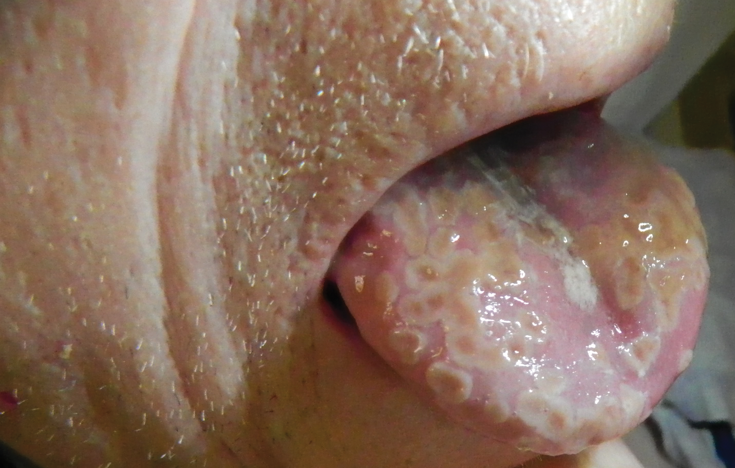
Painful Lesions on the Tongue
The Diagnosis: Herpetic Glossitis
Oral lesions of the tongue are common during primary herpetic gingivostomatitis, though most primary oral herpes simplex virus (HSV) infections occur during childhood or early adulthood. Reactivation of HSV type 1 most commonly manifests as herpes labialis.1 When recurrent HSV involves intraoral lesions, they are typically confined to the gingiva and palate, sparing the tongue.
Clinical presentation of herpetic glossitis varies. Recurrent herpetic glossitis has been described in immunocompromised patients, particularly those with hematologic malignancies and organ transplants.2 In addition, immunocompromised and human immunodeficiency virus–infected patients may present with deep and/or broad ulcers. A case of herpes infection presenting with nodules on the tongue has been reported in Hodgkin disease.3 Herpetic geometric glossitis also has been described, which is a linear, crosshatched, or sharply angled branching with painful fissuring of the tongue. Herpetic geometric glossitis has been reported to occur in both immunocompetent and immunocompromised individuals.4 Tongue involvement during oral reactivation of HSV is exceedingly rare and the pathogenesis remains elusive, though one hypothesis proposes a protective role of salivary-specific IgA and lysozyme.5 Here, we report a case in which a patient developed similar lingual HSV lesions following recent immunosuppression.
1. Arduino PG, Porter SR. Herpes simplex virus type 1 infection: overview on relevant clinico-pathological features. J Oral Pathol Med. 2008;37:107-121.
2. Nikkels AF, Piérard GE. Chronic herpes simplex virus type I glossitis in an immunocompromised man. Br J Dermatol. 1999;140:343-346.
3. Leming PD, Martin SE, Zwelling LA. Atypical herpes simplex (HSV) infection in a patient with Hodgkin’s disease. Cancer. 1984;54:3043-3047.
4. Mirowski GW, Goddard A. Herpetic geometric glossitis in an immunocompetent patient with pneumonia. J Am Acad Dermatol. 2009;61:139-142.
5. Heineman HS, Greenberg MS. Cell protective effect of human saliva specific for herpes simplex virus. Arch Oral Biol. 1980;25:257-261.
The Diagnosis: Herpetic Glossitis
Oral lesions of the tongue are common during primary herpetic gingivostomatitis, though most primary oral herpes simplex virus (HSV) infections occur during childhood or early adulthood. Reactivation of HSV type 1 most commonly manifests as herpes labialis.1 When recurrent HSV involves intraoral lesions, they are typically confined to the gingiva and palate, sparing the tongue.
Clinical presentation of herpetic glossitis varies. Recurrent herpetic glossitis has been described in immunocompromised patients, particularly those with hematologic malignancies and organ transplants.2 In addition, immunocompromised and human immunodeficiency virus–infected patients may present with deep and/or broad ulcers. A case of herpes infection presenting with nodules on the tongue has been reported in Hodgkin disease.3 Herpetic geometric glossitis also has been described, which is a linear, crosshatched, or sharply angled branching with painful fissuring of the tongue. Herpetic geometric glossitis has been reported to occur in both immunocompetent and immunocompromised individuals.4 Tongue involvement during oral reactivation of HSV is exceedingly rare and the pathogenesis remains elusive, though one hypothesis proposes a protective role of salivary-specific IgA and lysozyme.5 Here, we report a case in which a patient developed similar lingual HSV lesions following recent immunosuppression.
1. Arduino PG, Porter SR. Herpes simplex virus type 1 infection: overview on relevant clinico-pathological features. J Oral Pathol Med. 2008;37:107-121.
2. Nikkels AF, Piérard GE. Chronic herpes simplex virus type I glossitis in an immunocompromised man. Br J Dermatol. 1999;140:343-346.
3. Leming PD, Martin SE, Zwelling LA. Atypical herpes simplex (HSV) infection in a patient with Hodgkin’s disease. Cancer. 1984;54:3043-3047.
4. Mirowski GW, Goddard A. Herpetic geometric glossitis in an immunocompetent patient with pneumonia. J Am Acad Dermatol. 2009;61:139-142.
5. Heineman HS, Greenberg MS. Cell protective effect of human saliva specific for herpes simplex virus. Arch Oral Biol. 1980;25:257-261.

A 77-year-old man with a history of chronic obstructive pulmonary disease and recent pneumonia was treated with oral prednisone 40 mg daily, antibiotics, and a fluticasone-salmeterol inhaler. One week into treatment, the patient developed painful lesions limited to the oral cavity. Physical examination revealed many fixed, umbilicated, white-tan plaques on the lower lips, tongue, and posterior aspect of the oropharynx. The dermatology department was consulted because the lesions failed to respond to nystatin oral suspension.
In rosacea, flushing and inflammation need to be addressed
WAIKOLOA, HAWAII – The topical vasoconstrictor brimonidine 0.33% gel brings impressive improvement in the facial redness component of rosacea, but it does nothing to address the inflammatory lesions of the disease, Dr. Joseph F. Fowler, Jr., said at the Hawaii Dermatology Seminar provided by the Global Academy for Medical Education/Skin Disease Education Foundation.
Rebound flushing wasn’t seen in the 52-week, large, open-label safety study of brimonidine gel, but has been anecdotally reported since its approval. Flushing occurs 12-24 hours after application, and usually lasts a day or two, according to Dr. Fowler, who was the first author of several multicenter randomized trials of the therapy.

“The rebound phenomenon is not something that frightens me or others I know who use the medication routinely, because it’s not something that’s going to cause a long-term problem,” he declared. Indeed, once brimonidine gel (Mirvaso) has quelled the erythema, the inflammatory lesions will stand out even more prominently, noted Dr. Fowler, the conference co-director and a dermatologist at the University of Louisville. “You’ve got to do something about the inflammatory component.”
Two topical therapies that address the inflammatory component include ivermectin 1% cream (Soolantra), shown to reduce counts of papules and pustules in two 40-week extension studies of phase III trials (J Drugs Dermatol. 2014 Nov;13[11]:1380-6), and azelaic acid 15% foam (Finacea), which also achieved an impressive reduction in inflammatory lesion counts in a phase III trial (Cutis. 2015 Jul;96[1]:54-61).
Both agents were well tolerated in those studies. That’s a major treatment consideration because rosacea patients have elevated facial skin sensitivity and often can’t tolerate older off-label topical therapies because of stinging and burning, according to Dr. Fowler.
Some rosacea patients are so intolerant of topicals that they prefer oral therapy. For those patients, subantimicrobial-dose doxycycline is a good option, Dr. Fowler said.
In addition to its anti-inflammatory effects, topical ivermectin kills Demodex. While rosacea is an inflammatory disorder, not an infection, drugs like ivermectin and crotamiton 10% cream (Eurax) that kill Demodex also improve rosacea, he observed.
The initial irritation event rate with azelaic acid 15% foam in the phase III trial was in the 2%-5% range, and those events were short lived. Irritation is a much bigger problem with the older azelaic acid 15% gel, with event rates in the 15%-25% range.
Dr. Fowler has also had good results using topical calcineurin inhibitors off-label for rosacea. Pimecrolimus cream (Elidel) is in ongoing studies for treatment of seborrheic dermatitis, which often coexists with rosacea, he noted.
He reported serving as a consultant to half a dozen pharmaceutical companies, including Galderma, which markets ivermectin cream and brimonidine gel, and Bayer, which markets azelaic acid foam.
SDEF and this news organization are owned by the same parent company.
WAIKOLOA, HAWAII – The topical vasoconstrictor brimonidine 0.33% gel brings impressive improvement in the facial redness component of rosacea, but it does nothing to address the inflammatory lesions of the disease, Dr. Joseph F. Fowler, Jr., said at the Hawaii Dermatology Seminar provided by the Global Academy for Medical Education/Skin Disease Education Foundation.
Rebound flushing wasn’t seen in the 52-week, large, open-label safety study of brimonidine gel, but has been anecdotally reported since its approval. Flushing occurs 12-24 hours after application, and usually lasts a day or two, according to Dr. Fowler, who was the first author of several multicenter randomized trials of the therapy.
“The rebound phenomenon is not something that frightens me or others I know who use the medication routinely, because it’s not something that’s going to cause a long-term problem,” he declared. Indeed, once brimonidine gel (Mirvaso) has quelled the erythema, the inflammatory lesions will stand out even more prominently, noted Dr. Fowler, the conference co-director and a dermatologist at the University of Louisville. “You’ve got to do something about the inflammatory component.”
Two topical therapies that address the inflammatory component include ivermectin 1% cream (Soolantra), shown to reduce counts of papules and pustules in two 40-week extension studies of phase III trials (J Drugs Dermatol. 2014 Nov;13[11]:1380-6), and azelaic acid 15% foam (Finacea), which also achieved an impressive reduction in inflammatory lesion counts in a phase III trial (Cutis. 2015 Jul;96[1]:54-61).
Both agents were well tolerated in those studies. That’s a major treatment consideration because rosacea patients have elevated facial skin sensitivity and often can’t tolerate older off-label topical therapies because of stinging and burning, according to Dr. Fowler.
Some rosacea patients are so intolerant of topicals that they prefer oral therapy. For those patients, subantimicrobial-dose doxycycline is a good option, Dr. Fowler said.
In addition to its anti-inflammatory effects, topical ivermectin kills Demodex. While rosacea is an inflammatory disorder, not an infection, drugs like ivermectin and crotamiton 10% cream (Eurax) that kill Demodex also improve rosacea, he observed.
The initial irritation event rate with azelaic acid 15% foam in the phase III trial was in the 2%-5% range, and those events were short lived. Irritation is a much bigger problem with the older azelaic acid 15% gel, with event rates in the 15%-25% range.
Dr. Fowler has also had good results using topical calcineurin inhibitors off-label for rosacea. Pimecrolimus cream (Elidel) is in ongoing studies for treatment of seborrheic dermatitis, which often coexists with rosacea, he noted.
He reported serving as a consultant to half a dozen pharmaceutical companies, including Galderma, which markets ivermectin cream and brimonidine gel, and Bayer, which markets azelaic acid foam.
SDEF and this news organization are owned by the same parent company.
EXPERT ANALYSIS FROM THE SDEF HAWAII DERMATOLOGY SEMINAR

U.S. Surgeon General Encourages Hospitalists to Remain Hopeful, Motivated
Hopefully, many of you were able to attend the Society of Hospital Medicine’s annual meeting this year in San Diego. (I know at least 4,000 of you made it!) Each year, the annual meeting is a time of catching up with hospitalists from around the country (many of whom I only see once a year) and catching up on what is going on in the medical industry.

This year was not particularly unique in that many sessions focused on the myriad challenges we should expect to see in the medical industry in the coming years. There was much discussion about future payment models; although there is ongoing ambiguity about exactly how these models are going to be operationalized, there is certainly no ambiguity that the Centers for Medicare & Medicaid Services (CMS) is hard driving the amount of payments that will be tied to some form of alternative payment model (50% by 2018).
We also heard about ongoing challenges in quality and safety, where a stunning number of patients continue to suffer preventable harm on a daily basis within our hospital walls. And we heard much about the ongoing and mounting opiate abuse epidemic. All of these are monumentally difficult challenges that remain unsolved and without a clear path forward to resolution.
Contrast that with the message from the U.S. Surgeon General during the opening plenary of the annual meeting. Vivek Murthy, MD, was named Surgeon General at a time in the U.S. when all of the above challenges are being added to the abounding issues of chronic disease, mental illness, and extraordinary healthcare costs. He is the highest leader in the nation ordained with trying to improve the health of all Americans at a time when we have never been unhealthier. But despite these monumental challenges, his message was not about the average American body mass index (BMI), smoking status, or heroin addiction. Much different, his message was chock full of amazing stories of community engagement and resilience, focused on innovation and fresh thinking, and about creative problem-solving despite lean and unforgiving budgets.
What Dr. Murthy offered were endless stories of hope and goodness, which he was able to find in each and every city he has visited in his short time as the nation’s “top doc.”
During his tenure, he has visited innumerable communities and engaged with locals in listening sessions. His takeaway from these sessions is “you wouldn’t believe how much good is out there.” One of his many stories was of a hospital and a YMCA that joined forces to improve the health and well-being of the hospital patients, employees, and entire community. This was at a time when both were struggling with lean budgets and stagnant progress in healthy living.
This pragmatic optimism reminds me a bit of one of my life mentors, my Aunt Karen. She is extremely realistic and grounded and knows in great detail the trials and tribulations of being alive for 66 years (including being a 10-year survivor of recurrent ovarian rhabdomyosarcoma). What Aunt Karen does that is so uniquely different than anyone else I know is that she creates goodness. I did not fully understand this until a few years ago, but I noticed that she goes out of her way to create extreme goodness out of extreme ordinariness. I have often joked that she purposely befriends pregnant women just to have an excuse to host a baby shower. She goes overboard to make any and every excuse to celebrate relatively ordinary life milestones (anniversaries, Valentine’s Day, St. Patrick’s Day). In her words, “you have to have a buffer for the funerals.”
Flip Your Switch
And so while Dr. Murthy and Aunt Karen have little else in common, they do share the priceless ability to help others see the goodness in everything around them even when surrounded by remarkable challenges and uncertainty. What a unique gift they have.
But are there simple ways we can all incorporate such goodness into our lives and start to routinely build in these buffers?
In your own personal life and work life, what are your buffers? How could you routinely and repeatedly “find the good” in all things around you?
A few months ago, I started searching for what I call “inbox buffers” as I noticed my email inbox was routinely chock full of requests for time, advice, or resources (all of which can be limited). I found a daily email called “The Daily Good.” It comes into my inbox early each morning and typically covers a human-interest story that is short, interesting, and inspiring. I have found these help me reset my mindset and attitude toward one that is more resilient and forgiving; in other words, it helps me find the good even within the crevices of a cranky email inbox. I have many other buffers, but I cite this one as it is simple, easy, free, predictable, dependable, and routinely inspiring!
So in this time when hospitalists are facing monumental change, unpredictable conflict, and unending challenges, we all need to purposely and repeatedly build in buffers to keep us hopeful and motivated and to seamlessly and routinely find the good in all we do. TH

Hopefully, many of you were able to attend the Society of Hospital Medicine’s annual meeting this year in San Diego. (I know at least 4,000 of you made it!) Each year, the annual meeting is a time of catching up with hospitalists from around the country (many of whom I only see once a year) and catching up on what is going on in the medical industry.
This year was not particularly unique in that many sessions focused on the myriad challenges we should expect to see in the medical industry in the coming years. There was much discussion about future payment models; although there is ongoing ambiguity about exactly how these models are going to be operationalized, there is certainly no ambiguity that the Centers for Medicare & Medicaid Services (CMS) is hard driving the amount of payments that will be tied to some form of alternative payment model (50% by 2018).
We also heard about ongoing challenges in quality and safety, where a stunning number of patients continue to suffer preventable harm on a daily basis within our hospital walls. And we heard much about the ongoing and mounting opiate abuse epidemic. All of these are monumentally difficult challenges that remain unsolved and without a clear path forward to resolution.
Contrast that with the message from the U.S. Surgeon General during the opening plenary of the annual meeting. Vivek Murthy, MD, was named Surgeon General at a time in the U.S. when all of the above challenges are being added to the abounding issues of chronic disease, mental illness, and extraordinary healthcare costs. He is the highest leader in the nation ordained with trying to improve the health of all Americans at a time when we have never been unhealthier. But despite these monumental challenges, his message was not about the average American body mass index (BMI), smoking status, or heroin addiction. Much different, his message was chock full of amazing stories of community engagement and resilience, focused on innovation and fresh thinking, and about creative problem-solving despite lean and unforgiving budgets.
What Dr. Murthy offered were endless stories of hope and goodness, which he was able to find in each and every city he has visited in his short time as the nation’s “top doc.”
During his tenure, he has visited innumerable communities and engaged with locals in listening sessions. His takeaway from these sessions is “you wouldn’t believe how much good is out there.” One of his many stories was of a hospital and a YMCA that joined forces to improve the health and well-being of the hospital patients, employees, and entire community. This was at a time when both were struggling with lean budgets and stagnant progress in healthy living.
This pragmatic optimism reminds me a bit of one of my life mentors, my Aunt Karen. She is extremely realistic and grounded and knows in great detail the trials and tribulations of being alive for 66 years (including being a 10-year survivor of recurrent ovarian rhabdomyosarcoma). What Aunt Karen does that is so uniquely different than anyone else I know is that she creates goodness. I did not fully understand this until a few years ago, but I noticed that she goes out of her way to create extreme goodness out of extreme ordinariness. I have often joked that she purposely befriends pregnant women just to have an excuse to host a baby shower. She goes overboard to make any and every excuse to celebrate relatively ordinary life milestones (anniversaries, Valentine’s Day, St. Patrick’s Day). In her words, “you have to have a buffer for the funerals.”
Flip Your Switch
And so while Dr. Murthy and Aunt Karen have little else in common, they do share the priceless ability to help others see the goodness in everything around them even when surrounded by remarkable challenges and uncertainty. What a unique gift they have.
But are there simple ways we can all incorporate such goodness into our lives and start to routinely build in these buffers?
In your own personal life and work life, what are your buffers? How could you routinely and repeatedly “find the good” in all things around you?
A few months ago, I started searching for what I call “inbox buffers” as I noticed my email inbox was routinely chock full of requests for time, advice, or resources (all of which can be limited). I found a daily email called “The Daily Good.” It comes into my inbox early each morning and typically covers a human-interest story that is short, interesting, and inspiring. I have found these help me reset my mindset and attitude toward one that is more resilient and forgiving; in other words, it helps me find the good even within the crevices of a cranky email inbox. I have many other buffers, but I cite this one as it is simple, easy, free, predictable, dependable, and routinely inspiring!
So in this time when hospitalists are facing monumental change, unpredictable conflict, and unending challenges, we all need to purposely and repeatedly build in buffers to keep us hopeful and motivated and to seamlessly and routinely find the good in all we do. TH
Climate Change is Expected to Boost the Number of Annual Premature U.S Deaths
WASHINGTON (Reuters) - Climate change can be expected to boost the number of annual premature U.S. deaths from heat waves in coming decades and to increase mental health problems from extreme weather like hurricanes and floods, a U.S. study said on Monday.
"I don't know that we've seen something like this before, where we have a force that has such a multitude of effects," Surgeon General Vivek Murthy told reporters at the White House about the study. "There's not one single source that we can target with climate change, there are multiple paths that we have to address."
Heat waves were estimated to cause 670 to 1,300 U.S. deaths annually in recent years. Premature U.S. deaths from heat waves can be expected to rise more than 27,000 per year by 2100, from a 1990 baseline, one scenario in the study said. The rise outpaced projected decreases in deaths from extreme cold.
Extreme heat can cause more forest fires and increase pollen counts and the resulting poor air quality threatens people with asthma and other lung conditions. The report said poor air quality will likely lead to hundreds of thousands of premature deaths, hospital visits, and acute respiratory illness each year by 2030.
Climate change also threatens mental health, the study found. Post traumatic stress disorder, depression, and general anxiety can all result in places that suffer extreme weather linked to climate change, such as hurricanes and floods. More study needs to be done on assessing the risks to mental health, it said.
The peer-reviewed study by eight federal agencies can be found at: https://health2016.globalchange.gov/
Cases of mosquito and tick-borne diseases can also be expected to increase, though the study, completed over three years, did not look at whether locally-transmitted Zika virus cases would be more likely to hit the U.S.
President Barack Obama's administration has taken steps to cut carbon emissions by speeding a switch from coal and oil to cleaner energy sources. In February, the Supreme Court dealt a blow to the White House's climate ambitions by putting a hold on Obama's plan to cut emissions from power plants. Administration officials say the plan is on safe legal footing.John Holdren, Obama's senior science adviser, said steps the world agreed to in Paris last year to curb emissions through 2030 can help fight the risks to health.
"We will need a big encore after 2030 . . . in order to avoid the bulk of the worst impacts described in this report,"he said.
Treatment can produce durable responses in NHL

2016 AACR Annual Meeting
© AACR/Todd Buchanan
NEW ORLEANS—Administering an antibody-radionuclide conjugate after B-cell depletion with rituximab can produce lasting responses in patients with relapsed non-Hodgkin lymphoma (NHL), according to a phase 1/2 study.
The conjugate, 177Lu-DOTA-HH1 (Betalutin), consists of the tumor-specific antibody HH1, which targets the CD37 antigen on the surface of NHL cells, conjugated to the β-emitting isotope lutetium-177 (Lu-177) via the chemical linker DOTA.
In an ongoing phase 1/2 study, Betalutin given after rituximab produced an overall response rate of 63.2%.
The median duration of response has not yet been reached, and 1 patient has maintained a response for more than 36 months.
In addition, the researchers said Betalutin was well tolerated, with a predictable and manageable safety profile. Most adverse events were hematologic, and all have been transient and reversible.
These results were presented at the 2016 AACR Annual Meeting (abstract LB-252*). The study is sponsored by Nordic Nanovector ASA.
Patients and study design
The researchers presented data on 21 patients—19 with follicular lymphoma and 2 with mantle cell lymphoma. All tumors were positive for CD37.
The patients’ median age was 68 (range, 41-78). Sixty-seven percent were male, and they had received 1 to 8 prior treatment regimens.
In this dose-escalation study, patients received Betalutin at 3 different doses, but they were also divided into 2 arms according to predosing with cold HH1 antibody.
In Arm 1 (n=12), patients received rituximab (at 375 mg/m2) on day -28 and -21 to deplete circulating B cells. On day 0, predosing with 50 mg HH1 was given before Betalutin injection. Then, patients received Betalutin at 10 MBq/kg (n=3), 15 MBq/kg (n=6), or 20 MBq/kg (n=3).
In Arm 2 (n=4), patients received rituximab at the same dose and schedule as Arm 1, but Betalutin was administered without HH1 predosing on day 0 at either 10 MBq/kg (n=2) or 15 MBq/kg (n=2).
The first patient treated on this trial received 250 mg/m2 of rituximab on day -7 and day 0 prior to Betalutin administration and was included in the 10 MBq/kg group in Arm 2.
The 15 MBq/kg dose level of Arm 1 has been expanded into the phase 2 portion of the study, as dose-limiting toxicities occurred at the 20 MBq/kg dose. Five patients have been treated in the phase 2 portion.
Safety
Adverse events (AEs) from the phase 2 portion of the study were not reported, as the data are still being collected.
In the phase 1 portion, grade 3/4 AEs were hematologic in nature and included decreases in platelet counts (3 grade 3 and 6 grade 4) and neutrophil counts (5 grade 3 and 4 grade 4).
Serious AEs included decreases in platelet counts (n=2), atrial fibrillation (n=2), epistaxis (n=1), fractured sternum (n=1), decreased neutrophil count (n=1), pharyngitis (n=1), pneumonia (n=1), pulmonary embolism (n=1), and sepsis (n=1).
The pulmonary embolism was deemed unrelated to treatment, but the remaining events were considered possibly or probably related to Betalutin.
The researchers noted that 1 patient experienced pharyngitis, pneumonia, pulmonary embolism, epistaxis, sepsis, and a decrease in lymphocyte count.
All patients’ platelets and neutrophils recovered. Two patients required platelet transfusions—one patient in the 20 MBq/kg cohort of Arm 1 and one patient in the 15 MBq/kg cohort of Arm 2.
Efficacy
Nineteen patients were evaluable for response. The overall response rate was 63.2% (n=12) and included both complete (31.6%, n=6) and partial responses (31.6%, n=6). Progression occurred in 21.1% of patients (n=4), and 15.8% (n=3) had stable disease.
The researchers presented data on 9 patients treated at the recommended 15 MBq/kg dose level with 50 mg HH1 predosing. Five patients were treated in phase 1 and 4 in phase 2. One of these patients was excluded from the analysis due to transformed lymphoma.
Two patients in phase 1 responded—both complete responses—and 3 patients in phase 2 responded—2 complete and 1 partial response.
For the entire study cohort, the median duration of response has not yet been reached. Six responses are ongoing—2 for 3+ months, 1 for 6+ months, 1 for 18+ months, 1 for 24+ months, and 1 for 36+ months. ![]()
*Information in the abstract differs from that presented at the meeting.
Method detects SNVs better, group says

Researchers have developed a new method for detecting single-nucleotide variants (SNVs) in a single cell, and they believe it could have applications for cancer diagnosis and treatment.
The team said the method, known as Monovar, improves upon current single-cell sequencing (SCS) by more accurately detecting SNVs.
During testing, Monovar identified 28 new somatic SNVs in cells from a patient with acute lymphoblastic leukemia (ALL).
The researchers described Monovar in Nature Methods.
“To improve the SNVs in SCS datasets, we developed Monovar,” said study author Nicholas Navin, PhD, of the University of Texas MD Anderson Cancer Center in Houston.
“Monovar is a novel statistical method able to leverage data from multiple single cells to discover SNVs and provides highly detailed genetic data.”
Dr Navin and his colleagues found Monovar superior to standard algorithms for analyzing cells from previously studied patients with 3 different cancer types.
The team analyzed single cells from a patient with triple-negative breast cancer, a patient with muscle-invasive bladder cancer, and a child with ALL.
In cells from the ALL patient, Monovar discovered 57 somatic mutations, including 28 new somatic SNVs.
The researchers said Monovar identified significant mutations in OR4C3 and GPR107 (all subclones); LRFN5, PKD2L1, and ZNF781 (in subs 2, 4 and 5); DNAH7 (sub 1); LYAR and FMNL1 (sub 2); RGS3 (subs 4 and 5); and ADAMTS13, PRSS3, and PKD2L1 (subs 2-5).
The clonal mutations in OR4C3 and GPR107 and the subclonal mutations in PKD2L1, ADAMTS13, PRSS3, and RGS3 were not identified in the original study of the ALL patient (C Gawad et al. PNAS 2014).
Dr Navin and his colleagues said Monovar could have significant translational applications in cancer diagnosis and treatment, personalized medicine, and prenatal genetic diagnosis, where the accurate detection of SNVs is critical for patient care.
The researchers also believe Monovar could be used for studies in a range of biomedical fields.
“With the recent innovations in SCS methods to analyze thousands of single cells in parallel with RNA analysis, which will soon be extended to DNA analysis, the need for accurate DNA variant detection will continue to grow,” said Ken Chen, PhD, also of MD Anderson Cancer Center.
“Monovar is capable of analyzing large-scale datasets and handling different whole-genome protocols. Therefore, it is well-suited for many types of studies.” ![]()
Drug shows early promise for low-grade lymphoma

follicular lymphoma
NEW ORLEANS—The TLR9 agonist SD-101 has produced encouraging early results in patients with low-grade B-cell lymphoma, according to researchers.
In an ongoing phase 1/2 study, patients received low-dose radiation, followed by an injection of SD-101 into one of their tumors.
This prompted changes in the tumor microenvironment that potentially induced a systemic antitumor response and decreased the volume of both treated and untreated tumors.
In addition, SD-101 was considered well tolerated, with no dose-limiting toxicities or maximum-tolerated dose.
“We are pleased to have already demonstrated a safety profile, pharmacodynamics, and preliminary efficacy in this study,” said Ronald Levy, MD, of Stanford University in California.
Dr Levy and his colleagues presented these results at the 2016 AACR Annual Meeting (abstract CT047). The study is sponsored by Dynavax Technologies Corporation, the company developing SD-101.
The researchers reported data for 13 patients with untreated, low-grade B-cell lymphoma. They had a mean age of 63.2, and 53.8% were male.
Patients had follicular lymphoma (n=9), small lymphocytic lymphoma (n=2), chronic lymphocytic leukemia (n=1), and nodal marginal zone lymphoma (n=1).
At least 2 sites of measurable disease were required for participation in this study. One site was treated with low-dose radiation (2 Gy) and injected with SD-101 on days 1, 8, 15, 22, and 29. Other lesions received no treatment.
In Part 1—the dose-escalation portion of the study—patients received SD-101 at 1 mg (n=3), 2 mg (n=3), 4 mg (n=3), or 8 mg (n=4). The phase 2 expansion portion of the study is ongoing, with enrollment in 2 dose cohorts (1 mg and 8 mg).
“This clinical trial design is unique and takes advantage of the fact that lymphoma patients have easily injectable sites of disease,” Dr Levy said. “The local injections are conveniently added to low-dose radiotherapy, a standard treatment for low-grade lymphoma.”
Safety
All 13 patients experienced at least 1 adverse event (AEs), although nearly all were grade 1 or 2.
The most common treatment-related AEs were local injection-site reactions and flu-like symptoms, including fever, chills, and myalgia (n=11 for all 3). However, the researchers said these AEs were primarily short-lived and controlled by over-the-counter acetaminophen in most cases.
In the 8 mg dosing cohort, 1 patient had grade 3 neutropenia and 2 had grade 3 malaise, all of which were considered treatment-related. In addition, there was a case of grade 3 asymptomatic pulmonary embolism in the 4 mg dose cohort, which was deemed serious but unrelated to treatment.
Efficacy
The researchers observed induction of interferon-responsive genes at all dose levels 24 hours after the second dose of SD-101 was given (Day 9).
In addition, T-cell numbers increased at the treated site by Day 8. The total T cells increased in 7 of 10 evaluable patients by Day 8 (range, >300% to 18%).
CD4+ and CD8+ T cells increased simultaneously in 5 of 7 evaluable patients, regulatory T cells decreased in 8 of 10 evaluable patients by Day 8, and follicular T helper cells decreased in 9 of 9 evaluable patients by Day 8.
Furthermore, treated and untreated tumors decreased in volume across all dose groups.
At Day 90, 12 patients had a reduction of the product of diameters in treated tumors (median -45.3%; range, -87 to +100), and 11 patients had a reduction in untreated tumors (median -8.1%; range, -48 to +45).
In 9 patients, these abscopal effects were sustained for at least 180 to 360 days.
However, 6 patients discontinued from the study due to progression. Three had radiographic progression—1 at Day 92 (in the 4 mg cohort) and 2 at 1 year (1 in the 1 mg cohort and 1 in the 2 mg cohort).
Two patients had clinical progression—1 at Day 197 (4 mg) and 1 at Day 273 (2 mg). And 1 patient discontinued at Day 203 due to a combination of clinical and radiographic progression (8 mg).
The researchers pointed out that there was no evidence of a dose response with SD-101, but the study included a limited number of patients. ![]()
Team explains CD8 Treg dysfunction
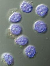
Image by Kathryn T. Iacono
Research published in The Journal of Clinical Investigation provides new insights regarding CD8 regulatory T cells (Tregs).
Investigators found that, in young, healthy individuals, CD8 Tregs suppress the activation and expansion of CD4 T cells.
However, older individuals and patients with a rare form of vasculitis exhibit CD8 Treg dysfunction, which is tied to a drop in production of an enzyme called NADPH oxidase 2 (NOX2).
Cornelia Weyand, MD, of Stanford University Medical Center in California, and her colleagues conducted this research.
First, the team found that CD8 Tregs preferentially take up residence in lymph nodes, the spleen, and other regions where there are huge “armies” of CD4 T cells. This proximity puts the CD8 Tregs in a position to stamp out CD4 T-cell activation early on.
Further experiments demonstrated that CD8 Tregs manufacture copious amounts of NOX2, which they package into tiny membrane-bound packets and transfer to the surfaces of abutting CD4 T cells.
These NOX2-laden packets are then taken up by the CD4 T cells. Inside their new home, the enzymes produce large volumes of highly reactive signaling substances that dial down CD4 T cells’ activation and proliferation.
The investigators noted that contact between CD8 Tregs and CD4 T cells in the early stages of activation shuts down the CD4 T cells’ activity and reduces their proliferation by half or more, even several days after the CD8 Tregs have been removed. Transferring NOX2 alone onto activated CD4 T cells also produces this effect.
Next, the team analyzed blood samples from healthy individuals and observed that CD8 Tregs were only about half as common in blood from people age 60 and older as in blood from 20- to 30-year-olds.
Both CD8 Treg numbers and their ability to suppress CD4 T-cell proliferation declined with advancing age. Experiments traced this to a drop in NOX2 production by older donors’ CD8 Tregs.
The investigators also discovered CD8 Treg failure in giant-cell arteritis (GCA). The team compared blood from GCA patients to blood from age-matched healthy control subjects and from patients with 2 other autoimmune diseases—psoriatic arthritis and small-vessel vasculitis. This revealed a severe deficit among GCA patients in NOX2-producing CD8 Tregs.
“This tells us that the deficit in NOX2-producing CD8 Tregs is specific to GCA, not just driving or driven by inflammation,” Dr Weyand said. “That’s good news for our patients who have this disease, which has been an enigma. Now we know something about what’s causing it.”
The discovery of NOX2 on the surface of CD8 Tregs—but not on other T-cell types—makes them much easier to identify and count, Dr Weyand added.
She and her colleagues are taking advantage of the new-found biomarker to tally CD8 Tregs in patients with age-associated disorders now understood to be driven by chronic inflammation to see if CD8 Treg deficits underlie some of these conditions’ pathology and whether they may be amenable to potential NOX2-restoring treatments. ![]()
UnitedHealth Group leaving most ACA marketplaces
UnitedHealth Group will not sell policies next year in most of the health insurance marketplaces created by the Affordable Care Act, CEO Stephen Helmsley announced during an April 19 earnings call.
“Next year, we will remain in only a handful of states, and we will not carry financial exposure from exchanges into 2017,” Mr. Helmsley said, citing the company’s inability to offset the “shorter-term, higher-risk” population covered by the ACA exchange plans with large enough risk pools.

Mr. Helmsley did not say in which state marketplaces UnitedHealth Group (UHG) will continue to offer coverage.
Areas that could be hardest hit by the UHG withdrawal include Alabama, Arizona, Arkansas, Nebraska, North Carolina, and Tennessee, according to an analysis from the Kaiser Family Foundation.
For now, the decision by UHG is not likely to impact the average benchmark premium by more than a 1% increase, according to the analysis. That’s because the insurer was less likely than its competitors to offer lower-cost silver plans; when silver plans were offered, they were offered at or very near to the competitors’ prices. However, in states where the withdrawal of UHG means that two or fewer insurers are participating in the marketplace, benchmark premiums could rise substantially, according to the study.
The long-term effect of the UHG exit from most marketplaces is not clear, according to Kaiser’s analysts. “In areas with limited insurer participation, the remaining plans after a United exit may have more market power relative to providers, but in the absence of insurer competition, those savings may not be passed along to consumers,” they wrote.
ACA measures such as rate reviews and medical loss ratio provisions could mitigate adverse effects on consumers in these markets, giving regulators the power to force insurers to issue rebates if premiums outstrip the cost of care.
The insurer’s decision is simply evidence that typical market forces are in play, according to Jonathan Gold, a spokesperson for the Centers for Medicare & Medicaid Services.
“As with any new market, we expect changes and adjustments in the early years with issuers both entering and exiting states,” Mr. Gold said in an interview. The UHG decision was not unexpected, as company officials said they were contemplating the move last November.
“We have full confidence, based on data, that the marketplaces will continue to thrive for years ahead,” he said, noting that in 2016, 39 insurers exited the marketplace, while 40 entered.
On Twitter @whitneymcknight
UnitedHealth Group will not sell policies next year in most of the health insurance marketplaces created by the Affordable Care Act, CEO Stephen Helmsley announced during an April 19 earnings call.
“Next year, we will remain in only a handful of states, and we will not carry financial exposure from exchanges into 2017,” Mr. Helmsley said, citing the company’s inability to offset the “shorter-term, higher-risk” population covered by the ACA exchange plans with large enough risk pools.
Mr. Helmsley did not say in which state marketplaces UnitedHealth Group (UHG) will continue to offer coverage.
Areas that could be hardest hit by the UHG withdrawal include Alabama, Arizona, Arkansas, Nebraska, North Carolina, and Tennessee, according to an analysis from the Kaiser Family Foundation.
For now, the decision by UHG is not likely to impact the average benchmark premium by more than a 1% increase, according to the analysis. That’s because the insurer was less likely than its competitors to offer lower-cost silver plans; when silver plans were offered, they were offered at or very near to the competitors’ prices. However, in states where the withdrawal of UHG means that two or fewer insurers are participating in the marketplace, benchmark premiums could rise substantially, according to the study.
The long-term effect of the UHG exit from most marketplaces is not clear, according to Kaiser’s analysts. “In areas with limited insurer participation, the remaining plans after a United exit may have more market power relative to providers, but in the absence of insurer competition, those savings may not be passed along to consumers,” they wrote.
ACA measures such as rate reviews and medical loss ratio provisions could mitigate adverse effects on consumers in these markets, giving regulators the power to force insurers to issue rebates if premiums outstrip the cost of care.
The insurer’s decision is simply evidence that typical market forces are in play, according to Jonathan Gold, a spokesperson for the Centers for Medicare & Medicaid Services.
“As with any new market, we expect changes and adjustments in the early years with issuers both entering and exiting states,” Mr. Gold said in an interview. The UHG decision was not unexpected, as company officials said they were contemplating the move last November.
“We have full confidence, based on data, that the marketplaces will continue to thrive for years ahead,” he said, noting that in 2016, 39 insurers exited the marketplace, while 40 entered.
On Twitter @whitneymcknight

AAN Updates Guidelines on Use of Botulinum Toxin for Four Disorders
VANCOUVER—The American Academy of Neurology (AAN) has updated its 2008 guidelines on the use of botulinum toxin for spasticity, cervical dystonia, blepharospasm, and migraine to reflect recent research. Unlike the 2008 guidelines, the current document describes the evidence for the four formulations of botulinum toxin individually. The guidelines were published online ahead of print April 18 in Neurology and presented at the 68th Annual Meeting of the AAN.

David M. Simpson, MD
Botulinum toxin blocks the release of substances at nerve endings, generally resulting in reduced muscle contraction and decreased transmission of pain signals. The four preparations available in the United States are abobotulinumtoxinA, incobotulinumtoxinA, onabotulinumtoxinA, and rimabotulinumtoxinB.
To develop the guideline, David M. Simpson, MD, Professor of Neurology and Neuromuscular Diseases at the Icahn School of Medicine at Mount Sinai Hospital in New York, and colleagues reviewed all available scientific studies on these formulations in the four conditions specified. The authors concluded that botulinum toxin is generally safe and effective for treating spasticity in adults, cervical dystonia, blepharospasm, and chronic migraine.
The new guideline’s recommendation on chronic migraine is a major change from the earlier document, said Dr. Simpson. In 2008, not enough evidence was available to support any recommendation on the use of botulinum toxin for chronic migraine. Since that time, two pivotal Class I studies demonstrated the effectiveness of onabotulinumtoxinA in the treatment of chronic migraine. The magnitude of the drug’s efficacy was small, however. In the four weeks after the first treatments, onabotulinumtoxinA was associated with an approximately 15% reduction in headache days per month, compared with placebo. The difference was statistically significant, and the new guideline contains a Level A recommendation of onabotulinumtoxinA for chronic migraine.
On the other hand, the authors found Level A evidence indicating a lack of effectiveness of onabotulinumtoxinA in episodic migraine. “It’s an interesting question as to whether there’s a pathophysiologic difference between episodic migraine and chronic migraine,” but the evidence is unclear, said Dr. Simpson. Another possible explanation for botulinum toxin’s lack of efficacy in this indication is that studies of episodic migraine may reflect a floor effect, he added. “If you have so few headaches per month … how much does that need to reduce to see a significant difference from placebo? That is perhaps one obvious concern about getting a positive result of those studies.” In addition, the authors concluded that abobotulinumtoxinA, incobotulinumtoxinA, and onabotulinumtoxinA are effective in reducing excess muscle tone and should be offered to patients with upper limb spasticity. RimabotulinumtoxinB is probably effective for this indication and should be considered, according to the guideline. For lower limb spasticity, abobotulinumtoxinA and onabotulinumtoxinA are effective and should be offered.
The guideline also indicates that abobotulinumtoxinA and rimabotulinumtoxinB are effective for cervical dystonia and should be offered. OnabotulinumtoxinA and incobotulinumtoxinA are probably effective and should be considered for this indication.Researchers have conducted few well-designed studies on blepharospasm. OnabotulinumtoxinA and incobotulinumtoxinA are probably effective and should be considered for this indication, according to the guideline. AbobotulinumtoxinA is possibly effective and may be considered. The 2008 guidelines also discussed disorders such as essential tremor, hemifacial spasm, and disorders of the voice. No new evidence about botulinum toxin’s effect in these disorders was available at the time that the update was initiated, and they were not included.
—Erik Greb
Suggested Reading
Simpson DM, Hallett M, Ashman EJ, et al. Practice guideline update summary: Botulinum neurotoxin for the treatment of blepharospasm, cervical dystonia, adult spasticity, and headache: Report of the Guideline Development Subcommittee of the American Academy of Neurology. Neurology. 2016 April 18 [Epub ahead of print].
VANCOUVER—The American Academy of Neurology (AAN) has updated its 2008 guidelines on the use of botulinum toxin for spasticity, cervical dystonia, blepharospasm, and migraine to reflect recent research. Unlike the 2008 guidelines, the current document describes the evidence for the four formulations of botulinum toxin individually. The guidelines were published online ahead of print April 18 in Neurology and presented at the 68th Annual Meeting of the AAN.
David M. Simpson, MD
Botulinum toxin blocks the release of substances at nerve endings, generally resulting in reduced muscle contraction and decreased transmission of pain signals. The four preparations available in the United States are abobotulinumtoxinA, incobotulinumtoxinA, onabotulinumtoxinA, and rimabotulinumtoxinB.
To develop the guideline, David M. Simpson, MD, Professor of Neurology and Neuromuscular Diseases at the Icahn School of Medicine at Mount Sinai Hospital in New York, and colleagues reviewed all available scientific studies on these formulations in the four conditions specified. The authors concluded that botulinum toxin is generally safe and effective for treating spasticity in adults, cervical dystonia, blepharospasm, and chronic migraine.
The new guideline’s recommendation on chronic migraine is a major change from the earlier document, said Dr. Simpson. In 2008, not enough evidence was available to support any recommendation on the use of botulinum toxin for chronic migraine. Since that time, two pivotal Class I studies demonstrated the effectiveness of onabotulinumtoxinA in the treatment of chronic migraine. The magnitude of the drug’s efficacy was small, however. In the four weeks after the first treatments, onabotulinumtoxinA was associated with an approximately 15% reduction in headache days per month, compared with placebo. The difference was statistically significant, and the new guideline contains a Level A recommendation of onabotulinumtoxinA for chronic migraine.
On the other hand, the authors found Level A evidence indicating a lack of effectiveness of onabotulinumtoxinA in episodic migraine. “It’s an interesting question as to whether there’s a pathophysiologic difference between episodic migraine and chronic migraine,” but the evidence is unclear, said Dr. Simpson. Another possible explanation for botulinum toxin’s lack of efficacy in this indication is that studies of episodic migraine may reflect a floor effect, he added. “If you have so few headaches per month … how much does that need to reduce to see a significant difference from placebo? That is perhaps one obvious concern about getting a positive result of those studies.” In addition, the authors concluded that abobotulinumtoxinA, incobotulinumtoxinA, and onabotulinumtoxinA are effective in reducing excess muscle tone and should be offered to patients with upper limb spasticity. RimabotulinumtoxinB is probably effective for this indication and should be considered, according to the guideline. For lower limb spasticity, abobotulinumtoxinA and onabotulinumtoxinA are effective and should be offered.
The guideline also indicates that abobotulinumtoxinA and rimabotulinumtoxinB are effective for cervical dystonia and should be offered. OnabotulinumtoxinA and incobotulinumtoxinA are probably effective and should be considered for this indication.Researchers have conducted few well-designed studies on blepharospasm. OnabotulinumtoxinA and incobotulinumtoxinA are probably effective and should be considered for this indication, according to the guideline. AbobotulinumtoxinA is possibly effective and may be considered. The 2008 guidelines also discussed disorders such as essential tremor, hemifacial spasm, and disorders of the voice. No new evidence about botulinum toxin’s effect in these disorders was available at the time that the update was initiated, and they were not included.
—Erik Greb
Suggested Reading
Simpson DM, Hallett M, Ashman EJ, et al. Practice guideline update summary: Botulinum neurotoxin for the treatment of blepharospasm, cervical dystonia, adult spasticity, and headache: Report of the Guideline Development Subcommittee of the American Academy of Neurology. Neurology. 2016 April 18 [Epub ahead of print].