User login
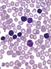
Risk of HBV reactivation ‘underappreciated’

Credit: CDC
Reactivation of the hepatitis B virus (HBV) may be more of a risk than we anticipated, investigators have reported in Hepatology.
Their research indicates that HBV reactivation is associated with the use of chemotherapy, high-dose corticosteroids, biologics targeting tumor necrosis factor-alpha (TNF-α), and agents that aren’t really considered immunosuppressive.
HBV reactivation is also fairly common after organ transplant and hematopoietic stem cell transplant (HSCT).
As reactivation of HBV can be fatal, the study authors suggest routine screening of HBV in all patients prior to the start of these treatments.
The researchers noted that, in September 2013, the US Food and Drug Administration (FDA) issued a Drug Safety Communication in an attempt to decrease the risk of HBV reactivation. The communication advised healthcare professionals to screen patients for HBV prior to the administration of ofatumumab or rituximab.
“[T]his may be just the tip of the iceberg,” said Adrian Di Bisceglie, MD, of Saint Louis University School of Medicine in Missouri.
“Our research suggests that the issue of HBV reactivation may be an underappreciated clinical challenge that extends well beyond the use of just two anti-CD20 medications.”
After a systematic literature review, Dr Di Bisceglie and his colleagues identified 504 studies pertaining to reactivation of HBV.
The investigators reviewed 14 studies in which the antiviral agent lamivudine was used to prevent HBV reactivation in HBsAg-positive patients receiving chemotherapy. Among patients who did not receive lamivudine, HBV reactivation occurred in 32%. Thirteen percent of patients experienced liver failure, and 7% died.
The researchers also looked at patients undergoing HSCT. In one study, 61 patients had resolved HBV infection before HSCT. But 12 of these patients (20%) developed reverse seroconversion (reappearance of HBsAg in a person who was HBsAg-negative, anti-HBc-positive prior to HSCT).
The cumulative probability of reverse seroconversion was 9% a year after HSCT, 21.7% at 2 years, and 42.9% at 4 years.
The investigators also noted that high-dose corticosteroids carry a significant risk of HBV reactivation, both as part of combination treatment for malignancies and when used alone to treat benign conditions.
In addition, the researchers found data showing that HBV reactivation has occurred with antitumor agents that are not thought to be particularly immunosuppressive, such as imatinib and thalidomide. The team said this raises questions about the mechanisms by which drugs are causing HBV reactivation.
The investigators also looked at data from 257 patients with active or recovered HBV infection who received treatment with biological therapies targeting TNF-α.
Forty-two percent of the patients had elevations in serum aminotransferase levels, 39% had reappearance of HBV DNA, 16% had signs and symptoms of liver disease, and 5% died of liver failure.
HBV reactivation was more frequent among patients receiving infliximab than etanercept. It was 7-fold higher among patients who were HBsAg-positive (38%) than those who were HBsAg-negative but anti-HBc-positive (5%).
While it remains unclear how HBV reactivation occurs, experts believe a loss of immune control over viral replication may trigger the process.
“Further study and cooperation between various medical disciplines will help broaden understanding of HBV reactivation,” Dr Di Bisceglie concluded. ![]()
Vaccine gets orphan designation for ATLL
The European Medicines Agency (EMA) has granted orphan drug designation for a therapeutic vaccine candidate known as THV02 to treat adult T-cell leukemia/lymphoma (ATLL).
THV02 is an experimental treatment composed of 2 lentiviral vectors to be used in a prime/boost regimen in ATLL patients infected by the HTLV-1 virus.
Both investigational drugs encode the same antigens, derived from 4 proteins of the HTLV-1 virus.
THV02 is intended to induce an immune response against HTLV antigens born by ATLL with the aim of enabling the patients’ immune system to fight leukemic cells.
Preclinical evaluation has suggested that THV02 is safe, and the vaccine has presented an “unprecedented” immunogenicity profile in several models, according to THERAVECTYS, the company developing THV02.
“Preclinical immunogenicity results obtained to date are very promising, and we are really excited by the prospect of bringing a safe and better-tolerated alternative to patients who are desperately in need of a treatment,” said Déborah Revaud, the senior scientist in charge of developing THV02.
The EMA grants orphan designation to drugs in development intended for the treatment, prevention, or diagnosis of life-threatening or chronically debilitating diseases occurring in fewer than 5 in 10,000 people.
The designation allows sponsors to benefit from an accelerated development process, financial incentives, and a 10-year period of market exclusivity once the drug is on the market.
“We are extremely pleased that the European Medicines Agency has granted an orphan drug status to our vaccine candidate against ATLL,” said Emmanuelle Sabbah-Petrover, PhD, head of regulatory affairs at THERAVECTYS.
“We expect to recruit our first patients towards the end of Q3 2015 in Europe and advance further developments in the US and in Japan in 2016.”
Should THV02 demonstrate a convincing safety and efficacy profile during its development against ATLL, THERAVECTYS said it will consider developing the vaccine for HTLV-related infections as treatment and possibly as prophylaxis. ![]()
Team creates new cells for modeling malaria

infection in iPSC-derived liver
cells 8 days after infection
Credit: Shengyong Ng et al.
Researchers say they’ve found a way to grow liver-like cells from induced pluripotent stem cells (iPSCs).
The liver-like cells can be infected with several strains of the malaria parasite and respond to existing drugs the same way mature human liver cells do.
The new cells, described in Stem Cell Reports, could allow scientists to test drugs on cells from people with different genetic backgrounds, who may respond differently to malaria infection and treatment.
Modeling infection
Until now, malaria researchers have not had many reliable ways to test new drugs in liver tissue.
“What’s historically been done is people have tried to make do with the systems that were available,” said study author Sangeeta Bhatia, MD, PhD, of the Massachusetts Institute of Technology in Cambridge.
In 2013, Dr Bhatia and her colleagues showed they could model malaria infection in hepatocytes from human donors. However, this generates only a limited supply from each donor, and not all of the cells work well for drug studies.
The researchers then turned to iPSCs, which can be generated from human skin cells by adding reprogramming factors. To create liver cells, the researchers added a series of growth factors, including hepatocyte growth factor, to the iPSCs.
The team generated these cells in 2012 and used them to model infection of hepatitis C. However, these cells, known as hepatocyte-like cells, did not seem to be as mature as real adult liver cells.
In the current study, the researchers found these cells could be infected with several strains of malaria. But, initially, the cells did not respond to drugs in the same way as adult liver cells.
In particular, they were not sensitive to primaquine, which works only if cells have a certain set of drug-metabolism enzymes found in mature liver cells.
To induce the cells to become more mature and turn on these metabolic enzymes, the researchers added a molecule they had identified in a previous study. This compound, which the researchers call a “maturin,” stimulated the cells to turn on those enzymes, which made them sensitive to primaquine.
Toward better drugs
The team is now working with the nonprofit foundation Medical Malaria Ventures to test about 10 potential malaria drugs that are in the pipeline, first using adult donor liver cells and then the hepatocyte-like cells generated in this study.
These cells could also prove useful to help identify new drug targets, the researchers said. In this study, they found the liver-like cells can be infected with malaria when they are still in the equivalent of fetal stages of development, when they become hepatoblasts, which are precursors to hepatocytes.
In future studies, the researchers plan to investigate which genes get turned on when the cells become susceptible to infection, which may suggest new targets for malaria drugs.
They also hope to compare the genes needed for malaria infection with those needed for hepatitis infection, in hopes of identifying common pathways to target for both diseases. ![]()
Neurological Rare Disease Special Report
Click here to download the PDF.

Click here to download the PDF.
Click here to download the PDF.

Weight Loss Achieved with Medication Can Delay Onset of Type 2 Diabetes in At-Risk Individuals
Study Overview
Objective. To determine the effect of phentermine and topiramate extended release (PHEN/TPM ER) treatment on progression to type 2 diabetes and/or cardiometabolic disease in subjects with prediabetes and/or metabolic syndrome (MetS) at baseline.
Design. Sub-group analysis of a larger double-blind, randomized, placebo-controlled trial of PHEN/TPM ER in overweight and obese adults.
Setting and participants. The larger study had 2 phases —a 56-week weight loss trial (CONQUER, n = 866), followed by an extension of the drug trial out to 108 weeks (SEQUEL, n = 675) in a sub-group of CONQUER participants. The CONQUER trial, based at 93 U.S. centers, enrolled overweight or obese patients with at least 2 obesity-related comorbidities and randomly assigned them to receive either placebo or PHEN/TPM ER at a lower (7.5 mg/46 mg) or higher (15 mg/92 mg) daily dose. All 3 groups also received lifestyle modification counseling that included an evidence-based diet and exercise curriculum. Participants received study drug and lifestyle counseling in the setting of monthly visits during the 60- (CONQUER) or 108-week (SEQUEL) follow-up period.
The analyses presented in this paper focus on the 475 participants who completed both CONQUER and SEQUEL and who were characterized as pre-diabetic or as having the metabolic syndrome (MetS) at baseline. Pre-diabetes was defined as having a blood glucose level of 100–125 mg/dL or higher while fasting, or 140–199 mg/dL after an oral glucose tolerance test (GTT). MetS was characterized in participants who displayed 3 or more of the following at baseline: waist circumference ≥ 102 cm in men or 88 cm in women; triglycerides ≥ 150 mg/dL or on a lipid-lowering medication; HDL < 40 mg/dL in men or < 50 mg/dL in women; systolic BP ≥ 130 mm Hg or diastolic BP ≥ 85 mm Hg (or on antihypertensive); and fasting glucose ≥ 100 mg/dL or on treatment for elevated glucose.
Main outcome measures. The primary outcome for this study was percent weight loss at 108 weeks of follow-up (or early termination). Secondary outcomes included cardiometabolic changes, such as development of type 2 diabetes and changes in lipid measures, blood pressure, and waist circumference. These were assessed at baseline, week 56, and week 108 (or at early termination). Rates of progression to type 2 diabetes were compared between the treatment groups using chi-square testing. Intention-to-treat (ITT) ANCOVA analysis was performed with multiple imputation techniques to address missing data, as well as with an alternative analysis using last observation carried forward.
Results. The study arms were similar with respect to baseline characteristics. Average age was 51 years in the high dose PHEN/TPM ER arm and 52 in the other arms. Over half (65%) of participants were women and 86% were Caucasian. Mean BMI was 36 kg/m2 (class II obesity). Over half of participants were on antihypertensive medications at baseline but with well-controlled blood pressure (mean 128/80 mm Hg). Of the 475 people in this analysis, 316 met criteria for prediabetes, 451 for MetS, and 292 for both prediabetes and MetS.
Weight loss at 2 years was significantly greater in subjects taking PHEN/TPM ER (10.9% in the lower dose group, 12.1% in the higher dose group) compared to those taking placebo (2.5%) (P < 0.001). Mirroring weight loss results, type 2 diabetes incidence was also significantly lower in the drug treatment arms than in the placebo arm at 2 years after randomization—annualized incidence was 6.1% for placebo vs. 1.8% for lower-dose drug and 1.3% for higher-dose drug (P < 0.05). Greater weight loss was associated with greater decrease in diabetes incidence across all 3 arms of the study. Those persons who did not achieve at least a 5% weight loss at 2 years had the highest annualized risk of developing diabetes (6.3%), compared with a 0.9% risk among those who lost at least 15% of their weight. Improvements in other cardiometabolic parameters, including HDL, triglycerides, waist circumference, and insulin sensitivity index, was more common among the PHEN/TPM ER participants compared with placebo. Blood pressure decreased slightly for all 3 groups and there was no significant difference between the drug arms and the placebo arm.
Discontinuation of study medication occurred in all 3 groups (3.1% in placebo, 6.1% in lower-dose medication, and 5.5% in higher-dose medication), with serious adverse events in 5%, 7%, and 8.5%, respectively. There were no deaths.
Conclusion. PHEN/TPM ER administered over a 2-year period significantly improved weight loss and decreased progression to type 2 diabetes relative to placebo in a group of at-risk participants.
Commentary
Diabetes and related cardiometabolic disease are major contributors to morbidity and mortality in adults. With the exception of invasive treatments such as bariatric surgery, reversal of diabetes once it is established has proven quite difficult [1,2], and thus there is an increased emphasis from the public health and medical communities on preventing the development of this disease in the first place. Complicating the picture, recently broadened criteria for pre-diabetes will likely result in a very large number of these at-risk individuals being identified [3,4]. Although intensive lifestyle interventions resulting in a 5% to 7% weight loss among pre-diabetics have been shown to delay progression to diabetes [5], the translation of these programs into real-world settings has, so far, shown less promise than the original randomized trials might have indicated [4]. Although there is ongoing work to try to improve results and uptake in community-based lifestyle intervention programs, for many patients and clinicians these resource-intensive programs currently prove difficult to do well on a large scale.
Alternative methods of helping patients achieve and maintain that critical > 5% weight loss are desperately needed, not only for preventing diabetes, but also for impacting the numerous other risks associated with obesity. This particular trial capitalized on the notion that it is probably successful weight loss, not the intervention format used to achieve that weight loss, which drives decreased diabetes risk. This study was a sub-analysis of a larger randomized trial, and many of the strengths of that larger study are therefore reflected in this paper. Participants and study staff were blinded to treatment arm with the use of placebo, a very important strength when adverse reactions and drug intolerances need to be measured. Furthermore, this likely equalized motivation to comply with the lifestyle recommendations across the treatment arms—this might not have been the case if patients were aware that they were or were not receiving study drug. Another key strength of the study is its duration. PHEN/TPM ER is unique in that it is approved by the FDA for long-term use. Whereas many studies of weight loss show maximum intervention effect at about 6 months followed by weight regain, this study showed sustained weight loss up to 2 years after starting therapy, presumably because participants could actually continue the therapy for the full 2 years. Most importantly, the intervention itself (medication plus low-intensity lifestyle counseling) is likely highly replicable in clinical practice.
There are some important limitations to consider when interpreting the results from this study. First, the participants analyzed in this paper were comprised entirely of people who had already participated in a full year of the parent study and therefore probably represent a sub-group that might have been experiencing greater success as a result of their participation, potentially generating an overly optimistic estimate of weight loss and health effect for all of the groups relative to what might be seen in a general population. This feature of the design also limits this study’s ability to comment on drug intolerance or early adverse reactions—those who didn’t stick with the pills for at least a year would not have been included in these analyses. In terms of generalizability, although the infrastructure required from a clinical standpoint is much lower for an intervention like this (prescribing a medication) compared to an intensive lifestyle intervention, these drugs are still costly, and many insurers/providers may not offer them on formulary. Thus, to realize the long-term benefits of sustained weight loss, patients may need to face significant out-of-pocket costs, which may limit uptake of this therapy to those with financial means. For this and other reasons, it will be important to do future studies looking at how quickly weight is re-gained once people stop taking the medication. Another threat to generalizability is the racial makeup of the participants—the vast majority of them were non-Hispanic white. Furthermore, although a majority of the participants had hypertension, it was well-controlled in all (a prerequisite for taking the medication), and it is unclear whether in a real-world patient population hypertension would be adequately controlled in a large number of patients.
Another issue to consider when looking at the use of weight loss medications for prevention of diabetes is the relative risk of prolonged medication use compared with the risk for developing diabetes. Clearly, for obese patients who are interested in losing weight for other reasons, prevention of diabetes is a wonderful side effect of achieving that goal. However, it is worth noting that even in the highest-risk group of participants in this study (those who lost < 5% of weight), the annualized risk of developing diabetes was about 6% (< 20% cumulative risk projected over 3 years). Compare this to the 7% to 8% serious adverse event rate observed in those on drug therapy. Although the medication did reduce annualized diabetes risk significantly, the vast majority of people in all the arms did not develop diabetes during follow-up. This drives home the point that our current categorization of pre-diabetes is far from perfect in identifying people who are at imminent risk of becoming diabetic, and reinforces the notion that any treatment we provide to them in the name of diabetes prevention should be free from risk of harm. Rather than applying a long-term medication with potentially harmful side effects to a large group of at-risk patients, more research is needed to provide tools for clinicians to think carefully about which of their patients are truly at highest risk of going on to develop diabetes in the near future.
Applications for Clinical Practice
Although clinicians ought not use PHEN/TPM ER exclusively for diabetes prevention based on the results from this trial, delay of diabetes onset is a possible and important benefit of the use of PHEN/TPM ER in obese patients, provided that they are willing to also make and sustain lifestyle changes in order to lose a clinically significant amount of weight.
—Kristina Lewis, MD, MPH
1. Gregg EW, Chen H, Wagenknecht LE, et al. Association of an intensive lifestyle intervention with remission of type 2 diabetes. JAMA 2012;308:2489-96.
2. Arterburn DE, O’Connor PJ. A look ahead at the future of diabetes prevention and treatment. JAMA 2012;308:2517–8.
3. Yudkin JS, Montori VM. The epidemic of pre-diabetes: the medicine and the politics. BMJ. 2014;349:g4485.
4. Kahn R, Davidson MB. The reality of type 2 diabetes prevention. Diabetes care 2014;37:943-9.
5. Knowler WC, Fowler SE, Hamman RF, et al. 10-year follow-up of diabetes incidence and weight loss in the Diabetes Prevention Program Outcomes Study. Lancet 2009;374:1677–86.
Study Overview
Objective. To determine the effect of phentermine and topiramate extended release (PHEN/TPM ER) treatment on progression to type 2 diabetes and/or cardiometabolic disease in subjects with prediabetes and/or metabolic syndrome (MetS) at baseline.
Design. Sub-group analysis of a larger double-blind, randomized, placebo-controlled trial of PHEN/TPM ER in overweight and obese adults.
Setting and participants. The larger study had 2 phases —a 56-week weight loss trial (CONQUER, n = 866), followed by an extension of the drug trial out to 108 weeks (SEQUEL, n = 675) in a sub-group of CONQUER participants. The CONQUER trial, based at 93 U.S. centers, enrolled overweight or obese patients with at least 2 obesity-related comorbidities and randomly assigned them to receive either placebo or PHEN/TPM ER at a lower (7.5 mg/46 mg) or higher (15 mg/92 mg) daily dose. All 3 groups also received lifestyle modification counseling that included an evidence-based diet and exercise curriculum. Participants received study drug and lifestyle counseling in the setting of monthly visits during the 60- (CONQUER) or 108-week (SEQUEL) follow-up period.
The analyses presented in this paper focus on the 475 participants who completed both CONQUER and SEQUEL and who were characterized as pre-diabetic or as having the metabolic syndrome (MetS) at baseline. Pre-diabetes was defined as having a blood glucose level of 100–125 mg/dL or higher while fasting, or 140–199 mg/dL after an oral glucose tolerance test (GTT). MetS was characterized in participants who displayed 3 or more of the following at baseline: waist circumference ≥ 102 cm in men or 88 cm in women; triglycerides ≥ 150 mg/dL or on a lipid-lowering medication; HDL < 40 mg/dL in men or < 50 mg/dL in women; systolic BP ≥ 130 mm Hg or diastolic BP ≥ 85 mm Hg (or on antihypertensive); and fasting glucose ≥ 100 mg/dL or on treatment for elevated glucose.
Main outcome measures. The primary outcome for this study was percent weight loss at 108 weeks of follow-up (or early termination). Secondary outcomes included cardiometabolic changes, such as development of type 2 diabetes and changes in lipid measures, blood pressure, and waist circumference. These were assessed at baseline, week 56, and week 108 (or at early termination). Rates of progression to type 2 diabetes were compared between the treatment groups using chi-square testing. Intention-to-treat (ITT) ANCOVA analysis was performed with multiple imputation techniques to address missing data, as well as with an alternative analysis using last observation carried forward.
Results. The study arms were similar with respect to baseline characteristics. Average age was 51 years in the high dose PHEN/TPM ER arm and 52 in the other arms. Over half (65%) of participants were women and 86% were Caucasian. Mean BMI was 36 kg/m2 (class II obesity). Over half of participants were on antihypertensive medications at baseline but with well-controlled blood pressure (mean 128/80 mm Hg). Of the 475 people in this analysis, 316 met criteria for prediabetes, 451 for MetS, and 292 for both prediabetes and MetS.
Weight loss at 2 years was significantly greater in subjects taking PHEN/TPM ER (10.9% in the lower dose group, 12.1% in the higher dose group) compared to those taking placebo (2.5%) (P < 0.001). Mirroring weight loss results, type 2 diabetes incidence was also significantly lower in the drug treatment arms than in the placebo arm at 2 years after randomization—annualized incidence was 6.1% for placebo vs. 1.8% for lower-dose drug and 1.3% for higher-dose drug (P < 0.05). Greater weight loss was associated with greater decrease in diabetes incidence across all 3 arms of the study. Those persons who did not achieve at least a 5% weight loss at 2 years had the highest annualized risk of developing diabetes (6.3%), compared with a 0.9% risk among those who lost at least 15% of their weight. Improvements in other cardiometabolic parameters, including HDL, triglycerides, waist circumference, and insulin sensitivity index, was more common among the PHEN/TPM ER participants compared with placebo. Blood pressure decreased slightly for all 3 groups and there was no significant difference between the drug arms and the placebo arm.
Discontinuation of study medication occurred in all 3 groups (3.1% in placebo, 6.1% in lower-dose medication, and 5.5% in higher-dose medication), with serious adverse events in 5%, 7%, and 8.5%, respectively. There were no deaths.
Conclusion. PHEN/TPM ER administered over a 2-year period significantly improved weight loss and decreased progression to type 2 diabetes relative to placebo in a group of at-risk participants.
Commentary
Diabetes and related cardiometabolic disease are major contributors to morbidity and mortality in adults. With the exception of invasive treatments such as bariatric surgery, reversal of diabetes once it is established has proven quite difficult [1,2], and thus there is an increased emphasis from the public health and medical communities on preventing the development of this disease in the first place. Complicating the picture, recently broadened criteria for pre-diabetes will likely result in a very large number of these at-risk individuals being identified [3,4]. Although intensive lifestyle interventions resulting in a 5% to 7% weight loss among pre-diabetics have been shown to delay progression to diabetes [5], the translation of these programs into real-world settings has, so far, shown less promise than the original randomized trials might have indicated [4]. Although there is ongoing work to try to improve results and uptake in community-based lifestyle intervention programs, for many patients and clinicians these resource-intensive programs currently prove difficult to do well on a large scale.
Alternative methods of helping patients achieve and maintain that critical > 5% weight loss are desperately needed, not only for preventing diabetes, but also for impacting the numerous other risks associated with obesity. This particular trial capitalized on the notion that it is probably successful weight loss, not the intervention format used to achieve that weight loss, which drives decreased diabetes risk. This study was a sub-analysis of a larger randomized trial, and many of the strengths of that larger study are therefore reflected in this paper. Participants and study staff were blinded to treatment arm with the use of placebo, a very important strength when adverse reactions and drug intolerances need to be measured. Furthermore, this likely equalized motivation to comply with the lifestyle recommendations across the treatment arms—this might not have been the case if patients were aware that they were or were not receiving study drug. Another key strength of the study is its duration. PHEN/TPM ER is unique in that it is approved by the FDA for long-term use. Whereas many studies of weight loss show maximum intervention effect at about 6 months followed by weight regain, this study showed sustained weight loss up to 2 years after starting therapy, presumably because participants could actually continue the therapy for the full 2 years. Most importantly, the intervention itself (medication plus low-intensity lifestyle counseling) is likely highly replicable in clinical practice.
There are some important limitations to consider when interpreting the results from this study. First, the participants analyzed in this paper were comprised entirely of people who had already participated in a full year of the parent study and therefore probably represent a sub-group that might have been experiencing greater success as a result of their participation, potentially generating an overly optimistic estimate of weight loss and health effect for all of the groups relative to what might be seen in a general population. This feature of the design also limits this study’s ability to comment on drug intolerance or early adverse reactions—those who didn’t stick with the pills for at least a year would not have been included in these analyses. In terms of generalizability, although the infrastructure required from a clinical standpoint is much lower for an intervention like this (prescribing a medication) compared to an intensive lifestyle intervention, these drugs are still costly, and many insurers/providers may not offer them on formulary. Thus, to realize the long-term benefits of sustained weight loss, patients may need to face significant out-of-pocket costs, which may limit uptake of this therapy to those with financial means. For this and other reasons, it will be important to do future studies looking at how quickly weight is re-gained once people stop taking the medication. Another threat to generalizability is the racial makeup of the participants—the vast majority of them were non-Hispanic white. Furthermore, although a majority of the participants had hypertension, it was well-controlled in all (a prerequisite for taking the medication), and it is unclear whether in a real-world patient population hypertension would be adequately controlled in a large number of patients.
Another issue to consider when looking at the use of weight loss medications for prevention of diabetes is the relative risk of prolonged medication use compared with the risk for developing diabetes. Clearly, for obese patients who are interested in losing weight for other reasons, prevention of diabetes is a wonderful side effect of achieving that goal. However, it is worth noting that even in the highest-risk group of participants in this study (those who lost < 5% of weight), the annualized risk of developing diabetes was about 6% (< 20% cumulative risk projected over 3 years). Compare this to the 7% to 8% serious adverse event rate observed in those on drug therapy. Although the medication did reduce annualized diabetes risk significantly, the vast majority of people in all the arms did not develop diabetes during follow-up. This drives home the point that our current categorization of pre-diabetes is far from perfect in identifying people who are at imminent risk of becoming diabetic, and reinforces the notion that any treatment we provide to them in the name of diabetes prevention should be free from risk of harm. Rather than applying a long-term medication with potentially harmful side effects to a large group of at-risk patients, more research is needed to provide tools for clinicians to think carefully about which of their patients are truly at highest risk of going on to develop diabetes in the near future.
Applications for Clinical Practice
Although clinicians ought not use PHEN/TPM ER exclusively for diabetes prevention based on the results from this trial, delay of diabetes onset is a possible and important benefit of the use of PHEN/TPM ER in obese patients, provided that they are willing to also make and sustain lifestyle changes in order to lose a clinically significant amount of weight.
—Kristina Lewis, MD, MPH
1. Gregg EW, Chen H, Wagenknecht LE, et al. Association of an intensive lifestyle intervention with remission of type 2 diabetes. JAMA 2012;308:2489-96.
2. Arterburn DE, O’Connor PJ. A look ahead at the future of diabetes prevention and treatment. JAMA 2012;308:2517–8.
3. Yudkin JS, Montori VM. The epidemic of pre-diabetes: the medicine and the politics. BMJ. 2014;349:g4485.
4. Kahn R, Davidson MB. The reality of type 2 diabetes prevention. Diabetes care 2014;37:943-9.
5. Knowler WC, Fowler SE, Hamman RF, et al. 10-year follow-up of diabetes incidence and weight loss in the Diabetes Prevention Program Outcomes Study. Lancet 2009;374:1677–86.
Real-world CAS results in Medicare patients not up to trial standards
The presence of competing risks and overall lower levels of provider proficiency appeared to limit the benefits of carotid artery stenting in Medicare beneficiaries, according to the results of a large retrospective cohort study of the Centers for Medicare & Medicaid Services CAS database (2005-2009).
Periprocedural mortality was more than twice the rate in this patient population than in those earlier patients those involved in the pivotal CREST and SAPPHIRE clinical trials, according to a report published online Jan. 12 in JAMA Neurology [doi:10.1001/jamaneurol.2014.3638].
“The higher risk of periprocedural complications and the burden of competing risks owing to age and comorbidity burden must be carefully considered when deciding between carotid stenosis treatments for Medicare beneficiaries,” according to Jessica J. Jalbert, Ph.D., of Brigham and Women’s Hospital and Harvard Medical School, Boston, and her colleagues.
Over 22,000 patients were assessed in the study. The mean patient age was just over 76 years, 60.5% were men, and 94% were white. Approximately half were symptomatic, 91.2% were at high surgical risk, and 97.4% had carotid stenosis of at least 70%.
Almost 80% of the patients undergoing carotid artery stenting (CAS) met the SAPPHIRE trial indications and about half met at least one of the SAPPHIRE criteria for high surgical risk.
In the mean follow-up of approximately 2 years, mortality risks exceeded one-third for patients who were 80 years of age or older (41.5% mortality risk), symptomatic (37.3% risk), at high surgical risk with symptomatic carotid stenosis of at least 50% (37.3% risk), or admitted nonelectively (36.2% risk). In addition, among asymptomatic patients, mortality after the periprocedural period exceeded one-third for patients at least 80 years old.
Of particular concern, few of these Medicare beneficiaries undergoing CAS as per the National Coverage Determinations were treated by providers with proficiency levels similar to those required in the clinical trials. This is a potential problem because lower annual volume and early operator experience are associated with increased periprocedural mortality, the authors wrote.
CAS was performed primarily by male physicians (98.4%), specializing in cardiology (52.9%), practicing within a group (79.4%), and residing in the South (42.5%). The mean number of past-year CAS procedures performed was only 13.9 for physicians and 29.8 for hospitals. This translated to more than 80% of the physicians not meeting the minimum CAS volume requirements and/or minimum complication rates of the SAPPHIRE trial, and more than 90% not meeting the requirements of the CREST trial.
“Our results may support concerns about the limited generalizability of [randomized clinical trial] findings,” the researchers stated.
“Real-world observational studies comparing CAS, carotid endarterectomy, and medical management are needed to determine the performance of carotid stenosis treatment options for Medicare beneficiaries,” Dr. Jalbert and her colleagues concluded.
The authors reported no relevant disclosures. The study was funded by the Agency for Healthcare Research and Quality, U.S. Department of Health & Human Services.
FROM JAMA NEUROLOGY
Key clinical point: Mortality risks exceeded one-third for patients who were 80 years of age or older, symptomatic, at high surgical risk with symptomatic carotid stenosis of at least 50%, or admitted nonelectively.
Major finding: More than 80% of the physicians performing CAS in the real world did not meet the minimum CAS volume requirements and/or minimum complication rates of the SAPPPHIRE trial.
Data source: Data were obtained from a large retrospective cohort study of the Centers for Medicare and Medicaid Services CAS database (2005-2009).
Disclosures: The authors reported no relevant disclosures.
MicroRNA may be therapeutic target for ALK- ALCL

SAN FRANCISCO—MicroRNA-155 (miR-155) can act as an oncogenic driver in ALK− anaplastic large-cell lymphoma (ALCL) and may therefore be a therapeutic target for the disease, according to a presentation at the 7th Annual T-cell Lymphoma Forum.
Analyzing patient samples and cell lines, researchers discovered that miR-155 is highly expressed in ALK− ALCL but is nearly absent in ALK+ ALCL.
They also found evidence suggesting that miR-155 drives tumor growth in mouse models of ALK− ALCL.
Philipp Staber, MD, PhD, of the Medical University of Vienna in Austria, presented these findings at the meeting.
Dr Staber and his colleagues previously found (Merkel et al, PNAS 2010) that miR-155 was highly expressed in ALK− ALCL. So they decided to take a closer look at the phenomenon.
They assessed miR-155 expression in samples from patients with ALK+ or ALK− ALCL, as well as 6 ALCL cell lines.
miR-155 expression was significantly higher in the ALK− patient samples than in the ALK+ samples (P<0.001). And it was significantly higher (P<0.001) in the ALK− cell lines (DL-40, Mac1, and Mac2a) than in the ALK+ cell lines (K299, SR789, and SUP-M2).
Dr Staber and his colleagues then overexpressed miR-155 in ALK+ ALCL cell lines (K299 and SR789). And they observed a decrease in known target genes of miR-155—C/EBPβ, SOCS1, and SHIP1.
The researchers also observed an inverse correlation between miR-155 host gene promoter methylation and miR-155 expression in an ALCL+ cell line, which suggested that ALK activity has no direct effect on miR-155 levels.
The team treated the K299 cell line with the ALK inhibitor crizotinib at 100 nM, 200 nM, and 400 nM doses and found that miR-155 did not increase at any dose. Dr Staber noted, however, that the researchers were only able to treat cells for a maximum of 24 hours.
The group then discovered that anti-miR-155 mimics could reduce tumor growth in mouse models of ALK- ALCL. Mice were injected with Mac1 or Mac2a cells transfected with anti-miR-155, control RNA, or pre-miR-155 oligos.
In both Mac1 and Mac2 models, tumors were substantially smaller in the anti-mir-155 mice than in the pre-miR-155 mice (P=0.038 and P=0.006, respectively). But tumor growth was not significantly reduced compared to controls.
“Immunohistochemistry in these tumors shows quite a clear picture,” Dr Staber said. “The C/EBPβ target gene is overexpressed when using anti-miR-155, and [expression is decreased] with overexpression of miR-155. And the same is true for SOCS1. STAT3 signaling is increased through overexpression of miR-155.”
The researchers observed the same effect in ALK− ALCL patient samples.
Using ALK+ cell lines (K299 and SR789), the team went on to show that miR-155 suppresses IL-8 expression and induces IL-22 expression, a finding they verified in mice.
“IL-8 is a direct target of C/EBPβ, and C/EBPβ, as shown before, is a target of miR-155, so this makes sense,” Dr Staber said. “On the other hand, IL-22 is a strong inducer of STAT3 signaling, which is strongly induced when we increase miR-155 expression.”
Dr Staber and his colleagues concluded that these findings, when taken together, suggest that miR-155 could be a therapeutic target for ALK− ALCL. ![]()
Memory T cells can fight CMV infection

CMV infection
Transplanting a small number of antiviral memory T cells along with donor hematopoietic stem cells can fight and may even prevent cytomegalovirus (CMV) disease in transplant recipients, according to preclinical research published in the Journal of Immunology.
To date, researchers have focused on developing anti-CMV immunotherapy with effector-phenotype CD8+ T cells (TEFF cells), which attack and kill virally infected host cells.
But Christopher Snyder, PhD, of Thomas Jefferson University in Philadelphia, Pennsylvania, and his colleagues found that CMV-specific TEFF cells divide poorly in response to CMV infection or reactivation in mouse models.
So they wondered if CMV-specific memory-phenotype CD8+ T cells (TM cells)—which act more like stem cells—could help control the infection long-term.
The group showed that a small number of TM cells were enough to produce and repeatedly replenish all of the TEFF cells needed to fight CMV.
The infused TM cells became major contributors to the recipient antiviral immune response, persisting for at least 3 months’ time and producing TEFF cells at a steady stream.
To determine whether these cells have counterparts in humans, Dr Snyder and his colleagues compared the genomic fingerprints of mouse and human TM cells that were specific for CMV. Results showed the two had similar profiles.
“This suggested that human and mouse CMV-specific memory T cells are very similar populations,” said study author Michael Quinn, an MD/PhD student at Thomas Jefferson University.
“Therefore, infusing similar cells into humans could improve on immunotherapeutic methods for controlling CMV infection. This may be a valuable approach to keep the disease from emerging in people.”
Dr Snyder added, “Our data argue for developing new clinical trials focused specifically on using these T memory cells, in order to determine if it would indeed be better than current therapeutic options.” ![]()
mAb shows ‘modest activity’ in rel/ref MM

Photo by Linda Bartlett
The monoclonal IgM antibody PAT-SM6 was “well-tolerated” and showed “modest clinical activity” in patients with relapsed or refractory multiple myeloma (MM), researchers reported in haematologica.
Adverse events occurred in all 12 patients enrolled in the phase 1/2a study, but most were considered unrelated to treatment.
A third of patients, all of whom had progressive disease upon study entry, achieved stable disease after receiving PAT-SM6. The remaining patients progressed.
Leo Rasche, MD, of University Hospital Wurzburg in Germany, and his colleagues conducted this study. It was funded, in part, by Patrys Limited, the company developing PAT-SM6.
The study included 12 heavily pretreated MM patients. They had a median age of 69.5 years and a long-standing history of MM (range, 3.25 to 15.75 years). They had received a median of 3.9 prior lines of therapy (range, 2-7).
Patients received 4 escalating doses of PAT-SM6, over a period of 2 weeks, via intravenous infusions at 0.3 mg/kg, 1 mg/kg, 3 mg/kg, and 6 mg/kg.
Safety data
There were 54 treatment-emergent adverse events in all 12 patients. However, there were no dose-limiting toxicities and no deaths. The maximum tolerated dose has not been reached.
More than 80% of the adverse events were of mild to moderate intensity. Two patients (16.6%) each experienced a single serious event. One patient had acute back pain, and one had a bile duct stone. Neither of these events was considered treatment-related.
Twenty-one adverse events were considered treatment-related. This included leukopenia (66.6%), neutropenia (50%), hypertension (16.6%), catheter-related thrombophlebitis (8.3%), injection site erythema (8.3%), slight headache (8.3%), C-reactive protein increase (8.3%), and hypertriglyceridemia (8.3%).
Efficacy data
Most patients progressed following treatment, but 4 (33.3%) had stable disease. The investigators noted that stable disease is not necessarily connected with a clinical benefit, so they analyzed the 4 patients in detail.
Patient 4, who received PAT-SM6 at 1 mg/kg, entered the study with high-risk disease. The patient had 13q deletion and 1q21 gain, had received 5 prior lines of therapy, and was refractory to novel agents, including pomalidomide and bortezomib.
The patient was treatment-free for 1 month prior to receiving PAT-SM6. During treatment, there were no symptoms of active myeloma, and the patient asked to continue salvage therapy 1 week after the end of the study.
Patient 7, who received PAT-SM6 at 3 mg/kg, had been diagnosed with MM for 15 years and had received 4 prior lines of therapy.
The patient’s treatment-free interval before receiving PAT-SM6 was 30 months. After PAT-SM6, the patient was therapy-free for 4.6 months.
Patient 10, who received PAT-SM6 at 6mg/kg, had 6 prior lines of therapy, including tandem autologous stem cell transplant and several polychemotherapeutic regimens.
The patient was therapy-free for 4 months prior to receiving PAT-SM6. The patient requested salvage therapy 1 month after receiving PAT-SM6.
Patient 11, who received PAT-SM6 at 6mg/kg, had 4 prior lines of therapy and was refractory to both lenalidomide and thalidomide.
The patient’s treatment-free interval prior to PAT-SM6 was 12 months. After PAT-SM6, the patient was therapy-free for 5.2 months.
The researchers said additional trials testing PAT-SM6 in combination with other MM therapies are planned. PAT-SM6 has received orphan drug designation for MM in the US and the European Union. ![]()
Vitamin A as malaria prophylaxis

Photo by Sarah Mattison
New research suggests vitamin A may reduce the risk of malaria in young children.
The study showed that children under 5 living in sub-Saharan Africa were 54% less likely to develop malaria if they had been given a single, large dose of vitamin A.
The finding, published in eLife, indicates that vitamin A may be able to protect children from the malaria parasite, especially if administered during the wet season, when malaria-infected mosquitos are most prevalent.
“Now, we need to test vitamin A in a randomized, controlled, clinical trial to better understand whether this could really be an effective way to prevent this disease,” said study author Maria-Graciela Hollm-Delgado, PhD, of the Johns Hopkins Bloomberg School of Public Health in Baltimore, Maryland.
Dr Hollm-Delgado and her colleagues analyzed national survey data from 4 sub-Saharan countries—Burkina Faso, Mozambique, Rwanda, and Senegal—on children between the ages of 6 months and 59 months.
The goal was to determine the risk of Plasmodium parasitemia (n=8390) and Plasmodium falciparum-specific antigenemia (n=6121) following vitamin A supplementation and vaccinations.
The researchers found the measles and polio vaccines were not associated with malaria. And Bacille Calmette Guerin vaccination was associated with an increased risk of antigenemia (relative risk [RR]=4.06) but not parasitemia.
Only vitamin A was protective against malaria. Children who received vitamin A were less likely to present with parasitemia (RR=0.46) and antigenemia (RR=0.23).
Vitamin A appeared to be more protective under certain circumstances, including when administered during the rainy season, as well as when given to older children and when more time had passed since supplementation.
The researchers aren’t certain why vitamin A would reduce the rate of malaria infection, but they suspect it is because vitamin A, which is known to boost immunity and improve the ability to fight off infection, may help the body clear out the malaria parasite more quickly.
Only 62% of children in the study had received vitamin A supplementation, even though World Health Organization guidelines recommend that all children in sub-Saharan Africa receive a single, large dose of vitamin A.
Rates were higher for many vaccinations, Dr Hollm-Delgado said, noting that the guidelines for vitamin A aren’t as specific as they are for most vaccinations. ![]()
Photo by Sarah Mattison
New research suggests vitamin A may reduce the risk of malaria in young children.
The study showed that children under 5 living in sub-Saharan Africa were 54% less likely to develop malaria if they had been given a single, large dose of vitamin A.
The finding, published in eLife, indicates that vitamin A may be able to protect children from the malaria parasite, especially if administered during the wet season, when malaria-infected mosquitos are most prevalent.
“Now, we need to test vitamin A in a randomized, controlled, clinical trial to better understand whether this could really be an effective way to prevent this disease,” said study author Maria-Graciela Hollm-Delgado, PhD, of the Johns Hopkins Bloomberg School of Public Health in Baltimore, Maryland.
Dr Hollm-Delgado and her colleagues analyzed national survey data from 4 sub-Saharan countries—Burkina Faso, Mozambique, Rwanda, and Senegal—on children between the ages of 6 months and 59 months.
The goal was to determine the risk of Plasmodium parasitemia (n=8390) and Plasmodium falciparum-specific antigenemia (n=6121) following vitamin A supplementation and vaccinations.
The researchers found the measles and polio vaccines were not associated with malaria. And Bacille Calmette Guerin vaccination was associated with an increased risk of antigenemia (relative risk [RR]=4.06) but not parasitemia.
Only vitamin A was protective against malaria. Children who received vitamin A were less likely to present with parasitemia (RR=0.46) and antigenemia (RR=0.23).
Vitamin A appeared to be more protective under certain circumstances, including when administered during the rainy season, as well as when given to older children and when more time had passed since supplementation.
The researchers aren’t certain why vitamin A would reduce the rate of malaria infection, but they suspect it is because vitamin A, which is known to boost immunity and improve the ability to fight off infection, may help the body clear out the malaria parasite more quickly.
Only 62% of children in the study had received vitamin A supplementation, even though World Health Organization guidelines recommend that all children in sub-Saharan Africa receive a single, large dose of vitamin A.
Rates were higher for many vaccinations, Dr Hollm-Delgado said, noting that the guidelines for vitamin A aren’t as specific as they are for most vaccinations.