User login
Patch solves problem plaguing malaria vaccination
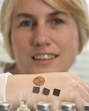
patches on her hand
Credit: Tómas Tyner
Delivering malaria vaccines via a skin patch can streamline the vaccination process, according to preclinical research published in Scientific Reports.
The patch has arrays of tiny silicon microneedles that painlessly create temporary pores in the outermost layer of the skin, permitting the vaccine to flow into the skin.
Researchers used the patch to vaccinate mice with a live adenovirus engineered to deliver a protein from the malaria parasite Plasmodium yoelii.
The team found the patch could overcome one of the main problems with this type of vaccine—namely, although it induces high levels of immunity to malaria, it can also induce a strong immune response to the adenovirus itself.
This anti-adenovirus immunity prevents its repeated use as a vaccine, as the immune system recognizes the adenovirus and prevents it from delivering the malaria protein. So another vaccine type or adenovirus strain needs to be used in the booster immunization.
“What’s exciting from this work is that administration of this vaccine with the microneedle patch did not induce this strong anti-adenovirus immunity, even though very potent immunity to the malaria antigen is generated,” said study author Anne Moore, PhD, of University College Cork in Ireland.
This suggests the patch can facilitate the repeated use of the same adenovirus vaccine, thereby potentially reducing manufacturing costs of multiple vaccines.
In their experiments with mice, Dr Moore and her colleagues demonstrated that using the microneedle patch in the primary immunization does indeed permit repeated use of the same adenovirus vaccine. And this immunization method induced potent and highly protective immune responses against malaria.
Specifically, the researchers delivered a vaccine known as HAdV5-PyMSP142 to mice. They found the patch induced equivalent or enhanced antibody responses but decreased anti-vector responses when compared to intradermal delivery of the vaccine.
The addition of a heterologous vaccine known as MVA-PyMSP142 also produced greater antibody responses in mice primed with HAdV5-PyMSP142 via the patch, compared to those vaccinated intradermally.
The researchers observed the highest protection against blood-stage malaria when mice were vaccinated first with the patch and then intradermally.
In an attempt to commercialize this research, Dr Moore is heading to Silicon Valley next week to meet with venture capitalists and technology companies. ![]()
Journals may fail to correct major errors in articles

Credit: CDC/James Gathany
A review of errata reports from medical publications revealed that nearly a quarter of the errors may have changed the way study data were interpreted.
And about half of the errors were not corrected in the original text, or the errata report did not specify whether a correction was made.
This suggests authors and journals must be more vigilant and consistent in identifying and reporting errors, said Paul Hauptman, MD, of Saint Louis University in Missouri.
He and his colleagues conducted this research and reported the results in The American Journal of Medicine.
The researchers reviewed 20 prominent English language journals in the fields of general medicine and cardiology. And they identified 577 error reports over an 18-month period.
More than 24% of these reports included an error the team rated as major. Major errors were associated with material changes in the interpretation of data in text, figures, or tables, or with significant alterations in the article’s conclusions.
One of the examples of a major error report in the study comes from a paper about depression. In the original article, the incidence of new depression cases was misquoted by a factor of 10. The article initially reported 15.8% of women with new depression, rather than the real figure of 1.58%, which was later corrected.
“As the volume of research publications continues to rise, the scientific community needs to examine how it manages its mistakes,” said Eric Armbrecht, PhD, of the Saint Louis University Center for Outcomes Research.
“Transparency, consistency, and clarity are essential. Our study found that these are not common among some of the top medical journals.”
In fact, the researchers found that 51% of the errors they identified were not corrected in the original text, or the errata report did not specify whether a correction was made.
The team noted that this study did not provide any definitive explanations for why such errors appear in publications, although one possibility is that most authors don’t read and edit the final version of their manuscripts prior to publication.
“It’s noteworthy that although final approval of an article may fall to the first or corresponding author, the criteria put forth by [the International Committee of Medical Journal Editors] specifies that each author must provide final approval of the version to be published,” Dr Hauptman said.
He added that, at this point, it’s not possible to measure how frequently a journal reader incorporates errata into clinical care or the extent to which patient outcomes may be affected. ![]()
Cellular RNA can template DNA repair in yeast

DNA has been repaired by
transcript RNA within the cells
Credit: Georgia Tech/Rob Felt
Scientists have shown that RNA produced within yeast cells can serve as a template for repairing the most devastating DNA damage—a break in both strands of a DNA helix.
The group believes their study is the first to show that a cell’s own RNA can be used for DNA recombination and repair.
If the phenomenon extends to human cells, it could potentially lead to new therapeutic or preventative strategies for genetic diseases.
The scientists described the phenomenon in Nature.
“We have found that genetic information can flow from RNA to DNA in a homology-driven manner, from cellular RNA to a homologous DNA sequence,” said study author Francesca Storici, PhD, of the Georgia Institute of Technology in Atlanta.
“This process is moving the genetic information in the opposite direction from which it normally flows. We have shown that when an endogenous RNA molecule can anneal to broken homologous DNA without being removed, the RNA can repair the damaged DNA. This finding reveals the existence of a novel mechanism of genetic recombination.”
Dr Storici’s team previously showed that synthetic RNA introduced into cells—including human cells—could repair DNA damage. But the process was inefficient, and there were questions about whether it could occur naturally.
To find out whether cells could use endogenous RNA transcripts to repair DNA damage, she and her colleagues devised experiments using the yeast Saccharomyces cerevisiae.
The team developed a strategy for distinguishing repair by endogenous RNA from repair by the normal DNA-based mechanisms in the budding yeast cells, including using mutants that lacked the ability to convert the RNA into a DNA copy.
They then induced a DNA double-strand break in the yeast genome and observed whether the organism could survive and grow by repairing the damage using only transcript RNA within the cells.
The DNA region that generates the transcript was constructed to contain a marker gene interrupted by an intron. Following intron removal during transcription, the transcript RNA sequence has no intron, while the DNA region that generates the transcript retains the intron.
Only the repair templated by the transcript devoid of the intron can restore the function of a homologous marker gene in which the DNA double-strand break is induced.
Dr Storici and her colleagues measured success by counting the number of yeast colonies growing on a Petri dish, indicating that the repair had been made by endogenous RNA.
They conducted testing on two types of breaks, one in the DNA from which the RNA transcript had been made, and the other in a homologous sequence from a different location in the DNA.
The team found that proximity of the RNA to the broken DNA increased the efficiency of the repair, and the repair occurred via a homologous recombination process. Dr Storici believes the repair mechanism may operate in cells beyond yeast, and many types of RNA can be used.
“We are showing that the flow of genetic information from RNA to DNA is not restricted to retro-elements and telomeres but occurs with a generic cellular transcript, making it more of a general phenomenon than had been anticipated,” she explained. “Potentially, any RNA in the cell could have this function.”
For the future, Dr Storici hopes to learn more about the mechanism, including what regulates it. She also wants to determine whether it takes place in human cells. If so, that could have implications for treating or preventing diseases caused by genetic damage.
“Cells synthesize lots of RNA transcripts during their life spans,” Dr Storici said. “Therefore, RNA may have an unanticipated impact on genomic stability and plasticity. We need to understand in which situations cells would activate RNA-DNA recombination. Better understanding this molecular process could also help us manipulate mechanisms for therapy, allowing us to treat a disease or prevent it altogether.” ![]()
VIDEO: Rivaroxaban provides advantages for cardioversion in AF
BARCELONA – The first-ever prospective, randomized trial of a novel oral anticoagulant in patients with atrial fibrillation undergoing elective cardioversion showed oral rivaroxaban at 20 mg once daily to be an effective and safe alternative to standard-of-care warfarin. But the study, known as X-VeRT, also showed rivaroxaban offers something in addition: more expeditious cardioversion amenable to reliable scheduling.
Dr. Riccardo Cappato, who presented the X-VeRT results at the annual congress of the European Society of Cardiology, explains in this video interview.
The video associated with this article is no longer available on this site. Please view all of our videos on the MDedge YouTube channel
AT THE ESC CONGRESS 2014
Successful Treatment of Schnitzler Syndrome With Canakinumab
To the Editor:
Schnitzler syndrome occurs with a triad of chronic urticaria, recurring fevers, and monoclonal gammopathy. It was recognized as a clinical entity in 1972; now nearly 200 patients are reported in the medical literature.1-3 Flulike symptoms, arthralgia, bone pain, lymphadenopathy, and hepatosplenomegaly also are clinical findings.4,5 The erythrocyte sedimentation rate (ESR) often is markedly elevated, as are other acute phase reactants. Leukocytosis with neutrophilia and IgM and IgG monoclonal gammopathies have been described.4
Schnitzler syndrome shares many clinical characteristics with a subset of autoinflammatory disorders referred to as cryopyrin-associated periodic syndromes (CAPS), which includes familial cold autoinflammatory syndrome and Muckle-Wellssyndrome. These syndromes are associated with mutations in the cold-induced autoinflammatory syndrome 1 gene, CIAS1, which encodes the NALP3 inflammasome, leading to overproduction of IL-1β.5 A gain-of-function mutation in CIAS1 has been described in a patient with Schnitzler syndrome.6
Treatment of urticaria and constitutional symptoms associated with Schnitzler syndrome is challenging. Antihistamines are ineffective, though high-dose systemic glucocorticosteroids control most of the clinical manifestations. Methotrexate sodium, cyclosporine, and tumor necrosis factor antagonists are utilized as glucocorticosteroid-sparing agents. Anakinra, an IL-1 receptor monoclonal antibody that is approved for use in CAPS, has been reported to induce complete resolution of Schnitzler syndrome when administered daily; however, it is not approved by the US Food and Drug Administration for this disorder.7 Canakinumab, an IL-1β monoclonal antibody that is dosed every 8 weeks, was approved by the US Food and Drug Administration in 2009 for the treatment of CAPS. Given the similar clinical characteristics and genetic mutations found in CAPS and Schnitzler syndrome, canakinumab may be an effective treatment of both disorders. We report successful treatment with this monoclonal antibody in 2 patients with Schnitzler syndrome.
A 63-year-old man reported having night sweats and fatigue but had no arthralgia or arthritis. He had a 1-year history of severe urticaria and recurrent fevers (temperature, up to 38.4°C) and he also had type 1 diabetes mellitus, hypothyroidism, and celiac disease. Physical examination revealed an elevated temperature (38.4°C) and generalized urticaria but no evidence of hepatosplenomegaly, adenopathy, or arthritis. Leukocytosis was revealed (white blood cell count, 12,400/μL [reference range, 4500–11,000/μL]) with neutrophilia (88.5% [reference range, 56%]), elevated ESR (81 mm/h [reference range, 0–20 mm/h]), and IgM κ monoclonal gammopathy (0.37 g/L [reference range, 0.4–2.3 g/L]). Clinical examination as well as laboratory and imaging studies did not show evidence of malignancy or autoimmune disease. A skin biopsy identified neutrophilic urticaria without vasculitis. Prednisone 20 mg daily controlled the urticaria and fever, but symptoms recurred within days of glucocorticosteroid withdrawal.
A 47-year-old woman presented with a 7-year history of severe urticaria, fever (temperature, 38.9°C), myalgia, and arthralgia. She had a medical history of allergic rhinitis, gastroesophageal reflux disease, chronic pain syndrome, and depression. Physical examination revealed generalized urticaria with cervical and axillary lymphadenopathy 1 to 2 cm in size but no hepatosplenomegaly or arthritis. Prior evaluations for fever of unknown origin as well as autoimmune and malignant disorders were negative. Skin biopsies reported neutrophilic urticaria without vasculitis, and a lymph node biopsy from the left axilla revealed neutrophilic inflammation. A white blood cell count of 17,800/μL with 61.6% neutrophils, elevated C-reactive protein (153.4 mg/L [reference range, 0.08–3.1 mg/L]) and ESR (90 mm/h), and an IgG λ monoclonal gammopathy were present. She was previously treated with etanercept, methotrexate sodium, golimumab, and adalimumab, with only a partial response. For more than 5 years, prednisone 20 to 50 mg daily was necessary to control her symptoms. Cyclosporine 200 mg twice daily was added as a corticosteroid-sparing drug with partial response.
Both patients were diagnosed with Schnitzler syndrome and were started on canakinumab 150 mg administered subcutaneously in the upper arm every 8 weeks. Resolution of the urticaria and fevers occurred within 2 weeks, and all other medications for the treatment of Schnitzler syndrome were withdrawn without recurrence of symptoms after 3 years. The neutrophil count and acute phase reactants returned within reference range in each patient, but the monoclonal gammopathies remained unchanged. Patient 2 noted worsening of arthralgia after initiation of canakinumab, but long-term corticosteroid withdrawal was considered the cause. Patient 1 has been able to increase the interval of dosing to every 3 to 4 months without recurrence of symptoms. Patient 2 has not tolerated similar changes in dosing interval.
Canakinumab given at 8-week intervals was a safe and effective treatment of Schnitzler syndrome in this open trial of 2 patients. Anakinra also induces remission, but daily dosing is required. Cost may be a notable factor in the choice of therapy, as canakinumab costs substantially more per year than anakinra. Further investigation is required to determine if treatment with canakinumab will result in long-term remission and if less-frequent dosing will provide continued efficacy.
1. Schnitzler L. Lésions urticariennes chroniques permanentes (érythème pétaloïde?). Cas cliniques. nº 46 B. Journee Dermatologique d’Angers. October 1972.
2. Schnitzler L, Schubert B, Boasson M, et al. Urticaire chronique, lésions osseuses, macroglobulinémie IgM: maladie de Waldenstrӧm. Bull Soc Fr Dermatol Syphiligr. 1974;81:363.
3. Simon A, Asli B, Braun-Falco M, et al. Schnitzler’s syndrome: diagnosis, treatment, and follow-up. Allergy. 2013;68:562-568.
4. de Koning HD, Bodar EJ, van der Meer JW, et al. Schnitzler syndrome: beyond the case reports: review and follow-up of 94 patients with an emphasis on prognosis and treatment [published online ahead of print June 21, 2007]. Semin Arthritis Rheum. 2007;37:137-148.
5. Lipsker D, Veran Y, Grunenberger F, et al. The Schnitzler syndrome. four new cases and review of the literature. Medicine (Baltimore). 2001;80:37-44.
6. Loock J, Lamprecht P, Timmann C, et al. Genetic predisposition (NLRP3 V198M mutation) for IL-1-mediated inflammation in a patient with Schnitzler syndrome. J Allergy Clin Immunol. 2010;125:500-502.
7. Ryan JG, de Koning HD, Beck LA, et al. IL-1 blockade in Schnitzler syndrome: ex vivo findings correlate with clinical remission [published online ahead of print October 22, 2007]. J Allergy Clin Immunol. 2008;121:260-262.
To the Editor:
Schnitzler syndrome occurs with a triad of chronic urticaria, recurring fevers, and monoclonal gammopathy. It was recognized as a clinical entity in 1972; now nearly 200 patients are reported in the medical literature.1-3 Flulike symptoms, arthralgia, bone pain, lymphadenopathy, and hepatosplenomegaly also are clinical findings.4,5 The erythrocyte sedimentation rate (ESR) often is markedly elevated, as are other acute phase reactants. Leukocytosis with neutrophilia and IgM and IgG monoclonal gammopathies have been described.4
Schnitzler syndrome shares many clinical characteristics with a subset of autoinflammatory disorders referred to as cryopyrin-associated periodic syndromes (CAPS), which includes familial cold autoinflammatory syndrome and Muckle-Wellssyndrome. These syndromes are associated with mutations in the cold-induced autoinflammatory syndrome 1 gene, CIAS1, which encodes the NALP3 inflammasome, leading to overproduction of IL-1β.5 A gain-of-function mutation in CIAS1 has been described in a patient with Schnitzler syndrome.6
Treatment of urticaria and constitutional symptoms associated with Schnitzler syndrome is challenging. Antihistamines are ineffective, though high-dose systemic glucocorticosteroids control most of the clinical manifestations. Methotrexate sodium, cyclosporine, and tumor necrosis factor antagonists are utilized as glucocorticosteroid-sparing agents. Anakinra, an IL-1 receptor monoclonal antibody that is approved for use in CAPS, has been reported to induce complete resolution of Schnitzler syndrome when administered daily; however, it is not approved by the US Food and Drug Administration for this disorder.7 Canakinumab, an IL-1β monoclonal antibody that is dosed every 8 weeks, was approved by the US Food and Drug Administration in 2009 for the treatment of CAPS. Given the similar clinical characteristics and genetic mutations found in CAPS and Schnitzler syndrome, canakinumab may be an effective treatment of both disorders. We report successful treatment with this monoclonal antibody in 2 patients with Schnitzler syndrome.
A 63-year-old man reported having night sweats and fatigue but had no arthralgia or arthritis. He had a 1-year history of severe urticaria and recurrent fevers (temperature, up to 38.4°C) and he also had type 1 diabetes mellitus, hypothyroidism, and celiac disease. Physical examination revealed an elevated temperature (38.4°C) and generalized urticaria but no evidence of hepatosplenomegaly, adenopathy, or arthritis. Leukocytosis was revealed (white blood cell count, 12,400/μL [reference range, 4500–11,000/μL]) with neutrophilia (88.5% [reference range, 56%]), elevated ESR (81 mm/h [reference range, 0–20 mm/h]), and IgM κ monoclonal gammopathy (0.37 g/L [reference range, 0.4–2.3 g/L]). Clinical examination as well as laboratory and imaging studies did not show evidence of malignancy or autoimmune disease. A skin biopsy identified neutrophilic urticaria without vasculitis. Prednisone 20 mg daily controlled the urticaria and fever, but symptoms recurred within days of glucocorticosteroid withdrawal.
A 47-year-old woman presented with a 7-year history of severe urticaria, fever (temperature, 38.9°C), myalgia, and arthralgia. She had a medical history of allergic rhinitis, gastroesophageal reflux disease, chronic pain syndrome, and depression. Physical examination revealed generalized urticaria with cervical and axillary lymphadenopathy 1 to 2 cm in size but no hepatosplenomegaly or arthritis. Prior evaluations for fever of unknown origin as well as autoimmune and malignant disorders were negative. Skin biopsies reported neutrophilic urticaria without vasculitis, and a lymph node biopsy from the left axilla revealed neutrophilic inflammation. A white blood cell count of 17,800/μL with 61.6% neutrophils, elevated C-reactive protein (153.4 mg/L [reference range, 0.08–3.1 mg/L]) and ESR (90 mm/h), and an IgG λ monoclonal gammopathy were present. She was previously treated with etanercept, methotrexate sodium, golimumab, and adalimumab, with only a partial response. For more than 5 years, prednisone 20 to 50 mg daily was necessary to control her symptoms. Cyclosporine 200 mg twice daily was added as a corticosteroid-sparing drug with partial response.
Both patients were diagnosed with Schnitzler syndrome and were started on canakinumab 150 mg administered subcutaneously in the upper arm every 8 weeks. Resolution of the urticaria and fevers occurred within 2 weeks, and all other medications for the treatment of Schnitzler syndrome were withdrawn without recurrence of symptoms after 3 years. The neutrophil count and acute phase reactants returned within reference range in each patient, but the monoclonal gammopathies remained unchanged. Patient 2 noted worsening of arthralgia after initiation of canakinumab, but long-term corticosteroid withdrawal was considered the cause. Patient 1 has been able to increase the interval of dosing to every 3 to 4 months without recurrence of symptoms. Patient 2 has not tolerated similar changes in dosing interval.
Canakinumab given at 8-week intervals was a safe and effective treatment of Schnitzler syndrome in this open trial of 2 patients. Anakinra also induces remission, but daily dosing is required. Cost may be a notable factor in the choice of therapy, as canakinumab costs substantially more per year than anakinra. Further investigation is required to determine if treatment with canakinumab will result in long-term remission and if less-frequent dosing will provide continued efficacy.
1. Schnitzler L. Lésions urticariennes chroniques permanentes (érythème pétaloïde?). Cas cliniques. nº 46 B. Journee Dermatologique d’Angers. October 1972.
2. Schnitzler L, Schubert B, Boasson M, et al. Urticaire chronique, lésions osseuses, macroglobulinémie IgM: maladie de Waldenstrӧm. Bull Soc Fr Dermatol Syphiligr. 1974;81:363.
3. Simon A, Asli B, Braun-Falco M, et al. Schnitzler’s syndrome: diagnosis, treatment, and follow-up. Allergy. 2013;68:562-568.
4. de Koning HD, Bodar EJ, van der Meer JW, et al. Schnitzler syndrome: beyond the case reports: review and follow-up of 94 patients with an emphasis on prognosis and treatment [published online ahead of print June 21, 2007]. Semin Arthritis Rheum. 2007;37:137-148.
5. Lipsker D, Veran Y, Grunenberger F, et al. The Schnitzler syndrome. four new cases and review of the literature. Medicine (Baltimore). 2001;80:37-44.
6. Loock J, Lamprecht P, Timmann C, et al. Genetic predisposition (NLRP3 V198M mutation) for IL-1-mediated inflammation in a patient with Schnitzler syndrome. J Allergy Clin Immunol. 2010;125:500-502.
7. Ryan JG, de Koning HD, Beck LA, et al. IL-1 blockade in Schnitzler syndrome: ex vivo findings correlate with clinical remission [published online ahead of print October 22, 2007]. J Allergy Clin Immunol. 2008;121:260-262.
Rivaroxaban shows advantages in AF cardioversion
BARCELONA – Rivaroxaban appears to be a safe and effective alternative to warfarin in patients with atrial fibrillation undergoing elective cardioversion, according to the findings of the first-ever prospective, randomized trial of a novel oral anticoagulant for this application.
Additionally, the X-VeRT trial showed that rivaroxaban (Xarelto) may offer an attractive, highly practical advantage over warfarin, the current guideline-recommended standard of care for cardioversion: Namely, rivaroxaban enabled patients to undergo the procedure more expeditiously, Dr. Riccardo Cappato noted in presenting the X-VeRT findings at the annual congress of the European Society of Cardiology.

Indeed, patients in the early-cardioversion arm of the trial safely underwent the procedure as early as 4 hours after taking their first 20-mg dose of rivaroxaban.
Moreover, patients in the delayed-conversion study arm, which required at least 3 consecutive weeks of effective oral anticoagulation preprocedurally, underwent cardioversion an average of 8 days earlier if randomized to rivaroxaban rather than to warfarin or another vitamin K antagonist. That’s because many warfarin-treated patients couldn’t maintain their international normalized ratio (INR) within the target range of 2.0-3.0 for 3 straight weeks, explained Dr. Cappato, professor of electrophysiology and chief of the arrhythmia and electrophysiology center at the University of Milan’s San Donato Polyclinic Hospital.
X-VeRT included 1,504 patients with nonvalvular atrial fibrillation (AF) at 141 centers in 16 countries. All were scheduled for elective cardioversion. They were randomized 2:1 to rivaroxaban 20 mg once daily or to warfarin at a dose adjusted to maintain an INR of 2.0-3.0.
Of those patients, 872 were assigned to an early-cardioversion strategy. Their cardioversion took place after 1-5 days on study medication, provided transesophageal echocardiography had ruled out a left atrial thrombus or they were already on chronic warfarin with their last three INRs in the target range. The other 632 patients were cardioverted using a delayed strategy in which they had to be on study medication for 3-8 weeks before the procedure.
The primary efficacy outcome in X-VeRT was the composite rate of stroke, TIA, peripheral embolism, MI, and cardiovascular death. The incidence was 0.51% in the rivaroxaban group and not statistically different at 1.02% in the warfarin group. The primary safety outcome – major bleeding – occurred in 0.6% of rivaroxaban-treated patients and 0.8% on warfarin.
The median time to cardioversion in the early-cardioversion strategy arm was similar regardless of which drug was used. Of note, however, in the delayed-strategy group, the median time to cardioversion was 22 days in patients on rivaroxaban, compared with 30 days with warfarin.
Of patients in the delayed-strategy group, 77% of those on rivaroxaban were cardioverted as scheduled, compared with 36% on warfarin. Only one patient in the rivaroxaban group was unable to undergo cardioversion before the 8-week cutoff because of inadequate anticoagulation as defined by less than 80% compliance in pill taking; in contrast, 95 warfarin-treated patients in the delayed-strategy group weren’t cardioverted because they missed the 8-week cutoff because of problematic INRs.
In an interview, ESC spokesman Dr. Jurrien Ten Berg predicted X-VeRT will be practice changing. He believes that on the basis of these study results, many physicians will view elective cardioversion as an excellent time to switch patients from warfarin, with all its inherent problems, to rivaroxaban, especially if they qualify for early cardioversion.
"I think this will absolutely change our policy. Here we’re talking about a once-daily pill that makes it possible to do a cardioversion early, and I think that’s a major advantage. If you delay the cardioversion, anything can happen. We know the vitamin K antagonists are unreliable, especially in the first weeks, when even if the INR is fine you don’t really know if the patient is well anticoagulated. For me, the totally of evidence, including the retrospective analyses of the large atrial fibrillation stroke prevention trials, is enough now to use a NOAC for several days and then do an early cardioversion," said Dr. Ten Berg, a cardiologist at St. Antonius Hospital, Nieuwegein, the Netherlands.
Discussant Dr. Christoph Bode called X-VeRT "a landmark trial" and "brilliant work."
"This should be included in the next update of the guidelines," said Dr. Bode, professor and chair of internal medicine and cardiology at the University of Freiburg, Germany.
However, Dr. Steven Nissen took a more skeptical, albeit clearly a minority, view.
"I think this study shows neither safety nor efficacy for this regimen. There were just a handful of events in both groups, too few to draw any conclusions. I don’t think the guidelines should be changed. I think the proper interpretation of the study is that it’s inconclusive," said Dr. Nissen, chair of the department of cardiovascular medicine at the Cleveland Clinic.
Dr. Cappato noted in response that he’d stated at the outset that X-VeRT, even at more than 1,500 patients, was underpowered statistically. Event rates in well-anticoagulated patients undergoing cardioversion are so low that a study to establish noninferiority for rivaroxaban would require 25,000-30,000 participants, which is not going to happen.
"Many physicians are already switching to NOACs [novel oral anticoagulants] for cardioversion despite the absence of good evidence. We thought bringing forward this solid, methodologically sound information from X-VeRT would provide more consistent support for those who are doing this or considering it," the cardiologist said.
Simultaneous with his presentation in Barcelona, the X-VeRT (Explore the Efficacy and Safety of Once-Daily Oral Rivaroxaban for the Prevention of Cardiovascular Events in Patients With Nonvalvular Atrial Fibrillation Scheduled for Cardioversion) study was published online (Eur. Heart J. 2014 [doi:10.1093/eurheart/ehu367]).
Dr. Cappato reported receiving investigator fees from and serving as a consultant to and on speakers bureaus for numerous pharmaceutical companies, including Bayer HealthCare, which sponsored X-VeRT. Dr. Bode has received honoraria from Bayer HealthCare. Dr. Ten Berg and Dr. Nissen reported having no financial conflicts.
BARCELONA – Rivaroxaban appears to be a safe and effective alternative to warfarin in patients with atrial fibrillation undergoing elective cardioversion, according to the findings of the first-ever prospective, randomized trial of a novel oral anticoagulant for this application.
Additionally, the X-VeRT trial showed that rivaroxaban (Xarelto) may offer an attractive, highly practical advantage over warfarin, the current guideline-recommended standard of care for cardioversion: Namely, rivaroxaban enabled patients to undergo the procedure more expeditiously, Dr. Riccardo Cappato noted in presenting the X-VeRT findings at the annual congress of the European Society of Cardiology.
Indeed, patients in the early-cardioversion arm of the trial safely underwent the procedure as early as 4 hours after taking their first 20-mg dose of rivaroxaban.
Moreover, patients in the delayed-conversion study arm, which required at least 3 consecutive weeks of effective oral anticoagulation preprocedurally, underwent cardioversion an average of 8 days earlier if randomized to rivaroxaban rather than to warfarin or another vitamin K antagonist. That’s because many warfarin-treated patients couldn’t maintain their international normalized ratio (INR) within the target range of 2.0-3.0 for 3 straight weeks, explained Dr. Cappato, professor of electrophysiology and chief of the arrhythmia and electrophysiology center at the University of Milan’s San Donato Polyclinic Hospital.
X-VeRT included 1,504 patients with nonvalvular atrial fibrillation (AF) at 141 centers in 16 countries. All were scheduled for elective cardioversion. They were randomized 2:1 to rivaroxaban 20 mg once daily or to warfarin at a dose adjusted to maintain an INR of 2.0-3.0.
Of those patients, 872 were assigned to an early-cardioversion strategy. Their cardioversion took place after 1-5 days on study medication, provided transesophageal echocardiography had ruled out a left atrial thrombus or they were already on chronic warfarin with their last three INRs in the target range. The other 632 patients were cardioverted using a delayed strategy in which they had to be on study medication for 3-8 weeks before the procedure.
The primary efficacy outcome in X-VeRT was the composite rate of stroke, TIA, peripheral embolism, MI, and cardiovascular death. The incidence was 0.51% in the rivaroxaban group and not statistically different at 1.02% in the warfarin group. The primary safety outcome – major bleeding – occurred in 0.6% of rivaroxaban-treated patients and 0.8% on warfarin.
The median time to cardioversion in the early-cardioversion strategy arm was similar regardless of which drug was used. Of note, however, in the delayed-strategy group, the median time to cardioversion was 22 days in patients on rivaroxaban, compared with 30 days with warfarin.
Of patients in the delayed-strategy group, 77% of those on rivaroxaban were cardioverted as scheduled, compared with 36% on warfarin. Only one patient in the rivaroxaban group was unable to undergo cardioversion before the 8-week cutoff because of inadequate anticoagulation as defined by less than 80% compliance in pill taking; in contrast, 95 warfarin-treated patients in the delayed-strategy group weren’t cardioverted because they missed the 8-week cutoff because of problematic INRs.
In an interview, ESC spokesman Dr. Jurrien Ten Berg predicted X-VeRT will be practice changing. He believes that on the basis of these study results, many physicians will view elective cardioversion as an excellent time to switch patients from warfarin, with all its inherent problems, to rivaroxaban, especially if they qualify for early cardioversion.
"I think this will absolutely change our policy. Here we’re talking about a once-daily pill that makes it possible to do a cardioversion early, and I think that’s a major advantage. If you delay the cardioversion, anything can happen. We know the vitamin K antagonists are unreliable, especially in the first weeks, when even if the INR is fine you don’t really know if the patient is well anticoagulated. For me, the totally of evidence, including the retrospective analyses of the large atrial fibrillation stroke prevention trials, is enough now to use a NOAC for several days and then do an early cardioversion," said Dr. Ten Berg, a cardiologist at St. Antonius Hospital, Nieuwegein, the Netherlands.
Discussant Dr. Christoph Bode called X-VeRT "a landmark trial" and "brilliant work."
"This should be included in the next update of the guidelines," said Dr. Bode, professor and chair of internal medicine and cardiology at the University of Freiburg, Germany.
However, Dr. Steven Nissen took a more skeptical, albeit clearly a minority, view.
"I think this study shows neither safety nor efficacy for this regimen. There were just a handful of events in both groups, too few to draw any conclusions. I don’t think the guidelines should be changed. I think the proper interpretation of the study is that it’s inconclusive," said Dr. Nissen, chair of the department of cardiovascular medicine at the Cleveland Clinic.
Dr. Cappato noted in response that he’d stated at the outset that X-VeRT, even at more than 1,500 patients, was underpowered statistically. Event rates in well-anticoagulated patients undergoing cardioversion are so low that a study to establish noninferiority for rivaroxaban would require 25,000-30,000 participants, which is not going to happen.
"Many physicians are already switching to NOACs [novel oral anticoagulants] for cardioversion despite the absence of good evidence. We thought bringing forward this solid, methodologically sound information from X-VeRT would provide more consistent support for those who are doing this or considering it," the cardiologist said.
Simultaneous with his presentation in Barcelona, the X-VeRT (Explore the Efficacy and Safety of Once-Daily Oral Rivaroxaban for the Prevention of Cardiovascular Events in Patients With Nonvalvular Atrial Fibrillation Scheduled for Cardioversion) study was published online (Eur. Heart J. 2014 [doi:10.1093/eurheart/ehu367]).
Dr. Cappato reported receiving investigator fees from and serving as a consultant to and on speakers bureaus for numerous pharmaceutical companies, including Bayer HealthCare, which sponsored X-VeRT. Dr. Bode has received honoraria from Bayer HealthCare. Dr. Ten Berg and Dr. Nissen reported having no financial conflicts.
AT THE ESC CONGRESS 2014
Key clinical point: Patients in atrial fibrillation were able to safely undergo cardioversion as early as 4 hours after taking their first 20-mg dose of oral rivaroxaban.
Major finding: Patients with AF assigned to rivaroxaban in conjunction with cardioversion had a 0.51% rate of major cardiovascular events, similar to the 1.02% rate in patients on warfarin or other vitamin K antagonists. These rates weren’t statistically different; nor were the major bleeding rates in the two groups.
Data source: X-VeRT was a randomized, prospective, open-label, phase IIIb clinical trial involving 1,504 patients at 141 centers in 16 countries.
Disclosures: The X-VeRT trial was sponsored by Bayer HealthCare. The presenter serves as a consultant to and on speakers bureaus for Bayer and other pharmaceutical companies.

Extending Therapy for Breast Cancer
Laronna Colbert, MD, discusses how recent breast cancer studies "have the potential to change current practice standards" for breast cancer.
"This is really a dynamic field of study," Colbert said during her 2013 AVAHO Meeting presentation. "Hopefully, we can continue to make advancements for these patients."
Treating Metastatic Lung Cancer
Rafael Santana-Davila, MD, discusses how understanding the treatment of non-small cell lung cancer (NSCLC) will help physicians better serve the veteran population.
"NSCLC is the second most common cancer—second to prostate cancer—in the VA and, more importantly, the most common cause of cancer death in our veterans." Santana-Davila said during his 2013 AVAHO meeting presentation. "The main goal of the treatment is to improve quality of life and extend survival as much as possible."
Calcium – Making deposits for a healthy adulthood
Likely, one of the most important roles of a pediatrician is to maximize health in childhood and positively impact health in adulthood. Bone density is one of the few things that can be maximized in adolescence. By maximizing bone density, we can directly slow and reduce the osteopenia that occurs later in life and the osteoporosis that 10 million Americans struggle with annually.
The physiology of calcium absorption changes throughout life. In early adolescence, the absorption is greater than the elimination. Between 30 and 50 years of age, absorption and elimination are about equal, but as we enter into the sixth decade of life, there is significant bone loss. Studies have shown that bone density is maximized by age 30 years, and little change is made later in life despite supplementation (Eur. J. Clin. Nutr. 1993;47:617-22). The greatest amount of bone loss occurs after the age of 65 years, and fractures after this age are predominantly at cortical sites.
Consumption of the appropriate amounts of calcium can be difficult given the inadequacies of most adolescents’ diet. The recommended daily intake is 1,200-1,500 mg of elemental calcium. But, absorption of calcium is quite variable and is dependent on other factors to be in place for it to be maximized.
The two most common form of calcium are calcium carbonate and calcium citrate malate. Calcium carbonate requires a higher pH of the stomach, and therefore needs to be taken with food. Calcium carbonate is more cost effective but is also associated with more side effects such as gas and bloating. Calcium citrate malate is found in many juices that are fortified with calcium, can be taken with or without food, is better absorbed with chronic conditions, and is thought to be protective against stone formation (J. Am. Coll. Nutr. 1996;15:313-6; Adv. Food. Nutr. Res. 2008;54:219-346).
Common sources of calcium include milk, yogurt, cheese, Chinese cabbage, kale, broccoli, and spinach. Appropriate levels of vitamin D are important to maximize the absorption of calcium, and recent studies have shown that 40% of adolescents are deficient in vitamin D (Arch. Pediatr. Adolesc. Med. 2004;158:531-7; Arch. Pediatr. Adolesc. Med. 2008;162:513-9). Many other adolescents are lactose intolerant or have a milk protein allergy, which also limit the calcium sources. Soymilk has similar levels of calcium, compared with whole milk. Almond-coconut milk has double the amount of calcium, compared with whole milk, so it is a great substitute for those who are lactose intolerant.
Oxalic acids are found in food such as spinach, collard greens, and sweet potatoes, all of which are rich in calcium, but the oxalic acid reduces the absorption of the calcium. Consumption of large amounts of tea and coffee also can reduce calcium absorption, so despite consuming appropriate amounts of calcium, limited amounts become bioavailable.
If using calcium supplements, ingesting less than or equal to 500 mg is better than taking 1,000 mg at once because it is better absorbed (Adv. Food Nutr. Res. 2008;54:219-346). Orange juice, apple juice, and cereals are fortified with calcium so these also are great sources that usually are accepted by adolescents.
Calcium is a critical dietary supplement that is needed for strong bones, metabolic functions, nerve transmission, and vascular contraction and vasodilation. Long-term deficiency will result in disease, and fragility of the bones. Early supplementation and calcium rich diets can ensure maximum bone development, but if parents are not educated on the appropriate delivery, this opportunity could be missed.
An excellent resource is the National Institutes of Health, Office of Dietary Supplements website. This site gives a wealth of information for sources and consumption of calcium. Another excellent resource for parents to use to guide them to make healthier choices is the U.S. Department of Agriculture site, www.choosemyplate.gov. Parents are looking for quick simple ways to maximize their children’s diet and ensure they are getting everything they need to be healthy adults. Becoming familiar with the basics will allow you to give informed advice that will significantly affect their children’s future.
Dr. Pearce is a pediatrician in Frankfort, Ill. E-mail her at [email protected].

Preclinical results support blood transfusions containing fibronectin
![]()
Credit: UAB Hospital
The protein fibronectin is instrumental in stopping bleeding and preventing life-threatening blood clots, according to preclinical research published in The Journal of Clinical Investigation.
Experiments in mice revealed that plasma fibronectin (pFn) can switch from supporting hemostasis to inhibiting thrombosis.
The researchers said these findings suggest transfusions containing pFn may control bleeding in humans, particularly in association with anticoagulant therapy.
“Most treatments that help the body stop bleeding can actually cause blood clots, and many treatments to prevent excessive blood clots increase [the] risk of bleeding out,” said study author Heyu Ni, MD, of St Michael’s Hospital in Toronto, Ontario, Canada.
“But when given to mice after an injury or to mice treated with blood thinners—which frequently lead to bleeding complications—fibronectin seems to offer a win-win solution.”
Dr Ni and his colleagues began this research by generating mice deficient in both fibrinogen (Fg) and von Willebrand factor (VWF), as well as mice deficient in both VWF and pFn. They then compared these mice to untreated wild-type mice and anticoagulant-treated wild-type mice.
The team discovered that pFn is vital for controlling bleeding in the context of Fg deficiency and supports hemostasis in the context of VWF deficiency. pFn deposition occurred before platelet accumulation (the first wave of hemostasis) in both wild-type and deficient mice.
However, pFn switched its function with regard to platelet aggregation based on the presence or absence of fibrin. pFn inhibited platelet aggregation when fibrin was absent and promoted aggregation when linked with fibrin.
The researchers found that pFn can control the diameter of fibrin fibers, increase the mechanical strength of a blood clot, and be actively involved in thrombosis.
The team said these results provide a solution for the controversy surrounding pFn’s function in hemostasis and establish pFn as a unique regulatory factor in thrombosis. The findings also suggest transfusions containing pFn may prove particularly effective in controlling bleeding.
“Fibrinogen has been shown to help the body stop bleeding, but our research indicates that less-refined blood products that include fibronectin and fibrinogen may help stop bleeding even more effectively,” Dr Ni said. “And, as an added bonus, fibronectin likely also reduces the risk of life-threatening blood clots from forming.”
“There is a lot of work to be done, but we might find that the less expensive and less processed form of donor blood may be more effective for transfusions. We’ve shown that fibronectin might play a role in improving results from transfusions and should not be discarded during blood product processing. It may be also an important protein in transfusions for stopping bleeding, particularly for patients who receive blood thinners during surgeries.” ![]()