User login
Repeat biopsy and long-term surveillance key for rare Hodgkin’s lymphoma subtype
Time to progression was inferior in patients with advanced-stage nodular lymphocyte-predominant Hodgkin’s lymphoma, compared with patients with classical Hodgkin’s lymphoma, in a study that compared outcomes between the two groups of Hodgkin’s lymphoma patients enrolled in the British Columbia Cancer Agency database.
Over 10 years, time to progression was 63% in the nodular lymphocyte-predominant Hodgkin’s lymphoma (NLPHL) group, vs. 73% in the classical Hodgkin’s lymphoma (CHL) group (P =.040), reported Dr. Katharine Xing of the Centre for Lymphoid Cancer at the BCCA and the University of British Columbia, Vancouver, and her associates.
Transformation to an aggressive non–Hodgkin’s lymphoma (NHL) over 15 years occurred in 24% of those with NLPHL, but in none of those with CHL (P = .00018), and the median time to transformation among those with NLPHL was 5.45 years (Blood 2014;123:3567-73).
The study compared 42 patients with advanced-stage NLPHL to 84 controls with advanced CHL, matched for age, sex, decade of diagnosis, stage, and chemotherapy type; all had been diagnosed between 1970 and 2011. Their mean age was 37 years, about two-thirds were men, most in both groups had stage III disease, and they were followed up for a median of about 11 years. Treatments included standard doxorubicin, bleomycin, vinblastine, and dacarbazine (ABVD) and most received standard ABVD or ABVD-equivalent chemotherapy. The study was conducted to "highlight the distinct natural history of this rare HL subtype," which accounts for 5% of HL cases, the authors noted.
Over 10 years, "HL freedom from treatment failure," which reflects only relapses from HL, was 75% among those with NLPHL and 73% among those with CHL. Overall survival was also similar between the two groups (83.5% among those with NLPHL and 81% among those with CHL at 10 years).
Among their other findings was a significantly higher incidence of transformation over 10 years among those who had splenic involvement at the time of NLPHL diagnosis, compared with those who did not have splenic involvement (29% vs. 7.8%). When they looked at only those NLPHL patients who had received ABVD-like treatment, the incidence of transformation over 10 years was 34% among those with splenic involvement at diagnosis, vs. 9% among those who did not have splenic involvement (P = .014).
Since NLPHL is rare, information on the optimal treatment is limited, particularly for those with advanced disease, the authors pointed out. The analysis "highlights the distinct disease behavior of NLPHL, compared with CHL, and the need for repeat biopsy at relapse as well as long-term surveillance," the authors concluded. "Given the strong expression of CD20" on the lymphocyte predominant cells that distinguishes NLPHL from CHL, the results also provide "a rationale for further evaluation" of CHOP (cyclophosphamide, doxorubicin, vincristine, and prednisone) with rituximab, they added.
FROM BLOOD
Key clinical point: Repeat biopsy and long-term surveillance are necessary in nodular lymphocyte-predominant Hodgkin’s lymphoma.
Major finding: Overall survival was similar between patients with advanced-stage NLPHL and those with advanced-stage CHL, but differences between the two groups included an inferior time to progression among those with NLPHL over 10 years (63% vs 73%).
Data source: The study compared outcomes in 42 patients with advanced-stage NLPHL and 84 matched controls with advanced CHL, who were diagnosed between 1970 and 2011 and were enrolled in a Canadian cancer database.
Disclosures: Fourauthors received research funding from Roche; the remaining seven authors, including the lead author, had no relevant disclosures.
Studies confirm importance of CALR mutation in PMF
MILAN—Two new studies appear to confirm the prognostic significance of CALR mutations in patients with primary myelofibrosis (PMF).
One study showed that PMF patients with mutated CALR had prolonged overall survival (OS) compared to patients with wild-type CALR. And additional subclonal mutations did not impair the positive impact of CALR mutations.
Another study suggested that indels in exon 9 of CALR are founding driver mutations in PMF. These mutations are independent predictors of clinical course, disease progression, and OS.
Both studies were presented at the 19th Congress of the European Hematology Association (EHA).
Paola Guglielmelli, MD, PhD, of the University of Florence in Italy, presented data on CALR mutations in the context of additional mutations (abstract S1355).
And Elisa Rumi, MD, of the University of Pavia in Italy, presented information on mutated CALR and other founding driver mutations in PMF (abstract S1356).
CALR & other subclonal mutations in PMF
To investigate the prognostic role of CALR mutations in relation to additional subclonal mutations, Dr Guglielmelli and her colleagues analyzed 274 samples from PMF patients.
The team genotyped the samples for mutations in 11 genes: JAK2, CALR, MPL, EZH2, ASXL1, SRSF2, IDH1, IDH2, CBL, TET2, and DNMT3A.
Two hundred and fifty-six patients (93.4%) presented with at least 1 somatic mutation, and 104 (38%) presented with at least 2.
The median follow-up was 3.8 years (range, 0.52-29.20 years). Among all patients, the median OS was 12.2 years (range, 5.6-18.8 years). Eighty-four patients died (30.7%), 44 (16.1%) of them due to leukemia.
The presence of CALR mutations was associated with better OS, independent of IPSS and molecular risk categories (hazard ratio [HR] 0.51, P=0.03). But CALR mutations did not impact the risk of progression to acute leukemia.
Among patients with a low- to intermediate-1-risk IPSS score, those with CALR mutations lived a median of 27.7 years, and those without lived a median of 21.7 years (HR 0.4, P=0.02). Among patients with intermediate-2 to high risk, those with CALR mutations lived a median of 4.2 years, and those without lived a median of 2.6 years (HR=0.5, P=0.09).
Among patients with high molecular risk, those with CALR mutations lived a median of 17.7 years, and those without lived a median of 4.3 years, (HR=0.3, P=0.008). High molecular risk was defined as at least 1 mutation in ASXL1, EZH2, SRSF2, or IDH1/2.
Among patients in the low-molecular-risk group, those with CALR mutations lived a median of 27.7 years, and those without lived a median of 21.7 years (HR=0.6, P=0.048).
Dr Guglielmelli said these results confirm the association between CALR mutation and favorable outcomes in PMF. They also show that additional subclonal mutations do not impair the positive impact of CALR mutation, thereby reinforcing the idea that CALR-mutated PMF is a distinct entity in terms of prognosis.
Founding driver mutations in PMF
In another presentation at the EHA Congress, Dr Rumi presented data on founding driver mutations in PMF. She and her colleagues analyzed 617 PMF patients, screening them for JAK2 V617F mutations, indels of CALR exon 9, and MPL exon 10 mutations.
The researchers assessed the impact of these mutations on thrombosis, progression to leukemia, and OS.
Their analysis suggested CALR-mutated PMF patients have a lower risk of thrombosis than JAK2-mutated patients (P=0.021). And this difference retained significance after adjusting for age.
Triple-negative PMF patients had a higher risk of leukemic evolution than CALR-mutated patients (P=0.016) and JAK2-mutated patients (P=0.043).
After adjusting for age, the risk remained significantly higher in triple-negative patients compared to JAK2-mutated patients (P=0.04) and retained borderline significance compared to CALR-mutated patients (P=0.052).
Patients with CALR-mutated PMF had a better OS than JAK2-mutated patients (P<0.001), MPL-mutated patients (P=0.009), and triple-negative patients (P<0.001).
In a multivariate analysis, CALR-mutated patients maintained a better OS than JAK2-mutated patients (P=0.019) and triple-negative patients (P<0.001).
Based on these results, Dr Rumi concluded that mutations in JAK2, CALR, and MPL are independent predictors of clinical course, disease progression, and OS in PMF. So screening patients for these mutations can likely improve upon the risk stratification provided by IPSS. ![]()
How genetics, race affect clopidogrel outcomes

Credit: Robert Boston
Washington University
New research helps explain the higher risk of death observed among some patients taking the anticoagulant clopidogrel after a heart attack.
Researchers identified genetic variants that increased the risk of dying in the year following a first heart attack, but they appeared to do so for different reasons depending on a patient’s race.
Two DNA variants common in African Americans were associated with an increased risk of both bleeding and death.
And in Caucasians, a different variant was linked to additional heart attacks and a higher risk of death.
The variations influence the way patients metabolize clopidogrel and can alter its effectiveness.
These findings were published in Circulation: Cardiovascular Genetics.
“The research is provocative,” said the study’s first author Sharon Cresci, MD, of the Washington University School of Medicine in St Louis.
“Knowing about potential genetic differences based on race can help physicians tailor drugs to patients based on their genetic makeup.”
Clopidogrel is metabolized in the liver, where it is turned into its active form via a group of enzymes called cytochrome P450 (CYP). Although clopidogrel is effective in many patients, earlier studies showed that some patients metabolize the drug better than others.
Indeed, in 2010, the US Food and Drug Administration added a black box warning to labels of clopidogrel after research (which primarily involved Caucasians) showed that people with a particular CYP genetic variant metabolized the drug poorly, which reduced the amount of the drug circulating in the blood. These patients had a higher risk of heart attack and stroke.
Additional studies showed that other CYP gene variants are linked to the rapid metabolism of clopidogrel, and patients with those variants had a higher risk of bleeding. But it has not been clear, until now, that the effects of these particular gene variations can vary by race.
To uncover this finding, Dr Cresci and her colleagues analyzed CYP variants among 2062 Caucasians and 670 African Americans who suffered heart attacks. Nearly 80% of the Caucasians and 65% of the African Americans were prescribed clopidogrel.
The patients were enrolled in a major study known as TRIUMPH (Translational Research Investigating Underlying disparities in acute Myocardial infarction Patients’ Health), which was conducted from 2005 to 2008 at 24 US hospitals.
Among patients taking clopidogrel, the 1-year mortality rate for African Americans was 7.2%, compared with 3.6% for Caucasians.
Caucasians who carried the CYP2C19*2 variant, which has been linked to poor metabolism of the drug, had a higher rate of repeat heart attacks and death. However, among African Americans treated with clopidogrel, the CYP2C19*2 variant was not associated with a higher rate of death.
African Americans had higher rates of bleeding and death if they carried either of 2 other variants: CYP1A2*1C or CYP2C19*17, the latter of which has been associated with the rapid metabolism of clopidogrel. Among Caucasians on clopidogrel, neither variant increased the risk of death.
“This is very novel information that begs for more research,” said study author Richard G. Bach, MD, also of Washington University.
Although genetic testing is available to identify CYP variants in a patient’s DNA, these tests are not widely used by cardiologists. Results of the current study suggest this practice may need to be reconsidered.
“This research is an important addition to the field because previous studies looking at CYP gene variants and their effects on risks of repeat heart attacks, bleeding, and death have included predominantly Caucasian patients of European ancestry,” Dr Cresci noted. “There is almost no data, until now, about these variants in African Americans.”
Research examining how genetic variants alter the effectiveness of clopidogrel remains somewhat controversial, Dr Bach said. Many physicians feel that before they can tailor therapy for heart attack patients, more data is needed to prove a clear link between genetic variants and negative health consequences and that tailoring therapy will improve patients’ outcomes, he explained.
His hope is that additional research would provide more definitive conclusions to help physicians choose the best medications for patients after a heart attack and, ultimately, “to reduce the too-high rate of death and disability for patients after a heart attack,” Dr Bach said.
Dr Cresci agreed, adding, “By focusing on genetic differences, we may be able to individualize therapies after heart attacks and achieve the best treatment for each patient.” ![]()
Restoring gene function can ‘reverse’ B-ALL

and Ross Dickins, PhD
Credit: Walter and Eliza Hall
Institute of Medical Research
Results of preclinical research suggest B-progenitor acute lymphoblastic leukemia (B-ALL) can be “reversed” by coaxing leukemic cells back into normal development.
Researchers found that switching off the gene Pax5 could induce B-ALL in mice, but restoring Pax5 function could prompt disease remission.
Grace Liu, of the Walter and Eliza Hall Institute of Medical Research in Victoria, Australia, and her colleagues detailed this research in Genes & Development.
“Pax5 is essential for normal development of [B cells],” Liu said. “When Pax5 function is compromised, developing B cells can get trapped in an immature state and become cancerous.”
The researchers used transgenic RNAi to suppress endogenous Pax5 expression in the hematopoietic compartment of mice, and this induced B-ALL.
“Along with other genetic changes, deactivating Pax5 drives normal blood cells to turn into leukemia cells, which has been shown before,” Liu said. “However, we showed, for the first time, that reactivating Pax5 enabled the cells to resume their normal development and lose their cancer-like qualities, effectively curing the leukemia.”
The team found that restoring endogenous Pax5 expression triggered immunophenotypic maturation and durable disease remission.
Even brief Pax5 restoration disabled B-ALL cells’ leukemia-initiating capacity in mice. And the researchers observed similar results in human B-ALL cell lines.
“This work shows how inactivating the tumor suppressor gene Pax5 contributes to B-ALL development and how leukemia cells become addicted to low Pax5 levels to continue proliferating,” said study author Ross Dickins, PhD, also of the Walter and Eliza Hall Institute of Medical Research.
“Even though the B-ALL cells have multiple genetic mutations, simply reactivating Pax5 causes tumor cells to resume normal development and lose their cancerous properties.”
Dr Dickins added that forcing B-ALL cells to resume their normal development could provide a new strategy for treating leukemia. However, genes lost in tumor cells are not traditionally considered suitable drug targets.
“It is very difficult to develop drugs that restore the function of genes that are lost during cancer development,” Dr Dickins said. “However, by understanding the mechanisms by which Pax5 loss causes leukemia, we can begin to look at ways of developing drugs that could have the same effect as restoring Pax5 function.” ![]()
Elderly males with DLBCL require increased rituximab dosing
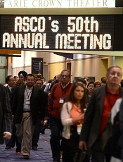
©ASCO/Phil McCarten
CHICAGO—Elderly males with non-Hodgkin lymphoma (NHL) may require one-third higher doses of rituximab than the current standard to attain optimal responses to rituximab-containing chemotherapy, a new study suggests.
Increasing the rituximab dose eliminated any gender-related differences in survival among elderly patients with aggressive, CD20+, B-cell lymphomas, said investigator Michael Pfreundschuh, MD, of Saarland University Medical Center in Germany.
He presented this finding at the 2014 ASCO Annual Meeting (abstract 8501).
Although rituximab has been used in NHL for nearly 2 decades, standard dosing of the drug is still largely empiric. Only recently have researchers examined whether responses to the drug may vary by gender and age.
New data show that rituximab clears the body more rapidly in elderly males than elderly females, suggesting that rituximab dosing may need to be increased in older men to maintain adequate drug exposure.
Dr Pfreundschuh noted that the RICOVER-60 trial established 6 cycles of rituximab plus cyclophosphamide, doxorubicin, vincristine, and prednisone every 14 days (R-CHOP-14), followed by 2 additional cycles of rituximab, as the new standard of care for elderly patients with NHL in Germany.
Further analysis of the trial’s results indicates that rituximab dosing may be inadequate in males older than 60, since they fare much worse than elderly females in terms of progression-free survival (PFS).
To test this gender difference, the German High-grade Non-Hodgkin Lymphoma Study Group designed the SEXIE-R-CHOP-14 trial.
The group tested 2 different rituximab doses in patients aged 61 to 80 with stage I to IV, CD20+, diffuse large B-cell lymphoma (DLBCL). A group of 120 females received rituximab at 375 mg/m2 per treatment cycle, and a group of 148 males received 500 mg/m2 per treatment cycle.
The increased rituximab dose in males resulted in slightly higher trough serum levels than in females. However, rituximab levels dropped faster in males, resulting in nearly identical serum levels thereafter and a very similar overall rituximab exposure time, Dr Pfreundschuh said.
The higher dose of rituximab given to elderly males did not result in increased toxicity.
Survival rates were similar between the male and female groups. The 3-year PFS was 74% in males and 68% in females, and 3-year overall survival was 82% in males and 72% in females.
A multivariate analysis that evaluated gender as a risk factor revealed that the male-vs-female hazard ratio for PFS was 0.8 in SEXIE-R-CHOP-14, compared to 1.6 in RICOVER-60. Similarly, the male-vs-female hazard ratio for overall survival was 0.7 in SEXIE-R-CHOP-14 and 1.4 in RICOVER-60.
“Increasing the rituximab dose by one-third eliminated the increased risk [of death and progression in] elderly males,” Dr Pfreundschuh concluded.
He also noted that younger males and females have faster rituximab clearance than elderly females. So increasing the dose in these populations should also result in a better outcome. ![]()
Passing stones with PDE5 inhibitors
Now that we have made the most likely diagnosis of a kidney stone, we can potentially avoid a urologic procedure if we expel it. Of the 22% of kidney stones that wind up in the ureter, two-thirds will be lodged in the distal ureter.
Tamsulosin works, and prednisolone may also be helpful. Anything else?
Dr. Kumar Jayant of Sudha Hospital and Medical Research Center, Kota, India, and associates investigated the efficacy of a phosphodiesterase type 5 (PDE5) inhibitor (tadalafil) to facilitate kidney stone expulsion. PDE5 inhibitors increase levels of cyclic guanosine monophosphate and cause ureteric relaxation (Int. J. Urol. 2014 June 3 [doi:10.1111/iju.12496]).
In this study, 244 patients with distal ureteral stones between 5 and 10 mm (about a 50% chance of passing) quantitated with noncontrast CT were randomized to two groups: tamsulosin (0.4 mg daily) or tamsulosin (0.4 mg daily) plus tadalafil (10 mg daily). Medications were given for 4 weeks.
The average patient age was about 37 years, and the mean stone size was 7 mm. Participants were included only if their pain was relieved within a day by diclofenac injection. Potential participants were excluded if they had fever, hydronephrosis, multiple kidney stones, a history of surgery or endoscopic procedures, or diabetes; were taking calcium channel blockers or nitrates; were pregnant or lactating; or required immediate treatment. If stones were not passed in 4 weeks, ureterorenoscopy was used to remove them.
The tamsulosin/tadalafil combination was associated with a statistically significantly higher rate of expulsion (83.6% vs. 65.5%; P = .031) and a shorter time to expulsion (14.9 days vs. 16.7 days; P =.003). Tamsulosin/tadalafil was associated with significantly fewer hospital visits and less need for pain medications. Not surprisingly, tamsulosin/tadalafil improved erectile function. However, patients taking the tamsulosin/tadalafil combination also had more headaches, dizziness, orthostatic hypotension, and backaches.
In certain patients, hypotension may be a concern with this combination. However, the researchers highlighted a study whose authors concluded, "in subjects on tamsulosin, tadalafil 10 and 20 mg produced mean maximal decreases in standing [systolic blood pressure] that were similar to placebo" (J. Urol. 2004;172(5, pt. 1):1935-40).
Out-of-pocket cost may be a barrier for some patients. But given the significance of these findings (an almost 20% difference in expulsion rate), total cost of care may be significantly reduced for these patients.
Dr. Ebbert is a professor of medicine and general internist at the Mayo Clinic in Rochester, Minn., and a diplomate of the American Board of Addiction Medicine. The opinions expressed are those of the author. He reports no disclosures.

Sticker Shock
A recent online study by Gerami et al in the Journal of the American Academy of Dermatology highlighted a new genomic method using messenger RNA to classify pigmented lesions as benign or malignant using a noninvasive adhesive patch developed by DermTech International. Patches were applied to the surface of pigmented lesions (42 melanomas; 22 nevi), vigorously rubbed, removed, frozen, and sent to the proprietary laboratory for RNA extraction and gene expression analysis. Then each lesion was excised for pathologic review. A 2-gene signature was discovered, including CMIP and LINC00518, differentiating melanoma from nevi with sensitivity of 97.6% and specificity of 72.7%.
What’s the issue?
Along with our evolving understanding and case-specific use of noninvasive modalities to diagnose difficult pigmented lesions, we add this test to the number of other tests and imaging approaches that seem perhaps too good to be true. A test that strips epithelial cells and involves no wound care but explores true gene differences likely sits better with us than surface microscopy, dermoscopy, and other imaging because, in this case, it provides a signature. A result. Similar to a pregnancy test, right? We wish. The diversity of pigmented lesions, especially the ones that stump us even on pathologic review, will likely prove too cryptic for 1 test to decode, but as these modalities evolve, their signatures will hopefully merge between researchers and industry to create a pigmented lesion map that we can all read. What noninvasive modalities do you use in your practices for pigmented lesions? How do you think this test will fit in?

Eltrombopag meets primary endpoint in children with ITP

Credit: Logan Tuttle
MILAN—Eltrombopag can elicit consistent platelet responses in children with immune thrombocytopenia (ITP), according to research presented at the 19th Congress of the European Hematology Association (EHA).
Results of the phase 3 PETIT2 study showed that eltrombopag can significantly improve platelet counts in pediatric ITP patients, when compared to placebo.
In fact, the drug enabled 61% of children to stop taking or reduce the dose of their other ITP medication.
John D. Grainger, MD, of the Royal Manchester Children’s Hospital in the UK, reported these results at the EHA Congress as abstract S732. The research was sponsored by GlaxoSmithKline, the makers of eltrombopag.
The PETIT2 trial, the largest study to date of pediatric patients with ITP, was conducted to establish eltrombopag’s efficacy, safety, and tolerability in children. The drug is not yet approved anywhere in the world for use in children.
The study included 92 children from 38 centers in 14 countries. All patients were 18 years old or younger, had chronic ITP for at least 12 months, had platelet counts less than 30 Gi/L, and had failed at least 1 prior treatment.
The researchers conducted the trial in 2 parts. The first lasted 13 weeks and randomized participants to receive either eltrombopag or placebo. The second phase lasted through week 24, and all participants received eltrombopag.
The primary endpoint was an increase in platelet count to 50 Gi/L or more. And eltrombopag met this endpoint, with a statistically significant improvement in platelet counts.
Almost 40% of patients maintained a consistent platelet count for 6 of 8 weeks, compared to 3% of patients who received placebo (P<0.001).
Fifty percent of eltrombopag-treated patients experienced reduced bleeding by week 12, and 66% did so by the end of the study.
Sixty-one percent of eltrombopag-treated patients were able to stop or reduce the other ITP medications they were taking.
These results were consistent across the ages enrolled.
The investigators did not observe any new safety concerns related to eltrombopag. The most common adverse events, which occurred more frequently in the eltrombopag-treated patients, were nasopharyngitis, rhinitis, cough, and respiratory tract infection.
Almost 13% of the eltrombopag-treated patients and 10% of the placebo-treated patients experienced grade 3/4 adverse events. Eight percent of eltrombopag-treated patients and 14% of placebo-treated patients had serious adverse events.
Four children discontinued the study due to a lack of response, and 5 children had abnormal liver tests that returned to normal after stopping the drug.
Given these results, the investigators concluded that eltrombopag has the potential to treat childhood ITP.
Eltrombopag is marketed as Promacta in the United States and Revolade in the European Union. ![]()
Thrombolytics decrease death, increase bleeding in PE, group finds

Credit: Andre E.X. Brown
Results of a large analysis indicate that adding thrombolytics to conventional anticoagulant therapy can lower the rates of death and recurrence in patients with pulmonary embolism (PE).
But the practice can also increase the risk of major bleeding and intracranial hemorrhage.
Saurav Chatterjee, MD, of St Luke’s-Roosevelt Hospital Center of the Mount Sinai Health System in New York, and his colleagues described these findings in JAMA.
The team performed a meta-analysis of 16 randomized, clinical trials of patients with PE (n=2115).
Roughly half of the patients received both thrombolytic therapy (drugs such as alteplase and tenecteplase) and conventional anticoagulation therapy (drugs such as heparin), and the other half received only conventional anticoagulation treatment.
Using criteria such as low blood pressure, heart damage revealed by diagnostic testing, and shortness of breath, physicians classified patients as being at high-risk, intermediate-risk, and low-risk of dying from PE.
Seventy-one percent of the studied patients had intermediate-risk PE, 9.9% had low-risk PE, and 1.5% had high-risk PE. Risk could not be classified in 18% of patients.
In all risk groups combined, thrombolytic therapy lowered the risk of recurrent PE when compared to anticoagulant therapy alone. The incidence of recurrent PE was 1.2% and 3%, respectively.
In addition, thrombolytic therapy was associated with a 47% decreased risk of death. There was a 2.2% incidence of mortality among patients who received thrombolytics and a 3.9% incidence among patients who received anticoagulants alone.
On the other hand, thrombolytic therapy was associated with a 2.7-times greater risk of major bleeding when compared to anticoagulants alone.
There was a 9.2% incidence of major bleeding in the thrombolytic group and a 3.4% incidence in the anticoagulant-only group. However, major bleeding was not significantly increased in patients 65 years of age and younger.
Thrombolytic therapy was also associated with a higher incidence of intracranial hemorrhage when compared to anticoagulant therapy alone, with rates of 1.5% and 0.2%, respectively.
“With this knowledge, future research can help identify subgroups of patients who are most likely to obtain [the] mortality benefit [of thrombolytic therapy] and least likely to be harmed by bleeding, particularly intracranial hemorrhage,” said study author Jay Giri, MD, MPH, of the University of Pennsylvania Perelman School of Medicine in Philadelphia.
“Research should focus on standardization of dosages of medication in thrombolysis, as well as explore the optimal method of administration—namely, intravenous vs catheter-directed therapy into the pulmonary arteries—to determine maximal clinical benefits with minimization of bleeding risk.” ![]()
Defect promotes CNS invasion of ALL in mice

Credit: Aaron Logan
Researchers say they’ve uncovered a genetic defect in acute lymphoblastic leukemia (ALL) cells that promotes their invasion of the brain.
The team believes the findings, recently published in Genes & Development, could lead to the development of therapies that specifically prevent ALL spread to the central nervous system (CNS).
Currently, curing children with ALL often requires injections of chemotherapy directly into the CNS to limit leukemia growth.
But these therapies can cause long-term health issues.
“If we can find therapies to prevent leukemia spread to the brain, instead of injecting chemotherapy into the brain, that would be huge,” said study author Cynthia Guidos, PhD, of The Hospital for Sick Children (SickKids) in Toronto, Canada.
“Our findings suggest that drugs targeting the functions controlled by a gene called Flt3 could help block leukemic cell growth in the CNS and may be less toxic than current treatments.”
Using a mouse model, the researchers found that CNS-invading leukemias expressed higher levels of Flt3 than leukemias that did not spread to the CNS.
And the CNS-invading leukemias expressed a defective version of Flt3 that cannot be turned off, suggesting that Flt3 allows the leukemic cells to invade the CNS.
The researchers transferred the defective Flt3 gene into mouse leukemia cells that don’t usually invade the brain. And this endowed them with the ability to spread to the CNS.
The team also showed that a Flt3 inhibitor halted growth of the CNS leukemia cells in vitro.
Dr Guidos said the next step for this research will be to investigate whether Flt3 also plays an important role in promoting CNS invasion of human ALL.
She and her colleagues plan to use frozen cells from the SickKids leukemia cell bank, which contains frozen samples from more than 100 patients, to do just that. ![]()
Credit: Aaron Logan
Researchers say they’ve uncovered a genetic defect in acute lymphoblastic leukemia (ALL) cells that promotes their invasion of the brain.
The team believes the findings, recently published in Genes & Development, could lead to the development of therapies that specifically prevent ALL spread to the central nervous system (CNS).
Currently, curing children with ALL often requires injections of chemotherapy directly into the CNS to limit leukemia growth.
But these therapies can cause long-term health issues.
“If we can find therapies to prevent leukemia spread to the brain, instead of injecting chemotherapy into the brain, that would be huge,” said study author Cynthia Guidos, PhD, of The Hospital for Sick Children (SickKids) in Toronto, Canada.
“Our findings suggest that drugs targeting the functions controlled by a gene called Flt3 could help block leukemic cell growth in the CNS and may be less toxic than current treatments.”
Using a mouse model, the researchers found that CNS-invading leukemias expressed higher levels of Flt3 than leukemias that did not spread to the CNS.
And the CNS-invading leukemias expressed a defective version of Flt3 that cannot be turned off, suggesting that Flt3 allows the leukemic cells to invade the CNS.
The researchers transferred the defective Flt3 gene into mouse leukemia cells that don’t usually invade the brain. And this endowed them with the ability to spread to the CNS.
The team also showed that a Flt3 inhibitor halted growth of the CNS leukemia cells in vitro.
Dr Guidos said the next step for this research will be to investigate whether Flt3 also plays an important role in promoting CNS invasion of human ALL.
She and her colleagues plan to use frozen cells from the SickKids leukemia cell bank, which contains frozen samples from more than 100 patients, to do just that.