User login
Method can track circulating cancer cells

Investigators have developed a technique that allowed them to track single tumor circulating in the blood of mice.
The method, described in Chemistry & Biology, involves photoswitchable fluorescent proteins that change color in response to light.
When one laser light hits the circulating tumor cells, they appear to be fluorescent green. A second laser makes the cells appear fluorescent red.
To label cells, the investigators use a violet laser beam aimed at small blood vessels.
The fluorescence from each cell is collected, detected, and reproduced on a computer monitor as real-time signal traces, allowing the team to count and track individual cells in the bloodstream.
“This technology allows for the labeling of just one circulating pathological cell among billions of other normal blood cells by ultrafast changing color of photosensitive proteins inside the cell in response to laser light,” said study author Ekaterina Galanzha, PhD, of the University of Arkansas for Medical Sciences in Little Rock.
In tumor-bearing mice, the investigators could monitor the real-time dynamics of circulating cancer cells released from a primary tumor.
They could also image the various final destinations of individual circulating cells and observe how these cells travel through circulation and colonize healthy tissue, existing sites of metastasis, or the site of the primary tumor.
“Therefore, the approach may give oncologists knowledge on how to intervene and stop circulating cancer cell dissemination that might prevent the development of metastasis,” Dr Galanzha said.
The investigators believe the approach might also prove useful for other areas of medicine—for example, tracking bacteria during infections or immune-related cells during the development of autoimmune disease. ![]()
Study reveals racial disparity in perioperative transfusion practices

Credit: Elise Amendola
Results of a large study showed that black patients were more likely than white patients to receive perioperative blood transfusions for 2 of 3 common surgical procedures.
Researchers evaluated transfusion practices in these 2 racial groups for coronary artery bypass surgery (CABG), total hip replacement (THR), and colectomy.
And they found that black patients undergoing CABG or THR had a significantly higher incidence of transfusion than white patients undergoing these procedures.
Feng Qian, PhD, of the University at Albany School of Public Health, and his colleagues reported these findings in BMC Health Services Research.
The team examined the use of perioperative red blood cell transfusion using patient data from the University Health System Consortium, a network of academic medical centers and affiliated hospitals. The data included hospitalizations occurring from 2009 to 2011.
The researchers’ final sample included 42,933 patients who underwent THR (37,888 white and 5045 black), 25,849 patients who underwent CABG (23,113 white and 2736 black), and 8255 patients who underwent colectomy (6861 white and 1394 black).
Black patients tended to be younger than white patients, with the overall age ranging from 48 to 73 years. Blacks were also less well-insured than whites and more likely to have comorbidities such as diabetes, renal failure, and anemia.
Dr Qian and his colleagues adjusted for these differences in their analysis, as well as for patient gender, admission status, and severity of illness.
The analysis revealed that black patients undergoing CABG had a 41% higher incidence of perioperative transfusion than white patients (P=0.002).
For THR, the incidence of transfusion was 39% higher among blacks than whites (P<0.001). And for colectomy, the incidence was 8% higher among blacks than whites (P=0.40).
The researchers then performed an analysis adjusted for the aforementioned factors as well as for hospital-fixed effects.
This revealed that black patients undergoing CABG had a 42% higher incidence of transfusion than whites (P<0.001). Blacks undergoing THR had a 43% higher incidence of transfusion (P<0.001). And blacks undergoing colectomy had a 1% higher incidence of transfusion (P=0.92)
The researchers noted that, although blood transfusion is widely employed in surgery, the practice is associated with adverse outcomes. So overuse of transfusions may pose serious health risks, specifically in black patients undergoing CABG and THR.
Dr Qian added that recognizing racial disparities related to the use of perioperative red blood cell transfusion may help reduce potentially unnecessary transfusions in minority patients. ![]()
Recycled RBCs prove more functional than banked ones
![]()
Credit: UAB Hospital
Reinfusing the blood a patient loses during cardiopulmonary bypass surgery confers benefits over transfusing the patient with banked blood, results of a small study suggest.
Investigators noted that both the surgery and red blood cell (RBC) storage are associated with changes in RBCs that can adversely affect oxygen delivery.
However, their study revealed minimal effects on RBC structure and function among patients who received their own recycled blood during surgery.
On the other hand, patients who received banked RBCs along with their own blood experienced a dose-dependent decrease in RBC cell membrane deformability that could persist beyond 3 days.
Steven Frank, MD, of the Johns Hopkins University School of Medicine in Baltimore, Maryland, and his colleagues reported these findings in Anesthesia & Analgesia.
The team studied 32 patients undergoing cardiopulmonary bypass, categorizing them by their transfusion status: those who received their own RBCs (n=12), those who received their own blood plus fewer than 5 units of banked blood (n=10), and those who received their own RBCs plus 5 or more units of stored blood (n=10).
All patients had blood samples drawn before, during, and for 3 days after surgery. The investigators examined samples for blood cell membrane stiffness and flexibility.
In patients who received only their own recycled blood, their cells behaved normally right away, as if they had never been outside the body.
But the more banked blood a patient received, the less flexible their entire population of RBCs. Three days after surgery, RBCs in the group that received the largest number of transfused units still had not recovered their full function.
“We now have more evidence that fresh blood cells are of a higher quality than what comes from a blood bank,” Dr Frank said.
“If banked blood, which is stored for up to 6 weeks, is now shown to be of a lower quality, it makes more sense to use recycled blood that has only been outside the body for 1 or 2 hours. It’s always been the case that patients feel better about getting their own blood, and recycling is also more cost-effective.”
The investigators used a cell saver machine to collect the material a patient lost during surgery. They then rinsed away the unneeded fat and tissue, centrifuged and separated the red cells, and returned them to the patient.
Dr Frank and his colleagues noted that disposable parts of the cell saver, which can be used to process multiple units of blood, cost around $120, compared to $240 for each unit of banked blood. Additionally, recycling blood reduces a patient’s risk of contracting infections and experiencing transfusion-related adverse reactions.
Dr Frank pointed out, however, that cell saver machines are not appropriate for all operations, and not all hospitals have access to round-the-clock perfusionists to run them. For heart surgeries, a perfusionist is already in the operating room to run the heart-lung bypass machine.
Dr Frank also noted that many operations are considered to be a low risk for blood loss, in which case, the cell saver is unnecessary. Nevertheless, he advocates wider use of recycled blood.
“In any patient where you expect to give 1 unit of red blood cells or more, it’s cost-effective and beneficial to recycle,” he said. ![]()
FDA approves antiplatelet agent despite bleeding risk

Credit: Andre E.X. Brown
The US Food and Drug Administration (FDA) has approved the antiplatelet agent vorapaxar (Zontivity) to reduce the risk of thrombotic cardiovascular events in patients with a history of myocardial infarction or peripheral arterial disease.
Results of a large study suggested the drug can be effective as prophylaxis but may also increase the risk of bleeding.
Vorapaxar’s label has a black box warning describing this risk, which includes intracranial hemorrhage and fatal bleeding.
The warning also states that vorapaxar should not be given to patients with a history of stroke, transient ischemic attack, intracranial hemorrhage, or active pathological bleeding.
The drug will be dispensed with an FDA-approved patient medication guide that provides instructions for its use and important safety information.
Vorapaxar tablets are intended to be given once daily, along with aspirin and/or clopidogrel, according to their indications or the standard of care.
“In patients who have had a heart attack or who have peripheral arterial disease, this drug will lower the risk of heart attack, stroke, and cardiovascular death,” said Ellis Unger, MD, director of the Office of Drug Evaluation in the FDA’s Center for Drug Evaluation and Research.
“In the study that supported the drug’s approval, [vorapaxar] lowered this risk from 9.5% to 7.9% over a 3-year period—about 0.5% per year.”
The study, which was published in NEJM, included 26,449 patients with a history of myocardial infarction (n=17,779), ischemic stroke (n=4883), or peripheral arterial disease (n=3787).
The patients were randomized to receive vorapaxar at 2.5 mg daily or matching placebo, in addition to standard antiplatelet therapy.
The study’s primary efficacy endpoint was the composite of death from cardiovascular causes, myocardial infarction, or stroke. At 3 years, 1028 patients (9.3%) in the vorapaxar group and 1176 patients (10.5%) in the placebo group reached the primary endpoint.
Both moderate and severe bleeding events were significantly higher in patients on vorapaxar than in the placebo group. Bleeding occurred in 4.2% and 2.5% of patients, respectively. And intracranial hemorrhage occurred in 1.0% and 0.5%, respectively.
The risk of intracranial bleeding was highest among patients with a history of stroke, so these patients were taken off of vorapaxar early.
Vorapaxar is made by Merck Sharp & Dohme Corp., a subsidiary of Merck & Co., Inc., of Whitehouse Station, New Jersey. For more information on the drug, see the full prescribing information. ![]()
Paper recounts failed attempt to create STAP cells
Credit: NIH
A group of researchers who tried, and failed, to replicate the STAP cell phenomenon have detailed their work in F1000Research.
Kenneth Ka Ho Lee, PhD, of the Chinese University of Hong Kong, and his colleagues attempted to create STAP (stimulus-triggered acquisition of pluripotency) cells using the method described in a recent Nature paper.
The paper’s authors had reported they could induce pluripotency in somatic cells by bathing them in acid.
However, not long after the paper and a related letter were published, members of the scientific community voiced concerns about the publications—such as suspicions of plagiarism and the possibility of doctored images—and began to question the findings.
Then, an investigation by the Japanese research institute RIKEN (where many of the researchers are employed) suggested that at least some of the paper’s authors were guilty of misconduct and/or negligence.
However, the study’s lead author, Haruko Obokata, PhD, has maintained that, despite errors in the paper, STAP cells can be created.
Dr Lee’s experiments suggest otherwise—or at least that the cells cannot be created using the methods outlined in the Nature paper.
Dr Lee and his colleagues reported that carefully replicating the original acid-treatment method does not induce pluripotency in 2 types of mouse somatic cells.
Using both white blood cells isolated from the spleen of neonatal mice—the same cells used in the original study—and lung fibroblasts, the team was unable to replicate the original findings.
They’ve published a full account of this work in F1000Research. The journal also employs open peer review by invited experts, which occurs after publication and is published in full online alongside the paper.
Dr Lee’s study will now undergo this process, and readers interested to see referees’ views as they come in can follow the paper by clicking “Track” on the published article. ![]()
RIKEN upholds misconduct allegations, calls for paper’s retraction

Credit: RIKEN
The Japanese research institute RIKEN has announced that it will not re-investigate the allegations of misconduct levelled against the researcher who claimed to have discovered a new method for inducing pluripotency in somatic cells.
In January, Haruko Obokata, PhD, and her colleagues reported the creation of stimulus-triggered acquisition of pluripotency (STAP) cells.
The team said they could induce pluripotency by exposing somatic cells to a low-pH environment.
But members of the scientific community voiced concerns about the research, so RIKEN launched an investigation.
In April, the investigative committee concluded that Dr Obokata and some of her colleagues were guilty of misconduct and/or negligence.
Dr Obokata appealed the findings, but the committee has decided another investigation is not warranted. RIKEN has also called for a retraction of the Nature paper in which the committee found evidence of misconduct.
Another RIKEN committee has already met to discuss possible disciplinary action for Dr Obokata and some of her colleagues. Potential punishments range from pay cuts to temporary suspension to disciplinary discharge.
In the meantime, a group headed by Shinichi Aizawa, PhD, special advisor to RIKEN, is attempting to verify the results of the STAP experiments and determine if the STAP phenomenon is, in fact, real.
As the events of this case unfolded, another possible case of misconduct surfaced at RIKEN.
The institute recently announced that Shunsuke Ishii, PhD, chairperson of the committee investigating misconduct in the STAP papers, had resigned from the committee amid concerns about one of his own papers.
RIKEN replaced Dr Ishii with Jun Watanabe, a lawyer already on the committee. And the institute has launched an investigation into Dr Ishii’s work.
Furthermore, RIKEN’s office for internal reform, headed by President Ryoji Noyori, PhD, has commissioned a committee of outside experts to deliberate and recommend measures to prevent research misconduct in the future. ![]()
Generalized Yellow Discoloration of the Skin
The Diagnosis: Carotenemia
Laboratory parameters including thyroid function testing as well as total protein and bilirubin levels were within reference range. Testing revealed multiple food allergies to almonds, oranges, cashews, garlic, peanuts, and cantaloupe. The patient was treated with a dietary expansion based on his allergy testing.
ß-Carotene converts to vitamin A in the intestine and acts as a lipochrome. Lack of conversion can be noted as an inborn error of metabolism.1 Many green, yellow, and orange fruits and vegetables contain ß-carotene, including carrots, sweet potatoes, squash, green beans, papayas, and pumpkins.1-3 ß-Carotene also is used as a vitamin supplement4 or therapeutic agent in photosensitive disorders such as genetic porphyrias.5
ß-Carotene can accumulate in the stratum corneum and impart a yellow color to the skin when the circulating levels are high; this coloration is termed carotenemia.1,4 Carotenemia is common in infants and young children who have diets rich in green and orange vegetable purees.6 Carotenemia limited to thick areas of the skin, such as the palms and soles, can be seen in adults who eat large amounts of carrots; generalized carotenemia is rare.1,4
Carotenemia is a benign condition of excess cutaneous buildup of ß-carotene through excessive intake of carotene-rich foods1-4 or nutritional supplements7 or through association with anorexia, liver disease, renal disease, hypothyroidism, or diabetes mellitus.1,4,8,9 Carotene deposits usually are most notable in areas with thick stratum corneum, such as the nasolabial folds, palms, and soles, as opposed to areas such as the conjunctivae and mucosa.1,4
Carotenemia may mimic jaundice and should be differentiated through scleral examination for icterus and bilirubin levels. Carotene levels can be tested but generally are unnecessary. Carotenemia can be seen in liver or renal disease and can exacerbate the yellow coloration seen in jaundiced individuals.1,4,9
Because it is a benign condition, the pathology usually is limited to skin discoloration, as seen in our patient. Although this condition can be reversed with a modified diet, our patient had multiple food allergies that further restricted his vegetarian diet, thereby limiting the modifications that he was willing to make to his diet.
1. Schwartz RA. Carotenemia. Emedicine. http://emedicine.medscape.com/article/1104368-overview. Updated April 8, 2014. Accessed April 30, 2014.
2. Sale TA, Stratman E. Carotenemia associated with green bean ingestion. Pediatr Dermatol. 2004;21:657-659.
3. Costanza DJ. Carotenemia associated with papaya ingestion. Calif Med. 1968;109:319-320.
4. Lascari AD. Carotenemia. a review. Clin Pediatr (Phila). 1981;20:25-29.
5. Puy H, Gouya L, Deybach JC. Porphyrias. Lancet. 2010;375:924-937.
6. Karthik SV, Campbell-Davidson D, Isherwood D. Carotenemia in infancy and its association with prevalent feeding practices. Pediatr Dermatol. 2006;23:571-573.
7. Takita Y, Ichimiya M, Hamamoto Y, et al. A case of carotenemia associated with ingestion of nutrient supplements. J Dermatol. 2006;2:132-134.
8. Thibault L, Roberge AG. The nutritional status of subjects with nervosa. Int J Vitam Nutr Res. 1987;57:447-452.
9. Matthews-Roth M, Gulbrandsen CL. Transport of beta-carotene in serum of individuals with carotenemia. Clin Chem. 1974;20:1578-1579.
The Diagnosis: Carotenemia
Laboratory parameters including thyroid function testing as well as total protein and bilirubin levels were within reference range. Testing revealed multiple food allergies to almonds, oranges, cashews, garlic, peanuts, and cantaloupe. The patient was treated with a dietary expansion based on his allergy testing.
ß-Carotene converts to vitamin A in the intestine and acts as a lipochrome. Lack of conversion can be noted as an inborn error of metabolism.1 Many green, yellow, and orange fruits and vegetables contain ß-carotene, including carrots, sweet potatoes, squash, green beans, papayas, and pumpkins.1-3 ß-Carotene also is used as a vitamin supplement4 or therapeutic agent in photosensitive disorders such as genetic porphyrias.5
ß-Carotene can accumulate in the stratum corneum and impart a yellow color to the skin when the circulating levels are high; this coloration is termed carotenemia.1,4 Carotenemia is common in infants and young children who have diets rich in green and orange vegetable purees.6 Carotenemia limited to thick areas of the skin, such as the palms and soles, can be seen in adults who eat large amounts of carrots; generalized carotenemia is rare.1,4
Carotenemia is a benign condition of excess cutaneous buildup of ß-carotene through excessive intake of carotene-rich foods1-4 or nutritional supplements7 or through association with anorexia, liver disease, renal disease, hypothyroidism, or diabetes mellitus.1,4,8,9 Carotene deposits usually are most notable in areas with thick stratum corneum, such as the nasolabial folds, palms, and soles, as opposed to areas such as the conjunctivae and mucosa.1,4
Carotenemia may mimic jaundice and should be differentiated through scleral examination for icterus and bilirubin levels. Carotene levels can be tested but generally are unnecessary. Carotenemia can be seen in liver or renal disease and can exacerbate the yellow coloration seen in jaundiced individuals.1,4,9
Because it is a benign condition, the pathology usually is limited to skin discoloration, as seen in our patient. Although this condition can be reversed with a modified diet, our patient had multiple food allergies that further restricted his vegetarian diet, thereby limiting the modifications that he was willing to make to his diet.
1. Schwartz RA. Carotenemia. Emedicine. http://emedicine.medscape.com/article/1104368-overview. Updated April 8, 2014. Accessed April 30, 2014.
2. Sale TA, Stratman E. Carotenemia associated with green bean ingestion. Pediatr Dermatol. 2004;21:657-659.
3. Costanza DJ. Carotenemia associated with papaya ingestion. Calif Med. 1968;109:319-320.
4. Lascari AD. Carotenemia. a review. Clin Pediatr (Phila). 1981;20:25-29.
5. Puy H, Gouya L, Deybach JC. Porphyrias. Lancet. 2010;375:924-937.
6. Karthik SV, Campbell-Davidson D, Isherwood D. Carotenemia in infancy and its association with prevalent feeding practices. Pediatr Dermatol. 2006;23:571-573.
7. Takita Y, Ichimiya M, Hamamoto Y, et al. A case of carotenemia associated with ingestion of nutrient supplements. J Dermatol. 2006;2:132-134.
8. Thibault L, Roberge AG. The nutritional status of subjects with nervosa. Int J Vitam Nutr Res. 1987;57:447-452.
9. Matthews-Roth M, Gulbrandsen CL. Transport of beta-carotene in serum of individuals with carotenemia. Clin Chem. 1974;20:1578-1579.
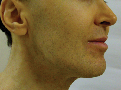
A 50-year-old man presented with yellow, pruritic, xerotic skin and lethargy. The patient also reported nasal congestion and sneezing, especially when eating peanuts. He was fearful of allergic reactions and restricted his diet to “safe foods” such as squash, green beans, and sweet potatoes. On examination the patient had marked generalized yellow discoloration of the skin with pale mucous membranes, nonicteric sclerae, infraocular violaceous and hyperpigmented skin (allergic shiners), and Dennie-Morgan folds.

Vascular Issues - Leadership: Getting buy-in through the art of persuasion
Scenario: The Chief Executive Officer (CEO) of a large hospital comes into the Chief Medical Officer's (CMO) office after a hospital board retreat meeting on strategy for faster physician integration. His marching orders include allowing daytime coverage but restricting after-hours house staff coverage only for patients of physicians who are fully employed by the health system. The CEO knows this will create an uproar but wants the CMO to get "buy-in" from all physicians on the medical staff. He shares the 5-year financial projections and insists that viability of the institution is at stake. The relatively new CMO not only realizes the challenge represented but also recognizes a great personal opportunity. From her efforts in the past 2 years, she has garnered a reputation for honesty and integrity. Relying on her education in leadership, communications, and negotiations, she formulates a strategy to achieve the CEO's directive.
Problem: "Getting buy-in" has become an unwelcome phrase for many physicians. The general implication is that hospital administration wants salaried physicians or other leaders to persuade the "troops" to agree to something for which a decision has already been made. Persuading others is often an art and not a show of strength, and furthermore, not always rational. The art involves putting yourself in the other person's shoes and asking yourself: "Would I buy this if the idea was offered to me?" If not, leaders first need to convince themselves of the benefits of the intended proposal. If it is always about you and your victories, you will acquire a reputation of being a self-serving leader and lose followers when you need them. Once a leader has convinced himself/herself, pushing hard against a timeline does not always work. Sometimes, being too aggressive can have the opposite reaction.
Solution: Physicians are scientists and, therefore, are influenced by data and empiric arguments. Most proposals advanced by hospitals involve finances to some degree. The problem is that most physicians do not have expertise in analyzing financial data. Thus, physician leaders must make the facts easily understandable and not use book-based accounting terms in attempts to impress physician groups. This may result in a reinforcement of the bias that everyone in administration is concerned only with money. Leaders must make the financial application easy to understand, possibly with charts or graphs, and seek to educate in the process.
Even though the proposal may be factual and well reasoned, the medical staff sometimes has not had time to process the pros and cons or separate their own parochial interests from those of the institution. Persuasion does indeed involve emotion at times but must be used wisely and at the right time when reason and rational arguments have hit home first. That is not to say that everyone in the audience will be convinced of the brilliance of your argument but it may persuade enough people to come over to your side. When emotion is used and timed correctly, it creates excitement and tends to spread through the audience so it completes the "sale."
If there is not enough support, it is best to let things rest and work on those who are opposed to the proposal as a knee-jerk response or based upon misperception. The leader needs to have established a reputation for honesty, integrity, and listening. A physician leader speaking on behalf of the institution may have already been deemed as having gone over to the "dark side" of administration and thus, sacrificed integrity. A successful physician leader will discuss the downsides of a proposal as well as inherent risks of the endeavor. If this is done, the opposition tends to then start looking at the positives you have presented.
Conclusion: Although presenting data to physicians is vital to getting their "buy-in" for an idea or proposal, the physician leader must build a reputation of honesty, integrity, and being a good listener. Convincing physician audiences almost always takes more time than hospital administrators think it should. Facts help to put the idea on a sound footing as well as a clear and rational explanation brings attention to the proposal. But genuine emotion displayed at the right time creates excitement and can lead to closing the deal.
PVSS is now VESS -The Vascular and Endovascular Surgery Society
Nearing 40 years of continued service to its membership, the Peripheral Vascular Surgery Society at its annual meeting elected a change in the society name to reflect the modern practice of its 1,000+ active and senior members. Originally founded in 1976, the society was designated as the Peripheral Vascular Surgery Club and later on invoked the PVSS. Now outdated, "peripheral" was once used to differentiate the emerging field of vascular surgery from "central" cardiovascular surgery. As evidenced by hundreds of VESS registrants at the most recent meeting and plenary sessions in Steamboat Springs, January 29–February 2, the membership has actively engaged and researched projects and outcomes across the spectrum of all vascular territories using the best of modern vascular and endovascular techniques.
Indeed, the VESS reflects our appropriate American Board Certifications, many division and practice names, as well as common journals in which the membership are often published and contribute. General knowledge of the role of vascular surgery in public health is increasing through a variety of mechanisms, and the importance of the marriage of endovascular surgery to traditional perceptions of vascular surgery has both timely and strategic implications for VESS going forward. The process of VESS name was researched since 2008 and carried forth with support of the past 26 PVSS presidents. An opinion poll was taken from the membership, and among respondents, the Vascular and Endovascular Surgery Society was the leading choice by an overwhelming margin. At our recent winter meeting, the membership voted overwhelmingly for the VESS moniker.
Our academic program at the winter meeting was very successful this year with 50 papers presented from institutions across the country and abroad. The topics discussed covered the entire scope of vascular and endovascular surgery including readmission rates after abdominal aortic aneurysm repair, hospital length of stay after carotid endarterectomy as a surrogate for quality, and methods for maintenance of end-stage dialysis access. The program also included case presentations that described new techniques for visceral vessel debranching and the management of large acquired arteriovenous fistulas. The plenary sessions promoted discussion and gave a forum for fellows and residents to present their work.
Building on the enthusiasm of a great winter program, the VESS inauguration has met great support, as Past PVSS President Chuck Anderson, M.D., commented, "Our specialty has evolved dramatically since the early meetings of the Peripheral Vascular Surgery Club. The original goal was to recognize the group of "new" surgeons that were fellowship trained in a "new" specialty and provide a forum for continued education and advocacy. The new name more appropriately reflects the evolution of the specialty and our mission remains the same." Jeb Hallett, M.D., a past PVSS president, echoes the sentiment: "Keep your focus on our mission for young surgeons!"
Without a doubt, VESS will continue in its mission to provide a forum for development of the young vascular and endovascular surgeon, with continued support for its spring meeting in conjunction with the Vascular Annual Meeting. The VESS Winter Meeting will remain the major venue for all its members with novel programs for residents and fellows, an exciting slate of plenary sessions and interchange, inspirational presidential addresses, and the unique and entertaining venues for its annual celebratory dinner.
James H. Black III, M.D., VESS Councilor-at-Large
Answering subpoenas poses legal, privacy risks for doctors
Receiving a subpoena for information or testimony can make any physician feel nervous and apprehensive. The official-looking notices often arrive as certified mail, have a stamped deadline, and include an attorney’s signature.
But immediately fulfilling a subpoena without first accessing the request could land doctors in legal trouble.
"Understandably, many physicians don’t recognize that no judge has reviewed the propriety of a subpoena," said Michael J. Schoppmann, a New York–based health law attorney and managing partner for a health care litigation firm. "Most judges are not even aware of the subpoena. There’s an assumption by the physician that it has been vetted by the court and, in many cases, that is not true. With good intentions, we find physicians getting into trouble out of a desire to be compliant."
Before answering a subpoena, physicians should evaluate where the request originated, the status of the case, and who is involved, legal experts say. Frequently, a request for patient information or records is objected to by the patient’s attorney, said Michael J. Sacopulos, a medical liability defense attorney and founder of Medical Risk Institute (MRI) in Terre Haute, Ind. Physicians should ensure record requests are authorized by the patient or are bidden through a court order.
"Let’s say you’re not going to court, but they want information," Mr. Sacopulos said. "You have HIPAA issues at play. You still have an independent duty to safeguard patient information."
Mr. Schoppmann recommends that physicians contact the judge affiliated with the case and request guidance about how to proceed with a subpoena. The judge may deny the request because the records are inappropriate or allow the subpoena to proceed.
"It’s great protection because if we get an order from the court, that (shields the doctor) from the litigants later saying, ‘We did not want you to release those records.’" Mr. Schoppmann said.
In other instances, physicians may be subpoenaed to discuss a patient’s condition or treatment in a third-party case. For example, a patient who is suing an insurance carrier or third-party over a vehicle accident. Doctors have a responsibility to offer honest and unbiased testimony about their patient’s medical condition, said Steven Fitzer, a medical malpractice defense attorney in Tacoma, Wash. and former chair of the Washington State Bar Association Litigation Section. However, physicians should steer clear of providing opinions on medical matters unrelated to their expertise.
"A family practice doctor should offer thoughts and opinions and facts that are within his or her specialty," Mr. Fitzer said. "If (the doctor) regularly treats people who have whiplash and cervical strain, you can talk about that. But the family practice doctor shouldn’t be talking about neck surgery unless you do neck surgeries."
Physicians should also consider and address how their involvement with a case may affect their relationship with patients, Mr. Fitzer adds. Often, patients view their doctor as their supporter and expect their alliance during in a third-party lawsuit.
"Patients sometimes misunderstand that the role of the doctor in a deposition is to call it the way he or she sees it, not to be an advocate for the patient," he said. Doctors should "communicate that to the patient" beforehand.

Be wary of subpoenas for information or records that stem from a malpractice case in which the doctor is a named defendant, Mr. Sacopulos notes. In such instances, a plaintiff’s attorney may request details about a patient visit or ask about a doctor’s role in treating the patient within the continuum of care. But the attorney could really be fishing for more physicians to sue.
"They may have sued one or two physicians, but the statute of limitations has yet to run out," Mr. Sacopulos said. "They’ll sue one or two, then depose others to see if they can’t get some finger pointing and increase the litigation."
In addition, plaintiffs’ attorneys may hope that the subpoenaed physician makes statements to further enhance their case against another doctor, without hiring them as an expert witness.
"My advice would be if you are requested to offer testimony in a case that you are not a party to, you immediately contact your liability carrier," Mr. Sacopulos said. "Your medical malpractice carrier will most often provide an attorney for a deposition, even though you’re not a party."
Physicians should not let attorneys intimidate them with subpoenas or excessive demands for information. Some lawyers request thousands of pages of documents in a short timeframe and pressure the physician to comply, Mr. Schoppmann said. Other attorneys try to convince physicians to speak with them over the phone about the request and to provide patient information verbally.
Ensure that the scope, the timing, and the overall content of the request are appropriate before responding, Mr. Schoppmann stresses.
"The subpoena has to be reasonable," he said. "Step back and do not respond unduly aggressively. You could be subject to criticism later if you respond to an inappropriate subpoena. We want right, rather than quick."
Receiving a subpoena for information or testimony can make any physician feel nervous and apprehensive. The official-looking notices often arrive as certified mail, have a stamped deadline, and include an attorney’s signature.
But immediately fulfilling a subpoena without first accessing the request could land doctors in legal trouble.
"Understandably, many physicians don’t recognize that no judge has reviewed the propriety of a subpoena," said Michael J. Schoppmann, a New York–based health law attorney and managing partner for a health care litigation firm. "Most judges are not even aware of the subpoena. There’s an assumption by the physician that it has been vetted by the court and, in many cases, that is not true. With good intentions, we find physicians getting into trouble out of a desire to be compliant."
Before answering a subpoena, physicians should evaluate where the request originated, the status of the case, and who is involved, legal experts say. Frequently, a request for patient information or records is objected to by the patient’s attorney, said Michael J. Sacopulos, a medical liability defense attorney and founder of Medical Risk Institute (MRI) in Terre Haute, Ind. Physicians should ensure record requests are authorized by the patient or are bidden through a court order.
"Let’s say you’re not going to court, but they want information," Mr. Sacopulos said. "You have HIPAA issues at play. You still have an independent duty to safeguard patient information."
Mr. Schoppmann recommends that physicians contact the judge affiliated with the case and request guidance about how to proceed with a subpoena. The judge may deny the request because the records are inappropriate or allow the subpoena to proceed.
"It’s great protection because if we get an order from the court, that (shields the doctor) from the litigants later saying, ‘We did not want you to release those records.’" Mr. Schoppmann said.
In other instances, physicians may be subpoenaed to discuss a patient’s condition or treatment in a third-party case. For example, a patient who is suing an insurance carrier or third-party over a vehicle accident. Doctors have a responsibility to offer honest and unbiased testimony about their patient’s medical condition, said Steven Fitzer, a medical malpractice defense attorney in Tacoma, Wash. and former chair of the Washington State Bar Association Litigation Section. However, physicians should steer clear of providing opinions on medical matters unrelated to their expertise.
"A family practice doctor should offer thoughts and opinions and facts that are within his or her specialty," Mr. Fitzer said. "If (the doctor) regularly treats people who have whiplash and cervical strain, you can talk about that. But the family practice doctor shouldn’t be talking about neck surgery unless you do neck surgeries."
Physicians should also consider and address how their involvement with a case may affect their relationship with patients, Mr. Fitzer adds. Often, patients view their doctor as their supporter and expect their alliance during in a third-party lawsuit.
"Patients sometimes misunderstand that the role of the doctor in a deposition is to call it the way he or she sees it, not to be an advocate for the patient," he said. Doctors should "communicate that to the patient" beforehand.
Be wary of subpoenas for information or records that stem from a malpractice case in which the doctor is a named defendant, Mr. Sacopulos notes. In such instances, a plaintiff’s attorney may request details about a patient visit or ask about a doctor’s role in treating the patient within the continuum of care. But the attorney could really be fishing for more physicians to sue.
"They may have sued one or two physicians, but the statute of limitations has yet to run out," Mr. Sacopulos said. "They’ll sue one or two, then depose others to see if they can’t get some finger pointing and increase the litigation."
In addition, plaintiffs’ attorneys may hope that the subpoenaed physician makes statements to further enhance their case against another doctor, without hiring them as an expert witness.
"My advice would be if you are requested to offer testimony in a case that you are not a party to, you immediately contact your liability carrier," Mr. Sacopulos said. "Your medical malpractice carrier will most often provide an attorney for a deposition, even though you’re not a party."
Physicians should not let attorneys intimidate them with subpoenas or excessive demands for information. Some lawyers request thousands of pages of documents in a short timeframe and pressure the physician to comply, Mr. Schoppmann said. Other attorneys try to convince physicians to speak with them over the phone about the request and to provide patient information verbally.
Ensure that the scope, the timing, and the overall content of the request are appropriate before responding, Mr. Schoppmann stresses.
"The subpoena has to be reasonable," he said. "Step back and do not respond unduly aggressively. You could be subject to criticism later if you respond to an inappropriate subpoena. We want right, rather than quick."