User login
The official dermatologist [YOUR NAME HERE]
Who do you call when your windshield’s busted?
Call Giant Glass!
There isn’t a Boston Red Sox fan on the planet who can’t sing that annoying jingle in his or her sleep. This is because, as they never tire of reminding us, Giant Glass is the Official Windshield Replacer of the Boston Red Sox.
Why does a baseball team need an Official Windshield Replacer? The announcers like to say, "Hey, Joe, that homer went over the Green Monster right onto Yawkey Way – somebody’s gonna have to fix their windshield!"
If that answer satisfies you, you might ponder why EMC is the Official Data Storage company for the team. Or why Benjamin Moore is the Official Paint. Or why Poland Spring is the Official Water.
Or why Beth Israel Deaconess is the Red Sox Official Hospital.
You can see where I’m going with this, can’t you?
In our increasingly complex and competitive environment (EHRs! ACOs!), your columnist is always on the lookout for ways to help you to get a leg up on the competition. (Branding! Online reviews!)
I have therefore embarked on an ambitious effort to become Official Dermatologist to the Official Sponsors of the Boston Red Sox. Follow my example, Colleagues.
*******************
Marriott Hotels
Dear Mr. or Ms. Marriott:
I salute you as Official Hotel of the Red Sox!
But suppose one of your guests uses a hotel Jacuzzi and comes down with nasty Pseudomonas folliculitis. It happens. Who ya gonna call?
Call Rockoff Dermatology! We’ll do the job right, fix up your guests fast, and explain why even state-of-the-art hot tub disinfection sometimes fails. Once the pustules go away, your guests will happily come back to you.
Our rates are reasonable. Give us a call!
*******************
Dunkin’ Donuts
Dear Donuts:
It has come to our notice that you are the Official Coffee of the Boston Red Sox. Good for you!
I should mention that I really like your coffee, especially the Pumpkin Blend you make around Thanksgiving. You might wonder why you need an Official Dermatologist. Well, most of your fine coffee beverages come with milk – and dairy products have been implicated in acne. Of course, the evidence is a little thin, but if one of your customers has a latte and breaks out in major zits, don’t you want to send them to a skin doctor who cares not just about the pimples, but about your corporate image?
That would be me! Let’s get together over a cup of Seattle’s Best. (Just kidding!)
*******************
John Hancock Insurance
Dear Mr. Hancock,
Congratulations on being the Official Insurance of the Boston Red Sox.
I just love your building, a real Boston landmark.
Here’s why you need an Official Dermatologist: You sell insurance – and we dermatologists know insurance. Between updating coverage, scanning insurance cards, and checking online eligibility, our patients spend way more time registering than they do being examined. (Hey, we’re skin doctors – How long do you think that takes?)
While patients are filling out all our forms, we can show them a list of all your fine insurance products. Synergy! Win-win! For faster service, you could even put an agent in our waiting room.
Let’s do lunch. Do you like Dunkin’ Donuts?
*******************
You get the idea. Just pick a popular institution in your area – opera company, sports team, bowling alley – whatever image you have in mind. Then contact them about sponsorship opportunities. Be the first one to do it, and have your agent nail down an exclusive.
Here’s a sample letter:
Toledo Mud Hens
Toledo, Ohio
Dear Mud Hens,
I am writing to suggest you consider having us [INSERT NAME] as Official Dermatology and Aesthetic Rejuvenation Center of the Toledo Mud Hens Baseball Club. We already have a close affiliation with Downtown Latte on South St. Clair Street, and are the exclusive providers of skin care to their clients who get breakouts from dairy products added to their fine coffees.
Let’s all get together and triangulate.
Go Mud Hens!
*******************
OK, colleagues, I’ve given you direction. Now get out there and make it happen!
Dr. Rockoff practices dermatology in Brookline, Mass. He is on the clinical faculty at Tufts University, Boston, and has taught senior medical students and other trainees for 30 years. Dr. Rockoff has contributed to the Under My Skin column in Skin & Allergy News since 1997.
Rise in VKDB cases prompts call for a tracking system

Credit: Petr Kratochvil
Physicians at a Tennessee hospital have seen a rise in late-onset vitamin K deficiency bleeding (VKDB) in young infants, due to parents declining a vitamin K shot at birth.
Over a period of 8 months, 7 infants were diagnosed with vitamin K deficiency, and 5 of them had VKDB.
Four of the infants experienced intracranial hemorrhaging, and 2 required urgent neurosurgical intervention.
These cases were diagnosed at Monroe Carell Jr. Children’s Hospital at Vanderbilt in Nashville.
And they were described in Pediatric Neurology.
Now, the authors are calling for a state and national tracking system to help them determine how many infants are not receiving a vitamin K shot at birth.
“There is no national tracking of this in the US, unfortunately, and cases are rarely reported,” said Robert Sidonio Jr, MD, of Vanderbilt University School of Medicine.
“We are probably just seeing the tip of the iceberg, and I worry that people are missing these cases often and not considering this diagnosis when presented with a sick infant.”
He and his colleagues are also calling for better education on this issue for healthcare providers and families. The group believes misinformation—that the shot causes leukemia, is a toxin, or is unnecessary in uncomplicated births—may be leading some families to decline the shot.
The American Academy of Pediatrics has recommended the single-dose shot of vitamin K at birth since 1961. Most cases of VKDB seen today occur in infants in the first 6 months of life who did not get the shot and are exclusively breastfed or who have an undiagnosed liver disorder.
Following a recent rise in cases in Tennessee, the US Centers for Disease Control and Prevention discovered that 28% (61/218) of parents of children born at private birthing centers in the state declined the shot.
Mark and Melissa Knotowicz declined the shot for their twins, Silas and Abel, following their birth last July at a Nashville hospital.
“From the information we had, we heard the main side effect was a preservative in the shot that could lead to childhood leukemia,” said Mark Knotowicz. “We thought, ‘We don’t want our kids to have childhood leukemia,’ so we declined it without really hearing any of the benefits.”
At about 6 weeks old, Silas was noticeably fussy. He vomited overnight, woke up extremely pale, and wouldn’t nurse.
The twins had a checkup scheduled that day. The pediatrician suspected sepsis and told the Knotowiczes to take Silas to the Emergency Department at Monroe Carell Jr. Children’s Hospital.
Because of a rash of VKBD cases, the staff there had received additional education. After Silas’s parents confirmed that he had not received the vitamin K shot, CT scans and blood work revealed he had suffered multiple brain bleeds.
Silas received a double dose of vitamin K to get the bleeding under control. His twin, Abel, was diagnosed with asymptomatic vitamin K deficiency and received the shot.
Silas spent a week in the hospital. Today, he undergoes physical therapy for neuromuscular development issues. Any effects on cognitive development aren’t yet known.
“The twins’ cases highlight our inability to determine which infants will go on to develop vitamin K deficiency bleeding, as they both had prolonged bleeding times at presentation,” Dr Sidonio said.
Mark Knotowicz wishes medical staff at the birthing hospital had more clearly defined the risks of declining the shot.
“Why didn’t they say, ‘There have been 4 other cases in Nashville, and we’re trying to prevent a lethal brain hemorrhage in your child?’”
Anna Morad, MD, of the Vanderbilt University School of Medicine, has worked to raise awareness about the vitamin K shot and the risk of VKDB, with some success.
“After our educational outreach, we have seen a decrease in our refusal rates [with about 3.4% of parents declining vitamin K],” she said. “Our goal is to fully educate parents on the risk of declining so they can make an informed choice for their baby.” ![]()
INR test strips linked to bleeding, deaths

Alere Inc., has initiated a Class 1 recall in the US of the Alere INRatio2 PT/INR Professional Test Strips (PN 99008G2).
The strips are used by healthcare professionals to determine the international normalized ratio (INR) in fresh capillary whole blood to monitor the effect of warfarin on clotting time.
Alere has received reports of patients who had a therapeutic or near therapeutic INR with the test strips but a significantly higher INR in tests performed by a central lab.
This error has been linked to 9 reports of serious adverse events, 3 of which described bleeding associated with patient deaths.
So the company has issued a recall of the Alere INRatio2 PT/INR Professional Test Strips (PN 99008G2). This recall does not include the Alere INRatio PT/INR Test Strip (PN 100071), which is used by patients for home INR monitoring.
Alere has not determined the root cause of the error but is concerned that the test strips may continue to report inaccurately low INR results. In the reports, the test strip results were between 3.1 and 12.2 INR units lower than the lab results.
Customers should stop using the Alere INRatio2 PT/INR Professional Test Strips immediately and return unused product to the company.
Alere will transition customers from the current Alere INRatio2 PT/INR Professional Test Strip to the Alere INRatio PT/INR Test Strip (PN 100139).
Alere said it has reported this issue to the US Food and Drug Administration and is conducting a thorough investigation into the adverse events.
Customers with questions about this recall and those who require replacement product can contact Alere at 844-292-5373. For additional information on the recall, visit www.inr-care.com.
Any adverse events or quality problems related to use of the test strips can be reported to the FDA’s MedWatch Adverse Event Reporting Program. ![]()
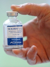
Eculizumab gets full FDA approval for aHUS
Credit: Globovision
The US Food and Drug Administration (FDA) has granted full approval for eculizumab (Soliris) to treat adult and pediatric patients with atypical
hemolytic uremic syndrome (aHUS).
The drug received accelerated approval for this indication in 2011.
Now, eculizumab has received full FDA approval based on the fulfillment of post-marketing requirements, including the submission of data from 2 additional prospective trials of eculizumab in patients with aHUS.
The revised eculizumab label now includes results with 2 years of ongoing treatment in aHUS patients and data on the use of eculizumab prior to supportive care with plasma or plasma exchange in prospective clinical trials.
The drug’s label also includes a boxed warning informing readers that life-threatening and fatal meningococcal infections have occurred in patients treated with eculizumab.
About aHUS and eculizumab
aHUS is a chronic, life-threatening disease in which a genetic deficiency in one or more complement regulatory genes causes chronic, uncontrolled complement activation. This results in complement-mediated thrombotic microangiopathy (TMA), the formation of blood clots in small blood vessels throughout the body.
Permanent, uncontrolled complement activation in aHUS causes a life-long risk for TMA, which leads to sudden and life-threatening damage to the kidney, brain, heart, and other vital organs, as well as premature death. Complement-mediated TMA also causes thrombocytopenia and hemolysis.
Eculizumab is a first-in-class terminal complement inhibitor indicated to inhibit complement-mediated TMA. The drug’s effectiveness in aHUS is based on its effects on TMA and renal function.
Eculizumab received accelerated FDA approval to treat aHUS in September 2011. The FDA granted this approval based on the results of 2 trials that suggested the drug was likely to provide a clinical benefit.
To achieve traditional FDA approval, the drug’s developer, Alexion Pharmaceuticals, was required to submit additional data that confirm the drug provides a clinical benefit.
To that end, the Clinical Studies section (Section 14.2) of the revised eculizumab prescribing information now contains results from 4 prospective, single-arm studies in patients with aHUS.
This includes updated data from the first 2 trials, as well as data from 2 new trials, 1 in pediatric patients with aHUS and the other in adolescents and adults with aHUS.
For details on these trials, see the full prescribing information. ![]()
Artificial bone marrow seems just like the real thing

Credit: James Weaver,
Harvard’s Wyss Institute
Researchers have created a device that reproduces the structure, function, and cellular make-up of bone marrow, according to a paper published
in Nature Methods.
The device, dubbed “bone marrow on a chip,” could serve as a new tool for testing the effects of radiation and other toxic agents on whole bone marrow.
The researchers believe this bone marrow on a chip could provide an alternative to animal testing, although the device itself was generated in mice.
The team also thinks that, in the future, the engineered bone marrow could be used to maintain a cancer patient’s own marrow temporarily during radiation or high-dose chemotherapy.
In an initial test, the engineered bone marrow withered in response to radiation, just as natural bone marrow does. And, as in natural marrow, granulocyte colony-stimulating factor conferred a protective effect on the engineered marrow.
“Bone marrow is an incredibly complex organ that is responsible for producing all of the blood cell types in our body, and our bone marrow chips are able to recapitulate this complexity in its entirety and maintain it in a functional form in vitro,” said study author Donald Ingber, MD, PhD, of the Wyss Institute for Biologically Inspired Engineering at Harvard University in Boston.
Dr Ingber leads an effort to develop human organs on chips—small microfluidic devices that mimic the physiology of living organs. To build these devices, researchers combine multiple types of cells from an organ on a plastic microfluidic device, while steadily supplying nutrients, removing waste, and applying mechanical forces the tissues would face in the body.
But bone marrow is so complex that Dr Ingber and his colleagues needed a new approach to mimic organ function. Rather than trying to reproduce such a complex structure cell by cell, the team used mice.
Specifically, the researchers packed dried bone powder into an open, ring-shaped mold the size of a coin battery and implanted the mold under the skin on the animal’s back.
After 8 weeks, the team surgically removed the disk-shaped bone that had formed in the mold and examined it with a specialized CAT scanner. The scan showed a honeycomb-like structure that looked identical to natural trabecular bone.
The marrow looked like the real thing as well. When the researchers stained the tissue and examined it under a microscope, the marrow was packed with blood cells, just like marrow from a living mouse.
And when the team sorted the bone marrow cells by type and tallied their numbers, the mix of different types of blood and immune cells in the engineered bone marrow was identical to that in a mouse thighbone.
To sustain the engineered bone marrow outside of a living animal, the researchers surgically removed the engineered bone from mice, then placed it in a microfluidic device that mimics the circulation the tissue would experience in the body.
Marrow in the device remained healthy for up to 1 week. This is typically long enough to test the toxicity and effectiveness of a new drug, the team said.
The device also passed an initial test of its drug-testing capabilities. Like marrow from live mice, this engineered marrow was susceptible to radiation, but granulocyte colony-stimulating factor conferred a protective effect.
The researchers believe that, in the future, they could potentially grow human bone marrow in immune-deficient mice. And their bone marrow on a chip could generate blood cells, which could circulate in an artificial circulatory system to supply a network of other organs on chips. ![]()
The ICU: From bed to bedside
I recently came to the realization that one doesn’t usually end up in an ICU unless the odds of making it out are not in one’s favor.
Now, I want to make it clear from the very first that my wife and I survived life-threatening medical experiences as a result of the superb care provided to both of us. Nevertheless, the experience made me aware of how ICU and hospital care has changed in the last 50 years. I have spent most of my life in ICUs from the "invention" of the Coronary Care Unit in the mid-1960s to its current iteration of an intensely monitored hospital room where emergency surgery could be performed if need be. Much of that change is a result of the variety of medical specialists who are players in the ICU drama. The other major changes have been the time restraints of house staff rotation to meet certification criteria and rotation of the senior staff in order to provide continuing on-site coverage of the ICU. As the acuteness of hospital admissions has increased, the ICU and its management have assumed a larger role in the care and the finances of major hospitals.
Some years ago, we hosted a distinguished European physician who spent 2 months with us as a visiting professor. It was at a time when we felt a need to begin to develop subspecialties in angiography, electrophysiology, and echocardiography in order to provide a research and training atmosphere for our fellowship program. Later, he wrote an editorial in his local medical journal criticizing cardiac care in the United States because of the lack of continuity. He was of the tradition that mandated that he would see the patient in the clinic, perform a cardiac catheterization himself, and follow his patient through surgery and manage their postoperative care, as was standard practice in the mid-20th century. He believed that the concept of delegating diagnostic and care responsibilities to specialty trained colleagues that he observed here was a major disaster. He should see the system now. Nevertheless, his plea for continuity in care resonates in my mind.
That need for continuity came back to me as I experienced the dizzying rotation of house staff and senior staff that takes place in the ICU today. Any semblance of continuity of care was lacking at a time when there was a need to provide information to anxious patients and their families. In the environment of medical uncertainty, when you would like to find a familiar physician to ask "How are we doing," the attending physician or medical resident in charge was either on another rotation or being covered by a colleague. No training or adherence to "sign-off" rounds can replace the need for that professional continuity. As competent and well meaning the covering doctor was, answers to questions seemed shallow. It was difficult even to express gratitude to "a" physician who had tipped the scales in my favor. One had to direct it to an amorphous team of doctors, nurses, and technicians who had participated in care. That is a reality that describes the methodology of ICU and its success. It is a reality that to a similar degree characterizes the current management of inpatient care.
It seemed that in the setting of a life-threatening experience, the link between the treating physicians and the patients or family has almost disappeared in the ICU. The challenge to us as we play our role in the ICU, and the CCU, is to establish and maintain a personal relationship with the patients and their family.
Dr. Goldstein, medical editor of Cardiology News, is professor of medicine at Wayne State University and division head emeritus of cardiovascular medicine at Henry Ford Hospital, both in Detroit. He is on data safety monitoring committees for the National Institutes of Health and several pharmaceutical companies.
Team describes protein’s antimyeloma effects

Researchers say they’ve discovered how a lack of the protein adiponectin promotes disease progression in patients with multiple myeloma (MM).
Obesity is known to increase the risk of developing MM, and obese individuals have lower-than-normal levels of adiponectin.
So the researchers set out to characterize the relationship between adiponectin and MM.
They found evidence to suggest the protein induces apoptosis in MM cells by suppressing lipogenesis. And this points to new treatment avenues for MM.
Edward Medina, MD, PhD, of University of Texas Health Science Center at San Antonio, and his colleagues described this research in Leukemia.
The team noted that adiponectin plays several roles in preserving health, including killing cancer cells. And, as levels of adiponectin are reduced in obese individuals, the protein has been implicated in MM progression.
Furthermore, adipocytes in obese individuals produce more fatty acids, and it’s likely that MM cells feed on these fatty acids.
“Synthesizing fatty acids is important for myeloma cells to build vital structures, including cell membranes, that enable them to keep on growing,” Dr Medina said.
With this in mind, he and his colleagues set out to determine how prolonged exposure to adiponectin affects MM cell survival and to describe exactly how the protein works.
Their experiments revealed that adiponectin activates protein kinase A (PKA). This leads to a decrease in AKT activity and an increase in AMP-activated protein kinase (AMPK) activation. Then, AMPK induces cell-cycle arrest and apoptosis.
The researchers said this apoptosis may be mediated, at least partly, by a decline in the expression of acetyl-CoA-carboxylase (ACC), which is essential to lipogenesis.
So the team introduced palmitic acid, the preliminary end product of fatty acid synthesis, to the MM cells and found it prevented adiponectin-induced apoptosis.
In addition, the ACC inhibitor 5-(tetradecyloxy)-2-furancarboxylic acid had an antiproliferative effect on MM cells. But an increase in fatty acids inhibited that effect.
The researchers said this new understanding of the pathways adiponectin uses to kill MM cells might lead to the development of drugs that would function in the same way.
“If we could pharmacologically suppress these fatty acid levels in obese myeloma patients, we could boost the effects of the chemotherapy that targets PKA or fatty acid synthesis and potentially decrease the chemotherapeutic dose,” Dr Medina said. “Also, it would give your own body’s protective measures more of a chance to work against the cancer.” ![]()
Portable spectrometers can detect malaria early, group says

Credit: St Jude Children’s
Research Hospital
An infrared spectroscopy technique can detect malaria parasites at early stages of development, according to preclinical research published in Analytical Chemistry.
A group of biochemists found this method could detect Plasmodium falciparum in red blood cells by picking up on a fatty acid signature.
This allowed the researchers to identify and quantify parasites at various stages of development, including the ring and gametocyte stages.
The team also pointed out that the spectrometer they used is portable, inexpensive, and does not require highly trained staff for operation. It could therefore prove useful in the field.
“Current tests for malaria suffer from serious limitations,” said study author Bayden Wood, PhD, of Monash University in Victoria, Australia.
“Many are expensive [and] require specialist instruments and highly trained staff to judge whether blood samples contain the parasite. What’s been holding us back is the lack of an accurate and inexpensive test to detect malaria early and stop it in its tracks. We believe we’ve found it.”
Dr Wood and his colleagues already knew that fatty acids were a marker for malaria from previous studies conducted at the Australian Synchrotron. The Synchrotron allowed the team to see the different life stages of the parasite and the variation in its fatty acids.
The researchers thought they might be able to use this information for diagnosis, but they needed a more portable detection method.
So they decided to test whether attenuated total reflectance Fourier transform infrared spectroscopy (ATR-FT-IR) could detect the fatty acid signature. The technique utilizes infrared light to pick up on the vibrations of molecules.
The researchers spiked red blood cells with parasites of different numbers and life stages and observed them using ATR-FT-IR.
Dr Wood said the method produced results within minutes. And it gave a clear indication of malaria at a much earlier stage of infection than current tests on the market—at the ring and gametocyte stages.
The absolute detection limit was 0.00001% parasitemia (<1 parasite/μL of blood) for cultured early ring-stage parasites in a suspension of normal red blood cells.
“Now that we can detect the early stages of a parasite’s life in the bloodstream, the disease will be much easier to test and treat,” Dr Wood said. “The big advantage of our test is that it doesn’t need scientists and expensive equipment. This has the potential to dramatically reduce the number of people dying from this disease in remote communities.”
The method also shows the potential to detect a number of other blood-borne diseases, according to the researchers. Dr Wood and his colleagues are now planning a clinical trial of ATR-FT-IR in Thailand. ![]()
Hospira issues Class I recall of infusion pumps
Credit: Daniel Gay
Hospira, Inc., has issued a Class I recall of Abbott Acclaim infusion pumps and Hospira Acclaim Encore infusion pumps, after receiving reports of broken door assemblies on these products.
If a door assembly breaks, the door may not close properly and an over-infusion or a delay of therapy may occur.
If the door cannot be closed, the pump cannot be used, and this can result in a delay of therapy.
Use of these products may cause serious adverse events, including death.
These pumps are used to deliver hydration fluids, drugs, blood and blood fractions, intravenous nutritionals, and enteral nutritionals.
The affected Abbott Acclaim Infusion Pumps, list Number 12032, were manufactured from February 1998 to November 1998 and distributed from September 1998 through February 2004.
The affected Hospira Acclaim Encore infusion pumps, list Number 12237, were manufactured from February 1997 to February 2010 and distributed from July 1999 through November 2013.
Hospira is recommending that users inspect each Hospira/Abbott Acclaim Encore infusion pump for door handle cracks prior to programming a therapy, by taking the following steps:
1. After inserting the tubing (with the roller clamp closed) and closing the door handle against the infusion pump, check that the door is fully closed.
If a pump has a door that does not close properly and a gap or separation exists between the completely closed door and the pump itself, remove the pump from clinical service, and call Hospira at 1-800-441-4100 (M-F, 8am-5pm, CT). If the door closes correctly, proceed to Step 2.
2. If the door closes correctly and a gap or separation does not exist between the completely closed door and the pump itself, check that there is no free flow activity in the drip chamber of the administration set by opening the roller clamp.
If free flow is detected, close the roller clamp, remove the pump from clinical service, and call Hospira at 1-800-441-4100 (M-F, 8am-5pm, CT).
3. If no issues are found through steps 1 and 2, the pump is acceptable for use. However, healthcare professionals should still ensure that anyone in their facility who might use these products is made aware of this safety notification and the recommended actions.
Healthcare professionals and patients can report adverse events or side effects related to the use of these products to the FDA’s MedWatch Program. ![]()
Credit: Daniel Gay
Hospira, Inc., has issued a Class I recall of Abbott Acclaim infusion pumps and Hospira Acclaim Encore infusion pumps, after receiving reports of broken door assemblies on these products.
If a door assembly breaks, the door may not close properly and an over-infusion or a delay of therapy may occur.
If the door cannot be closed, the pump cannot be used, and this can result in a delay of therapy.
Use of these products may cause serious adverse events, including death.
These pumps are used to deliver hydration fluids, drugs, blood and blood fractions, intravenous nutritionals, and enteral nutritionals.
The affected Abbott Acclaim Infusion Pumps, list Number 12032, were manufactured from February 1998 to November 1998 and distributed from September 1998 through February 2004.
The affected Hospira Acclaim Encore infusion pumps, list Number 12237, were manufactured from February 1997 to February 2010 and distributed from July 1999 through November 2013.
Hospira is recommending that users inspect each Hospira/Abbott Acclaim Encore infusion pump for door handle cracks prior to programming a therapy, by taking the following steps:
1. After inserting the tubing (with the roller clamp closed) and closing the door handle against the infusion pump, check that the door is fully closed.
If a pump has a door that does not close properly and a gap or separation exists between the completely closed door and the pump itself, remove the pump from clinical service, and call Hospira at 1-800-441-4100 (M-F, 8am-5pm, CT). If the door closes correctly, proceed to Step 2.
2. If the door closes correctly and a gap or separation does not exist between the completely closed door and the pump itself, check that there is no free flow activity in the drip chamber of the administration set by opening the roller clamp.
If free flow is detected, close the roller clamp, remove the pump from clinical service, and call Hospira at 1-800-441-4100 (M-F, 8am-5pm, CT).
3. If no issues are found through steps 1 and 2, the pump is acceptable for use. However, healthcare professionals should still ensure that anyone in their facility who might use these products is made aware of this safety notification and the recommended actions.
Healthcare professionals and patients can report adverse events or side effects related to the use of these products to the FDA’s MedWatch Program.
FDA approves CML drug for home administration

The US Food and Drug Administration (FDA) has expanded the approval of omacetaxine mepesuccinate (Synribo) to include home administration.
The drug is already FDA-approved to treat adults with chronic or accelerated phase chronic myeloid leukemia (CML) who do not respond to or cannot tolerate 2 or more tyrosine kinase inhibitors.
The new approval allows CML patients to self-administer subcutaneous injections of omacetaxine mepesuccinate at home.
“It had been necessary for adults living with chronic or accelerated phase CML who are prescribed Synribo to travel to their doctor’s office twice a day for 2 weeks, which can be extremely burdensome and inconvenient to both patients and their caregivers,” said Meir Wetzler, MD, FACP, Chief of the Leukemia Section at Roswell Park Cancer Institute in Buffalo, New York.
“Now, physicians can decide if their patients are candidates for self-administration and, if so, provide their patients with guidance on how to properly administer reconstituted Synribo in the home.”
The drug’s maker, Teva Pharmaceutical Industries, Ltd., is working to finalize a pharmacy support program that will help facilitate successful home administration of omacetaxine mepesuccinate. The program is expected to “go live” this month or next.
About omacetaxine mepesuccinate
Omacetaxine mepesuccinate is a protein synthesis inhibitor. Although the drug’s mechanism of action is not fully understood, it is known to prevent the production of Bcr-Abl and Mcl-1, which help drive CML.
In October 2012, the FDA granted omacetaxine mepesuccinate accelerated approval for the treatment of adult patients with chronic or accelerated phase CML with resistance and/or intolerance to 2 or more tyrosine kinase inhibitors. Omacetaxine mepesuccinate gained full FDA approval in February.
The drug has been associated with severe and fatal myelosuppression, including thrombocytopenia, neutropenia, and anemia in some patients. So healthcare professionals should monitor patients’ complete blood counts weekly during induction and initial maintenance cycles and every 2 weeks during later maintenance cycles, as clinically indicated.
Omacetaxine mepesuccinate has been known to cause severe thrombocytopenia, which increases the risk of hemorrhage. Fatalities from cerebral hemorrhage have occurred. And severe, non-fatal gastrointestinal hemorrhages have occurred.
So healthcare professionals should monitor platelet counts as part of the complete blood count as recommended. Patients should not receive anticoagulants, aspirin, or non-steroidal anti-inflammatory drugs when their platelet counts are <50,000/μL, as these drugs may increase the risk of bleeding.
Omacetaxine mepesuccinate can induce glucose intolerance as well. So healthcare professionals should monitor blood glucose levels frequently, especially in patients with diabetes or risk factors for diabetes. Patients with poorly controlled diabetes mellitus should not receive omacetaxine mepesuccinate until good glycemic control has been established.
Omacetaxine mepesuccinate can cause fetal harm when administered to a pregnant woman. So women should be advised to avoid becoming pregnant while using the drug.
For more details on omacetaxine mepesuccinate, see the full prescribing information.