User login
Gordon Guyatt, MD: The Guru of Evidence-Based Medicine
Hospitalists Get Lessons in Quality Improvement Techniques
Tanya Boldenow, MD, a hospitalist at St. Joseph Mercy Hospital in Ann Arbor, Mich., attended yesterday’s HM13 Quality Improvement pre-course because she had been inspired by SHM’s participation in the ABIM Foundation’s Choosing Wisely campaign that identifies common treatments worth questioning by physicians and patients.
“I feel that I got some quality training in residency and have some support for it at the hospital, but for taking that next step to actually implement a project, it was important for me to have additional training and tools,” Dr. Boldenow said. SHM’s five recommended treatments to question “were things that I had a sense we ought to be looking at,” she added, but they weren’t being actively pursued at her hospital.
The full-day pre-course offered a review of QI principles and techniques. Participants then planned how concepts could be applied to a project targeting one of the SHM recommendations.
Dr. Boldenow chose the practice of avoiding or removing unnecessary urinary catheters, with a focus on preventing catheter-related urinary tract infections. Her group discussed baseline data to collect and analyze, where and how to pilot an initiative in the hospital, how to mobilize electronic health records, and what might persuade other professionals to change their habits.“It’s important to keep our eyes on the prize,” noted faculty member Ian Jenkins, MD, a hospitalist at the University of California at San Diego. “What are we trying to reduce; catheters or catheter-related complications?”
Dr. Boldenow sent an email to her department head a month ago proposing such a project. “I got funding from the residency program to come to the pre-course,” she said, “with the idea that I’d go back and make a presentation to our core faculty—and initiate a quality project.”
HM13 Session Analysis: Diagnostic Errors & Hospitalists: Why they happen and how to avoid them
Jennifer Myers, the patient safety officer at the Hospital of the University of Pennsylvania in Philadelphia, and James Reilly, assistant professor of clinical medicine, at UPenn, expertly facilitated a hands-on workshop exploring cognitive biases and common heuristics encountered in the daily practice of hospital medicine. Techniques to identify potential cognitive errors and de-biasing strategies were explored.
Specific methods included:
- Ishikawa diagrams (aka, cause-and-effect diagram, fish-bone diagram);
- Review of common heuristics; and
- Four double-check questions we can ask ourselves on every patient (Is this a case where I need to “slow down”?, What else could it be? What doesn’t fit? Could there be more than one thing going on?).
My favorite recommendation was: Practice “worst-case scenario” medicine. Consider the life threatening even if you don’t immediately test for it. TH
Dr. Lindsey is COO and strategist of Synergy Surgicalists, lead consultant of Asynd Consulting, and a Team Hospitalist member
HM13 Session Analysis: The Multicenter Medication Reconciliation Quality Improvement Study (MARQUIS)
The authors conducted a 6-site mentored implementation study to assess the effects of implementing a best practice med-rec toolkit on unintentional medication discrepancies.
The best practice toolkit included a number of interventions:
- Provider education (how to take a good history, how to get an accurate med lists);
- Patient education (teach-back method on medication education and how to keep their own medication list); and
- Risk stratifying patients regarding their need for additional resources (based on what medications they are currently prescribed and the total number of medications).
Stay tuned for the outcomes which are currently being collected. Click here for the toolkit resources.
HM13 Session Analysis: Strategies for Promoting Clinical Reasoning to Avoid Diagnostic Errors
Diagnostic reasoning is an essential skill for all physicians. There are multiple tools to refine this skill in physicians and to teach diagnostic reasoning to learners.
The session “Strategies for Promoting Clinical Reasoning to Avoid Diagnostic Errors” sought to review these skills in depth. According to Mary Ottolini, MD, of Children’s National Medical Center, the fundamentals of diagnostic reasoning are 1) Co-selection, in which 2-3 hypotheses or diagnoses are actively considered, 2) looking at the “big picture”, and 3) analyzing the information.
Looking at the big picture includes using appropriate adjectives to describe the patient and the illness presentation in medical and efficient terms. A well worded “one-liner” can frame the patient well for the team and for the next steps in diagnosis. Careful problem representation promotes thoughtful case presentations.
Analyzing the information includes comparing and contrasting key findings. Discriminating features should be discussed and competing evidence should be acknowledged.
Illness scripts is a method of looking at an illness in its entirety as a diagnosis is approached. The four parts of an illness script are mechanism of disease, epidemiology, clinical presentation (signs and symptoms), and time course.
Presentations can include diagnostic reasoning. The PBEAR format consists of:
- P- Problem Presentation
- BE – Background Evidence
- A- Analysis (including differential diagnoses)
- R- Recommendations (including goals and plan)
Key Takeaways:
- Diagnostic reasoning during case presentations is a valuable tool for patient care.
- Three fundamentals of diagnostic reasoning are 1) Co-selection of potential diagnoses, 2) looking at the “big picture”, and 3) analyzing the information.
- The PBEAR format (Problem Presentation, Background Evidence, Analysis , and Recommendations) can streamline presentations.
- Illness scripts (mechanism of disease, epidemiology, clinical presentation, and time course) are a helpful approach in diagnosis.
Dr. Hale is a pediatric hospitalist at the Floating Hospital for Children at Tufts Medical Center in Boston
HM13 Session Analysis: Overdoses and Other Taxing Toxicology
I attended a presentation titled, “Overdoses and Other Taxing Toxicology,” presented by Kennon Heard, MD, of the University of Colorado Department of Emergency Medicine and the Rocky Mountain Poison and Drug Center. He updated HM13 attendees on current overdose management in a rapid-fire, question-and-answer format.
Here is a summary of Dr. Heard’s key points:
- Modest QTc prolongation can be common after many overdoses, but most do not require prolonged cardiac monitoring. The key is to optimize electrolytes, such as magnesium, phosphorus, and potassium, and to treat other effects caused by the overdose, such as central nervous depression.
- Bath salts are not truly bath salts or plant food, but are actually substituted amphetamines. Treatment is mainly supportive, similar to methamphetamine overdose. Most patients do not require treatment, but providers may need to treat for agitation, seizures or rhabdomyolysis.
Key Takeaways
- Physicians need to remember to treat the symptoms, not necessarily the poisons. First, you may not always be able to accurately identify the poison--there can be both false positives and false negatives on toxicology screens. Second, co-ingestions are common and again may not be easily identified. Toxidromes are specific, but not sensitive.
- Current acetaminophen overdose management is to treat based on clinical endpoints, rather than on a time-based protocol. Treat until all acetaminophen has been metabolized and markers of liver injury, such as liver enzymes and coagulation factors, are improving. TH
Dr. O’Callaghan is pediatric hospitalist and clinical assistant professor of pediatrics at Seattle Children’s Hospital and the University of Washington School of Medicine. He also is a Team Hospitalist member.
Fecal microbiota transplant designated a biologic, requiring IND status
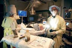
BETHESDA, MD. – The Food and Drug Administration has designated stool for transplant as a biologic drug, necessitating that any gastroenterologist performing fecal microbiota transplants obtain an Investigational New Drug permit.
By designating stool for transplant as a biologic drug, anyone who performs fecal microbiota transplants (FMTs) – whether they perform a single transplant for one patient or recruit dozens for a study – needs to have an IND permit.
The announcement was made at a 2-day public workshop convened by the National Institutes of Health and the Food and Drug Administration to sift through some of the evidence surrounding FMTs.
The intent of the IND requirement, said Dr. Jay Slater, director of FDA’s Division of Bacterial, Parasitic, and Allergenic Products, is not to stamp out the care that patients can now receive, but to make sure it’s safe, effective, and data driven.
"This is a low-tech procedure that already has a CPT code. A ‘how-to’ guide recently appeared online, and it walks you right through how to do the procedure. There are a very large number of people doing this off the grid. What we need are long-term controlled trials on this that will enhance our understanding of its safety and efficacy."
Although neither the biologic designation nor the IND requirement is brand new, they have not been well publicized, according to one of the innovators of FMT, Dr. Lawrence Brandt, of Albert Einstein Medical Center in New York.
"I am struck by the fact that FDA wants these INDs and yet FDA has never publicly set forth any message on it. So from this moment on, all of us who continue to do this without an IND are violating FDA’s policy," he said at the meeting.
One of the primary concerns now, according to Dr. Brandt, is a burgeoning public interest in FMTs, even to the point of do-it-yourself treatments. With online instructions for self-treatment, and a procedure that is rife with variations, regulators can no longer tacitly ignore the issue.
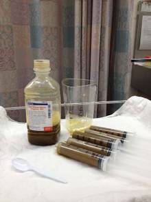
There’s no standardization of how the stool is prepared and filtered – it could be blended in a kitchen blender or by hand with a tongue depressor, or strained through gauze or a coffee filter, he said. Dosing is all over the place, listed as spoonfuls, grams, and milliliters. Different institutions screen donors in different ways. Some patients get a bowel prep, which can be mild or aggressive, and some don’t get one at all. Should the stool be fresh, and if so, how fresh? Is frozen okay? These are all issues that need to be examined from a safety and efficacy perspective and standardized.
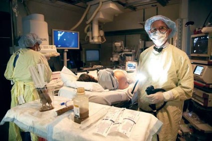
Even the method of delivery varies. The transplant can be administered via nasogastric tube or colonoscope, or by enema. There’s no standardization of data collection either. Some large institutions keep records of everything from the first workup to the last visit. Doctors who perform transplants occasionally may not be as conscientious. And no one knows anything about the do-it-yourselfers, at home with a family member’s donation and a squeeze enema.
Dr. Brandt – and a number of clinicians at the meeting – agreed that answers must be found for all these questions. And they agree that well-conducted clinical trials are the best way to go forward.
Dr. Colleen Kelly, one of the gastroenterologists launching one of 22 currently recruiting studies on FMT, started doing the procedure 5 years ago. Her first patient was a 26-year-old medical student with recurrent bouts of a Clostridium difficile infection.
"I’d heard of [FMT], but never, ever thought of doing one," said Dr. Kelly, of Brown University Medical Center, Providence, R.I. "I thought it was something at the far fringes of medicine."
At her patient’s insistence, Dr. Kelly contacted Dr. Brandt and learned about his process for performing FMT. The following year, she performed 10 FMTs. She is now undertaking a study with Dr. Brandt to recruit about 48 patients with relapsing C. difficile to be randomized to FMT or to a sham treatment with their own stool. Patients who have clinical failure in the sham group will be offered FMT; patients in the active arm who fail on initial FMT will get another FMT from a different donor.
While most trials are examining the utility of FMT in patients with recurrent C. difficile, a few are investigating FMT for use in patients with Crohn’s disease and inflammatory bowel disease. Some clinicians and individual patients are now using FMTs for this condition outside a clinical trial, with no understanding of how a compromised intestinal mucosa might react to transplanted stool.
It will take years to accumulate the data necessary to fully understand FMT and all its implications, Dr. Kelly said. In the interim, the IND requirement will likely shrink the already-small pool of gastroenterologists performing FMTs. "Some will be motivated to get an IND, but the average person in practice won’t," she predicted. "You need to put hours and hours of work into it, and then you’re still under FDA’s oversight because this is not an approved therapy. So that means you have to submit adverse events reports, keep records, and report annually on your program. And at any time, without any warning, [the FDA] can come and inspect your facility."
Dr. Brandt agreed. "It’s a huge amount of paperwork documentation, record-keeping, and follow-up that the average practitioner is simply not going to do." The requirement for an IND means there are simply going to be fewer and fewer physicians who do them, he said.
Admittedly, though, the risks of no regulation can endanger patients, Dr. Kelly said. "If things go on completely unregulated, stupid things will happen," including the spread of infectious diseases like hepatitis C and parasitic infestations.
Indeed, Dr. Alexander Khoruts of the University of Minnesota, Minneapolis, who spoke at the workshop, described the case of an FMT "do-it-yourselfer" who called for some advice on improving her outcomes. Specifically, she had mixed stool from a neighbor and her son’s mother-in-law and administered it to herself without results. "She wanted to know if maybe the chlorine in the water killed off everything. ... Six months later she called me back and said her C. diff was gone, but now she had parasites."
"There are already predatory practices out there [performing FMTs]," Dr. Khoruts said. "I got an e-mail from someone who couldn’t make the drive up to see me, but she found someone near her who would do it for $10,000."
Well-designed and well-executed studies would not only address these immediate safety questions, but would also examine the more nebulous concerns about the long-term effects of tampering with an individual’s unique ecosystem of gut microbes. In recent years, research has begun to document how the balance and proportion of microbial species in the gut can either protect from – or predispose to – metabolic syndrome, obesity, diabetes, cardiovascular disease, arthritis, and even cognitive disorders.
An engrafting microbial transplant could predispose the recipient to develop illnesses that would otherwise never have been destined to occur, Dr. Slater said. "All of the evidence we have suggests that manipulating the gut microbiome is a powerful act that may have long-reaching and subtle effects."
The move toward a standardized FMT product and process is inevitable, Dr. Brandt said. "We’re not going to be doing fecal transplants much longer. This is a temporary situation. We’re already developing compounds that will do the same thing."
Researchers at the University of Guelph, Ontario, have developed a machine that distills and cultures microbes from human feces, producing a kind of super-probiotic that can be used in place of fresh stool.
Also, the Canadian biotech company Rebiotix is working on a similar product, which it intends to test in a phase II randomized controlled trial.
But until those machine-made products are available, physicians and patients with have to stay with the man-made version. "We have access to a substance that is free and has a virtually unlimited supply," Dr. Kelly said. "We cannot deny this effective therapy to these patients who’ve failed all other available treatments."
None of the sources quoted in this article had any financial declarations.
BETHESDA, MD. – The Food and Drug Administration has designated stool for transplant as a biologic drug, necessitating that any gastroenterologist performing fecal microbiota transplants obtain an Investigational New Drug permit.
By designating stool for transplant as a biologic drug, anyone who performs fecal microbiota transplants (FMTs) – whether they perform a single transplant for one patient or recruit dozens for a study – needs to have an IND permit.
The announcement was made at a 2-day public workshop convened by the National Institutes of Health and the Food and Drug Administration to sift through some of the evidence surrounding FMTs.
The intent of the IND requirement, said Dr. Jay Slater, director of FDA’s Division of Bacterial, Parasitic, and Allergenic Products, is not to stamp out the care that patients can now receive, but to make sure it’s safe, effective, and data driven.
"This is a low-tech procedure that already has a CPT code. A ‘how-to’ guide recently appeared online, and it walks you right through how to do the procedure. There are a very large number of people doing this off the grid. What we need are long-term controlled trials on this that will enhance our understanding of its safety and efficacy."
Although neither the biologic designation nor the IND requirement is brand new, they have not been well publicized, according to one of the innovators of FMT, Dr. Lawrence Brandt, of Albert Einstein Medical Center in New York.
"I am struck by the fact that FDA wants these INDs and yet FDA has never publicly set forth any message on it. So from this moment on, all of us who continue to do this without an IND are violating FDA’s policy," he said at the meeting.
One of the primary concerns now, according to Dr. Brandt, is a burgeoning public interest in FMTs, even to the point of do-it-yourself treatments. With online instructions for self-treatment, and a procedure that is rife with variations, regulators can no longer tacitly ignore the issue.
There’s no standardization of how the stool is prepared and filtered – it could be blended in a kitchen blender or by hand with a tongue depressor, or strained through gauze or a coffee filter, he said. Dosing is all over the place, listed as spoonfuls, grams, and milliliters. Different institutions screen donors in different ways. Some patients get a bowel prep, which can be mild or aggressive, and some don’t get one at all. Should the stool be fresh, and if so, how fresh? Is frozen okay? These are all issues that need to be examined from a safety and efficacy perspective and standardized.
Even the method of delivery varies. The transplant can be administered via nasogastric tube or colonoscope, or by enema. There’s no standardization of data collection either. Some large institutions keep records of everything from the first workup to the last visit. Doctors who perform transplants occasionally may not be as conscientious. And no one knows anything about the do-it-yourselfers, at home with a family member’s donation and a squeeze enema.
Dr. Brandt – and a number of clinicians at the meeting – agreed that answers must be found for all these questions. And they agree that well-conducted clinical trials are the best way to go forward.
Dr. Colleen Kelly, one of the gastroenterologists launching one of 22 currently recruiting studies on FMT, started doing the procedure 5 years ago. Her first patient was a 26-year-old medical student with recurrent bouts of a Clostridium difficile infection.
"I’d heard of [FMT], but never, ever thought of doing one," said Dr. Kelly, of Brown University Medical Center, Providence, R.I. "I thought it was something at the far fringes of medicine."
At her patient’s insistence, Dr. Kelly contacted Dr. Brandt and learned about his process for performing FMT. The following year, she performed 10 FMTs. She is now undertaking a study with Dr. Brandt to recruit about 48 patients with relapsing C. difficile to be randomized to FMT or to a sham treatment with their own stool. Patients who have clinical failure in the sham group will be offered FMT; patients in the active arm who fail on initial FMT will get another FMT from a different donor.
While most trials are examining the utility of FMT in patients with recurrent C. difficile, a few are investigating FMT for use in patients with Crohn’s disease and inflammatory bowel disease. Some clinicians and individual patients are now using FMTs for this condition outside a clinical trial, with no understanding of how a compromised intestinal mucosa might react to transplanted stool.
It will take years to accumulate the data necessary to fully understand FMT and all its implications, Dr. Kelly said. In the interim, the IND requirement will likely shrink the already-small pool of gastroenterologists performing FMTs. "Some will be motivated to get an IND, but the average person in practice won’t," she predicted. "You need to put hours and hours of work into it, and then you’re still under FDA’s oversight because this is not an approved therapy. So that means you have to submit adverse events reports, keep records, and report annually on your program. And at any time, without any warning, [the FDA] can come and inspect your facility."
Dr. Brandt agreed. "It’s a huge amount of paperwork documentation, record-keeping, and follow-up that the average practitioner is simply not going to do." The requirement for an IND means there are simply going to be fewer and fewer physicians who do them, he said.
Admittedly, though, the risks of no regulation can endanger patients, Dr. Kelly said. "If things go on completely unregulated, stupid things will happen," including the spread of infectious diseases like hepatitis C and parasitic infestations.
Indeed, Dr. Alexander Khoruts of the University of Minnesota, Minneapolis, who spoke at the workshop, described the case of an FMT "do-it-yourselfer" who called for some advice on improving her outcomes. Specifically, she had mixed stool from a neighbor and her son’s mother-in-law and administered it to herself without results. "She wanted to know if maybe the chlorine in the water killed off everything. ... Six months later she called me back and said her C. diff was gone, but now she had parasites."
"There are already predatory practices out there [performing FMTs]," Dr. Khoruts said. "I got an e-mail from someone who couldn’t make the drive up to see me, but she found someone near her who would do it for $10,000."
Well-designed and well-executed studies would not only address these immediate safety questions, but would also examine the more nebulous concerns about the long-term effects of tampering with an individual’s unique ecosystem of gut microbes. In recent years, research has begun to document how the balance and proportion of microbial species in the gut can either protect from – or predispose to – metabolic syndrome, obesity, diabetes, cardiovascular disease, arthritis, and even cognitive disorders.
An engrafting microbial transplant could predispose the recipient to develop illnesses that would otherwise never have been destined to occur, Dr. Slater said. "All of the evidence we have suggests that manipulating the gut microbiome is a powerful act that may have long-reaching and subtle effects."
The move toward a standardized FMT product and process is inevitable, Dr. Brandt said. "We’re not going to be doing fecal transplants much longer. This is a temporary situation. We’re already developing compounds that will do the same thing."
Researchers at the University of Guelph, Ontario, have developed a machine that distills and cultures microbes from human feces, producing a kind of super-probiotic that can be used in place of fresh stool.
Also, the Canadian biotech company Rebiotix is working on a similar product, which it intends to test in a phase II randomized controlled trial.
But until those machine-made products are available, physicians and patients with have to stay with the man-made version. "We have access to a substance that is free and has a virtually unlimited supply," Dr. Kelly said. "We cannot deny this effective therapy to these patients who’ve failed all other available treatments."
None of the sources quoted in this article had any financial declarations.
HM13 Session Analysis: The Business of Medicine
Denice Cora-Bramble, MD, of Children’s National Medical Center in Washington, D.C., presented “The Business of Medicine” breakout Friday at HM13.
Key Points
- Whether you are salaried, work for productivity, or have a combination of the two, it is important for hospitalists to understand the business side of medicine.
- Even if you are not a hospitalist group leader, there are several things that you should know about the finances of your hospitalist program. Dr. Cora-Bramble reviewed the basics of financial statements, hospital revenue reports, and expense reports. She also reviewed how the hospitalist division partners with the entire hospital.
- After understanding the basic finances of your program, there are ways to enhance your financial performance. These include noting any lack of payments, billing and patient trends, and looking at program losses.
Key Takeaways
- It is important to understand the general principles of financial statements, budgets and financial decision making.
- There are multiple strategies to improve your division’s financial performance.
- There are financial challenges inherent in leading an academic division.
Dr. Hale is a pediatric hospitalist at the Floating Hospital for Children at Tufts Medical Center in Boston, and a Team Hospitalist member.
Medical Consultation and Co-Management in Perioperative Medicine
NATIONAL HARBOR, MD—Amir Jaffer, MD, and Steven Cohn, MD, chaired an excellent pre-course centered on common problems hospitalists encounter in managing medical problems in surgical patients. Topics covered were: the pre-operative evaluation and role of the consultant, cardiac risk assessment, pulmonary risk assessment, perioperative medication management, old and new antithrombotic therapy, diabetes management, co-management of the hip fracture, VTE prevention, and management of perioperative anemia.
Among other takeaway points, the physicians emphasized the importance of personal communication between surgeons and hospitalists. Frank Michota, MD, made an analogy using the patient as the plane, the surgeon as the pilot, the anesthesiologist as the co-pilot, and the hospitalist as the mechanic.
Check out The Hospitalist's Day One video from Hospital Medicine 2013
Others commented on cardiac management of the surgical patient, the risk assessment calculators available, and how noninvasive testing is rarely needed. Physicians also noted that partial functional dependence is the strongest predictor for patients needing recommendations regarding risk of postoperative respiratory failure. In reviewing patients’ medications in the perioperative phase, the speakers noted it is important to ask about herbal remedies and OTC drugs, as these may cause problems during and after surgery.
Additional topics addressed included: bridge therapy for anti coagulation, and the need to take into account the risk of bleeding among some, but not all, patients; how diabetic management should focus on a pre-meal glucose measurement of less than 140 mg/dl, and that all other glucose readings be under 180 mg/dl; how extended prophalyxis for VTE prevention is needed for elective hip surgery; and that hospitalists should be versed in the latest recommendations regarding restricted blood transfusion criteria. TH
Hands-On Training Helps Prepare Hospitalists for Procedures
Sally Wang MD, FHM, director of procedure education at Brigham and Women’s Hospital in Boston, and Brad Rosen, MD, MBA, FHM, medical director of the Inpatient Specialty Program (ISP) at Cedars-Sinai Hospital in Los Angeles, led another rapid-fire pre-course in ultrasound-guided procedures for the hospitalist at HM13.
Drs. Wang, Rosen, and a veteran group of faculty and trainers brought hands-on training in core bedside procedures, plus training in relatively new procedures to hospitalists such as intraosseous lines and skin biopsies. All attendees received close interaction with faculty and trainers, and participated in training exercises on tissue models, training models, and live models.
Additional discussion was focused on developing a proceduralist program. Experts explained the required commitment to proficiency and ongoing data collection, quality improvement, and “customer service” to stakeholders. But the basics of ongoing experiential learning are invaluable, they said, and often begin with simulation exercises.
Hospitalists thinking of becoming a proceduralist should start by making sure that they are proficient and experienced, and have invested the time necessary to maintain that experience. Beyond personal interest in procedures, administering an HM program that encourages and fosters procedural experience requires input from multiple stakeholders, as well as ongoing efforts to promote a climate of safety surrounding bedside procedures. TH
Dr. Chang is med-peds hospitalist at Univeristy of California San Diego, and a Team Hospitalist member.