User login
Brd4: Potential new target in AML

Researchers at Cold Spring Harbor Laboratory (CSHL) and 5 other laboratories may have identified a new drug target for the treatment of acute myeloid leukemia (AML).
They pinpointed a protein called Brd4, which contains a distinct region known as bromodomain and is a member of the BET protein family, known for regulating gene expression.
A new drug candidate that inhibits Brd4 was able to suppress AML in experimental models.
“The drug candidate not only displays remarkable anti-leukemia activity in aggressive disease models and against cells derived from patients with diverse, genetic subtypes of AML, but is also minimally toxic to non-cancerous cells,” said Chris Vakoc, MD, PhD, leader of the study at CSHL.
“The drug is currently being developed for therapeutic use for cancer patients by Tensha Therapeutics and is expected to enter clinical trials within 2 years.”
The team used RNAi screening in an AML murine model. The RNAi screen introduced small hairpin-shaped pieces of RNA (shRNA) that encode epigenetic proteins into mice that harbor leukemia-causing mutations.
The mice in this study carried the oncogene Nras and rearranged forms of the MLL gene, both of which are mutations often found in patients whose leukemia is resistant to standard chemotherapy.
The drinking water for the mice was also supplemented with doxycyline.
“Inducing shRNA that shuts down a gene required for the survival of leukemic cells can lead to complete disease remission,” said Johannes Zuber, MD, former postdoctoral researcher at CSHL. “This ability to use shRNA to simulate the effect of an anti-cancer drug illustrates the power of this approach.”
The team also identified another potential target in AML called Myb. The team found that suppressing the activity of Myb also eliminated AML in mice. The team screened more than 1000 shRNAs targeting 243 known epigenetic regulators of chromatin.
They homed in on Brd4 as a target and found that suppressing the protein led to the most dramatic changes in AML. The cell cycle was arrested, the leukemic cells died, leukemia progression was delayed, and the mice survived longer.
Previously, James Bradner, MD, at Dana-Farber Cancer Institute in Boston, and his research team had developed a small molecule inhibitor of Brd4 called JQ1. The two groups collaborated, reproducing the antileukemic effects in the Brd4 shRNA experiments. They consider JQ1 to be an ideal drug candidate.
Their findings were published online in Nature.![]()
Power Through Afternoon Energy Slumps

Late-afternoon slowdowns are natural, explains Susan Swadener, PhD, RD, dietetic internship director and lecturer in the Food Science and Nutrition Department at California State University San Luis Obispo. "Your enzyme levels go down, which is part of your diurnal pattern to slow down the body's processes to get ready for the evening and sleep," she says.
Common-Sense Nutrition
To fight afternoon fatigue, adopt good nutritional habits throughout your day. It's essential to have a healthy breakfast in the morning, advises Dr. Swadener, who's also a registered dietitian in private practice. Make sure you eat lunch, too, with a balance of protein, carbohydrates, and fats.
Afternoon snacks are a good idea, especially if they incorporate some protein. Foods high in protein can increase norepinephrine and epinephrine production, which helps you stay alert. Some examples of quick and nutritious snacks: string cheese and an apple; sliced cheese or peanut butter on whole-wheat crackers; yogurt; a handful of almonds or walnuts; or trail mix.
And don't forget one of the most common directives to your patients: "Push the fluids." Have a glass of water or nonfat milk with your lunch, and make sure you keep your water bottle handy at your desk.
As for coffee, "you don't want to be drinking it constantly to keep your energy level up, because you'll just crash afterwards," Dr. Swadener says. If you don't abuse caffeine, one cup of coffee in the morning and one in the afternoon is found to be most effective in increasing your alertness.
Change It Up
Desk tasks can make you drowsy. Daniel Markovitz is president of TimeBack Management, which specializes in applying Lean manufacturing principles to increase personal productivity for healthcare workers. He's found—and research such as a 2003 study in Ergonomics and a 2007 National Institute for Occupational Safety and Health study in the American Journal of Industrial Medicine supports these conclusions—that taking mini-breaks and then returning to the task at hand can refresh you and make you more productive.
You also get energy by using it, so a brisk walk around the hospital or walking up and down a couple of flights of stairs can increase circulation and blood flow to the brain. "You don't even have to get up from your desk," Markovitz says. "Just by changing the nature of the work you're doing, it's refreshing to your brain." That might mean switching from dictation to administrative work, or from scheduling to research.
And remember the value of play, Markovitz advises. "We tend to discourage going on Facebook or playing a video game at work. But if you take a 15-minute break to do something that's pleasurable, that causes your brain to fire in different ways, that can be another helpful adaptation."
Gretchen Henkel is a freelance writer based in California.
Interactions are Engaging
You've just finished a brain-numbing administrative report, and you've got 10 minutes before your next task. Don't just fill that time with checking your email.
Corporate consultant Daniel Markovitz advises another tack: Walk down the hall to touch base with colleagues. "You don't have to get into an involved conversation about their QI project. Just ask them how it's going for them," he advises.
Breaking up a busy day with exercise or a social call can get you over the doldrums hump, he says, because "you're getting up and interacting with someone. At the same time, you're doing something that's really important for the hospital: You're strengthening those bonds and interrelationships with people.—GH
Paraneoplastic Autoimmune Multiorgan Syndrome Proves Rapidly Fatal
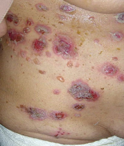
The skin may hold the key to differentiating classic pemphigus from the heterogenous autoimmune syndrome known as paraneoplastic autoimmune multiorgan syndrome.
It is a distinction of critical prognostic importance, because paraneoplastic autoimmune multiorgan syndrome (PAMS) typically is rapidly fatal, according to Dr. Sergei A. Grando, professor of dermatology and biologic chemistry at the University of California, Irvine. "The vast majority of patients die within several months of diagnosis, usually due to infections or respiratory failure, often taking the form of multiorgan system failure."
Two-thirds of patients with PAMS have a known internal malignancy at the time of their first mucocutaneous eruption. The most common of these neoplasms are non-Hodgkin’s lymphoma, which is present in more than 40% of PAMS patients; chronic lymphocytic leukemia, present in 30%; Castleman disease, present in 10%; and thymoma, present in 6%.
When a patient meets the diagnostic criteria for PAMS without having a known cancer, it is appropriate to launch a search for hidden malignancy, said Dr. Grando.
A key distinction between the skin lesions of PAMS and classic pemphigus is that PAMS involves inflammatory macules, papules, plaques, and blisters occurring on an inflammatory background over the trunk and extremities, including the palms and soles, but sparing the scalp.
In contrast, the generally more vesicular blisters and crusted erosions of pemphigus vulgaris display little erythema and usually occur on a noninflammatory background on the scalp, trunk, and extremities (but sparing the palms and soles). The most common location for skin lesions in PAMS is the palms; in pemphigus vulgaris, it is the scalp.
Also, Nikolsky's sign is positive in pemphigus vulgaris, but negative in PAMS, added Dr. Grando, who was among the investigators who first described PAMS a decade ago (Arch. Dermatol. 2001;137:193-206).
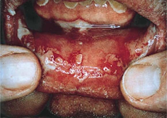
PAMS is characterized by severe and diffuse oral mucous membrane involvement, with persistent painful stomatitis because of blisters and erosions, and frequent involvement of other mucous membranes, including the eyes and genitalia. Cicatrizing conjunctivitis is particularly common in PAMS. In contrast, oral mucous membrane involvement in pemphigus is more discrete, with the eyes or other nonoral mucosa rarely involved.
Another key in the differential diagnosis: PAMS is associated with the HLA-DRB1*03 allele, whereas pemphigus vulgaris and foliaceous are strongly associated with the -04 and -14 alleles, respectively.
A hallmark of PAMS is respiratory involvement, with the sloughing of bronchial epithelial cells contributing to small airway occlusion and bronchiolitis obliterans. Classic pemphigus is free of respiratory involvement.
Both the esophagus and colon may be involved in PAMS, whereas only the esophagus is affected in pemphigus vulgaris.
In a published series of 28 PAMS patients, painful and generalized oral stomatitis was present in all 28, respiratory involvement was in 26, death from respiratory failure occurred in 22, and lichenoid skin involvement was present in 19. Only seven patients had no skin lesions (Br. J. Dermatol. 2003;149:1143-51).
At least five different subtypes of PAMS can be distinguished on the basis of the skin disease manifestations they most resemble. These subtypes of pemphiguslike PAMS include paraneoplastic pemphigus, bullous pemphigoid–like, erythema multiforme–like, lichen planus–like, and graft-vs.-host-disease–like versions of PAMS.
Dr. Grando eschews the term "paraneoplastic pemphigus" as too restrictive. After all, true pemphigus doesn’t usually affect the lungs, he noted.
Whereas most patients with classic pemphigus respond well to high-dose corticosteroids and recalcitrant disease can be effectively treated with cytotoxic agents, cyclosporine, intravenous gamma globulin, and other second-line agents, PAMS is resistant to all conventional forms of therapy.
"What can we offer? Really not much," Dr. Grando said during his presentation at the World Congress of Dermatology in Seoul, South Korea.
The preferred treatment regimen is a combination of prednisone and cyclosporine, with or without cyclophosphamide. Monthly courses of IVIG can buy patients a few extra months, he added.
Dr. Grando declared having no relevant financial interests.
The skin may hold the key to differentiating classic pemphigus from the heterogenous autoimmune syndrome known as paraneoplastic autoimmune multiorgan syndrome.
It is a distinction of critical prognostic importance, because paraneoplastic autoimmune multiorgan syndrome (PAMS) typically is rapidly fatal, according to Dr. Sergei A. Grando, professor of dermatology and biologic chemistry at the University of California, Irvine. "The vast majority of patients die within several months of diagnosis, usually due to infections or respiratory failure, often taking the form of multiorgan system failure."
Two-thirds of patients with PAMS have a known internal malignancy at the time of their first mucocutaneous eruption. The most common of these neoplasms are non-Hodgkin’s lymphoma, which is present in more than 40% of PAMS patients; chronic lymphocytic leukemia, present in 30%; Castleman disease, present in 10%; and thymoma, present in 6%.
When a patient meets the diagnostic criteria for PAMS without having a known cancer, it is appropriate to launch a search for hidden malignancy, said Dr. Grando.
A key distinction between the skin lesions of PAMS and classic pemphigus is that PAMS involves inflammatory macules, papules, plaques, and blisters occurring on an inflammatory background over the trunk and extremities, including the palms and soles, but sparing the scalp.
In contrast, the generally more vesicular blisters and crusted erosions of pemphigus vulgaris display little erythema and usually occur on a noninflammatory background on the scalp, trunk, and extremities (but sparing the palms and soles). The most common location for skin lesions in PAMS is the palms; in pemphigus vulgaris, it is the scalp.
Also, Nikolsky's sign is positive in pemphigus vulgaris, but negative in PAMS, added Dr. Grando, who was among the investigators who first described PAMS a decade ago (Arch. Dermatol. 2001;137:193-206).
PAMS is characterized by severe and diffuse oral mucous membrane involvement, with persistent painful stomatitis because of blisters and erosions, and frequent involvement of other mucous membranes, including the eyes and genitalia. Cicatrizing conjunctivitis is particularly common in PAMS. In contrast, oral mucous membrane involvement in pemphigus is more discrete, with the eyes or other nonoral mucosa rarely involved.
Another key in the differential diagnosis: PAMS is associated with the HLA-DRB1*03 allele, whereas pemphigus vulgaris and foliaceous are strongly associated with the -04 and -14 alleles, respectively.
A hallmark of PAMS is respiratory involvement, with the sloughing of bronchial epithelial cells contributing to small airway occlusion and bronchiolitis obliterans. Classic pemphigus is free of respiratory involvement.
Both the esophagus and colon may be involved in PAMS, whereas only the esophagus is affected in pemphigus vulgaris.
In a published series of 28 PAMS patients, painful and generalized oral stomatitis was present in all 28, respiratory involvement was in 26, death from respiratory failure occurred in 22, and lichenoid skin involvement was present in 19. Only seven patients had no skin lesions (Br. J. Dermatol. 2003;149:1143-51).
At least five different subtypes of PAMS can be distinguished on the basis of the skin disease manifestations they most resemble. These subtypes of pemphiguslike PAMS include paraneoplastic pemphigus, bullous pemphigoid–like, erythema multiforme–like, lichen planus–like, and graft-vs.-host-disease–like versions of PAMS.
Dr. Grando eschews the term "paraneoplastic pemphigus" as too restrictive. After all, true pemphigus doesn’t usually affect the lungs, he noted.
Whereas most patients with classic pemphigus respond well to high-dose corticosteroids and recalcitrant disease can be effectively treated with cytotoxic agents, cyclosporine, intravenous gamma globulin, and other second-line agents, PAMS is resistant to all conventional forms of therapy.
"What can we offer? Really not much," Dr. Grando said during his presentation at the World Congress of Dermatology in Seoul, South Korea.
The preferred treatment regimen is a combination of prednisone and cyclosporine, with or without cyclophosphamide. Monthly courses of IVIG can buy patients a few extra months, he added.
Dr. Grando declared having no relevant financial interests.
Study: Post-Discharge Costs Negate HM's In-Hospital Savings
A new report that suggests the lower costs and reduced length of stay (LOS) associated with hospitalist care of hospitalized Medicare beneficiaries is offset by higher costs post-discharge highlights progress and opportunities for hospitalists, SHM's president says.
The federally-funded study published Monday in Annals of Internal Medicine showed that while hospitalists clearly reduced LOS and cut in-hospital spending (by $282 per hospital visit), Medicare costs in the 30 days post-discharge were $332 higher than those followed up by primary care physicians. Patients discharged by hospitalists were less likely to be sent straight home and more likely to be admitted to a nursing home.
SHM President Joseph Li, MD, SFHM, associate professor of medicine at Harvard Medical School and director of the hospital medicine division at Beth Israel Deaconess Medical Center in Boston, says the data underscores the need for programs like SHM's Project BOOST, which helps hospitals improve discharge and reduce readmissions.
Dr. Li notes two limitations to the research. First, the patients reviewed were all from 2001-2006, long before the current healthcare debate surrounding cost containment and patient safety. Second, the review does not account for the quality of medical care.
“The study was done at a time which was early in the hospitalist movement,” Dr. Li says. “It’s fair to say at that point we weren’t talking as a field as much about trying to prevent unnecessary readmissions. ... When I look at this study, I think it’s an opportunity for hospitalists to think how we can improve on communications with post-discharge facilities. How we can improve transitions.”
An accompanying editorial agreed, noting that while hospitalists and outpatient care might be viewed in different silos, “patients regularly move back and forth across that divide.”
“It is important to better understand the association between hospitalist care and costs,” editorial co-author Lena Chen, MD, MS, Division of General Medicine, University of Michigan, writes in an email to The Hospitalist. “This paper takes a big step towards addressing this subject with a large study, and will hopefully spur additional related research. That would be a good thing for hospital medicine.”
CMS Ups the Stakes for Coordinated Care
Health organizations and providers experienced in providing care across multiple settings have until Aug. 19 to apply for a new accountable care organizations (ACO) program through the Centers for Medicare & Medicaid Services (CMS) that promises to offer a higher level of savings than that offered through a previous initiative.
“Hospital medicine groups in and of themselves won’t be applying for it,” says Ron Greeno, MD, MHM, chief medical officer of Brentwood, Tenn.-based Cogent HMG, and chair of SHM's Public Policy Committee. “But the groups that are looking to participate will have hospitalists working for them.”
The new program, tiled the Pioneer ACO Model, was created to offer potentially higher payments to providers and organizations who have already worked under contracts tied to shared savings or care coordination. A related model, the Medicare Shared Savings Program, does not require any previous experience with such contracts. The latter program has completed accepting public comments, but no application deadline has yet been set. (updated Aug. 15, 2011)
Just how many groups apply for the Pioneer program will be interesting: Initially, CMS will enter participation agreements with up to 30 organizations, and each must serve at least 15,000 beneficiaries (5,000 in rural areas). The capped cohort size didn’t seem to spur additional interest, as CMS originally set an application deadline for July, but pushed it back a month after providers questioned whether they were given enough time.
Still, Dr. Greeno, for one, is anxious to see the first round of applications. “It won’t interest me in terms of who jumps in,” he says. “The robustness of the response is what I’m interested in…It’s not going to do (CMS) any good to build a program nobody participates in.”
VATS, Open Lobectomy Produce Similar Survival in Early Lung Cancer
AMSTERDAM – Video-assisted thoracoscopic surgery worked as well as open lobectomy for 5-year survival in early-stage lung cancer, based on a secondary analysis of nonrandomized patients who underwent surgery as part of a multicenter trial.
"These data demonstrate that VATS, when properly done, can achieve long-term survival that is similar to open lobectomy," Dr. Walter J. Scott said at the World Conference on Lung Cancer. He stressed that study included only patients with early-stage lung cancer that was either node negative or had nonhilar N1 disease, and hence the finding is specific for only these patients. Until now, questions existed about the oncologic efficacy of VATS, noted Dr. Scott, chief of the division of thoracic surgery at Fox Chase Cancer Center in Philadelphia.But "VATS lobectomy provides comparable oncologic outcomes" for this group of patients, he said.
His analysis used data collected from 964 lung cancer patients who participated in a multicenter study during 1999-2004 that compared two different strategies for lymph node assessment in early-stage lung cancer (Ann. Thorac. Surg. 2006;81:1013-20). Although most surgeons did not perform VATS during this time, a few surgeons did, and 5-year outcome results were available for 66 patients in the study underwent VATS. Five-year data also existed for 898 of the patients who underwent open lobectomy.
To adjust for baseline differences among the patients, Dr. Scott and his associates ran a propensity score analysis that took into account age, sex, performance status, tumor histology, location, and tumor size and invasion. The analysis excluded about a fifth of the open lobectomy patients because their propensity scores fell outside the range of the VATS patients, so the final survival comparison included 66 VATS and 686 open lobectomy patients.
With propensity adjustment, the results showed no significant differences between the VATS and open lobectomy patients in their 5-year rates of overall survival, disease-free survival, local disease-free survival, or freedom from new primary tumors (see table), Dr. Scott reported at the meeting, which was sponsored by the International Association for the Study of Lung Cancer.
Dr. Scott said that he is a shareholder in Biogen Idec, Celgene, and Johnson & Johnson.
FROM THE WORLD CONFERENCE ON LUNG CANCER
Major Finding: Patients with early-stage lung cancer who underwent VATS had a 72% 5-year overall survival rate, statistically similar to the 66% rate among matched patients who underwent open lobectomy.
Data Source: Secondary analysis of 752 early-stage patients with node-negative or nonhilar N1 lung cancer enrolled in a thoracic surgery trial designed to compare two approaches to lymph node assessment. Outcome comparisons for the 66 patients treated with VATS and the 686 treated with open lobectomy involved propensity-score matching for age, sex, performance status, tumor histology, tumor location, size, and invasion.
Disclosures: Dr. Scott said that he is a shareholder in Biogen Idec, Celgene, and Johnson & Johnson.
ONLINE EXCLUSIVE: Scheduling Rules of Thumb
John Krisa, MD, medical director of the hospitalist group at Albany Memorial Hospital in New York, pictures his HM group as an organic whole when he draws up the schedule. He tries to avoid a strict 50-50 parceling out of night and day shifts. The hospitalist group makes liberal use of per-diem hospitalists and moonlighters, and has a few nocturnists.
“The vast majority of the work at night is processing new admissions, so these tend to be single encounters. You want your full-time people there multiple consecutive days for continuity and to represent the face of your program,” he says.
But for the required, ’round-the-clock coverage, he and other group members are expected to pull their share of nights as well. “I was always more of a nighttime person, in terms of my body clock,” Dr. Krisa says, “but now that I have more daytime nonclinical duties [as regional site director for Cogent HMG], it’s been more of a challenge to juggle home responsibilities, night shifts, and multiple administrative meetings.”
There are some basic principles of sleep hygiene and lessons learned from industrial settings that are good to keep in mind, says Christopher P. Landrigan, MD, SFHM, MPH, associate professor of medicine and pediatrics at Harvard Medical School and director of the Sleep and Patient Safety Program at Brigham and Women’s Hospital in Boston. “It’s really incumbent upon hospitalist group leaders to recognize the hazards of scheduling people for too many nights in a row, which conveys a risk both to the patients and to the hospitalists themselves,” Dr. Landrigan says. “We know that if hospitalists are driving home after night shifts, particularly multiple night shifts, that they’re at risk for motor vehicle crashes and at risk of sticking themselves with needles and scalpels toward the tail end of their shifts. None of us want that.”
Dr. Landrigan advises hospitalist groups to be cognizant of the hazards and think about the schedule “proactively.”
ONLINE EXCLUSIVE: The “Weak Link” in Patient Handoffs
Increased handoffs are often viewed as a byproduct of the growth in hospital medicine, with heightened scrutiny on the quality of communication that accompanies these transfers of care. As research suggests, though, finding and fixing the weak links can require persistence.
A study led by Siddhartha Singh, MD, MS, associate chief medical officer of Medical College Physicians, the adult practice for Medical College of Wisconsin in Milwaukee, compared a traditional, resident-based model of care to one involving a hospitalist-physician assistant team. Initially, his study found a 6% higher length of stay (LOS) for the hospitalist-physician assistant teams, with no differences in costs or readmission rates.1
But when the researchers pored over their results, they discovered that the increased LOS was limited to patients admitted overnight. Those patients, Dr. Singh says, were admitted by other providers—a night-float resident or faculty hospitalist—and then transferred to the hospitalist-physician assistant teams when they arrived in the morning. These “overflow patients” also were admitted only during busy periods, when limits on the number of admissions by house staff required other arrangements.
To make a direct comparison, Dr. Singh focused on a window from 11 a.m. to 4 p.m., when patients would have an equal probability of being admitted by a resident team or a hospitalist-physician assistant team. From a pool of about 3,000 admitted patients, the study found no significant difference in LOS, cost, readmission rates, or mortality. Instead of highlighting significant differences in models of care, then, Dr. Singh says, his study highlighted a potential weak link in the “treacherous” overnight-to-morning handoffs during busy periods that should be addressed.
“There have been a lot of studies implicating poor communication as a cause of patient-safety issues,” notes Sunil Kripalani, MD, MSc, FHM, chief of the section of hospital medicine and an associate professor of medicine at Vanderbilt University Medical Center in Nashville, Tenn. But fewer studies, he says, have shown how to effectively improve communication in a way that improves patient safety.
One focal point is the often incomplete and inadequate nature of discharge summaries. Several models are emerging on how to build a better discharge summary, Dr. Kripalani says, with researchers offering solid recommendations (as multiple presentations at SHM’s annual meeting suggest). The trick is ensuring that those plans can be implemented into practice on a consistent and timely basis.
Dr. Kripalani says at least one straightforward strategy might help improve handoffs, however: building time into the schedule for them, such as 15-minute overlaps between shifts.—BN
Dr. Hospitalist: Your Hospital Medicine Questions Answered

What’s up with the dress code in hospitals these days? Some of my colleagues wear white coats, some wear ties, some have short-sleeved shirts. Some even wear scrubs in the daytime, and they swear they are right as to “the most clinically appropriate attire.” Any thoughts?
Attirely Concerned in Los Angeles
Dr. Hospitalist responds:
There are a lot of suggestions out there regarding attire. The United Kingdom’s National Health Service is probably most famous for instituting a “bare below elbows” (BBE for short) dress code in 2007.
Although lots of studies have shown bacterial colonization on the items doctors wear or carry (e.g. pagers, pens, neckties, coats, scrubs), none of them truly show causality. The Journal of Hospital Medicine just published a study on scrubs versus white coats, which showed no real difference in contamination.1
Even the BBE policy was meant to promote hand-washing more than anything else. On that point, there is little disagreement, as there is a substantial amount of data to show that good hand hygiene is a patient-care imperative. We all should spend more time thinking about “clean in/clean out” when it comes to patient rooms than deciding which article of clothing carries the fewest bacteria.
There is another issue at play here, though, and that is the question of how hospitalists are expected to dress. Certainly, there is some regional variation. I don’t think you’ll find that physicians at the Mayo Clinic in Rochester, Minn., are going to dress the same as physicians in San Diego or Hawaii.
So, setting aside the cultural expectations for your region, I do think it’s a good idea for your group to agree on some standards. These policies might vary from white coats for everyone to scrubs after hours, or that blue jeans are OK only on weekends.
Why bother?
Well, for starters, a little consistency will promote the professionalism of your group, and it also sets some baseline expectations for everyone involved. Think about how many healthcare providers wander into a patient’s room during the day: You want to be readily identifiable as the treating physician. No, it’s not just how you dress (a voice, a name badge, and putting your name on the white board also count), but it is part of the picture.
As a hospitalist, not only are you a professional, but, by definition, you are going to meet patients with whom you have no prior relationship. Like it or not, perception matters, and when you need to quickly gain the trust of a patient (and a family) to make urgent clinical decisions, being dressed professionally will help. Looking like a slob won’t.
My advice? First, wash your hands where the patient can see you. If you have to use that gel 40 times a day, you might as well make a show of it. Two, dress professionally within the parameters that your group outlines.
Beyond that, I don’t think you need to autoclave your peripherals and go through a decontamination room just yet.
Reference
- Burden M, Cervantes L, Weed D, Keniston A, Price CS, Albert RK. Newly cleaned physician uniforms and infrequently washed white coats have similar rates of bacterial contamination after an 8-hour workday: A randomized controlled trial. J Hosp Med. 2011;6(4):177-182.
Power Struggles

Many hospitalist practices are started by “traditionalists”: primary-care physicians (PCPs) active in the outpatient and hospital settings. The practice typically grows due in large part to the leadership of the founders. Ultimately, the practice is made up of both the founders and a cadre of part- or full-time hospitalists who don’t work in the outpatient setting. And sometimes they have different incentives and ideas about how the practice should operate.
When these individuals disagree, which group should break the tie—the founding “hybrid” or “rotating” doctors who work part time on the hospitalist service or the doctors who work only as hospitalists?
This is a reasonably common issue for “medical” hospitalist groups, and in many cases is becoming an issue for groups in other specialties that adopt the hospitalist model, such as surgical hospitalists, laborists, etc.
A Common Scenario
Let me illustrate this issue with a composite of several former consulting clients. Let’s say this is a hospitalist practice that serves a 250-bed community hospital. One large private internal medicine group adopted a “rotating hospitalist” model there in the late 1990s. One of the internists provided the daytime hospital coverage for all the group’s patients one week out of every six. Their hospital volume grew quickly. They were asked to take on responsibility for admitting an increasing portion of the unassigned patients, provide care for patients referred by other PCPs who wanted to drop out of hospital work, and increasingly were asked to consult on patients admitted by surgeons.
When faced with this situation, many PCP groups decided to exit the hospital themselves and turn that work over to hospitalists. This group stuck it out. At first, the one doctor in the group covering the hospital each week kept up with the growing volume by simply working harder and longer every day. Eventually, the group sought financial help from the hospital to hire hospitalists who didn’t have outpatient responsibilities.
Years passed, and this PCP group transitioned to employment by the hospital, just like the full-time hospitalists. And by this time, the hospitalist practice was seen as distinct from the original PCP group. About 80% of the staffing was provided by hospitalists who didn’t work in the outpatient setting, the remainder by PCPs who essentially founded the practice. The PCPs chose to continue providing hospital care, both because they found it professionally satisfying and their compensation formula made it attractive for generating production in the hospital.
Tensions arose between the hospitalists and the “hybrids.” The hybrids refused to work night shifts and generally were unable to fill in for unplanned absences by the hospitalists. And because of the PCPs’ compensation formula, and possibly the work ethic of more senior doctors, they favored managing larger patient volumes and decreasing weekend staffing significantly to allow more weekends off in total for everyone. The hospitalists had other ideas about these things, and they were unhappy that the PCPs would have first say about when they could work hospital shifts, thereby decreasing the hospitalists’ scheduling flexibility.
The hospitalists were all within a few years of their residency training, and most of the PCPs were midcareer. This created a social divide, making it that much more difficult for the two groups to work through the issues. While the hybrid doctors saw the hospitalists as good clinicians, and vice versa, each group said: “The other guys are difficult to work with. They don’t understand what it is like for us.”
Need for Paradigm Shift
At many sites, the doctors and their administrative counterparts get stuck in a stalemate and have the same, unhappy conversations repeatedly. These conversations are really gripe sessions more than anything else.
I think the best solution is for everyone to acknowledge the valuable contribution of the hybrid doctors in founding and leading the hospitalist practice through years of growth, but also to begin seeing the hospitalist practice as being owned and governed primarily by the hospitalists who do most of the work. For most issues in which the two factions can’t agree, the hospitalists should have the tie-breaking vote.
While this approach reduces the autonomy of the hybrid doctors to make operational decisions, it doesn’t mean they have zero influence. In fact, the practice usually has a critical need for the hybrid doctors to continue providing some of the staffing. This usually means that the practice will need to ensure it puts together a package of compensation and available shifts on the schedule to ensure the hybrids want to remain active in the practice.
In most cases, all involved should ensure that those hybrid doctors who want to remain active in the hospital, and perform well in the hospitalist practice, should have the opportunity to do so indefinitely.
Compensation Methods for Hybrids
Even if the hybrids and hospitalists are able to harmoniously agree on things like work schedules, the hybrid doctors often have compensation schemes such that when working in the hospital, they have different financial incentives from the hospitalists. (I’m using “hybrid” to describe physicians who work in both inpatient and outpatient settings, usually more time in the office practice.) A common situation is that the production (i.e. wRVUs) generated in the hospital counts toward their office productivity. So the hybrids and the hospitalists will have different ideas about how hard they want to work.
The solution here is to divorce the office and hospital compensation schemes. There should be no connection between the compensation in the two settings, and both should be designed to ensure a competitive amount of money and performance incentives appropriate for that setting. Such methods usually mean that a day of work in the office will result in a different-sized paycheck than what comes with a day of work in the hospital.
Dr. Nelson has been a practicing hospitalist since 1988 and is cofounder and past president of SHM. He is a principal in Nelson Flores Hospital Medicine Consultants, a national hospitalist practice management consulting firm. He is course codirector and faculty for SHM’s “Best Practices in Managing a Hospital Medicine Program” course. This column represents his views and is not intended to reflect an official position of SHM.