User login
Stop using the hCG discriminatory zone of 1,500 to 2,000 mIU/mL to guide intervention during early pregnancy
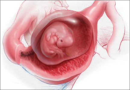
Approximately 15% of early pregnancies are complicated by pelvic or abdominal pain and uterine bleeding or spotting. In these situations, you must determine whether your patient has a viable intrauterine pregnancy, a pregnancy that will end in a miscarriage (spontaneous abortion), or an ectopic pregnancy.
To guide you to the correct diagnosis, a medical history and physical examination can be helpful. For example, a woman with a prior ectopic pregnancy who now has an early pregnancy complicated by pelvic pain and uterine bleeding is at high risk for an ectopic pregnancy. Physical examination also is important; if the pelvic examination reveals a dilated cervix with pregnancy tissue in the cervical os it is likely that a miscarriage is in progress. For most cases of early pregnancy complicated by pelvic pain and/or uterine bleeding, however, a pelvic sonogram and serial quantitative measurement of human chorionic gonadotropin (hCG) are needed to achieve the correct diagnosis.
Here I outline the clinical markers for transvaginal ultrasonography that indicate a viable or failed intrauterine pregnancy as well as an ectopic pregnancy. I also present data on single vs serial hCG measurement and discuss serial hCG levels that indicate viable or nonviable intrauterine or ectopic pregnancy.
Is an early gestation viable? Clinical evaluationTransvaginal pelvic ultrasoundTransvaginal and transabdominal ultrasonography play a critical role in evaluating early pregnancy problems. In a normal pregnancy, key developmental milestones that can be observed reliably on ultrasound are1,2:
- intrauterine gestational sac at 5 weeks
- yolk sac at 5.5 weeks
- embryonic pole and fetal heart beat at 6 to 6.5 weeks’ gestation.
A pelvic ultrasound also may provide evidence that an intrauterine pregnancy will fail and result in a miscarriage. Findings diagnostic of a failed intrauterine pregnancy include3:
- crown-rump length ≥7 mm and no fetal heartbeat
- mean sac diameter ≥25 mm and no embryo
- absence of an embryo with a heartbeat more than 2 weeks after an ultrasound scan that showed a gestational sac without a yolk sac
- absence of an embryo with a heartbeat more than 11 days after a scan that showed a gestational sac with a yolk sac.
Findings suspicious for a failing intrauterine pregnancy include3:
- crown-rump length <7 mm and no fetal heartbeat
- mean sac diameter of 16 to 24 mm and no embryo
- no heartbeat 7 to 13 days after an ultrasound scan that showed a gestational sac without a yolk sac
- no heartbeat 7 to 10 days after an ultrasound scan that showed a gestational sac with a yolk sac.
When it’s an ectopic pregnancy. Definitive ultrasonographic evidence of an ectopic pregnancy is identification of a fetal heartbeat outside the uterus or a gestational sac and yolk sac outside of the uterus. An adnexal mass can be identified on ultrasonography in most cases of ectopic pregnancy. In one study of 291 ectopic pregnancies, an adnexal mass was identified in 94% of cases, and a moderate to large amount of free pelvic fluid was found in 36% of cases.4 The adnexal masses included nonspecific (54% of all ectopic cases), a tubal ring without a yolk sac or embryo (25%), a yolk sac but no embryonic heartbeat (8%), and an embryo with cardiac activity (7%).
In clinical units with high-quality gynecologic ultrasonography available, most ectopic pregnancies will be detected on initial scan and only 10% to 15% of ectopic pregnancies will have an ultrasound finding of no intrauterine pregnancy and no evidence for an extrauterine pregnancy.5

Almost all (in the range of 99%) viable intrauterine pregnancies demonstrate an increase in hCG level of 53% or more over 48 hours, whereas only 21% of ectopic pregnancies demonstrate a rise of 53% or more.7
Most pregnancies that will end in a miscarriage demonstrate a decrease in hCG level over 48 hours. If the initial hCG value is 2,000 mIU/mL, 90% of pregnancies that will end in miscarriage will have an approximate 30% decrease in hCG over 48 hours. If the initial hCG is 1,000 mIU/mL, 95% of spontaneous abortions will have a 28% decline in 48 hours.7 About 10% of ectopic pregnancies also will demonstrate a 30% decrease in hCG over 48 hours.
A minor disadvantage of serial hCG measurements is that patients may become anxious and fearful as they await the result of life-altering test results.
When a gestation is found to be nonviable A viable intrauterine pregnancy is highly unlikely in a woman with no ultrasound evidence of an intrauterine pregnancy or an adnexal mass and an hCG level that rises very little, plateaus, or decreases over 48 hours. In this situation, a Karman cannula aspiration of uterine contents with rush pathology analysis can help clarify the likely diagnosis and guide therapy.
Women with documented villi on pathology likely are experiencing a miscarriage and can have their hCG level followed to resolution. Women with no documented villi and no decrease in hCG after the Karman
cannula aspiration can be presumed to have an ectopic pregnancy. If stable, these women may be candidates for treatment with methotrexate.8,9
Many experts have counseled against the use of a single hCG measurement in the discriminatory zone of 1,500 to 2,000 mIU/mL to trigger methotrexate treatment. Here is a sampling of their advice:
“An hCG level of 2,000 mIU/mL, without ultrasound findings of intrauterine pregnancy, while suggestive of abnormal pregnancy, is not diagnostic. Per the results of recent studies, it is reasonable to closely follow up rather than treat many of these early, stable cases of ectopic pregnancy.”
—Mehta et al.1
“Our data demonstrate that using a single value of serum hCG in a pregnancy of unknown location (PUL) population is of limited value.... A significant proportion of failing PULs and early intrauterine pregnancies in a PUL population have high serum hCG levels at presentation.”
—Condus et al.2
“The hCG discriminatory level should not be used to determine the management of a hemodynamically stable patient with suspected ectopic pregnancy, if sonography demonstrates no findings of intrauterine or ectopic pregnancy.”
—Doubilet et al.3
“There is almost no reason to give methotrexate on first encounter with a patient. If a patient is symptomatic with severe pain or signs of rupture, a surgical approach is indicated and methotrexate is contraindicated.”
—Barnhart et al.4
[When using the discriminatory zone]... “there is a chance of harming a viable intrauterine pregnancy, especially if the hCG level is 2000 to 3000 mIU/mL.... There is limited risk in taking a few extra days to make a definitive diagnosis in a woman with a pregnancy of unknown location who has no signs or symptoms of rupture and no ultrasonographic evidence of ectopic pregnancy.”
—Doubliet et al.3
“Viable intrauterine pregnancy is possible in patients with pregnancy of unknown location and hCG levels above the generally accept discriminatory zone, strict adherence to which can potentially disrupt a normal pregnancy. We support the need for judicious use of the hCG discriminatory level in hemodynamically stable patients with pregnancy of unknown location, and the decision to intervene should not be based solely on a single hCG level.”
—Ko and Cheung.5
References
- Mehta TS, Levine D, Beckwith B. Treatment of ectopic pregnancy: is a human chorionic gonadotropin level of 2,000 mIU/mL a reasonable threshold? Radiology. 1997;205(2):569–573.
- Condous G, Kirk E, Lu C, et al. Diagnostic accuracy of varying discriminatory zones for the prediction of ectopic pregnancy in women with pregnancy of unknown location. Ultrasound Obstet Gynecol. 2005;26(7):770–775.
- Doubilet PM, Benson CB, Bourne T, et al. Diagnostic criteria for nonviable pregnancy early in the first trimester. N Engl J Med. 2013;369(15):1443–1451.
- Barnhart KT. Early pregnancy failure: beware of the pitfalls of modern management. Fertil Steril. 2012;98(5):1061–1065.
- Ko JK, Cheung VY. Time to revisit the human chorionic gonadotropin discriminatory level in the management of pregnancy of unknown location. J Ultrasound Med. 2014;33(3):465–471.
Stop using the discriminatory zone and a single hCG measurement to trigger clinical intervention As noted above, a single hCG measurement is of very little value in determining whether an early pregnancy is a viable or nonviable intrauterine pregnancy or is ectopic. Many experts have reported that if a single hCG measurement shows a value of more than 1,500 mIU/mL and a pelvic ultrasound shows no intrauterine pregnancy, an ectopic or nonviable intrauterine pregnancy is likely. Some experts have used the presence of an hCG value of more than 1,500 mIU/mL plus an ultrasound scan without evidence of an intrauterine pregnancy to clinically diagnose the absence of a viable intrauterine pregnancy and administer methotrexate to treat a presumptive ectopic pregnancy. Many experts believe, however, that this approach will necessarily result in the treatment of viable intrauterine pregnancies with methotrexate.5,10
Based on one analysis, for 100 women with an initial hCG value between 2,000 and 3,000 mIU/mL and no intrauterine pregnancy or adnexal mass seen on ultrasound, follow-up will reveal that 65.5% had a failed intrauterine pregnancy, 33% had an ectopic pregnancy, and 1.5% had a viable intrauterine pregnancy.3,10,11 If all of these 100 women had been treated with methotrexate for a presumed ectopic pregnancy, approximately two women with a viable intrauterine pregnancy would have been exposed to methotrexate. This exposure would likely result in either a pregnancy loss or, if the pregnancy continues, an increased risk of fetal anomalies.
If the patient is obese, has fibroids, or has adenomyosis, she has an increased risk of an ultrasound failing to detect an early intrauterine pregnancy when the hCG value ranges from 1,500 to 3,000 mIU/mL.12 If the discriminatory zone is raised to 4,000 mIU/mL, the likelihood of mistakenly diagnosing a viable intrauterine pregnancy as a failed or ectopic pregnancy is much less (but not zero).
There is almost no clinical situation in which methotrexate should be given to a patient suspected of having an ectopic pregnancy on the first visit, unless ultrasound demonstrates an adnexal mass indicative of ectopic pregnancy.13,14 If the patient has severe pain or bleeding, or has signs consistent with a ruptured ectopic pregnancy, surgical intervention likely is warranted. If the patient is clinically stable, a safe option is to repeat the hCG measurement in 48 hours, with an ultrasound if indicated.
The discriminatory zone is an interesting and elegant idea. But in practice it is fraught with serious dangers, the greatest of which is methotrexate administration to a patient with a viable intrauterine gestation. My advice is that gynecologists should stop relying on a discriminatory zone of 1,500 to 2,000 mIU/mL to trigger clinical intervention.
Share your thoughts on this article! Send your Letter to the Editor to [email protected]. Please include your name and the city and state in which you practice.
1. Bree RL, Edwards M, Böhm-Vélez M, et al. Transvaginal sonography in the evaluation of normal early pregnancy: correlation with hCG level. AJR Am J Roentgenol. 1989;153(1):75–79.
2. Goldstein I, Zimmer EA, Tamir A, Peretz BA, Paldi E. Evaluation of normal gestational sac growth: appearance of embryonic heartbeat and embryo body movements using the transvaginal technique. Obstet Gynecol. 1991;77(6):885–888.
3. Doubilet PM, Benson CB, Bourne T, et al. Diagnostic criteria for nonviable pregnancy early in the first trimester. N Engl J Med. 2013;369(15):1443–1451.
4. Frates MC, Doubilet PM, Peters HE, Benson CR. Adnexal sonographic findings in ectopic pregnancy and their correlation with tubal rupture and human chorionic gonadotropin levels. J Ultrasound Med. 2014;33(4):697–703.
5. Condous G, Kirk E, Lu C, et al. Diagnostic accuracy of varying discriminatory zones for the prediction of ectopic pregnancy in women with pregnancy of unknown location. Ultrasound Obstet Gynecol. 2005;26(7):770–775.
6. Barnhart KT. Clinical practice. Ectopic pregnancy. N Engl J Med. 2009;361(4):379–387.
7. Silva C, Sammel MD, Zhou L, et al. Human chorionic gonadotropin profile for women with ectopic pregnancy. Obstet Gynecol. 2006;107(3):605–610.
8. Shaunik A, Kulp J, Appleby DH, Sammel MD, Barnhart KT. Utility of dilation and curettage in the diagnosis of pregnancy of unknown location. Am J Obstet Gynecol. 2011;204(2):130.e1–6.
9. Brady P, Imudia AN, Awonuga AO, Wright DL, Syter AK, Toth TL. Pregnancies of unknown location after in vitro fertilization: minimally invasive management with Karman cannula aspiration. Fertil Steril. 2014;101(2):420–426.
10. Doubilet PM, Benson CB. Further evidence against the reliability of the human chorionic discriminatory level. J Ultrasound Med. 2011;30(12):1637–1642.
11. Benson CB, Doubilet PM, Peters HE, Frates MC. Intrauterine fluid with ectopic pregnancy: a reappraisal. J Ultrasound Med. 2013;32(3):389–393.
12. Ko JK, Cheung VY. Time to revisit the human chorionic gonadotropin discriminatory level in the management of pregnancy of unknown location. J Ultrasound Med. 2014;33(3):465–471.
13. Barnhart KT. Early pregnancy failure: beware of the pitfalls of modern management. Fertil Steril. 2012;98(5):1061–1065.
14. Mehta TS, Levine D, Beckwith B. Treatment of ectopic pregnancy: is a human chorionic gonadotropin level of 2,000 mIU/mL a reasonable threshold? Radiology. 1997;205(2):569–573.
Dr. Barbieri is Editor in Chief, OBG Management; Chair, Obstetrics and Gynecology, at Brigham and Women’s Hospital, Boston, Massachusetts; and Kate Macy Ladd Professor of Obstetrics, Gynecology, and Reproductive Biology at Harvard Medical School, Boston.
Dr. Barbieri reports no financial relationships relevant to this article.
Approximately 15% of early pregnancies are complicated by pelvic or abdominal pain and uterine bleeding or spotting. In these situations, you must determine whether your patient has a viable intrauterine pregnancy, a pregnancy that will end in a miscarriage (spontaneous abortion), or an ectopic pregnancy.
To guide you to the correct diagnosis, a medical history and physical examination can be helpful. For example, a woman with a prior ectopic pregnancy who now has an early pregnancy complicated by pelvic pain and uterine bleeding is at high risk for an ectopic pregnancy. Physical examination also is important; if the pelvic examination reveals a dilated cervix with pregnancy tissue in the cervical os it is likely that a miscarriage is in progress. For most cases of early pregnancy complicated by pelvic pain and/or uterine bleeding, however, a pelvic sonogram and serial quantitative measurement of human chorionic gonadotropin (hCG) are needed to achieve the correct diagnosis.
Here I outline the clinical markers for transvaginal ultrasonography that indicate a viable or failed intrauterine pregnancy as well as an ectopic pregnancy. I also present data on single vs serial hCG measurement and discuss serial hCG levels that indicate viable or nonviable intrauterine or ectopic pregnancy.
Is an early gestation viable? Clinical evaluationTransvaginal pelvic ultrasoundTransvaginal and transabdominal ultrasonography play a critical role in evaluating early pregnancy problems. In a normal pregnancy, key developmental milestones that can be observed reliably on ultrasound are1,2:
- intrauterine gestational sac at 5 weeks
- yolk sac at 5.5 weeks
- embryonic pole and fetal heart beat at 6 to 6.5 weeks’ gestation.
A pelvic ultrasound also may provide evidence that an intrauterine pregnancy will fail and result in a miscarriage. Findings diagnostic of a failed intrauterine pregnancy include3:
- crown-rump length ≥7 mm and no fetal heartbeat
- mean sac diameter ≥25 mm and no embryo
- absence of an embryo with a heartbeat more than 2 weeks after an ultrasound scan that showed a gestational sac without a yolk sac
- absence of an embryo with a heartbeat more than 11 days after a scan that showed a gestational sac with a yolk sac.
Findings suspicious for a failing intrauterine pregnancy include3:
- crown-rump length <7 mm and no fetal heartbeat
- mean sac diameter of 16 to 24 mm and no embryo
- no heartbeat 7 to 13 days after an ultrasound scan that showed a gestational sac without a yolk sac
- no heartbeat 7 to 10 days after an ultrasound scan that showed a gestational sac with a yolk sac.
When it’s an ectopic pregnancy. Definitive ultrasonographic evidence of an ectopic pregnancy is identification of a fetal heartbeat outside the uterus or a gestational sac and yolk sac outside of the uterus. An adnexal mass can be identified on ultrasonography in most cases of ectopic pregnancy. In one study of 291 ectopic pregnancies, an adnexal mass was identified in 94% of cases, and a moderate to large amount of free pelvic fluid was found in 36% of cases.4 The adnexal masses included nonspecific (54% of all ectopic cases), a tubal ring without a yolk sac or embryo (25%), a yolk sac but no embryonic heartbeat (8%), and an embryo with cardiac activity (7%).
In clinical units with high-quality gynecologic ultrasonography available, most ectopic pregnancies will be detected on initial scan and only 10% to 15% of ectopic pregnancies will have an ultrasound finding of no intrauterine pregnancy and no evidence for an extrauterine pregnancy.5
Almost all (in the range of 99%) viable intrauterine pregnancies demonstrate an increase in hCG level of 53% or more over 48 hours, whereas only 21% of ectopic pregnancies demonstrate a rise of 53% or more.7
Most pregnancies that will end in a miscarriage demonstrate a decrease in hCG level over 48 hours. If the initial hCG value is 2,000 mIU/mL, 90% of pregnancies that will end in miscarriage will have an approximate 30% decrease in hCG over 48 hours. If the initial hCG is 1,000 mIU/mL, 95% of spontaneous abortions will have a 28% decline in 48 hours.7 About 10% of ectopic pregnancies also will demonstrate a 30% decrease in hCG over 48 hours.
A minor disadvantage of serial hCG measurements is that patients may become anxious and fearful as they await the result of life-altering test results.
When a gestation is found to be nonviable A viable intrauterine pregnancy is highly unlikely in a woman with no ultrasound evidence of an intrauterine pregnancy or an adnexal mass and an hCG level that rises very little, plateaus, or decreases over 48 hours. In this situation, a Karman cannula aspiration of uterine contents with rush pathology analysis can help clarify the likely diagnosis and guide therapy.
Women with documented villi on pathology likely are experiencing a miscarriage and can have their hCG level followed to resolution. Women with no documented villi and no decrease in hCG after the Karman
cannula aspiration can be presumed to have an ectopic pregnancy. If stable, these women may be candidates for treatment with methotrexate.8,9
Many experts have counseled against the use of a single hCG measurement in the discriminatory zone of 1,500 to 2,000 mIU/mL to trigger methotrexate treatment. Here is a sampling of their advice:
“An hCG level of 2,000 mIU/mL, without ultrasound findings of intrauterine pregnancy, while suggestive of abnormal pregnancy, is not diagnostic. Per the results of recent studies, it is reasonable to closely follow up rather than treat many of these early, stable cases of ectopic pregnancy.”
—Mehta et al.1
“Our data demonstrate that using a single value of serum hCG in a pregnancy of unknown location (PUL) population is of limited value.... A significant proportion of failing PULs and early intrauterine pregnancies in a PUL population have high serum hCG levels at presentation.”
—Condus et al.2
“The hCG discriminatory level should not be used to determine the management of a hemodynamically stable patient with suspected ectopic pregnancy, if sonography demonstrates no findings of intrauterine or ectopic pregnancy.”
—Doubilet et al.3
“There is almost no reason to give methotrexate on first encounter with a patient. If a patient is symptomatic with severe pain or signs of rupture, a surgical approach is indicated and methotrexate is contraindicated.”
—Barnhart et al.4
[When using the discriminatory zone]... “there is a chance of harming a viable intrauterine pregnancy, especially if the hCG level is 2000 to 3000 mIU/mL.... There is limited risk in taking a few extra days to make a definitive diagnosis in a woman with a pregnancy of unknown location who has no signs or symptoms of rupture and no ultrasonographic evidence of ectopic pregnancy.”
—Doubliet et al.3
“Viable intrauterine pregnancy is possible in patients with pregnancy of unknown location and hCG levels above the generally accept discriminatory zone, strict adherence to which can potentially disrupt a normal pregnancy. We support the need for judicious use of the hCG discriminatory level in hemodynamically stable patients with pregnancy of unknown location, and the decision to intervene should not be based solely on a single hCG level.”
—Ko and Cheung.5
References
- Mehta TS, Levine D, Beckwith B. Treatment of ectopic pregnancy: is a human chorionic gonadotropin level of 2,000 mIU/mL a reasonable threshold? Radiology. 1997;205(2):569–573.
- Condous G, Kirk E, Lu C, et al. Diagnostic accuracy of varying discriminatory zones for the prediction of ectopic pregnancy in women with pregnancy of unknown location. Ultrasound Obstet Gynecol. 2005;26(7):770–775.
- Doubilet PM, Benson CB, Bourne T, et al. Diagnostic criteria for nonviable pregnancy early in the first trimester. N Engl J Med. 2013;369(15):1443–1451.
- Barnhart KT. Early pregnancy failure: beware of the pitfalls of modern management. Fertil Steril. 2012;98(5):1061–1065.
- Ko JK, Cheung VY. Time to revisit the human chorionic gonadotropin discriminatory level in the management of pregnancy of unknown location. J Ultrasound Med. 2014;33(3):465–471.
Stop using the discriminatory zone and a single hCG measurement to trigger clinical intervention As noted above, a single hCG measurement is of very little value in determining whether an early pregnancy is a viable or nonviable intrauterine pregnancy or is ectopic. Many experts have reported that if a single hCG measurement shows a value of more than 1,500 mIU/mL and a pelvic ultrasound shows no intrauterine pregnancy, an ectopic or nonviable intrauterine pregnancy is likely. Some experts have used the presence of an hCG value of more than 1,500 mIU/mL plus an ultrasound scan without evidence of an intrauterine pregnancy to clinically diagnose the absence of a viable intrauterine pregnancy and administer methotrexate to treat a presumptive ectopic pregnancy. Many experts believe, however, that this approach will necessarily result in the treatment of viable intrauterine pregnancies with methotrexate.5,10
Based on one analysis, for 100 women with an initial hCG value between 2,000 and 3,000 mIU/mL and no intrauterine pregnancy or adnexal mass seen on ultrasound, follow-up will reveal that 65.5% had a failed intrauterine pregnancy, 33% had an ectopic pregnancy, and 1.5% had a viable intrauterine pregnancy.3,10,11 If all of these 100 women had been treated with methotrexate for a presumed ectopic pregnancy, approximately two women with a viable intrauterine pregnancy would have been exposed to methotrexate. This exposure would likely result in either a pregnancy loss or, if the pregnancy continues, an increased risk of fetal anomalies.
If the patient is obese, has fibroids, or has adenomyosis, she has an increased risk of an ultrasound failing to detect an early intrauterine pregnancy when the hCG value ranges from 1,500 to 3,000 mIU/mL.12 If the discriminatory zone is raised to 4,000 mIU/mL, the likelihood of mistakenly diagnosing a viable intrauterine pregnancy as a failed or ectopic pregnancy is much less (but not zero).
There is almost no clinical situation in which methotrexate should be given to a patient suspected of having an ectopic pregnancy on the first visit, unless ultrasound demonstrates an adnexal mass indicative of ectopic pregnancy.13,14 If the patient has severe pain or bleeding, or has signs consistent with a ruptured ectopic pregnancy, surgical intervention likely is warranted. If the patient is clinically stable, a safe option is to repeat the hCG measurement in 48 hours, with an ultrasound if indicated.
The discriminatory zone is an interesting and elegant idea. But in practice it is fraught with serious dangers, the greatest of which is methotrexate administration to a patient with a viable intrauterine gestation. My advice is that gynecologists should stop relying on a discriminatory zone of 1,500 to 2,000 mIU/mL to trigger clinical intervention.
Share your thoughts on this article! Send your Letter to the Editor to [email protected]. Please include your name and the city and state in which you practice.
1. Bree RL, Edwards M, Böhm-Vélez M, et al. Transvaginal sonography in the evaluation of normal early pregnancy: correlation with hCG level. AJR Am J Roentgenol. 1989;153(1):75–79.
2. Goldstein I, Zimmer EA, Tamir A, Peretz BA, Paldi E. Evaluation of normal gestational sac growth: appearance of embryonic heartbeat and embryo body movements using the transvaginal technique. Obstet Gynecol. 1991;77(6):885–888.
3. Doubilet PM, Benson CB, Bourne T, et al. Diagnostic criteria for nonviable pregnancy early in the first trimester. N Engl J Med. 2013;369(15):1443–1451.
4. Frates MC, Doubilet PM, Peters HE, Benson CR. Adnexal sonographic findings in ectopic pregnancy and their correlation with tubal rupture and human chorionic gonadotropin levels. J Ultrasound Med. 2014;33(4):697–703.
5. Condous G, Kirk E, Lu C, et al. Diagnostic accuracy of varying discriminatory zones for the prediction of ectopic pregnancy in women with pregnancy of unknown location. Ultrasound Obstet Gynecol. 2005;26(7):770–775.
6. Barnhart KT. Clinical practice. Ectopic pregnancy. N Engl J Med. 2009;361(4):379–387.
7. Silva C, Sammel MD, Zhou L, et al. Human chorionic gonadotropin profile for women with ectopic pregnancy. Obstet Gynecol. 2006;107(3):605–610.
8. Shaunik A, Kulp J, Appleby DH, Sammel MD, Barnhart KT. Utility of dilation and curettage in the diagnosis of pregnancy of unknown location. Am J Obstet Gynecol. 2011;204(2):130.e1–6.
9. Brady P, Imudia AN, Awonuga AO, Wright DL, Syter AK, Toth TL. Pregnancies of unknown location after in vitro fertilization: minimally invasive management with Karman cannula aspiration. Fertil Steril. 2014;101(2):420–426.
10. Doubilet PM, Benson CB. Further evidence against the reliability of the human chorionic discriminatory level. J Ultrasound Med. 2011;30(12):1637–1642.
11. Benson CB, Doubilet PM, Peters HE, Frates MC. Intrauterine fluid with ectopic pregnancy: a reappraisal. J Ultrasound Med. 2013;32(3):389–393.
12. Ko JK, Cheung VY. Time to revisit the human chorionic gonadotropin discriminatory level in the management of pregnancy of unknown location. J Ultrasound Med. 2014;33(3):465–471.
13. Barnhart KT. Early pregnancy failure: beware of the pitfalls of modern management. Fertil Steril. 2012;98(5):1061–1065.
14. Mehta TS, Levine D, Beckwith B. Treatment of ectopic pregnancy: is a human chorionic gonadotropin level of 2,000 mIU/mL a reasonable threshold? Radiology. 1997;205(2):569–573.
Vitamin D deficiency
Most physicians can recall the impressive x-ray of the rickets rosary or the flared radial head seen in medical school. On almost every exam, there was an a reference to the infant who was solely breastfed which led to seizures and abnormal finding on physical exam, but in practice, most of us would be hard pressed to recall an infant that presented with any of those symptoms.
With recent guideline changes to require that all infants be supplemented with vitamin D from birth, extreme presentations of vitamin D deficiency such as rickets are rare, but has vitamin D deficiency really gone away?

It is hard to pin down the prevalence of vitamin D deficiency in adolescents; it depends on the cut-off you use. In one study of 307 healthy Boston adolescents presenting for primary care, 24% were vitamin D deficient (serum 25[OH]D level, ≤15 ng/mL); of these 4.6% were severely vitamin D deficient (25[OH]D level, ≤8 ng/mL) (Arch. Pediatr. Adolesc. Med. 2004;158:531-7). Broader studies have demonstrated the prevalence of vitamin D deficiency to be 7% for adolescent males aged 14-18 years and 10% for females that age (National Center for Health Statistics Data Brief No. 59, March 2011). This makes sense given that the natural sources of vitamin D are oily fishes such as salmon and sardines, cod liver oil, liver, egg yolk, and organ meat, none of which are big favorites in the adolescent population.
Other factors that contribute to the rise in D deficiency are the promotion of sunscreen use to block UV rays which are essential for cutaneous synthesis. Skin cancer prevention and the promotion of the use of sunscreen and a direct correlation of lower vitamin D levels have been identified(Am. J. Clin. Nutr. 2008;88:1519-27).
Obesity plays a unique role, in that it causes sequestration of the vitamin D into the fat cells. With the dramatic rise in adolescent obesity plus poor nutritional intake, vitamin D deficiency is the natural result.
Clinically it is easy for vitamin D deficiency to go unnoticed. Bones have mineralized, so the typical changes seen in the infant do not occur in the adolescent. But what is apparent is the generalized fatigue, muscle or bone pain, and increased upper respiratory infections. Adolescents with low vitamin D levels also are at increased risk for hypertension, elevated blood sugars, and metabolic syndrome.
Screening for vitamin D deficiency is imperative for the adolescent age group who have been identified to have risk factors because the deficiency can compound other illnesses, prevents appropriate bone mineralization, and decreases the patient’s generalized sense of well-being. Serum 25(OH)D has a long half-life and is a better predictor of serum levels (Am. J. Clin. Nutr. 2008;88:582S-6S).
Supplementation of Vitamin D should be in the form of D3, which has been shown to be more efficient in raising the serum level (Am. J. Clin. Nutr. 2012;95:1357-64), and dose will vary depending on the severity. Complimenting it with a calcium supplement also will improve bone mineralization and absorption.
Vitamin D deficiency is not a thing of the past. A simple nutritional review with every encounter allows for the educational opportunity for healthy eating and appropriate supplementation. The patient with recurrent upper respiratory infections should be encouraged to maintain a daily intake of at least 600 IU, but in patients with increased risk factors an actual blood test should be done to determine the amount that should be prescribed.
Dr. Pearce is a pediatrician in Frankfort, Ill. She had no relevant financial disclosures. E-mail her at [email protected]. Scan this QR code or go to pediatricnews.com to view similar articles.
Most physicians can recall the impressive x-ray of the rickets rosary or the flared radial head seen in medical school. On almost every exam, there was an a reference to the infant who was solely breastfed which led to seizures and abnormal finding on physical exam, but in practice, most of us would be hard pressed to recall an infant that presented with any of those symptoms.
With recent guideline changes to require that all infants be supplemented with vitamin D from birth, extreme presentations of vitamin D deficiency such as rickets are rare, but has vitamin D deficiency really gone away?
It is hard to pin down the prevalence of vitamin D deficiency in adolescents; it depends on the cut-off you use. In one study of 307 healthy Boston adolescents presenting for primary care, 24% were vitamin D deficient (serum 25[OH]D level, ≤15 ng/mL); of these 4.6% were severely vitamin D deficient (25[OH]D level, ≤8 ng/mL) (Arch. Pediatr. Adolesc. Med. 2004;158:531-7). Broader studies have demonstrated the prevalence of vitamin D deficiency to be 7% for adolescent males aged 14-18 years and 10% for females that age (National Center for Health Statistics Data Brief No. 59, March 2011). This makes sense given that the natural sources of vitamin D are oily fishes such as salmon and sardines, cod liver oil, liver, egg yolk, and organ meat, none of which are big favorites in the adolescent population.
Other factors that contribute to the rise in D deficiency are the promotion of sunscreen use to block UV rays which are essential for cutaneous synthesis. Skin cancer prevention and the promotion of the use of sunscreen and a direct correlation of lower vitamin D levels have been identified(Am. J. Clin. Nutr. 2008;88:1519-27).
Obesity plays a unique role, in that it causes sequestration of the vitamin D into the fat cells. With the dramatic rise in adolescent obesity plus poor nutritional intake, vitamin D deficiency is the natural result.
Clinically it is easy for vitamin D deficiency to go unnoticed. Bones have mineralized, so the typical changes seen in the infant do not occur in the adolescent. But what is apparent is the generalized fatigue, muscle or bone pain, and increased upper respiratory infections. Adolescents with low vitamin D levels also are at increased risk for hypertension, elevated blood sugars, and metabolic syndrome.
Screening for vitamin D deficiency is imperative for the adolescent age group who have been identified to have risk factors because the deficiency can compound other illnesses, prevents appropriate bone mineralization, and decreases the patient’s generalized sense of well-being. Serum 25(OH)D has a long half-life and is a better predictor of serum levels (Am. J. Clin. Nutr. 2008;88:582S-6S).
Supplementation of Vitamin D should be in the form of D3, which has been shown to be more efficient in raising the serum level (Am. J. Clin. Nutr. 2012;95:1357-64), and dose will vary depending on the severity. Complimenting it with a calcium supplement also will improve bone mineralization and absorption.
Vitamin D deficiency is not a thing of the past. A simple nutritional review with every encounter allows for the educational opportunity for healthy eating and appropriate supplementation. The patient with recurrent upper respiratory infections should be encouraged to maintain a daily intake of at least 600 IU, but in patients with increased risk factors an actual blood test should be done to determine the amount that should be prescribed.
Dr. Pearce is a pediatrician in Frankfort, Ill. She had no relevant financial disclosures. E-mail her at [email protected]. Scan this QR code or go to pediatricnews.com to view similar articles.

A night in the tropicals
In a recent column, I considered the different meanings some words we use every day can have when patients use them. The word I discussed was “biopsy.” There are, of course, many other words our patients use, or at least pronounce, differently than we do.
Many middle-aged men, for instance, have troubles with their prostrate.
Patients of both genders may be quite outgoing in general, but the cells in their skin cancers are squeamish.
And lots of people ask me to take a look at their molds. Or remove them. Or they write as a reason for “Why are you seeing the doctor today?” the answer “Check molds.”
Or sometimes patients tell me that the medicine I prescribed for their eczema not only hadn’t helped, but had exasperated things. (This works both ways. The other day a friend complained that his kids were really exacerbating him. As a parent, I can relate.)
And then there was Jim, who came in last month. “Dr. Skirball sent me over to have you look at this rash,” he said. “He wants you to do an autopsy.”
Well, Dr. Skirball was just going to have to wait, wasn’t he?
But then I saw Emma, who presented me with a linguistic insight I never heard before. Even after many years, patients can surprise you.
Emma is 17. She has acne. One glance showed that after 2 months of treatment, Emma wasn’t getting any better.
“Is the cream irritating you at all?” I asked.
“No,” she said. “I’m not using it, Doctor.”
OK, I thought. That happens often enough. I needed to find out why, though. Maybe I could convince her to try it after all.
“How come you didn’t use it?” I asked.
“I read the instructions that came with it,” Emma said, brightly. “And I followed them!”
“That’s great,” I said. “What do you mean?”
“Well, I read the small print at the end, and I saw that there was a warning: ‘Only for tropical use.’ ”
“What?”
“It said it was just for tropical use. And just around then it got kind of chilly, so I decided not to take a chance.”
I’ve seen plenty of people who read a label warning that says, “Avoid excessive sun exposure,” (whatever that means) and think they should stop the medicine every time the sun comes out. In fact, I always tell patients up front to ignore that warning, to follow routine sun precautions when relevant, and take the medicine.
And I’ve also heard plenty of people pronounce topical treatment, “tropical treatment.” Or refer to the branded version of desoximetasone as “Tropicort.”
But never, ever, had I met someone who not only mispronounced “topical” as “tropical,” but understood it as “of or pertaining to the tropics.” And then didn’t use the product, because they live in the temperate zone.
Besides, it’s late fall in Boston. What was Emma planning to do? Wait till next spring? Move to the Cayman Islands?
While we’re at it, why don’t many patients bother calling to tell us that the reason they’ve decided to stop using something we prescribed? But that’s another story.
“Emma,” I explained. “It’s not ‘tropical use.’ It’s ‘topical use.’ That just means you use it externally. On top of the skin.”
“Oh, I get it,” Emma said.
As I said, patients never cease to amaze. The weather’s gotten even chillier around here, but now that Emma will use the cream, we’ll see how she does. If she goes to Mexico for winter break, she’ll do even better.
Where is global warming when you need it?
Dr. Rockoff practices dermatology in Brookline, Mass., and is a longtime contributor to Dermatology News. He serves on the clinical faculty at Tufts University, Boston, and has taught senior medical students and other trainees for 30 years.

Insomnia and e-books
Seems that none of my patients sleeps, or at least not very well. Indeed, population-based studies suggest that almost one-third of adults report difficulty initiating or maintaining sleep, waking up too early, and/or nonrestorative or poor quality of sleep.
Light-emitting electronic “e-readers” or “e-books” have exploded as a favorite medium for reading. Many of my patients tell me that when they are unable to sleep, they read. Much of this reading occurs on e-books. But artificial light can produce alerting effects and suppress melatonin. This might be causing or worsening the national insomnia epidemic.
Dr. Anne-Marie Chang and colleagues conducted a randomized, crossover study evaluating the impact of an e-book and a traditional book on sleep (PNAS 2014 Dec. 22 [doi:10.1073/pnas.1418490112]). In the study, 12 healthy young adults were randomized to two conditions: 1) reading an e-book for 4 hours before bedtime; or 2) reading a printed book for 4 hours before bedtime on 5 consecutive evenings. Participants then switched conditions.
The e-book suppressed evening levels of melatonin by 55%, whereas the printed book showed no suppression, and the e-book shifted the melatonin onset to more than 1.5 hours later. Compared with the printed book, the e-book also significantly increased sleep latency (10 minutes longer), decreased REM sleep by11 minutes, decreased evening sleepiness, and increased morning sleepiness.
But not all e-book readers are created equal. Screens of devices used in the current study emit blue light (wavelength 452 nm), the type of light most implicated in melatonin suppression. Newer “e-ink” readers do not emit this light and are “front lit,” with light cast inward rather than outward.
People struggling with insomnia should be encouraged to explore the e-ink options. The table included in the study can provide some guidance as to the type of e-book that may be most beneficial to our sleepy patients.
Dr. Ebbert is professor of medicine, a general internist at the Mayo Clinic in Rochester, Minn., and a diplomate of the American Board of Addiction Medicine. The opinions expressed are those of the author. The opinions expressed in this article should not be used to diagnose or treat any medical condition nor should they be used as a substitute for medical advice from a qualified, board-certified practicing clinician.

Qualifying as an expert
Question: A patient alleges that an ophthalmologist was negligent in performing blepharoplasty. The surgical site became infected and left the patient with a disfigured eye. An infectious disease specialist was to testify as plaintiff’s expert, but the defense, relying on a state statute, objected that the expert-to-be had no specialized training in ophthalmology.
Correct statements about expert medical testimony include the following, except:
A. The federal Rules of Evidence require that an expert possess the relevant knowledge, skill, education, training, or experience.
B. Only a few states have enacted statutes specifying that an expert must be in the same or similar specialty as the defendant.
C. Advocates contend that such statutes reduce frivolous lawsuits by limiting expert shopping and the use of “hired guns.”
D. It is the sitting judge, not the jury, who interprets the statute and decides whether a witness qualifies as an expert.
E. In some cases, a nondoctor such as a nurse or pharmacist may be allowed to offer expert testimony against a doctor.
Answer: B. Lay testimony is usually insufficient to define the standard of care in a claim of medical malpractice, and “the question of negligence must be decided by reference to relevant medical standards of care for which the plaintiff carries the burden of proving through expert medical testimony” (Craft v. Peebles, 893 P.2d 138 [Haw.1995]). Court rules of evidence dictate that the expert must possess the knowledge, skill, experience, training, or education for establishing that standard.
In coming to an opinion, the expert may rely on external evidence in the form of a book, treatise, or article; and although these sources represent hearsay evidence, they are admissible to enable the expert witness to form his/her opinion.
The facts of the multiple-choice question above are taken from a recent case (Edwards v. Sunrise Ophthalmology ASC, LLC, 134 So. 3d 1056 [Fla. 4th DCA 2013]), whose appeal is yet to be heard by the Florida Supreme Court. The plaintiff alleged that the surgical site was infected with nocardia during a lower-lid blepharoplasty, which caused her to undergo additional surgery with resulting disfigurement of the eye.
A lower court disqualified the plaintiff’s expert, an infectious disease specialist, based on a Florida statute stipulating that expert medical opinion can be offered only by one in the “same or similar specialty” (Section 766.102, Florida Statutes (2009)). In the words of the court, “Simply put, the infectious disease doctor is not an eye surgeon, nor is the ophthalmologist an infectious disease doctor.”
More than half of all the states have a medical-expert law, many with language comparable to the Florida statute. The idea behind such a statute, frequently enacted as part of a state’s tort reform, is to limit expert shopping and the use of hired guns and “junk science.”
Unsurprisingly, litigation abounds over the statutory language.
For example, a Maryland court ruled that a vascular surgeon was qualified to set the standard of care when an orthopedic surgeon’s alleged negligence caused a patient to lose a leg following knee surgery. The court found the two specialties to be “related,” because the orthopedic complication was vascular in origin.
In some jurisdictions without strict statutory requirements, doctors are more likely to be allowed to testify outside their specialty.
Instances of professionals of unlike specialties qualifying as experts include a nephrologist testifying against a urologist, an infectious disease specialist offering an expert opinion in a stroke case, a pharmacist testifying on the issue of a medication side effect, and a nurse on bedsores. Georgia requires only that an expert show significant familiarity with the area of practice in which the expert opinion is to be given. Still, in a sleep apnea case (Nathans v. Diamond, 654 S.E.2d 121 [Ga. 2007]), the court held that a pulmonologist was not qualified to testify on the standard of surgical care provided by an otolaryngologist.
Besides arguing over the statutory language, litigants have also raised questions of constitutionality. For example, Arizona’s statute ARS §12-2604 (A) requires a medical expert to be a specialist who is actively practicing or teaching in that area of medicine. The state court of appeals held that this violated the separation of powers doctrine (conflicting with Arizona Rule of Evidence 702), but the Supreme Court of Arizona subsequently reversed and reinstated the law (Seisinger v. Siebel, 203 P.3d 483 [Ariz. 2009]).
More recently, the same court upheld the constitutionality of the requirement that an expert share “the same specialty” as the treating physician, and disqualified an adult hematologist from serving as an expert because the defendant was a pediatric hematologist, not an adult hematologist (Baker v. University Physicians Healthcare, 296 P.3d 42 [Ariz. 2013]).
Another issue deals with the locality rule. In an Illinois case, the court accepted an out-of-state plaintiff expert based on his qualifications, competency, and familiarity with standards in the defendant’s community. The case dealt with the development of a rectovaginal fistula that complicated an episiotomy during delivery (Purtill v. Hess, 489 N.E.2d 867 [Ill. 1986]).
The defense attempted to exclude the expert, alleging the lack of familiarity with the standards in the community (Rantoul, Ill.). However, the expert stated that he was familiar with the minimum standards of medical practice in relation to the diagnosis and treatment of rectovaginal fistulae, and those minimum standards were uniform throughout the country.
It is not necessary that the expert witness has the highest possible qualifications to testify about a particular matter. Still, in Domingo v. T.K. (289 F.3d 600 [9th Cir. 2002]), a federal court excluded the testimony of the plaintiff’s expert witness, because it lacked reliability. The plaintiff developed brain damage from fat embolism following hip surgery, and alleged that prolonged malleting of a hip prosthesis was the cause of the fat embolism syndrome (FES).
The court found that “there was no evidence of widespread acceptance of Dr. Harrington’s theory linking extended malleting to FES; indeed, no theory linking extensive malleting to FES had ever been published.” It also noted the lack of any objective source, peer review, clinical tests, establishment of an error rate or other evidence to show that Dr. Harrington followed a valid, scientific method in developing his theory.
Being disqualified as an expert is one thing, but a recent case goes further. In addition to dismissing an expert’s testimony, a state judge barred the expert from ever testifying in his courtroom after it was determined that the testimony was untruthful.
Dr. Tan is professor emeritus of medicine and former adjunct professor of law at the University of Hawaii, and currently directs the St. Francis International Center for Healthcare Ethics in Honolulu. This article is meant to be educational and does not constitute medical, ethical, or legal advice. Some of the articles in this series are adapted from the author’s 2006 book, “Medical Malpractice: Understanding the Law, Managing the Risk,” and his 2012 Halsbury treatise, “Medical Negligence and Professional Misconduct.” For additional information, readers may contact the author at [email protected]

Doctors and patients are ping-pong balls to those in power
It’s 2015. Where am I? I have no idea anymore. The state of American medicine is less predictable and arguably more adversarial than ever before.
Quality of care and (when possible) good outcomes aren’t the benchmarks they used to be. In the modern era of pay for performance, we’re graded on things such as reducing stroke risk, blood sugar control, whether we use a meaningless computer system instead of one that works, and how many patients we can cram into a fixed time frame. Things that we can’t control, such as patients continuing to smoke or flat-out refusing to take their medications, are often considered to be our fault, even though we’ve clearly emphasized the importance of our advice.
Rate-a-doc” sites continue to proliferate. Any of us can be given a terrible review by a patient who is upset that we didn’t give them enough Percocet, didn’t like our office building, or never even met us and is upset that our kid made honor band and theirs didn’t. And we’re powerless to respond with the truth. Yet, a frightening number of people trying to choose a physician will base their decisions on such sites.
Most of us are going to get penalized by our government (you know, the one we support with our taxes) because we can’t afford to upgrade to an electronic health record program that does nothing to improve quality of care. Based on my experience with them, I’d have to say they make things worse. Instead of telling what’s going on with the patient and showing the physician’s reasoning in the case, they give you a list of check boxes for negatives and positives, and an unhelpful string of ICD codes. In my opinion, it simply continues the degradation of a patient from a person to a number.
My salary has gone down every year since 2007. My staff hasn’t had a raise in that time, either, and I’m grateful they’re still with me. All my other expenses have gone up. Besides the above-mentioned penalty, the year starts (as all do) with threatened large-scale cuts in Medicare (and, by extension, all insurance payments). Hopefully, they’ll get canceled quickly as they are every year, but you never know. It’s been easier for the last 15 years or so for the government to simply slap on an expensive bandage than to actually fix the problem, and so the overall cost of a real repair keeps going up. The shift in Congress this year likely won’t change anything as our patients, careers, and livelihoods are simply ping-pong balls to those in power, bounced back and forth to score political points against each other.
A year ago, I didn’t know I’d still be here when 2014 ended, but, battered, I am. Like other doctors, I’m trying to see more patients and find other ways of supporting my practice and family, but no one is increasing reimbursements to keep up with inflation or adding more hours to the day. I can’t predict how 2015 will play out any more than you can. But I hope it will be better. My wonderful staff deserves a raise, and my family needs me more than my office does.
Dr. Block has a solo neurology practice in Scottsdale, Ariz.

Umbilical cord clamping
Embryogenesis is an incredible phenomenon. It begins with fertilization and implantation of the blastocyst into the uterine wall, followed by development of the extraembryonic membrane which gives rise to the placenta, which is, literally, the lifeblood of the fetus. Although physicians often focus on the health of the placenta during pregnancy, the umbilical cord is an equally important organ.
The umbilical cord acts as a conduit through which metabolic products and biproducts, such as nutrients, antibodies, iron, and blood, pass bidirectionally between a mother and her baby. Many problems can arise if the cord becomes altered. For example, true umbilical cord knot is associated with small-for-gestational-age fetuses, premature birth, neonatal intensive care unit admissions, and fetal death (Int. J. Gynaecol. Obstet. 2013;122:18-21). In addition, short or long cord length may lead to fetal heart rate anomalies and higher risk for birth asphyxia (J. Obstet. Gynaecol. India. 2012;62:520-5).

While the health of the umbilical cord during pregnancy closely correlates to successful outcomes, the cord’s functions conclude at birth. Physicians clamp the umbilical cord at parturition as a routine part of the delivery process. Many ob.gyns. may not even stop to think about cord clamping. To borrow the phrase from the old Nike slogan, they “just do it.”
However, exactly when physicians should clamp the umbilical cord remains a topic for debate. Should ob.gyns. clamp the cord immediately or shortly after birth? Proponents of immediate clamping might argue that waiting too long could cause an influx of placental* blood in the neonate, leading to risk for jaundice. Those on the side of delayed cord clamping might say that waiting to terminate the cord prevents the baby’s oxygen supply from being prematurely cut off. Also, how long should the delay last: One minute? Two minutes? More? Less?
Clearly, the issue of cord clamping requires discussion. Therefore, we have devoted the first Master Class of 2015 to this important topic. We have invited Dr. George A. Macones, the Mitchell and Elaine Yanow Professor and Chair, and director of the division of maternal-fetal medicine and ultrasound in the department of obstetrics and gynecology at Washington University, St. Louis, to explore the debate. As the recent vice chair for the Committee on Practice Bulletins – Obstetrics for the American College of Obstetricians and Gynecologists, Dr. Macones has a unique perspective on the arguments for and against immediate or delayed cord clamping, as well as significant experience as a practicing ob.gyn. and leader at a vibrant academic medical center.
Dr. Reece, who specializes in maternal-fetal medicine, is vice president for medical affairs at the University of Maryland, Baltimore, as well as the John Z. and Akiko K. Bowers Distinguished Professor and dean of the school of medicine. Dr. Reece said he had no relevant financial disclosures. He is the medical editor of this column. Contact him at [email protected].
Correction, 4/14/15: An earlier version of this article misstated the potential consequences of waiting too long to clamp the umbilical cord.
Embryogenesis is an incredible phenomenon. It begins with fertilization and implantation of the blastocyst into the uterine wall, followed by development of the extraembryonic membrane which gives rise to the placenta, which is, literally, the lifeblood of the fetus. Although physicians often focus on the health of the placenta during pregnancy, the umbilical cord is an equally important organ.
The umbilical cord acts as a conduit through which metabolic products and biproducts, such as nutrients, antibodies, iron, and blood, pass bidirectionally between a mother and her baby. Many problems can arise if the cord becomes altered. For example, true umbilical cord knot is associated with small-for-gestational-age fetuses, premature birth, neonatal intensive care unit admissions, and fetal death (Int. J. Gynaecol. Obstet. 2013;122:18-21). In addition, short or long cord length may lead to fetal heart rate anomalies and higher risk for birth asphyxia (J. Obstet. Gynaecol. India. 2012;62:520-5).
While the health of the umbilical cord during pregnancy closely correlates to successful outcomes, the cord’s functions conclude at birth. Physicians clamp the umbilical cord at parturition as a routine part of the delivery process. Many ob.gyns. may not even stop to think about cord clamping. To borrow the phrase from the old Nike slogan, they “just do it.”
However, exactly when physicians should clamp the umbilical cord remains a topic for debate. Should ob.gyns. clamp the cord immediately or shortly after birth? Proponents of immediate clamping might argue that waiting too long could cause an influx of placental* blood in the neonate, leading to risk for jaundice. Those on the side of delayed cord clamping might say that waiting to terminate the cord prevents the baby’s oxygen supply from being prematurely cut off. Also, how long should the delay last: One minute? Two minutes? More? Less?
Clearly, the issue of cord clamping requires discussion. Therefore, we have devoted the first Master Class of 2015 to this important topic. We have invited Dr. George A. Macones, the Mitchell and Elaine Yanow Professor and Chair, and director of the division of maternal-fetal medicine and ultrasound in the department of obstetrics and gynecology at Washington University, St. Louis, to explore the debate. As the recent vice chair for the Committee on Practice Bulletins – Obstetrics for the American College of Obstetricians and Gynecologists, Dr. Macones has a unique perspective on the arguments for and against immediate or delayed cord clamping, as well as significant experience as a practicing ob.gyn. and leader at a vibrant academic medical center.
Dr. Reece, who specializes in maternal-fetal medicine, is vice president for medical affairs at the University of Maryland, Baltimore, as well as the John Z. and Akiko K. Bowers Distinguished Professor and dean of the school of medicine. Dr. Reece said he had no relevant financial disclosures. He is the medical editor of this column. Contact him at [email protected].
Correction, 4/14/15: An earlier version of this article misstated the potential consequences of waiting too long to clamp the umbilical cord.

5 ways digital health technologies are patient advocacy tools
When technology is mentioned in the context of health care, it is often received as an impersonal, profit- or regulatory-driven interface between a provider and patient. If designed – hopefully with a clinician involved – with the purpose of actually solving a problem, digital technology will ultimately gain favor. Examples of such tools include links and apps which provide reference information. Epocrates and doximity on the provider side and WebMD on the consumer/patient side are prime examples. There are increasingly more digital tools for patients and caregivers to help them improve self-participation in their health care as well as to navigate the system. The challenge in the health care technology space is to make people (both providers and patients) aware of them, to facilitate use, and to incorporate relevant and actionable data seamlessly into the patient’s electronic record. Technology needs to be designed in a way in which it conforms to the clinical work flow between the patient and provider. I will give examples of available tools that can improve a patient’s daunting journey. I do not have any financial or other affiliation with any companies mentioned.

1. They can help prepare for the office visit.
I don’t know why, but patients have evolved a belief that they need to present a self-diagnosed condition at the office visit. They often feel guilty not providing the diagnosis. I believe firmly (and tell patients) that their responsibility is to know when something isn’t right and to call the provider. Notwithstanding this, I encourage patients to do online research into their symptoms. Tools found at FamilyDoctor.org, the Mayo Clinic Symptom Checker, or iTriage can help frame thoughts or prompt a discussion with a caregiver prior to a visit, which can then serve as a foundation for the office encounter.
2. Patient education content.
The term “patient engagement” is used commonly today. It implies the active participation of the patient in health care and disease management. Many believe that patient engagement should be focused on medication adherence. While this is critical, it remains a reflection of a patient’s understanding of diagnosis; long-term treatment goals (which need be personalized per a discussion about them); and the components of the treatment itself, which include lifestyle changes and nontraditional pharmacologic therapies as well. Seeing disease through a patient’s eyes (empathy) is the key to good relationships that in turn promote engagement. Excellent digital patient education tools are now available for download and review by patients and caregivers. They explain diagnoses, tests, procedures, and medications. Some are proprietary and made by pharmaceutical and medical device companies, while others are produced by third-party companies that allow the provider to white label the product or even customize the content. One excellent example is Liberate Health. (Ed. note: This publication’s parent company has a relationship with Liberate Health.)
3. Social media.
This is where the patients and caregivers are. It follows then that social media is where providers should be. There are some excellent online patient communities that contain disease-specific groups. Examples are Smart Patients and Treatment Diaries. Social media is a big part of motivating patients and giving support to them and to caregivers. It allows for information exchange in a convenient, relaxing, and nonthreatening setting. While skeptics might question the validity of medical information and advice on these sites, I would say that encouraging patients to participate shows empathy. If a disclaimer is offered stating that this is not a substitute for a health care provider, it can be a significant source of support.
4. Connections to caregivers.
Caregivers are left out of many digital health tools. A good working definition of a caregiver is “an unpaid individual (a spouse, partner, family member, friend, or neighbor) involved in assisting others with activities of daily living and/or medical tasks.” About 29% of the U.S. adult population (65.7 million) provides care to someone who is ill, disabled, or aged. Other statistics about caregivers are more impressive. Health and medical apps are promising tools that can be offered to patients. The rubber has yet to fully meet the road in this arena for a few reasons, many of which are tied to the reputation, usability, and priorities of present electronic health record vendors who represent the face of digital health technology to most physicians and other health care providers. However, there is little denial that they (and other mobile health tools) will play an important role in health care’s future. Both patients and caregivers have expressed what is desired in a mobile app. As aging at home becomes a necessary goal of health care from social, financial, and societal standpoints, caregivers will assume an even greater portion of care.
5. Provide for better continuity of care.
Lack of continuity of care leading to medical errors is not a new topic of discussion. This is relevant in both the inpatient and the outpatient setting. Mobile digital technologies can reduce errors by improving communication to both providers and patients as well as among providers themselves. Use of digital tablets at the bedside by patients can improve provider-patient communication and decrease errors. Handoff of patients among providers is another opportunity for mobile health tools to decrease errors. One such app is Smart Sign Out. Ultimately, any tool that decreases errors is a patient advocate tool.
While some physicians believe that patient advocacy is distinct from patient care, I submit that patient advocacy is something any good physician does every day with every patient, including conveying empathy, providing easy to understand explanations of conditions, and offering advice to be considered in a shared decision-making process. We all enter the field of medicine because we want to contribute to the well-being of others. Let’s not lose sight of that, and let’s look to available and emerging technologies to assist us in this mission.
Dr. Scher is an electrophysiologist with the Heart Group of Lancaster (Pa.) General Health. He is also director of DLS Healthcare Consulting, Harrisburg, Pa., and clinical associate professor of medicine at the Pennsylvania State University, Hershey.
When technology is mentioned in the context of health care, it is often received as an impersonal, profit- or regulatory-driven interface between a provider and patient. If designed – hopefully with a clinician involved – with the purpose of actually solving a problem, digital technology will ultimately gain favor. Examples of such tools include links and apps which provide reference information. Epocrates and doximity on the provider side and WebMD on the consumer/patient side are prime examples. There are increasingly more digital tools for patients and caregivers to help them improve self-participation in their health care as well as to navigate the system. The challenge in the health care technology space is to make people (both providers and patients) aware of them, to facilitate use, and to incorporate relevant and actionable data seamlessly into the patient’s electronic record. Technology needs to be designed in a way in which it conforms to the clinical work flow between the patient and provider. I will give examples of available tools that can improve a patient’s daunting journey. I do not have any financial or other affiliation with any companies mentioned.
1. They can help prepare for the office visit.
I don’t know why, but patients have evolved a belief that they need to present a self-diagnosed condition at the office visit. They often feel guilty not providing the diagnosis. I believe firmly (and tell patients) that their responsibility is to know when something isn’t right and to call the provider. Notwithstanding this, I encourage patients to do online research into their symptoms. Tools found at FamilyDoctor.org, the Mayo Clinic Symptom Checker, or iTriage can help frame thoughts or prompt a discussion with a caregiver prior to a visit, which can then serve as a foundation for the office encounter.
2. Patient education content.
The term “patient engagement” is used commonly today. It implies the active participation of the patient in health care and disease management. Many believe that patient engagement should be focused on medication adherence. While this is critical, it remains a reflection of a patient’s understanding of diagnosis; long-term treatment goals (which need be personalized per a discussion about them); and the components of the treatment itself, which include lifestyle changes and nontraditional pharmacologic therapies as well. Seeing disease through a patient’s eyes (empathy) is the key to good relationships that in turn promote engagement. Excellent digital patient education tools are now available for download and review by patients and caregivers. They explain diagnoses, tests, procedures, and medications. Some are proprietary and made by pharmaceutical and medical device companies, while others are produced by third-party companies that allow the provider to white label the product or even customize the content. One excellent example is Liberate Health. (Ed. note: This publication’s parent company has a relationship with Liberate Health.)
3. Social media.
This is where the patients and caregivers are. It follows then that social media is where providers should be. There are some excellent online patient communities that contain disease-specific groups. Examples are Smart Patients and Treatment Diaries. Social media is a big part of motivating patients and giving support to them and to caregivers. It allows for information exchange in a convenient, relaxing, and nonthreatening setting. While skeptics might question the validity of medical information and advice on these sites, I would say that encouraging patients to participate shows empathy. If a disclaimer is offered stating that this is not a substitute for a health care provider, it can be a significant source of support.
4. Connections to caregivers.
Caregivers are left out of many digital health tools. A good working definition of a caregiver is “an unpaid individual (a spouse, partner, family member, friend, or neighbor) involved in assisting others with activities of daily living and/or medical tasks.” About 29% of the U.S. adult population (65.7 million) provides care to someone who is ill, disabled, or aged. Other statistics about caregivers are more impressive. Health and medical apps are promising tools that can be offered to patients. The rubber has yet to fully meet the road in this arena for a few reasons, many of which are tied to the reputation, usability, and priorities of present electronic health record vendors who represent the face of digital health technology to most physicians and other health care providers. However, there is little denial that they (and other mobile health tools) will play an important role in health care’s future. Both patients and caregivers have expressed what is desired in a mobile app. As aging at home becomes a necessary goal of health care from social, financial, and societal standpoints, caregivers will assume an even greater portion of care.
5. Provide for better continuity of care.
Lack of continuity of care leading to medical errors is not a new topic of discussion. This is relevant in both the inpatient and the outpatient setting. Mobile digital technologies can reduce errors by improving communication to both providers and patients as well as among providers themselves. Use of digital tablets at the bedside by patients can improve provider-patient communication and decrease errors. Handoff of patients among providers is another opportunity for mobile health tools to decrease errors. One such app is Smart Sign Out. Ultimately, any tool that decreases errors is a patient advocate tool.
While some physicians believe that patient advocacy is distinct from patient care, I submit that patient advocacy is something any good physician does every day with every patient, including conveying empathy, providing easy to understand explanations of conditions, and offering advice to be considered in a shared decision-making process. We all enter the field of medicine because we want to contribute to the well-being of others. Let’s not lose sight of that, and let’s look to available and emerging technologies to assist us in this mission.
Dr. Scher is an electrophysiologist with the Heart Group of Lancaster (Pa.) General Health. He is also director of DLS Healthcare Consulting, Harrisburg, Pa., and clinical associate professor of medicine at the Pennsylvania State University, Hershey.

Editorial: Urgent Care and the Urgent Need for Care

The proliferation of urgent care centers (UCCs) in many urban areas during the past few years is beginning to significantly affect the practice of emergency medicine in both anticipated and unanticipated ways. The demand for rapid care of minor but urgent medical problems along with the diminishing availability of primary care providers, continued hospital closings, and overcrowded EDs make efficient, conveniently-located UCCs that accept most forms of insurance an attractive and profitable way to deliver needed health care.
In some areas, UCCs located in close proximity to hospital-based EDs are relieving the burden to divert resources from the care of seriously ill and injured patients to those with relatively minor emergencies. In other areas, however, UCCs are competing with EDs for patients who are relatively easy to care for and are a source of income that mitigates the financial burden of providing costly and poorly reimbursed care for other patients.
Freed of the need to comply with federal and state regulatory requirements imposed on hospital-based EDs -- particularly the obligation to evaluate and stabilize everyone who comes to an ED for care -- UCCs can offer less expensive care that is attractive to patients and third party payers alike. Many health insurers are now encouraging their patients to utilize UCCs instead of EDs by listing on their insurance cards lower co-payments for UCC visits directly next to those for EDs.
Many, if not most, health care providers who staff and frequently own UCCs are residency-trained, board certified emergency physicians, which undoubtedly offers prospective patients reassurance that the care provided in a store-front UCC is of the highest quality. Alternatives to UCC and ED care, such as retail pharmacy walk-in “clinics” staffed by NPs or PA’s, do not offer their self-triaging patients the safety of stabilization or treatment by trained EPs prior to transfer to a nearby ED, should the patient’s condition turn out to be life threatening or more serious than initially thought.
None of the foregoing is necessarily bad for the specialty of emergency medicine. For those who worry about the “fragmentation” of EM into subspecialty areas such as pediatric emergency medicine and now urgent care, it is worth recalling Heraclitus’ observation that change is the only thing that is constant, and that the scope and practice of EM has been expanding, not shrinking, to now include the evaluation and possible admission of many more patients referred by PMDs (See the 2012 Rand report, “The Evolving Role of Emergency Departments in the United States”), and to the provision of observation services for patients who require short hospital stays. Nor is it bad that UCCs are providing another venue for EPs to practice emergency medicine outside of EDs, i.e. to be emergency physicians instead of “emergency room doctors”
But in some cases, UCCs are beginning to jeopardize the future financial viability of hospital EDs — a shaky situation to begin with, based on a poor national model of attributing all costs, but only a small percentage of revenues to the ED. Moreover, a truly worrisome unanticipated consequence of the rapid proliferation of independent UCCs is the drain on the number of emergency physicians available to staff EDs, including many young EPs who opt for UCC positions offering higher compensation and no night duty, upon completing their residencies.
Should these trends continue, the ability to adequately staff the nation’s EDs with well-trained emergency physicians will become a severe problem. Ironically, the survival of all of the alternative venues to EDs, including UCCs and pharmacy-based walk-in clinics, depends on having well-functioning EDs to refer seriously ill patients to. This impending EP shortage should be addressed now by adjusting compensation formulas for hospital-based EPs where necessary to compete successfully with those of UCCs, and by increasing the number of available emergency medicine residency positions.

Short and sweet: Writing better consult notes in the era of the electronic medical record
After 4 decades of clinical practice in a teaching hospital, I believe that the notes we write to document medical consultations are too long. When I review them for my own patients, the only part I read is the consultant’s assessment and diagnostic and therapeutic recommendations. Many of my colleagues and trainees do the same.
In the old days, when medical records were handwritten, the first three pages of my hospital’s four-page consultation form were for the history, review of systems, physical examination, and test results. The top two-thirds of the last page was for diagnostic impressions and recommendations for additional testing and treatment, to be completed by the trainee performing the consultation.
This left only the bottom third of this page for attestation and additional remarks from the senior consultant. Often, this last (but most used) page was just a bullet list of diagnostic possibilities and suggested tests and treatments, with nothing about the critical reasoning underlying the differential diagnosis and recommendations. This was probably the result of fatigue from having to fill in the first three pages by hand, and then having only limited space on the final page.
Even though the written record has been replaced by the electronic medical record in my hospital, consult notes continue to be at least as long as before, without any change in the length of the assessment and recommendations section. I would guess this is true in most institutions and practices that have switched to an electronic record system.
WHY ARE CONSULT NOTES SO LONG?
The main factor contributing to the lengthy consultation document is that the Center for Medicare and Medicaid Services, with other third-party payers following suit, ties the level of reimbursement to detailed documentation of the history (present, past medical, past surgical, medications, allergies, social, and family), review of systems, and physical examination in the consultation.1 Physicians are under constant pressure from professional fee-coders to meet these requirements.
Since most of this information is already in the medical record, to require that it be documented again in the consultation note is unnecessary duplication. I believe that consultants comply with this requirement mainly to ensure adequate reimbursement, even though they know that the referring medical team will probably not read the repeated information.
Electronic medical record systems, which focus disproportionately on meeting insurers’ requirements governing reimbursement,2–5 have made it easier to create a lengthy consult note by checking boxes in templates and copying and pasting from other parts of the electronic record.2,6–12 Although verbatim copying and pasting may result in punitive audits by insurers, this practice remains common,13 including, in my experience, in consultations.
WHAT ARE THE NEGATIVE EFFECTS OF A NEEDLESSLY LONG CONSULT NOTE?
Time spent on repeating information—even if less time is required when using an electronic system—is clearly time wasted, since this part of the consult note is hardly ever read. Equally bad, the assessment and recommendations section in consult notes continues to be very short, probably because long-standing physician practices change slowly.
An ideal consult note has been described as one that, in addition to addressing the patient care issues, is as brief as possible, avoids duplication of already documented information, and has educational value to the person requesting it.14,15 The educational value of the consultation is especially important in teaching hospitals.
If the only part of the consultation perused in depth consists merely of lists of diagnoses, recommended tests, and therapy and does not include the consultant’s critical reasoning underlying them, the educational value of the consultation is lost.
HOW CAN THE FORMAT BE MADE SHORTER, YET MORE USEFUL?
The note should begin by briefly documenting the reason the consultation was requested. Ideally, institutions should train their staff to state this very specifically. For example, instead of “clearance for surgery,” it is better to ask, “Please identify risks involved in proposed surgery and suggest ways to reduce them.” The former steers the consultant to merely say “cleared for surgery, but with increased risk,” whereas the latter ensures a more specific and detailed response.
The consulting team must review in detail and verify the accuracy of all available information in the patient’s record. Once this is done, instead of repeating it, a statement that all existing information has been thoroughly reviewed should suffice, with mention in a separate paragraph of only the additional relevant positive or negative points in the history related to the issue the consultant has been asked to address.
The consultant shares with all users of the medical record the responsibility of pointing out and correcting any errors in the previously recorded information, thereby decreasing perpetuation of erroneous “chart lore,” an undesirable consequence of copying and pasting. If only previously unrecorded data and corrections to existing information are documented, the referring team is more likely to read the note because it points out relevant information that has been overlooked.
The main part of the document should consist of a detailed assessment and recommendations section, which should include not only a list of diagnoses and recommendations for testing and treatment, but also the consultant’s reasoning behind them, the results of tests already obtained that support the consultant’s conclusions, and information of value for teaching and cost-effective practice. A critically reasoned assessment and recommendation section not only will prove very educational, but by challenging the consultant to justify his or her choices, may discourage unnecessary testing and questionable therapy4,14 and thereby contribute to cost-saving.
My suggestions would not shorten the time spent by the consulting team in evaluating the patient, but only eliminate redundant documentation. I believe the consultation document will be shorter but adequate for patient care, the referring team will read and use the entire document, its educational value will be enhanced, and the time spent on redundant documentation will be saved.
A CASE VIGNETTE
The following vignette (from my own subspecialty) of a patient with acute kidney injury illustrates how a consult note can be made shorter but more useful and educational.
A 78-year-old man had a history of long-standing insulin-requiring diabetes mellitus, hypertension (treated with lisinopril and amlodipine), and benign prostatic hypertrophy. One month earlier, his blood urea nitrogen level had been 15 mg/dL and his serum creatinine had been 1.2 mg/dL.
He presented with a 3-day history of vomiting, diarrhea, and fever, presumed to be viral gastroenteritis. His blood urea nitrogen level was 100 mg, serum creatinine 2.5 mg, and blood glucose 450 mg/dL. Urinalysis revealed 2+ albuminuria, 3+ glucosuria, and 6 red blood cells per high-power field.
In the emergency department he received 2 L of normal saline and regular insulin intravenously, and an indwelling bladder catheter was inserted. He was admitted after 6 hours.
Tests obtained on arrival on the inpatient floor revealed a urinary fractional excretion of sodium of 2.5% and a blood glucose level of 295 mg/dL. His admission history and physical listed his home medications as insulin glargine, amlodipine, lisinopril, and tamsulosin. It also listed the differential diagnosis for acute kidney injury as:
- Prerenal azotemia due to volume depletion
- Rapidly progressive glomerulonephritis to be ruled out in view of proteinuria and microhematuria
- Obstructive uropathy to be ruled out.
Ultrasonography the morning after admission showed normal kidneys and no hydronephrosis. The absence of hydronephrosis was interpreted by the primary team as ruling out obstruction secondary to benign prostatic hypertrophy. The nephrology team saw the patient in consultation the day after admission and discovered the following additional information: urinalysis done 6 months earlier had also shown albuminuria and microhematuria, and the patient had been taking over-the-counter ibuprofen 400 mg three times daily for several days prior to admission.
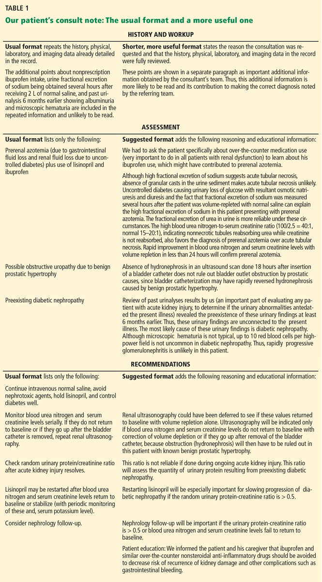
Table 1 compares consultation documentation in the usual format and in the format I am suggesting. The revised format has much more information of educational value (eg, the importance of reviewing past urinalysis results, asking about over-the-counter medications, factors affecting fractional excretion of sodium, effect of bladder catheterization on hydronephrosis due to benign prostatic hypertrophy, and measuring urine protein only after acute kidney injury resolves). It also encourages cost-effective care (ultrasonography could have been delayed or avoided, and the patient could have been cautioned about ibuprofen-like drugs to decrease the risk of recurrent acute kidney injury).
FINAL THOUGHTS
The modifications I have suggested in consult notes will be accepted only if they are reimbursement-neutral. I hope insurers will not equate a shorter note with an opportunity to lower reimbursement and will see the value in not paying for things almost never read. I hope they will recognize and pay for the effort that went into creating a shorter document that contributes adequately to patient care, provides greater educational value, and may promote cost-effective medical practice. Also, not requiring redundant documentation may reduce or even eliminate undesirable copying and pasting.
Accountable-care organizations are an important part of the Affordable Care Act,16 which went into effect in 2014. Many organizations had already come into existence in the United States before the act became effective, and their numbers and the number of patients covered by them are projected to grow enormously over the next few years.17
Since the accountable-care organization model will rely heavily on capitated reimbursement to contain costs, these organizations are likely to scrutinize and curtail the use of consultations. I believe that a shorter consultation note—yet one that is more useful for patient care, education, and cost-containment—is more likely to pass such scrutiny, especially if it decreases time spent on documentation. Furthermore, unlike the fee-for-service model, in a capitated-payment system it may not be necessary to lengthen consultation documentation just to ensure adequate reimbursement.
- Department of Health and Human Services; Office of Inspector General. Consultations in Medicare: coding and reimbursement. http://oig.hhs.gov/oei/reports/oei-09-02-00030.pdf. Accessed November 24, 2014.
- Hartzband P, Groopman J. Off the record—avoiding the pitfalls of going electronic. N Engl J Med 2008; 358:1656–1658.
- O’Malley AS, Grossman JM, Cohen GR, Kemper NM, Pham HH. Are electronic medical records helpful for care coordination? Experiences of physician practices. J Gen Intern Med 2010; 25:177–185.
- The Center for Public Integrity; Schulte F. Electronic medical records probed for over-billing. Critics question credibility of federal panel charged with investigating. www.publicintegrity.org/2013/02/14/12208/electronic-medical-records-probed-over-billing. Accessed November 24, 2014.
- Li B. Cracking the codes: do electronic medical records facilitate hospital revenue enhancement? www.kellogg.northwestern.edu/faculty/b-li/JMP.pdf. Accessed November 24, 2014.
- Hirschtick RE. A piece of my mind. Copy-and-paste. JAMA 2006; 295:2335–2336.
- Thielke S, Hammond K, Helbig S. Copying and pasting of examinations within the electronic medical record. Int J Med Inform 2007; 76(suppl 1):S122–S128.
- Hanlon JT. The electronic medical record: diving into a shallow pool? Cleve Clin J Med 2010; 77:408–411.
- Fitzgerald FT. The emperor’s new clothes. Ann Intern Med 2012; 156:396–397.
- Bernat JL. Ethical and quality pitfalls in electronic health records. Neurology 2013; 80:1057–1061.
- Thornton JD, Schold JD, Venkateshaiah L, Lander B. Prevalence of copied information by attendings and residents in critical care progress notes. Crit Care Med 2013; 41:382–388.
- Foote RS. The challenge to the medical record. JAMA Intern Med 2013; 173:1171–1172.
- Tamburello LM. The road to EMR noncompliance and fraud is paved with cut and paste. MD Advis 2013; 6:24–30.
- Goldman L, Lee T, Rudd P. Ten commandments for effective consultations. Arch Intern Med 1983; 143:1753–1755.
- Salerno SM, Hurst FP, Halvorson S, Mercado DL. Principles of effective consultation: an update for the 21st-century consultant. Arch Intern Med 2007; 167:271–275.
- Longworth DL. Accountable care organizations, the patient-centered medical home, and health care reform: what does it all mean? Cleve Clin J Med 2011; 78:571–582.
- Meyer H. Many accountable care organizations are now up and running, if not off to the races. Health Aff (Millwood) 2012; 31:2363–2367.
After 4 decades of clinical practice in a teaching hospital, I believe that the notes we write to document medical consultations are too long. When I review them for my own patients, the only part I read is the consultant’s assessment and diagnostic and therapeutic recommendations. Many of my colleagues and trainees do the same.
In the old days, when medical records were handwritten, the first three pages of my hospital’s four-page consultation form were for the history, review of systems, physical examination, and test results. The top two-thirds of the last page was for diagnostic impressions and recommendations for additional testing and treatment, to be completed by the trainee performing the consultation.
This left only the bottom third of this page for attestation and additional remarks from the senior consultant. Often, this last (but most used) page was just a bullet list of diagnostic possibilities and suggested tests and treatments, with nothing about the critical reasoning underlying the differential diagnosis and recommendations. This was probably the result of fatigue from having to fill in the first three pages by hand, and then having only limited space on the final page.
Even though the written record has been replaced by the electronic medical record in my hospital, consult notes continue to be at least as long as before, without any change in the length of the assessment and recommendations section. I would guess this is true in most institutions and practices that have switched to an electronic record system.
WHY ARE CONSULT NOTES SO LONG?
The main factor contributing to the lengthy consultation document is that the Center for Medicare and Medicaid Services, with other third-party payers following suit, ties the level of reimbursement to detailed documentation of the history (present, past medical, past surgical, medications, allergies, social, and family), review of systems, and physical examination in the consultation.1 Physicians are under constant pressure from professional fee-coders to meet these requirements.
Since most of this information is already in the medical record, to require that it be documented again in the consultation note is unnecessary duplication. I believe that consultants comply with this requirement mainly to ensure adequate reimbursement, even though they know that the referring medical team will probably not read the repeated information.
Electronic medical record systems, which focus disproportionately on meeting insurers’ requirements governing reimbursement,2–5 have made it easier to create a lengthy consult note by checking boxes in templates and copying and pasting from other parts of the electronic record.2,6–12 Although verbatim copying and pasting may result in punitive audits by insurers, this practice remains common,13 including, in my experience, in consultations.
WHAT ARE THE NEGATIVE EFFECTS OF A NEEDLESSLY LONG CONSULT NOTE?
Time spent on repeating information—even if less time is required when using an electronic system—is clearly time wasted, since this part of the consult note is hardly ever read. Equally bad, the assessment and recommendations section in consult notes continues to be very short, probably because long-standing physician practices change slowly.
An ideal consult note has been described as one that, in addition to addressing the patient care issues, is as brief as possible, avoids duplication of already documented information, and has educational value to the person requesting it.14,15 The educational value of the consultation is especially important in teaching hospitals.
If the only part of the consultation perused in depth consists merely of lists of diagnoses, recommended tests, and therapy and does not include the consultant’s critical reasoning underlying them, the educational value of the consultation is lost.
HOW CAN THE FORMAT BE MADE SHORTER, YET MORE USEFUL?
The note should begin by briefly documenting the reason the consultation was requested. Ideally, institutions should train their staff to state this very specifically. For example, instead of “clearance for surgery,” it is better to ask, “Please identify risks involved in proposed surgery and suggest ways to reduce them.” The former steers the consultant to merely say “cleared for surgery, but with increased risk,” whereas the latter ensures a more specific and detailed response.
The consulting team must review in detail and verify the accuracy of all available information in the patient’s record. Once this is done, instead of repeating it, a statement that all existing information has been thoroughly reviewed should suffice, with mention in a separate paragraph of only the additional relevant positive or negative points in the history related to the issue the consultant has been asked to address.
The consultant shares with all users of the medical record the responsibility of pointing out and correcting any errors in the previously recorded information, thereby decreasing perpetuation of erroneous “chart lore,” an undesirable consequence of copying and pasting. If only previously unrecorded data and corrections to existing information are documented, the referring team is more likely to read the note because it points out relevant information that has been overlooked.
The main part of the document should consist of a detailed assessment and recommendations section, which should include not only a list of diagnoses and recommendations for testing and treatment, but also the consultant’s reasoning behind them, the results of tests already obtained that support the consultant’s conclusions, and information of value for teaching and cost-effective practice. A critically reasoned assessment and recommendation section not only will prove very educational, but by challenging the consultant to justify his or her choices, may discourage unnecessary testing and questionable therapy4,14 and thereby contribute to cost-saving.
My suggestions would not shorten the time spent by the consulting team in evaluating the patient, but only eliminate redundant documentation. I believe the consultation document will be shorter but adequate for patient care, the referring team will read and use the entire document, its educational value will be enhanced, and the time spent on redundant documentation will be saved.
A CASE VIGNETTE
The following vignette (from my own subspecialty) of a patient with acute kidney injury illustrates how a consult note can be made shorter but more useful and educational.
A 78-year-old man had a history of long-standing insulin-requiring diabetes mellitus, hypertension (treated with lisinopril and amlodipine), and benign prostatic hypertrophy. One month earlier, his blood urea nitrogen level had been 15 mg/dL and his serum creatinine had been 1.2 mg/dL.
He presented with a 3-day history of vomiting, diarrhea, and fever, presumed to be viral gastroenteritis. His blood urea nitrogen level was 100 mg, serum creatinine 2.5 mg, and blood glucose 450 mg/dL. Urinalysis revealed 2+ albuminuria, 3+ glucosuria, and 6 red blood cells per high-power field.
In the emergency department he received 2 L of normal saline and regular insulin intravenously, and an indwelling bladder catheter was inserted. He was admitted after 6 hours.
Tests obtained on arrival on the inpatient floor revealed a urinary fractional excretion of sodium of 2.5% and a blood glucose level of 295 mg/dL. His admission history and physical listed his home medications as insulin glargine, amlodipine, lisinopril, and tamsulosin. It also listed the differential diagnosis for acute kidney injury as:
- Prerenal azotemia due to volume depletion
- Rapidly progressive glomerulonephritis to be ruled out in view of proteinuria and microhematuria
- Obstructive uropathy to be ruled out.
Ultrasonography the morning after admission showed normal kidneys and no hydronephrosis. The absence of hydronephrosis was interpreted by the primary team as ruling out obstruction secondary to benign prostatic hypertrophy. The nephrology team saw the patient in consultation the day after admission and discovered the following additional information: urinalysis done 6 months earlier had also shown albuminuria and microhematuria, and the patient had been taking over-the-counter ibuprofen 400 mg three times daily for several days prior to admission.
Table 1 compares consultation documentation in the usual format and in the format I am suggesting. The revised format has much more information of educational value (eg, the importance of reviewing past urinalysis results, asking about over-the-counter medications, factors affecting fractional excretion of sodium, effect of bladder catheterization on hydronephrosis due to benign prostatic hypertrophy, and measuring urine protein only after acute kidney injury resolves). It also encourages cost-effective care (ultrasonography could have been delayed or avoided, and the patient could have been cautioned about ibuprofen-like drugs to decrease the risk of recurrent acute kidney injury).
FINAL THOUGHTS
The modifications I have suggested in consult notes will be accepted only if they are reimbursement-neutral. I hope insurers will not equate a shorter note with an opportunity to lower reimbursement and will see the value in not paying for things almost never read. I hope they will recognize and pay for the effort that went into creating a shorter document that contributes adequately to patient care, provides greater educational value, and may promote cost-effective medical practice. Also, not requiring redundant documentation may reduce or even eliminate undesirable copying and pasting.
Accountable-care organizations are an important part of the Affordable Care Act,16 which went into effect in 2014. Many organizations had already come into existence in the United States before the act became effective, and their numbers and the number of patients covered by them are projected to grow enormously over the next few years.17
Since the accountable-care organization model will rely heavily on capitated reimbursement to contain costs, these organizations are likely to scrutinize and curtail the use of consultations. I believe that a shorter consultation note—yet one that is more useful for patient care, education, and cost-containment—is more likely to pass such scrutiny, especially if it decreases time spent on documentation. Furthermore, unlike the fee-for-service model, in a capitated-payment system it may not be necessary to lengthen consultation documentation just to ensure adequate reimbursement.
- Department of Health and Human Services; Office of Inspector General. Consultations in Medicare: coding and reimbursement. http://oig.hhs.gov/oei/reports/oei-09-02-00030.pdf. Accessed November 24, 2014.
- Hartzband P, Groopman J. Off the record—avoiding the pitfalls of going electronic. N Engl J Med 2008; 358:1656–1658.
- O’Malley AS, Grossman JM, Cohen GR, Kemper NM, Pham HH. Are electronic medical records helpful for care coordination? Experiences of physician practices. J Gen Intern Med 2010; 25:177–185.
- The Center for Public Integrity; Schulte F. Electronic medical records probed for over-billing. Critics question credibility of federal panel charged with investigating. www.publicintegrity.org/2013/02/14/12208/electronic-medical-records-probed-over-billing. Accessed November 24, 2014.
- Li B. Cracking the codes: do electronic medical records facilitate hospital revenue enhancement? www.kellogg.northwestern.edu/faculty/b-li/JMP.pdf. Accessed November 24, 2014.
- Hirschtick RE. A piece of my mind. Copy-and-paste. JAMA 2006; 295:2335–2336.
- Thielke S, Hammond K, Helbig S. Copying and pasting of examinations within the electronic medical record. Int J Med Inform 2007; 76(suppl 1):S122–S128.
- Hanlon JT. The electronic medical record: diving into a shallow pool? Cleve Clin J Med 2010; 77:408–411.
- Fitzgerald FT. The emperor’s new clothes. Ann Intern Med 2012; 156:396–397.
- Bernat JL. Ethical and quality pitfalls in electronic health records. Neurology 2013; 80:1057–1061.
- Thornton JD, Schold JD, Venkateshaiah L, Lander B. Prevalence of copied information by attendings and residents in critical care progress notes. Crit Care Med 2013; 41:382–388.
- Foote RS. The challenge to the medical record. JAMA Intern Med 2013; 173:1171–1172.
- Tamburello LM. The road to EMR noncompliance and fraud is paved with cut and paste. MD Advis 2013; 6:24–30.
- Goldman L, Lee T, Rudd P. Ten commandments for effective consultations. Arch Intern Med 1983; 143:1753–1755.
- Salerno SM, Hurst FP, Halvorson S, Mercado DL. Principles of effective consultation: an update for the 21st-century consultant. Arch Intern Med 2007; 167:271–275.
- Longworth DL. Accountable care organizations, the patient-centered medical home, and health care reform: what does it all mean? Cleve Clin J Med 2011; 78:571–582.
- Meyer H. Many accountable care organizations are now up and running, if not off to the races. Health Aff (Millwood) 2012; 31:2363–2367.