User login
Welcome to three new Editorial Advisory Board members!
We are pleased to welcome Dr. Joseph B. Domachowske, Dr. Howard Smart, and Dr. Francis E. Rushton Jr. to the Pediatric News Editorial Advisory Board.

Dr. Domachowske is professor of pediatrics and professor of microbiology and immunology at the State University of New York Upstate Medical University in Syracuse. He serves on the New York State American Academy of Pediatrics Chapter 1 executive committee, volunteers as his district’s immunization champion, and is an appointed member of the New York State Immunization Advisory Council. He also enjoys his work as an AAP PREP-ID editorial board member. His overlapping clinical and research interests include immunization advocacy and studies related to the treatment and prevention of viral respiratory tract infections, particularly respiratory syncytial virus. He has published more than 120 papers and book chapters in these areas. Dr. Domachowske has had the privilege of presenting on his global vaccine advocacy efforts with funding through AAP’s Shot@Life program.

Dr. Rushton Jr. is a clinical professor of pediatrics at the University of South Carolina, Columbia, and medical director of the Quality Through Innovation in Pediatrics (QTIP) network. He has practiced pediatrics in Beaufort, S.C., for 32 years and is the author of “Family Support in Community Pediatrics, Confronting the Challenge. “Dr. Rushton’s academic interests include quality improvement, community pediatrics, early brain development, home visitation, and group well child care.

Dr. Smart practices general pediatrics and adolescent medicine as a member of the Sharp Rees-Stealy Medical Group in San Diego. He is a voluntary assistant clinical professor of pediatrics at the University of California, San Diego, and is currently chief of pediatrics at Sharp Mary Birch Hospital for Women & Newborns. Dr. Smart’s interests include medical informatics, health care IT, and specifically clinical decision support and the use of data to drive clinical quality improvement.
We are pleased to welcome Dr. Joseph B. Domachowske, Dr. Howard Smart, and Dr. Francis E. Rushton Jr. to the Pediatric News Editorial Advisory Board.
Dr. Domachowske is professor of pediatrics and professor of microbiology and immunology at the State University of New York Upstate Medical University in Syracuse. He serves on the New York State American Academy of Pediatrics Chapter 1 executive committee, volunteers as his district’s immunization champion, and is an appointed member of the New York State Immunization Advisory Council. He also enjoys his work as an AAP PREP-ID editorial board member. His overlapping clinical and research interests include immunization advocacy and studies related to the treatment and prevention of viral respiratory tract infections, particularly respiratory syncytial virus. He has published more than 120 papers and book chapters in these areas. Dr. Domachowske has had the privilege of presenting on his global vaccine advocacy efforts with funding through AAP’s Shot@Life program.
Dr. Rushton Jr. is a clinical professor of pediatrics at the University of South Carolina, Columbia, and medical director of the Quality Through Innovation in Pediatrics (QTIP) network. He has practiced pediatrics in Beaufort, S.C., for 32 years and is the author of “Family Support in Community Pediatrics, Confronting the Challenge. “Dr. Rushton’s academic interests include quality improvement, community pediatrics, early brain development, home visitation, and group well child care.
Dr. Smart practices general pediatrics and adolescent medicine as a member of the Sharp Rees-Stealy Medical Group in San Diego. He is a voluntary assistant clinical professor of pediatrics at the University of California, San Diego, and is currently chief of pediatrics at Sharp Mary Birch Hospital for Women & Newborns. Dr. Smart’s interests include medical informatics, health care IT, and specifically clinical decision support and the use of data to drive clinical quality improvement.

Incidentalomas: You can hate them but can’t ignore them
We order a lot of MRIs. Patients, in general, want a lot of MRIs (as long as insurance covers them). We have all this cool technology, so why not use it?
For the most part we’re doing them to rule-out bad things such as gliomas and ginormous aneurysms so we can say: “It looks fine, so I think you’re just having headaches/migraines/anxiety attacks, whatever.”
Unfortunately, as technology improves, we end up with a whole new issue that previous generations of neurologists didn’t have to deal with: the hated incidentaloma. And, often, this can be insanely frustrating. Just because an abnormality isn’t related to the symptoms doesn’t mean you can forget about it, either.
It’s amazing how many scans come back with small meningiomas, aneurysms, microadenomas, etc. Once you find them, you (and the patient) are stuck with it.
I usually try to downplay these findings, as they’re typically irrelevant. But, even then, you’re now obligated to repeat the scans every 1-5 years (depending on what you found) to make sure the thingamajig is stable. Which only drives up costs for the patient and their insurance.
Then there’s the aspect of how the patient sees this. Most are perfectly fine when you explain it to them, but you get some who are panicked (“OMG! I have a brain tumor!”) and require quite a bit of time to calm down.
There are others who latch onto it, and insist, against all rational evidence, that it’s the sole cause of their symptoms. They will often call at 2:00 a.m. for the slightest change in their symptoms or just go straight to an emergency department “because I have an aneurysm.” Trying to get them to accept that the finding is incidental is often a challenge, with them often seeking multiple other opinions.
In the best case, though, the finding is a nuisance to all involved. I have to enter that patient in my scheduled reminders to order a follow-up study. If they don’t respond to a phone call, or regular letter, I have to send them a certified letter. From their view they have to work another MRI into what’s probably a busy schedule. Depending on their deductible, they may have to pay a decent amount of money for it. And then it may add paperwork next time they apply for life insurance.
What’s to be done for it? Nothing that I can think of. If we don’t pursue the testing, we become legally liable if the lesion grows. The patient could decline it, but most don’t. And, as scans improve, the number of incidentalomas will increase.
The revolution that MRI has brought to neurology can’t be understated. But, at the same time, it has its drawbacks. For both patients and neurologists, dealing with the incidentals and their consequences is one of them.
Dr. Block has a solo neurology practice in Scottsdale, Ariz.

Nitrous oxide for labor pain
Neuraxial anesthesia, including epidural and combined spinal-epidural anesthetics, are the “gold standard” interventions for pain relief during labor because they provide a superb combination of reliable pain relief and safety for the mother and child.1 Many US birthing centers also offer additional options for managing labor pain, including continuous labor support,2 hydrotherapy,3 and parenteral opioids.4 In 2012, the US Food and Drug Administration (FDA) approved equipment to deliver a mixture of 50% nitrous oxide and 50% oxygen, which has offered a new option for laboring mothers.
Nitrous oxide is widely used for labor pain in the United Kingdom, Finland, Sweden, Canada, Australia, and New Zealand.5 In the United States, nitrous oxide has been a long-standing and common adjunct to general anesthetics, although it recently has fallen out of favor in place of better, more rapidly acting inhalation and intravenous general anesthetics. With these agents not suitable for labor analgesic use, however, nitrous oxide is undergoing a resurgence in popularity for obstetric analgesia in the United States, and we believe that it will evolve to have a prominent place among our interventions for labor pain.6 In this editorial, we detail the mechanism of action and the equipment’s use, as well as benefits for patients and cautions for clinicians.
How does nitrous oxide work?
Pharmacology. Nitrous oxide (N2O) was first synthesized by Joseph Priestley in 1772 and was used as an anesthetic for dental surgery in the mid-1800s. In the late 19th Century, nitrous oxide was tested as an agent for labor analgesia.7 It was introduced into clinical practice in the United Kingdom in the 1930s.8
The mechanism of action of nitrous oxide is not fully characterized. It is thought that the gas may produce analgesia by activating the endogenous opioid and noradrenergic systems, which in turn, modulate spinal cord transmission of pain signals.5

Administration to the laboring mother. For labor analgesia, nitrous oxide is typically administered as a mix of 50% N2O and 50% O2 using a portable unit with a gas mixer that is fed by small tanks of N2O and O2 or with a valve fed by a single tank containing a mixture of both N2O and O2. The portable units approved by the FDA contain an oxygen fail-safe system that ensures delivery of an appropriate oxygen concentration. The portable unit also contains a gas scavenging system that is attached to wall suction. The breathing circuit has a mask or a mouthpiece (according to patient preference) and demand valve. The patient places the mask over her nose and mouth, or uses just her mouth for the mouthpiece. With inhalation, the demand valve opens, releasing the gas mixture. On exhalation, the valve shunts the exhaled gases to the scavenging system.
Proper and safe use requires adherence to the principles of a true “patient-controlled” protocol. Only the patient is permitted to place the mask or mouthpiece over her nose and/or mouth. If the patient becomes drowsy, such that she cannot hold the mask to her face, then the internal demand valve will not deliver nitrous oxide and she will return to breathing room air. No one should hold the mask over the patient’s nose or mouth, and the mask should not be fixed in place with elastic bands because these actions may result in the inhalation of too much nitrous oxide.
Nitrous oxide has a rapid onset of action after inhalation and its action quickly dissipates after discontinuing inhalation. There is likely a dose-response relationship, with greater use of the nitrous oxide producing more drowsiness. With the intermittent inhalation method, the laboring patient using nitrous oxide is advised to initiate inhalation of nitrous oxide about 30 seconds before the onset of a contraction and discontinue inhalation at the peak of the contraction.
There is no time limit to the use of nitrous oxide. It can be used for hours during labor or only briefly for a particularly painful part of labor, such as during rapid cervical dilation or during the later portions of the second stage.
Patients report that nitrous oxide does not completely relieve pain but creates a diminished perception of the pain.9 As many as one-third of women are nonresponders and report no significant pain improvement with nitrous oxide use.10
The main side effects of inhalation of the gas are nausea, vomiting, dizziness, and drowsiness. Nausea has been reported in 5% to 40% of women, and vomiting has been reported in up to 15% of women using nitrous oxide.11
Cautions
Contraindications to nitrous oxide include a baseline arterial oxygenation saturation less than 95% on room air, acute asthma, emphysema, or pneumothorax, or any other air-filled compartment within the body, such as bowel obstruction or pneumocephalus. (Nitrous oxide can displace nitrogen from closed body spaces, which may lead to an increase in the volume of the closed space.12)
Nitrous oxide inactivates vitamin B12 by oxidation; therefore, vitamin B12 deficiency or related disorders may be considered a relative contraindication. However, compared with more extensive continuous use, such as during prolonged general anesthesia, intermittent use for a limited time during labor is associated with minimal to no hematologic effects.
If a laboring woman is using N2O, parenteral opioids should be administered only with great caution by an experienced clinician.
What do the data indicate?
The Agency for Healthcare Research and Quality (AHRQ) recently invited the Vanderbilt Evidence-based Practice Center to review the world literature on nitrous oxide for labor pain and to provide a summary of the research. Fifty-eight publications were identified, with 46 rated as poor quality.11,13 Given this overall poor quality of available research, many of the recommendations concerning the use of nitrous oxide for labor pain are based on clinical experience and expert opinion.
The experts concluded that, for the relief of labor pain, neuraxial anesthesia was more effective than nitrous oxide inhalation. In one randomized trial included in their systematic review, nulliparous laboring women were randomly assigned to neuraxial anesthesia or nitrous oxide plus meperidine.14 About 94% of nulliparous laboring women reported satisfaction with neuraxial anesthesia, compared with 54% treated with nitrous oxide and meperidine.14
Nitrous oxide is believed to be generally safe for mother and fetus. Its use does not impact the newborn Apgar score15 or alter uterine
contractility.16
Considering a nitrous oxide program for your birthing unit? Helpful hints to get started.
Catherine McGovern, RN, MSN, CNM
- Do your research to determine which type of equipment is right for the size and volume of your organization.
You need to consider ease of access and use for staff to bring this option to the bedside in a prompt and safe manner. Initial research includes visiting or speaking with practitioners on units currently using nitrous oxide. Use of nitrous oxide is growing, and networking is helpful in terms of planning your program. Making sure you have the correct gas line connectors for oxygen as well as for suction when using a scavenger system is a preliminary necessity. - Determine storage ability.
Your environmental safety officer is a good resource to determine location and regulations regarding safe storage as well as tank capacity. He or she also can help you determine where else in your organization nitrous oxide is used so you may be able to develop your unit-specific protocol from hospital-wide policy that is already in place. - Collaborate on a protocol.
After determining which type of equipment is best for you, propose the idea to committees that can contribute to the development of pain and sedation management protocols. The anesthesia department, pain committee, and postoperative pain management teams are knowledgeable resources and can help you write a safe protocol. Keep as the main focus the safe application and use of nitrous oxide for various patient populations. Potential medication interactions and contraindications for use should be discussed and included in a protocol.
One more department you want to include in your planning is infection control. For our unit, reviewing various types of equipment to determine the best infection control revealed some interesting design benefits to reduce infection risk. Because the nitrous oxide equipment would be mobile, the types of filter options, disposal options, and cleaning ability are important components for final equipment choice. - Include all parties in training and final roll out.
Once you develop your policy with input from all stakeholders, make sure you share it early and often before you go live. Include midwives, physicians, nurses, technicians, and administrative staff in training, which will help to dispel myths and increase awareness of availability within your unit. Provide background information to all trainees to ensure safe use and appropriate patient selection.
The most important determinant of success is the formation of an interprofessional team that works well together to develop a safe clinician- and patient-friendly program for the use of nitrous oxide.
Nitrous oxide, a bridge to an epidural or a natural childbirth
Many women start labor unsure about whether they want to use an epidural. For these women, nitrous oxide may be an option for reducing labor pain, thereby giving the woman more time to make a decision about whether to have an epidural anesthetic. In our practice, a significant percentage of women who use nitrous oxide early in labor subsequently request a neuraxial anesthetic. However, many women planning natural childbirth use nitrous oxide to reduce labor pain and successfully achieve their goal.
Postpartum pain reliever
Some women deliver without the use of any pain medicine. Sometimes birth is complicated by perineal lacerations requiring significant surgical repair. If a woman does not have adequate analgesia after injection of a local anesthetic, nitrous oxide may help reduce her pain during the perineal repair and facilitate quick completion of the procedure by allowing her to remain still. N2O also has been used to facilitate analgesia during manual removal of the placenta.
We predict an expanding role
There are many pharmacologic and nonpharmacologic options for managing labor pain, including a supportive birth environment, touch and massage, maternal positioning, relaxation and breathing techniques, continuous labor support, hydrotherapy, opioids, and neuraxial anesthesia. Midwives, labor nurses, and physicians have championed increasing the availability of nitrous oxide to laboring women in US birthing centers.17–20 With the FDA approval of inexpensive portable nitrous oxide units, it is likely that we will witness a resurgence of its use and gain important clinical experience in the role of nitrous oxide for managing labor pain.
Share your thoughts on this article! Send your Letter to the Editor to [email protected]. Please include your name and the city and state in which you practice.
1. Amin-Somuah M, Smyth R, Jones L. Epidural versus non-epidural or no analgesia in labour. Cochrane Database Syst Rev. 2011;(12):CD000331.
2. Hodnett ED, Gates S, Hofmeyr JG, Sakala C. Continuous support for women during childbirth. Cochrane Database Syst Rev. 2013;(7):CD003766.
3. Cluett ER, Burns E. Immersion in water in labour and birth. Cochrane Database Syst Rev. 2009;(2):CD000111.
4. Ullman R, Smith LA, Burns E, Mori R, Dowswell T. Parenteral opioids for maternal pain management in labor. Cochrane Database Syst Rev. 2010;(9):CD007396.
5. Rosen MA. Nitrous oxide for relief of labor pain: a systematic review. Am J Obstet Gynecol. 2002;186(5 suppl Nature):S110–S126.
6. Klomp T, van Poppel M, Jones L, Lazet J, Di Nisio M, Lagro-Janssen A. Inhaled analgesia for pain management in labor. Cochrane Database Syst Rev. 2012;(9):CD009351.
7. Richards W, Parbrook G, Wilson J. Stanislav Klikovitch (1853-1910). Pioneer of nitrous oxide and oxygen analgesia. Anaesthesia. 1976;31(7):933–940.
8. Minnitt R. Self-administered anesthesia in childbirth. Br Med J. 1934;1:501–503.
9. Camann W, Alexander K. Easy labor: Every Woman’s Guide to Choosing Less Pain and More Joy during Childbirth. New York: Ballantine Books; 2007.
10. Rosen M, Mushin WW, Jones PL, Jones EV. Field trial of methoxyflurane, nitrous oxide, and trichloroethylene as obstetric analgesics. Br Med J. 1969;3(5665):263–267.
11. Likis FE, Andrews JC, Collins MR, et al. Nitrous oxide for the management of labor pain: a systematic review. Anesth Analg. 2014;118(1):153–167.
12. Eger EI 2nd, Saidman LJ. Hazards of nitrous oxide anesthesia in bowel obstruction and pneumothorax. Anesthesiology. 1965;26:61–66.
13. Agency for Healthcare Research and Quality. Nitrous oxide for the management of labor pain. Comparative Effectiveness Review Number 67. August 2012. http://www.effectivehealthcare.ahrq.gov/ehc/products/260/1175/CER67_NitrousOxideLaborPain_FinalReport_20120817.pdf. Accessed November 7, 2014.
14. Leong EW, Sivanesaratnam V, Oh LL, Chan YK. Epidural analgesia in primigravidae in spontaneous labor at term: a prospective study. J Obstet Gynaecol Res. 2000;26(4):271–275.
15. Clinical trials of different concentrations of oxygen and nitrous oxide for obstetric analgesia. Report to the Medical Research Council of the Committee on Nitrous Oxide and Oxygen Analgesia in Midwifery. Br Med J. 1970;1(5698):709–713.
16. Vasicka A, Kretchmer H. Effect of conduction and inhalation anesthesia on uterine contractions. Am J Obstet Gynecol. 1961;82:600–611.
17. Rooks JP. Labor pain management other than neuraxial: what do we know and where do we go next? Birth. 2012;39(4):318–322.
18. American College of Nurse-Midwives. From the American College of Nurse-Midwives. Nitrous oxide for labor analgesia. J Midwifery Womens Health. 2010;55(3):292–296.
19. Bishop JT. Administration of nitrous oxide in labor: expanding the options for women. J Midwifery Womens Health. 2007;52(3):308–309.
20. Rooks JP. Nitrous oxide for pain in labor—why not in the United States? Birth. 2007;34(1):3–5.
Robert L. Barbieri, MD; William Camann, MD; and Catherine McGovern, RN, MSN, CNM

Dr. Barbieri is Editor in Chief, OBG Management; Chair, Obstetrics and Gynecology, at Brigham and Women’s Hospital, Boston, Massachusetts; and Kate Macy Ladd Professor of Obstetrics, Gynecology, and Reproductive Biology at Harvard Medical School, Boston.

Dr. Camann is Associate Professor, Anesthesia and Pain Management, Harvard Medical School; Director, Obstetric Anesthesiology, Brigham and Women’s Hospital, Boston, Massachusetts.

Ms. McGovern is Clinical Educator, Center for Labor and Birth, at Brigham and Women’s Hospital, Boston, Massachusetts.
The authors report no financial relationships relevant to this article.
Robert L. Barbieri, MD; William Camann, MD; and Catherine McGovern, RN, MSN, CNM
Dr. Barbieri is Editor in Chief, OBG Management; Chair, Obstetrics and Gynecology, at Brigham and Women’s Hospital, Boston, Massachusetts; and Kate Macy Ladd Professor of Obstetrics, Gynecology, and Reproductive Biology at Harvard Medical School, Boston.
Dr. Camann is Associate Professor, Anesthesia and Pain Management, Harvard Medical School; Director, Obstetric Anesthesiology, Brigham and Women’s Hospital, Boston, Massachusetts.
Ms. McGovern is Clinical Educator, Center for Labor and Birth, at Brigham and Women’s Hospital, Boston, Massachusetts.
The authors report no financial relationships relevant to this article.
Neuraxial anesthesia, including epidural and combined spinal-epidural anesthetics, are the “gold standard” interventions for pain relief during labor because they provide a superb combination of reliable pain relief and safety for the mother and child.1 Many US birthing centers also offer additional options for managing labor pain, including continuous labor support,2 hydrotherapy,3 and parenteral opioids.4 In 2012, the US Food and Drug Administration (FDA) approved equipment to deliver a mixture of 50% nitrous oxide and 50% oxygen, which has offered a new option for laboring mothers.
Nitrous oxide is widely used for labor pain in the United Kingdom, Finland, Sweden, Canada, Australia, and New Zealand.5 In the United States, nitrous oxide has been a long-standing and common adjunct to general anesthetics, although it recently has fallen out of favor in place of better, more rapidly acting inhalation and intravenous general anesthetics. With these agents not suitable for labor analgesic use, however, nitrous oxide is undergoing a resurgence in popularity for obstetric analgesia in the United States, and we believe that it will evolve to have a prominent place among our interventions for labor pain.6 In this editorial, we detail the mechanism of action and the equipment’s use, as well as benefits for patients and cautions for clinicians.
How does nitrous oxide work?
Pharmacology. Nitrous oxide (N2O) was first synthesized by Joseph Priestley in 1772 and was used as an anesthetic for dental surgery in the mid-1800s. In the late 19th Century, nitrous oxide was tested as an agent for labor analgesia.7 It was introduced into clinical practice in the United Kingdom in the 1930s.8
The mechanism of action of nitrous oxide is not fully characterized. It is thought that the gas may produce analgesia by activating the endogenous opioid and noradrenergic systems, which in turn, modulate spinal cord transmission of pain signals.5
Administration to the laboring mother. For labor analgesia, nitrous oxide is typically administered as a mix of 50% N2O and 50% O2 using a portable unit with a gas mixer that is fed by small tanks of N2O and O2 or with a valve fed by a single tank containing a mixture of both N2O and O2. The portable units approved by the FDA contain an oxygen fail-safe system that ensures delivery of an appropriate oxygen concentration. The portable unit also contains a gas scavenging system that is attached to wall suction. The breathing circuit has a mask or a mouthpiece (according to patient preference) and demand valve. The patient places the mask over her nose and mouth, or uses just her mouth for the mouthpiece. With inhalation, the demand valve opens, releasing the gas mixture. On exhalation, the valve shunts the exhaled gases to the scavenging system.
Proper and safe use requires adherence to the principles of a true “patient-controlled” protocol. Only the patient is permitted to place the mask or mouthpiece over her nose and/or mouth. If the patient becomes drowsy, such that she cannot hold the mask to her face, then the internal demand valve will not deliver nitrous oxide and she will return to breathing room air. No one should hold the mask over the patient’s nose or mouth, and the mask should not be fixed in place with elastic bands because these actions may result in the inhalation of too much nitrous oxide.
Nitrous oxide has a rapid onset of action after inhalation and its action quickly dissipates after discontinuing inhalation. There is likely a dose-response relationship, with greater use of the nitrous oxide producing more drowsiness. With the intermittent inhalation method, the laboring patient using nitrous oxide is advised to initiate inhalation of nitrous oxide about 30 seconds before the onset of a contraction and discontinue inhalation at the peak of the contraction.
There is no time limit to the use of nitrous oxide. It can be used for hours during labor or only briefly for a particularly painful part of labor, such as during rapid cervical dilation or during the later portions of the second stage.
Patients report that nitrous oxide does not completely relieve pain but creates a diminished perception of the pain.9 As many as one-third of women are nonresponders and report no significant pain improvement with nitrous oxide use.10
The main side effects of inhalation of the gas are nausea, vomiting, dizziness, and drowsiness. Nausea has been reported in 5% to 40% of women, and vomiting has been reported in up to 15% of women using nitrous oxide.11
Cautions
Contraindications to nitrous oxide include a baseline arterial oxygenation saturation less than 95% on room air, acute asthma, emphysema, or pneumothorax, or any other air-filled compartment within the body, such as bowel obstruction or pneumocephalus. (Nitrous oxide can displace nitrogen from closed body spaces, which may lead to an increase in the volume of the closed space.12)
Nitrous oxide inactivates vitamin B12 by oxidation; therefore, vitamin B12 deficiency or related disorders may be considered a relative contraindication. However, compared with more extensive continuous use, such as during prolonged general anesthesia, intermittent use for a limited time during labor is associated with minimal to no hematologic effects.
If a laboring woman is using N2O, parenteral opioids should be administered only with great caution by an experienced clinician.
What do the data indicate?
The Agency for Healthcare Research and Quality (AHRQ) recently invited the Vanderbilt Evidence-based Practice Center to review the world literature on nitrous oxide for labor pain and to provide a summary of the research. Fifty-eight publications were identified, with 46 rated as poor quality.11,13 Given this overall poor quality of available research, many of the recommendations concerning the use of nitrous oxide for labor pain are based on clinical experience and expert opinion.
The experts concluded that, for the relief of labor pain, neuraxial anesthesia was more effective than nitrous oxide inhalation. In one randomized trial included in their systematic review, nulliparous laboring women were randomly assigned to neuraxial anesthesia or nitrous oxide plus meperidine.14 About 94% of nulliparous laboring women reported satisfaction with neuraxial anesthesia, compared with 54% treated with nitrous oxide and meperidine.14
Nitrous oxide is believed to be generally safe for mother and fetus. Its use does not impact the newborn Apgar score15 or alter uterine
contractility.16
Considering a nitrous oxide program for your birthing unit? Helpful hints to get started.
Catherine McGovern, RN, MSN, CNM
- Do your research to determine which type of equipment is right for the size and volume of your organization.
You need to consider ease of access and use for staff to bring this option to the bedside in a prompt and safe manner. Initial research includes visiting or speaking with practitioners on units currently using nitrous oxide. Use of nitrous oxide is growing, and networking is helpful in terms of planning your program. Making sure you have the correct gas line connectors for oxygen as well as for suction when using a scavenger system is a preliminary necessity. - Determine storage ability.
Your environmental safety officer is a good resource to determine location and regulations regarding safe storage as well as tank capacity. He or she also can help you determine where else in your organization nitrous oxide is used so you may be able to develop your unit-specific protocol from hospital-wide policy that is already in place. - Collaborate on a protocol.
After determining which type of equipment is best for you, propose the idea to committees that can contribute to the development of pain and sedation management protocols. The anesthesia department, pain committee, and postoperative pain management teams are knowledgeable resources and can help you write a safe protocol. Keep as the main focus the safe application and use of nitrous oxide for various patient populations. Potential medication interactions and contraindications for use should be discussed and included in a protocol.
One more department you want to include in your planning is infection control. For our unit, reviewing various types of equipment to determine the best infection control revealed some interesting design benefits to reduce infection risk. Because the nitrous oxide equipment would be mobile, the types of filter options, disposal options, and cleaning ability are important components for final equipment choice. - Include all parties in training and final roll out.
Once you develop your policy with input from all stakeholders, make sure you share it early and often before you go live. Include midwives, physicians, nurses, technicians, and administrative staff in training, which will help to dispel myths and increase awareness of availability within your unit. Provide background information to all trainees to ensure safe use and appropriate patient selection.
The most important determinant of success is the formation of an interprofessional team that works well together to develop a safe clinician- and patient-friendly program for the use of nitrous oxide.
Nitrous oxide, a bridge to an epidural or a natural childbirth
Many women start labor unsure about whether they want to use an epidural. For these women, nitrous oxide may be an option for reducing labor pain, thereby giving the woman more time to make a decision about whether to have an epidural anesthetic. In our practice, a significant percentage of women who use nitrous oxide early in labor subsequently request a neuraxial anesthetic. However, many women planning natural childbirth use nitrous oxide to reduce labor pain and successfully achieve their goal.
Postpartum pain reliever
Some women deliver without the use of any pain medicine. Sometimes birth is complicated by perineal lacerations requiring significant surgical repair. If a woman does not have adequate analgesia after injection of a local anesthetic, nitrous oxide may help reduce her pain during the perineal repair and facilitate quick completion of the procedure by allowing her to remain still. N2O also has been used to facilitate analgesia during manual removal of the placenta.
We predict an expanding role
There are many pharmacologic and nonpharmacologic options for managing labor pain, including a supportive birth environment, touch and massage, maternal positioning, relaxation and breathing techniques, continuous labor support, hydrotherapy, opioids, and neuraxial anesthesia. Midwives, labor nurses, and physicians have championed increasing the availability of nitrous oxide to laboring women in US birthing centers.17–20 With the FDA approval of inexpensive portable nitrous oxide units, it is likely that we will witness a resurgence of its use and gain important clinical experience in the role of nitrous oxide for managing labor pain.
Share your thoughts on this article! Send your Letter to the Editor to [email protected]. Please include your name and the city and state in which you practice.
1. Amin-Somuah M, Smyth R, Jones L. Epidural versus non-epidural or no analgesia in labour. Cochrane Database Syst Rev. 2011;(12):CD000331.
2. Hodnett ED, Gates S, Hofmeyr JG, Sakala C. Continuous support for women during childbirth. Cochrane Database Syst Rev. 2013;(7):CD003766.
3. Cluett ER, Burns E. Immersion in water in labour and birth. Cochrane Database Syst Rev. 2009;(2):CD000111.
4. Ullman R, Smith LA, Burns E, Mori R, Dowswell T. Parenteral opioids for maternal pain management in labor. Cochrane Database Syst Rev. 2010;(9):CD007396.
5. Rosen MA. Nitrous oxide for relief of labor pain: a systematic review. Am J Obstet Gynecol. 2002;186(5 suppl Nature):S110–S126.
6. Klomp T, van Poppel M, Jones L, Lazet J, Di Nisio M, Lagro-Janssen A. Inhaled analgesia for pain management in labor. Cochrane Database Syst Rev. 2012;(9):CD009351.
7. Richards W, Parbrook G, Wilson J. Stanislav Klikovitch (1853-1910). Pioneer of nitrous oxide and oxygen analgesia. Anaesthesia. 1976;31(7):933–940.
8. Minnitt R. Self-administered anesthesia in childbirth. Br Med J. 1934;1:501–503.
9. Camann W, Alexander K. Easy labor: Every Woman’s Guide to Choosing Less Pain and More Joy during Childbirth. New York: Ballantine Books; 2007.
10. Rosen M, Mushin WW, Jones PL, Jones EV. Field trial of methoxyflurane, nitrous oxide, and trichloroethylene as obstetric analgesics. Br Med J. 1969;3(5665):263–267.
11. Likis FE, Andrews JC, Collins MR, et al. Nitrous oxide for the management of labor pain: a systematic review. Anesth Analg. 2014;118(1):153–167.
12. Eger EI 2nd, Saidman LJ. Hazards of nitrous oxide anesthesia in bowel obstruction and pneumothorax. Anesthesiology. 1965;26:61–66.
13. Agency for Healthcare Research and Quality. Nitrous oxide for the management of labor pain. Comparative Effectiveness Review Number 67. August 2012. http://www.effectivehealthcare.ahrq.gov/ehc/products/260/1175/CER67_NitrousOxideLaborPain_FinalReport_20120817.pdf. Accessed November 7, 2014.
14. Leong EW, Sivanesaratnam V, Oh LL, Chan YK. Epidural analgesia in primigravidae in spontaneous labor at term: a prospective study. J Obstet Gynaecol Res. 2000;26(4):271–275.
15. Clinical trials of different concentrations of oxygen and nitrous oxide for obstetric analgesia. Report to the Medical Research Council of the Committee on Nitrous Oxide and Oxygen Analgesia in Midwifery. Br Med J. 1970;1(5698):709–713.
16. Vasicka A, Kretchmer H. Effect of conduction and inhalation anesthesia on uterine contractions. Am J Obstet Gynecol. 1961;82:600–611.
17. Rooks JP. Labor pain management other than neuraxial: what do we know and where do we go next? Birth. 2012;39(4):318–322.
18. American College of Nurse-Midwives. From the American College of Nurse-Midwives. Nitrous oxide for labor analgesia. J Midwifery Womens Health. 2010;55(3):292–296.
19. Bishop JT. Administration of nitrous oxide in labor: expanding the options for women. J Midwifery Womens Health. 2007;52(3):308–309.
20. Rooks JP. Nitrous oxide for pain in labor—why not in the United States? Birth. 2007;34(1):3–5.

Uterus ruptures at home: My most memorable experience of a transferred home birth

Uterus ruptures at home
A woman (G4P3) had undergone two cesarean deliveries followed by successful vaginal birth after cesarean (VBAC) in hospital. During her second trimester, the patient decided, against the advice of her ObGyn, to have a home delivery. Her midwife was present when, after several hours, the mother felt a sudden sharp pain. When the midwife detected fetal heart-rate tones of 60–70 bpm, she called 911. The patient was transported to the labor and delivery (L&D) unit where I was the in-house ObGyn on call for unattached patients.
In triage, the baby’s heart rate was in the 60-bpm range. I found no presenting part of the fetus on vaginal examination; the patient had a surgical abdomen on palpation. She was immediately taken for an emergency cesarean delivery. We found the baby halfway out of a uterine rupture. The placenta was still partially attached to the fundus.
The baby’s blood gases were too low to register on the machine. She was resuscitated, but still had no suck or gag reflex when discharged from the neonatal intensive care unit (NICU), with minimal brainstem function.
The mother didn’t require blood products because the rupture was barely bleeding. The uterine rupture occurred over the entire scar area from previous cesarean deliveries. The mother was in our hospital for a total of 14 minutes before delivery occurred.
I have no collaborative agreement with any midwife for accepting their failed home birth attempts.
Kevin Fulford, MD
San Diego, California
A perfect candidate for home birth?
In my last call as a resident, a patient arrived at L&D after arrest of labor at 5 cm for many hours. The patient was a G1P0 at term with twins and the presenting twin was in a known breech presentation. The patient quickly agreed to a cesarean delivery and has two healthy babies.
I will never forget the patient’s lay midwife telling me that the mother was the “perfect candidate for a home birth.” She seemed so disappointed by the patient’s decision to head to the hospital. It made for a memorable last night as a resident!
Erinn Hoekstra, MD
Grand Rapids, Michigan
Why women choose out-of-hospital birth
As a Board-Certified ObGyn who attends women at home, at a birthing center, and in hospital, I find this article lacking in two ways: why women choose out-of-hospital birth and why maternal outcomes at home are never discussed.
Women choose out-of-hospital birth because they are refused meaningful decision making in their labor and birth processes. They are refused VBACs, vaginal delivery of breech presentations, food and drink, and are forced to accept continuous electronic fetal monitoring (EFM), to name but a few. They also choose to stay away from the hospital because of the cascade of unscientific interventions that lead to unnecessary cesarean deliveries.
Noted in the article are neonatal outcomes, but not maternal outcomes, which are universally better outside the hospital. Fewer than 2% of women birth at home. Therefore, they can’t be responsible for the skyrocketing maternal mortality rate, the ever-increasing induction and cesarean delivery rates, and attendant accretas, etc. We have reached the point that Semmelweiss noted in Vienna in the 1800s: women who deliver outside the hospital are less likely to die than those who deliver inside.
So much hand-wringing over the few who stay away from the hospital rather than reforming the practice of obstetrics to make it safer for the majority of women who go to hospital.
Katharine Morrison, MD
The Birthing Center of Buffalo
Buffalo, New York
Dr. Barbieri responds
I appreciate the perspectives and case histories provided by Drs. Fulford, and Hoekstra. As these case reports indicate, home birth can be dangerous for both the newborn and mother. Drs. Fulford, Hoekstra, and I agree with the American College of Obstetricians and Gynecologists (ACOG) and the American Academy of Pediatrics (AAP) that pregnant women should deliver at certified birth centers or hospital-based obstetrics units in order to optimize outcomes for newborns and mothers.
Dr. Morrison supports the ACOG and AAP conclusion that women have a right to exercise their autonomy and choose a planned home birth. I know that all clinicians are deeply dedicated to continually advancing the quality of care we provide to pregnant women, regardless of their perspectives on home birth.
Additional tips for vaginal hysterectomy
I concur with Dr. Kho’s recommendations regarding vaginal hysterectomy. Having been in practice for 32 years, I have performed more than 2,000 vaginal hysterectomies and it continues to be my preferred method.
As a volunteer faculty member in the benign gynecology division of a medical school, I am dismayed at the lack of adequate training gynecologists have received upon graduation—admittedly, some because they choose something else or will perform only laparoscopic surgery.
Here are a few tips that I use to improve my success:
- place indigo carmine in the bladder before starting the case
- inject the vaginal mucosa with vasopressin in saline solution to decrease bleeding
- wear a headlamp like vascular surgeons use
- use an electrical sealer (Ligasure Vessel Sealing Generator, Covidien) to reduce knot tying.
I developed what I call a “baby-lap” that is one-third the size of a regular laparotomy that can be used to push the bowel and other organs away to gain better visualization while removing the ovaries and performing a McCall culdoplasty.
I tell residents not to miss these vaginal hysterectomy cases instead of attending lectures, but sometimes it falls in deaf ears. I consider it my moral obligation to pass on this expertise to young gynecologists for the betterment of future generations.
Richard Nuila-Crouse, MD
Houston Texas
Why does vaginal hysterectomy have to be so complicated?
Adding more technology to a formerly straightforward procedure causes additional problems for most vaginal hysterectomies. I am a proponent of the “KISS” (Keep It Simple, Stupid) philosophy!
I don’t agree with several points that Dr. Kho presents in her article and video:
- The Magrina-Bookwalter Retractor as used decreases exposure. On the video, Dr. Kho could not even get her small fingers into the vagina.
- Positioning the surgeon in the upright position (not sitting) makes it much easier to perform the procedure, and aids the assistants.
- Use a headlamp for a better lighting source. A properly fixed overhead lamp can also provide excellent light to the operative field.
- In well over 550 vaginal hysterectomies, I never had to enlarge the introitus with an incision. What were Dr. Kho’s complications by doing so? Has she ever entered the rectal area with this incision?
- In the video, Dr. Kho used a cautery, something that used to be unheard of due to its associated complications. What complications has she had by using it?
New technology has not improved the technique of performing a vaginal hysterectomy. Why make this procedure so complicated when, in reality, it is a simple, straightforward surgical procedure that can usually be performed in less than 60 minutes.
Rudi Ansbacher, MD, MS
Professor Emeritus of Obstetrics and Gynecology
University of Michigan Health System
Ann Arbor, Michigan
Dr. Kho responds
I appreciate the comments of Dr. Nuila-Crouse and Dr. Ansbacher.
The “baby-lap” that Dr. Nuila-Crouse describes sounds very similar to the vaginal packing I use, manufactured by Dukal Corporation. It is an 8-ply, 4” x 46” packing that greatly facilitates the case, particularly with the adnexectomy and placement of the sutures on the uterosacral ligaments to support the vaginal apex.
As I mentioned in the article, it is time that we update our techniques and incorporate available surgical innovation and devices to facilitate the vaginal procedure and prevent its continued decline. The use of the Magrina-Bookwalter vaginal retractor system eliminates the need for two bedside assistants. The self-retracting blades are also significantly narrower than the weighted speculum and deaver retractors traditionally used.
In addition, electro-energy has been available in laparoscopy for more than 20 years. The same principles are applied in the vaginal approach to prevent risks associated with the use of energy.
In addition to the use of the vessel-sealing device for hemostasis, I described the use of electrocautery to create a superficial relaxing incision in the mucosa of the distal posterior vagina. This incision is no more than 2–3 mm deep and does not disrupt the levator ani, much less the rectum. I have not had any complications associated with the use of this relaxing incision.

Suggestions to boost safety
The patient positioning for minimally invasive procedures demonstrated in Dr. Advincula’s video has worked well for me. However, I would like to offer a few additional maneuvers to increase safety:
- Preoperatively, tell the patient that she will have received intravenous (IV) medication to relieve anxiety before entering the operating room. Explain that she will be placed in stirrups and covered.
- Once the patient is in stirrups, ask if she is comfortable before she receives general anesthesia. This helps to identify pressure points on the lower back.
- Undo the snaps/buttons at the top of the hospital gown and remove the gown from beneath the shoulders to help prevent pressure points on the shoulder girdle.
- Before wrapping and tucking the arms, cut off any plastic clips that control flow from the IV line at the wrist or forearm; the clips are not needed and could potentially cause pressure point injury. Also place a piece of gauze between the arm and IV connections to prevent pressure point injury.
- Prevent calf pressure by placing the heel against the back of the stirrup foot-piece.
Ray Wertheim, MD
Fairfax, Virginia
Share your thoughts on these letters or on another article! Send your Letter to the Editor to [email protected]. Please include your name and the city and state in which you practice.
Uterus ruptures at home
A woman (G4P3) had undergone two cesarean deliveries followed by successful vaginal birth after cesarean (VBAC) in hospital. During her second trimester, the patient decided, against the advice of her ObGyn, to have a home delivery. Her midwife was present when, after several hours, the mother felt a sudden sharp pain. When the midwife detected fetal heart-rate tones of 60–70 bpm, she called 911. The patient was transported to the labor and delivery (L&D) unit where I was the in-house ObGyn on call for unattached patients.
In triage, the baby’s heart rate was in the 60-bpm range. I found no presenting part of the fetus on vaginal examination; the patient had a surgical abdomen on palpation. She was immediately taken for an emergency cesarean delivery. We found the baby halfway out of a uterine rupture. The placenta was still partially attached to the fundus.
The baby’s blood gases were too low to register on the machine. She was resuscitated, but still had no suck or gag reflex when discharged from the neonatal intensive care unit (NICU), with minimal brainstem function.
The mother didn’t require blood products because the rupture was barely bleeding. The uterine rupture occurred over the entire scar area from previous cesarean deliveries. The mother was in our hospital for a total of 14 minutes before delivery occurred.
I have no collaborative agreement with any midwife for accepting their failed home birth attempts.
Kevin Fulford, MD
San Diego, California
A perfect candidate for home birth?
In my last call as a resident, a patient arrived at L&D after arrest of labor at 5 cm for many hours. The patient was a G1P0 at term with twins and the presenting twin was in a known breech presentation. The patient quickly agreed to a cesarean delivery and has two healthy babies.
I will never forget the patient’s lay midwife telling me that the mother was the “perfect candidate for a home birth.” She seemed so disappointed by the patient’s decision to head to the hospital. It made for a memorable last night as a resident!
Erinn Hoekstra, MD
Grand Rapids, Michigan
Why women choose out-of-hospital birth
As a Board-Certified ObGyn who attends women at home, at a birthing center, and in hospital, I find this article lacking in two ways: why women choose out-of-hospital birth and why maternal outcomes at home are never discussed.
Women choose out-of-hospital birth because they are refused meaningful decision making in their labor and birth processes. They are refused VBACs, vaginal delivery of breech presentations, food and drink, and are forced to accept continuous electronic fetal monitoring (EFM), to name but a few. They also choose to stay away from the hospital because of the cascade of unscientific interventions that lead to unnecessary cesarean deliveries.
Noted in the article are neonatal outcomes, but not maternal outcomes, which are universally better outside the hospital. Fewer than 2% of women birth at home. Therefore, they can’t be responsible for the skyrocketing maternal mortality rate, the ever-increasing induction and cesarean delivery rates, and attendant accretas, etc. We have reached the point that Semmelweiss noted in Vienna in the 1800s: women who deliver outside the hospital are less likely to die than those who deliver inside.
So much hand-wringing over the few who stay away from the hospital rather than reforming the practice of obstetrics to make it safer for the majority of women who go to hospital.
Katharine Morrison, MD
The Birthing Center of Buffalo
Buffalo, New York
Dr. Barbieri responds
I appreciate the perspectives and case histories provided by Drs. Fulford, and Hoekstra. As these case reports indicate, home birth can be dangerous for both the newborn and mother. Drs. Fulford, Hoekstra, and I agree with the American College of Obstetricians and Gynecologists (ACOG) and the American Academy of Pediatrics (AAP) that pregnant women should deliver at certified birth centers or hospital-based obstetrics units in order to optimize outcomes for newborns and mothers.
Dr. Morrison supports the ACOG and AAP conclusion that women have a right to exercise their autonomy and choose a planned home birth. I know that all clinicians are deeply dedicated to continually advancing the quality of care we provide to pregnant women, regardless of their perspectives on home birth.
Additional tips for vaginal hysterectomy
I concur with Dr. Kho’s recommendations regarding vaginal hysterectomy. Having been in practice for 32 years, I have performed more than 2,000 vaginal hysterectomies and it continues to be my preferred method.
As a volunteer faculty member in the benign gynecology division of a medical school, I am dismayed at the lack of adequate training gynecologists have received upon graduation—admittedly, some because they choose something else or will perform only laparoscopic surgery.
Here are a few tips that I use to improve my success:
- place indigo carmine in the bladder before starting the case
- inject the vaginal mucosa with vasopressin in saline solution to decrease bleeding
- wear a headlamp like vascular surgeons use
- use an electrical sealer (Ligasure Vessel Sealing Generator, Covidien) to reduce knot tying.
I developed what I call a “baby-lap” that is one-third the size of a regular laparotomy that can be used to push the bowel and other organs away to gain better visualization while removing the ovaries and performing a McCall culdoplasty.
I tell residents not to miss these vaginal hysterectomy cases instead of attending lectures, but sometimes it falls in deaf ears. I consider it my moral obligation to pass on this expertise to young gynecologists for the betterment of future generations.
Richard Nuila-Crouse, MD
Houston Texas
Why does vaginal hysterectomy have to be so complicated?
Adding more technology to a formerly straightforward procedure causes additional problems for most vaginal hysterectomies. I am a proponent of the “KISS” (Keep It Simple, Stupid) philosophy!
I don’t agree with several points that Dr. Kho presents in her article and video:
- The Magrina-Bookwalter Retractor as used decreases exposure. On the video, Dr. Kho could not even get her small fingers into the vagina.
- Positioning the surgeon in the upright position (not sitting) makes it much easier to perform the procedure, and aids the assistants.
- Use a headlamp for a better lighting source. A properly fixed overhead lamp can also provide excellent light to the operative field.
- In well over 550 vaginal hysterectomies, I never had to enlarge the introitus with an incision. What were Dr. Kho’s complications by doing so? Has she ever entered the rectal area with this incision?
- In the video, Dr. Kho used a cautery, something that used to be unheard of due to its associated complications. What complications has she had by using it?
New technology has not improved the technique of performing a vaginal hysterectomy. Why make this procedure so complicated when, in reality, it is a simple, straightforward surgical procedure that can usually be performed in less than 60 minutes.
Rudi Ansbacher, MD, MS
Professor Emeritus of Obstetrics and Gynecology
University of Michigan Health System
Ann Arbor, Michigan
Dr. Kho responds
I appreciate the comments of Dr. Nuila-Crouse and Dr. Ansbacher.
The “baby-lap” that Dr. Nuila-Crouse describes sounds very similar to the vaginal packing I use, manufactured by Dukal Corporation. It is an 8-ply, 4” x 46” packing that greatly facilitates the case, particularly with the adnexectomy and placement of the sutures on the uterosacral ligaments to support the vaginal apex.
As I mentioned in the article, it is time that we update our techniques and incorporate available surgical innovation and devices to facilitate the vaginal procedure and prevent its continued decline. The use of the Magrina-Bookwalter vaginal retractor system eliminates the need for two bedside assistants. The self-retracting blades are also significantly narrower than the weighted speculum and deaver retractors traditionally used.
In addition, electro-energy has been available in laparoscopy for more than 20 years. The same principles are applied in the vaginal approach to prevent risks associated with the use of energy.
In addition to the use of the vessel-sealing device for hemostasis, I described the use of electrocautery to create a superficial relaxing incision in the mucosa of the distal posterior vagina. This incision is no more than 2–3 mm deep and does not disrupt the levator ani, much less the rectum. I have not had any complications associated with the use of this relaxing incision.
Suggestions to boost safety
The patient positioning for minimally invasive procedures demonstrated in Dr. Advincula’s video has worked well for me. However, I would like to offer a few additional maneuvers to increase safety:
- Preoperatively, tell the patient that she will have received intravenous (IV) medication to relieve anxiety before entering the operating room. Explain that she will be placed in stirrups and covered.
- Once the patient is in stirrups, ask if she is comfortable before she receives general anesthesia. This helps to identify pressure points on the lower back.
- Undo the snaps/buttons at the top of the hospital gown and remove the gown from beneath the shoulders to help prevent pressure points on the shoulder girdle.
- Before wrapping and tucking the arms, cut off any plastic clips that control flow from the IV line at the wrist or forearm; the clips are not needed and could potentially cause pressure point injury. Also place a piece of gauze between the arm and IV connections to prevent pressure point injury.
- Prevent calf pressure by placing the heel against the back of the stirrup foot-piece.
Ray Wertheim, MD
Fairfax, Virginia
Share your thoughts on these letters or on another article! Send your Letter to the Editor to [email protected]. Please include your name and the city and state in which you practice.
Featured letters:
– Uterus ruptures at home
– A perfect candidate for home birth?
– Why women choose out-of-hospital birth
– Additional tips for vaginal hysterectomy
– Why does vaginal hysterectomy have to be so complicated?
– Suggestions to boost safety in patient positioning for MIGS
Square pegs and round holes
How many times have you been asked by the parent of a child with attention-deficit/hyperactivity disorder when he will outgrow it? Or even, if he will ever outgrow it? My answer has always been, “I suspect that your son will always have whatever the brain structure or chemistry is contributing to the behaviors you are seeing now. But, we can hope that as an adult he will have found a job and an environment that better suits his talents and vulnerabilities.”
It turns out that like many of my responses to parents, my answer was only half right. In a long essay in the New York Times (“A Natural Fix for ADHD,” Nov 2, 2014), Dr. Richard A. Friedman, professor of clinical psychiatry at Cornell University, New York, writes that there is some evidence that adults who were diagnosed with ADHD as children will outgrow the condition. And, that they will have MRIs that no longer demonstrate the asynchrony that was present when they had symptoms. However, the adults whose ADHD symptoms and behaviors have persisted continue to have abnormal scans.
This sounds like a typical chicken-and-egg situation. Did the brains of the lucky children who stumbled onto a path that better suited their strengths and vulnerabilities “normalize” in response to the more compatible environment? Or, did some maturational process occur in their neural connections that now allows them to thrive in an environment that they would have found so challenging as children?
Dr. Friedman doesn’t offer us an answer, but his conclusion echoes the advice that I had been peddling. He recommends that “we should be doing everything we can to help young people with ADHD select situations – whether school now or professions later – that are a better fit for their novelty-seeking behavior.” Behaviors that may have helped us survive as we wandered the environment as nomads now get those of us prone to distraction into trouble within the confines of our modern “civilized” societies.
Education should not just involve teaching students about the world they inhabit. It also must strive to help them learn more about themselves, both their strengths and their weaknesses. With this information, the well-educated student will be more likely to find a path on which he feels successful.
Unfortunately, our one-size-doesn’t-fit-all educational system is failing when it comes to helping students find careers in which they can thrive and be rewarded. Although industries across the country are crying out for skilled workers, the students who chose the “vocational” path continue to face the stigma of not having a 4-year college education. Unreasonable concerns about workplace safety and memories about the horrors of child labor make it difficult for young people to experience a variety of work environments and role models that open the door to a career in which they would thrive.
There is always the risk of channeling young people into an educational path based on their apparent aptitudes. However, as it stands today, we are guilty of not offering students a chance to experience a broad variety of options. At present, we are trying to fit square pegs into round holes. Although education has focused on rounding off some of those sharp edges, it also must help students find niches into which they can more comfortably fit.
Dr. Wilkoff practiced primary care pediatrics in Brunswick, Maine, for nearly 40 years. He has authored several books on behavioral pediatrics, including “How to Say No to Your Toddler.” E-mail him at [email protected].

Human experimentation: The good, the bad, and the ugly
Ever since the earliest medical practitioners treated the first patients, a tension has existed between potentially beneficial innovation and unintentional harm. For many centuries doctors relied on their own experience or intuition to determine what was best for those whom they treated. It was not until the 17th century that Francis Bacon introduced the scientific method that consisted of systematic observation and testing of hypotheses. In the case of clinical science, this provided an objective means of determining which treatments would be in the best interest of patients. Since then, society has greatly benefited from remarkable medical advancements based on what is essentially human experimentation, much of it noble, but unfortunately some episodes quite tragic, misguided, and even demonic.
The most notorious human research abuses were those perpetrated by the Nazi regime during the Holocaust. There were only 200 survivors from the 1,500 sets of twins forced to participate in Josef Mengele’s infamous twin experiments at the Auschwitz concentration camp. Many of these investigations were genetic experiments intended to prove the superiority of the Aryan race. Little useful scientific information was gained from these inhumane and evil studies.
However, totalitarianism is not a prerequisite for mistreatment of human subjects. The American research community has its own checkered past. Possibly the most well-known abuse is the Tuskegee syphilis experiments that were conducted between 1932 and 1972 by the U.S. Public Health Service. Four hundred impoverished African American males infected with syphilis, who were not fully informed about their disease, were closely followed in order to record the natural history of this deadly and debilitating illness. These patients were not treated with penicillin although the drug became available in 1947. As a result, over one-third of the subjects died of their disease, many of their wives contracted syphilis, and numerous children were unnecessarily born with congenital syphilis.
On the other end of the ethical scale are a number of noble researchers scattered throughout history who insisted on experimenting on themselves before submitting others to their treatments or procedures. A prime example is a courageous and creative German surgical intern, Werner Forssmann, who paved the path to heart surgery through self-experimentation. Even into the 20th century, it was taboo for a physician to touch the living heart. Thus, much of its physiology and pathophysiology remained shrouded in mystery. In 1929, Dr. Forssmann did a cut-down on his antecubital vein, inserted a ureteral catheter into the right side of his heart, and then descended a flight of stairs to confirm its position by x-ray. Later experiments, also performed on him, resulted in the first cardiac angiograms. Although heavily criticized by his superiors and the German medical establishment, Dr. Forssmann, an obscure urologist and general surgeon at the time, was eventually rewarded by sharing the Nobel Prize in 1956.
From the very beginning of surgery as a clinical science, surgeons have sat on the precipice of beneficial innovation versus unintentional harm to their patients. Because of the very nature of what they do, it has not usually been possible for them to self-experiment before testing their ideas on others. Every operation ever devised, occasionally with, but often without, animal experimentation, has had its initial human guinea pigs. In fact, surgeons have generally been given freer rein to try new and untested procedures or to modify older accepted ones. They have had greater license than have their counterparts who innovate with drugs and medical devices and are thus more tightly regulated by agencies such as the Food and Drug Administration.
In the best of circumstances, surgical patients are fully informed as to the potential consequences of a novel operation, both good and bad, and the results are carefully recorded to determine the benefit/harm ratio of the procedure. Ideally, though it is often not possible, the new approach is compared to a proven alternative therapy in a carefully designed trial. Unfortunately, such careful analysis has not always been done.
A glaring example of surgical human experimentation gone wrong is the frontal lobotomy story. In the early part of the 20th century, mental institutions in this country and throughout the world were filled with desperate patients for whom there were few therapeutic alternatives available. Many of these patients were incapable of giving meaningful informed consent. In 1935, frontal lobotomy was introduced by Antonio Moniz, a Portuguese neurologist, who later shared in a highly controversial Nobel Prize for his discovery. In 1946, an American neuropsychiatrist, Walter Freeman, modified the procedure so it could be done by psychiatrists with an ice pick–like instrument via a transorbital approach. A neurosurgeon performing a craniotomy, general anesthesia, and an operating room were no longer necessary, resulting in the rapid proliferation of this simpler operation despite its increasingly well-known and devastating side effects of loss of personality, decreased cognition, and even death. Only after more than 40,000 procedures were done in the United States did mounting criticism eventually lead to a ban on most lobotomies..
On the more noble side of surgical innovation, if Dr. Thomas Starzl and Dr. C. Walton Lillehei had not persisted despite failure after failure and death after death, liver transplantation and cardiac surgery would not have evolved to the lifesaving therapies they are today. These surgical pioneers and many others like them, who have persisted in the face of failure to develop new and useful approaches to surgical disease, can hardly be condemned for their human experiments that were disasters in the short term but enduring medical advancements in the long-term. Their initial patients were courageous, desperate, and hopefully well informed.
What separates these successful forerunners from those who promoted the lobotomy debacle? One factor may be history itself. Passed by Congress in response to the atrocities that had occurred earlier in the century, the National Research Act of 1974 mandated Institutional Review Boards (IRBs) in institutions conducting human research. Although the initial attempts at operating on the heart and transplanting the liver predated IRBs, much of the development of these specialties as we know them today took place under the watchful eye of these committees.
Whereas Freeman’s modifications made lobotomy a procedure that could be performed by almost anyone, cardiac surgery and liver transplantation required resources that could be provided only by major academic institutions.
While lobotomy almost became a traveling sideshow with poor documentation of results, the earliest attempts at heart surgery and liver transplantation were carefully recorded in the surgical literature for the entire academic community to analyze and ponder.
We owe much to those surgeons who persisted against great odds to develop our craft and to those patients with the courage to be a part of the great enterprise of surgical innovation. Without their daring, perseverance, and creativity, surgery would not have evolved to the diverse and noble specialty it is today. It is now incumbent upon us to make certain that future surgical innovation transpires only under an umbrella of safe, well-informed, and satisfactorily documented and controlled human experimentation.
Dr. Rikkers is the Editor in Chief of ACS Surgery News.

Preventing recurrent staphylococcal skin and soft tissue infection
A frequent referral to our pediatric infectious disease outpatient program at Boston Medical Center is the child with recurrent skin and soft tissue infection. Most often, the child is an infant, toddler, or adolescent; the child is otherwise well but has had two or three prior episodes of skin infection; the infections are typically peri-inguinal including the buttocks, but may involve the face, back, thighs, or scalp. The families are often frustrated and hoping for a solution. Are there effective strategies for reducing recurrences?
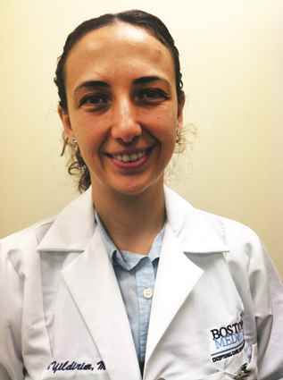
Several recent studies provide insights and can be helpful in forming an evidence-based approach that offers modest benefit for reducing the risk of recurrence. Most recently, Kaplan et al. (Clin. Inf. Dis. 2014;58:679-82) reported on a clinical trial of sodium hypochlorite bleach baths combined with hygienic measures (frequent hand washing with soap, cutting fingernails short, using towels or washcloths and clothing without sharing, and daily bathing or showering), compared with hygienic measures alone. The treatment group received twice-weekly hypochlorite baths with 5 mL household bleach (Clorox-Regular 6.0% hypochlorite) per gallon of bath water, followed by moisturizer. Most children were colonized with methicillin-resistant Staphylococcus aureus (MRSA)(approximately 70%) or methicillin-susceptible S. aureus (MSSA)(approximately 30%). In the 12-month follow-up, 20% of children had recurrent skin or soft tissue infection (SSTI). Risk factors for recurrence were young age (<6 years) and burden of colonization (number of colonized sites). A small, nonstatistically significant benefit was observed in the treatment group with a 17% incidence of SSTI, compared with 20.9% in controls (P = 0.15). The authors concluded a bleach bath plus hygiene measures was associated with about a 20% nonstatistically significant decrease in recurrent community-acquired SSTI. No adverse effects of bleach baths were identified.
A second open-label, randomized study by Fritz et al. (Clin. Inf. Dis. 2012;54:743-51) evaluated the value of individual decolonization, compared with household decolonization, in children 6 months through 20 years of age with prior community-acquired SSTI. Cases were randomized to individual decolonization regimens (hygiene, 2% mupirocin for 5 days and 4% chlorhexidine daily body washes) or to household decolonization. Staphylococcal colonization was evaluated at 1, 3, 6, and 12 months. No differences in the rate of eradication of S. aureus were observed between the two strategies, except at 3 months where a greater proportion of children randomized to household decolonization were culture negative. Despite the lack of impact on colonization, SSTI documented by a physician was less common in children where decolonization was householdwide. After 12 months, 36% of children in the household decolonization sites had recurrent SSTI, compared with 55% in the individual decolonization stratum (P = .03). The authors concluded that household decolonization reduces SSTI in both the individual and household contacts.

Another approach to decolonization has been the use of oral antibiotics in combination with mupirocin and hexachloradine. Although data are limited, Miller et al. (Antimicrob. Agents Chemother. 2012;56:1084-6) reported on a small cohort of 31 prospectively evaluated patients with recurrent community-acquired MRSA skin infections. Individuals received nasal mupirocin, topical hexachlorophene body wash, and an oral antibiotic based on susceptibility testing (doxycycline, minocycline, or trimethoprim-sulfamethoxazole). In the 6 months prior to enrollment, the mean rate of SSTI was three infections per person (range, 2-30). The mean number of MRSA infections after the intervention decreased significantly from 0.84 infections per month to 0.03 infections per month during the 5.2-month follow-up. In general, the regimens were well tolerated with minor gastrointestinal complaints. The authors concluded that the combination of systemic and topical antimicrobials was associated with subsequent decreases in community-acquired MRSA SSTI; however, they acknowledged that without a control group, they were unable to be certain that the decrease was due to the prescribed regimen.
Our current approach for children referred with recurrent SSTI is household decolonization with nasal mupirocin and daily hexachloradine baths or showers or hypochlorite baths. The mupirocin is prescribed for 5-10 days; the hexachloradine/hypochlorite baths, for several months. We also stress the need for hygiene, including washing towels and linens in hot water, and cleaning surfaces and items such as remote controls with hypochlorite solutions. Although the value of environmental decontamination is unknown, studies by Uhlemann et al. (PLOS ONE 2011;6: e22407) demonstrated excess contamination of household surfaces in homes of SSTI cases. If recurrences continue, the addition of an antimicrobial agent is considered. We reserve doxycycline for children over 8 years of age and prescribe trimethoprim-sulfamethoxazole for those younger than 8 years. We also will ask about pets although we are aware of only anecdotal reports where treating the family dog or cat has aborted recurrent disease in the patients.
In summary, recurrent SSTI is common, especially among young children. The burden of colonization appears related to both the risk for recurrent disease and the risk for transmission within the household. Reducing colonization is valuable for decreasing the incidence of recurrent SSTI both for the individual as well as the household members. The current strategies demonstrate modest success, but as many as 30%-40% of patients will continue to have recurrent SSTI. Education about the early signs of infection, early evaluation of SSTI, and appropriate management (topical treatment, incision and drainage, or systemic antibiotics) are successful strategies for limiting progression to invasive staphylococcal disease.
Dr. Pelton is chief of pediatric infectious disease and coordinator of the maternal-child HIV program at Boston Medical Center. Dr. Yildirim is a fellow in pediatric infectious disease and an epidemiologist, at Boston Medical Center. To comment, e-mail Dr. Pelton and Dr. Yildirim at [email protected].
A frequent referral to our pediatric infectious disease outpatient program at Boston Medical Center is the child with recurrent skin and soft tissue infection. Most often, the child is an infant, toddler, or adolescent; the child is otherwise well but has had two or three prior episodes of skin infection; the infections are typically peri-inguinal including the buttocks, but may involve the face, back, thighs, or scalp. The families are often frustrated and hoping for a solution. Are there effective strategies for reducing recurrences?
Several recent studies provide insights and can be helpful in forming an evidence-based approach that offers modest benefit for reducing the risk of recurrence. Most recently, Kaplan et al. (Clin. Inf. Dis. 2014;58:679-82) reported on a clinical trial of sodium hypochlorite bleach baths combined with hygienic measures (frequent hand washing with soap, cutting fingernails short, using towels or washcloths and clothing without sharing, and daily bathing or showering), compared with hygienic measures alone. The treatment group received twice-weekly hypochlorite baths with 5 mL household bleach (Clorox-Regular 6.0% hypochlorite) per gallon of bath water, followed by moisturizer. Most children were colonized with methicillin-resistant Staphylococcus aureus (MRSA)(approximately 70%) or methicillin-susceptible S. aureus (MSSA)(approximately 30%). In the 12-month follow-up, 20% of children had recurrent skin or soft tissue infection (SSTI). Risk factors for recurrence were young age (<6 years) and burden of colonization (number of colonized sites). A small, nonstatistically significant benefit was observed in the treatment group with a 17% incidence of SSTI, compared with 20.9% in controls (P = 0.15). The authors concluded a bleach bath plus hygiene measures was associated with about a 20% nonstatistically significant decrease in recurrent community-acquired SSTI. No adverse effects of bleach baths were identified.
A second open-label, randomized study by Fritz et al. (Clin. Inf. Dis. 2012;54:743-51) evaluated the value of individual decolonization, compared with household decolonization, in children 6 months through 20 years of age with prior community-acquired SSTI. Cases were randomized to individual decolonization regimens (hygiene, 2% mupirocin for 5 days and 4% chlorhexidine daily body washes) or to household decolonization. Staphylococcal colonization was evaluated at 1, 3, 6, and 12 months. No differences in the rate of eradication of S. aureus were observed between the two strategies, except at 3 months where a greater proportion of children randomized to household decolonization were culture negative. Despite the lack of impact on colonization, SSTI documented by a physician was less common in children where decolonization was householdwide. After 12 months, 36% of children in the household decolonization sites had recurrent SSTI, compared with 55% in the individual decolonization stratum (P = .03). The authors concluded that household decolonization reduces SSTI in both the individual and household contacts.
Another approach to decolonization has been the use of oral antibiotics in combination with mupirocin and hexachloradine. Although data are limited, Miller et al. (Antimicrob. Agents Chemother. 2012;56:1084-6) reported on a small cohort of 31 prospectively evaluated patients with recurrent community-acquired MRSA skin infections. Individuals received nasal mupirocin, topical hexachlorophene body wash, and an oral antibiotic based on susceptibility testing (doxycycline, minocycline, or trimethoprim-sulfamethoxazole). In the 6 months prior to enrollment, the mean rate of SSTI was three infections per person (range, 2-30). The mean number of MRSA infections after the intervention decreased significantly from 0.84 infections per month to 0.03 infections per month during the 5.2-month follow-up. In general, the regimens were well tolerated with minor gastrointestinal complaints. The authors concluded that the combination of systemic and topical antimicrobials was associated with subsequent decreases in community-acquired MRSA SSTI; however, they acknowledged that without a control group, they were unable to be certain that the decrease was due to the prescribed regimen.
Our current approach for children referred with recurrent SSTI is household decolonization with nasal mupirocin and daily hexachloradine baths or showers or hypochlorite baths. The mupirocin is prescribed for 5-10 days; the hexachloradine/hypochlorite baths, for several months. We also stress the need for hygiene, including washing towels and linens in hot water, and cleaning surfaces and items such as remote controls with hypochlorite solutions. Although the value of environmental decontamination is unknown, studies by Uhlemann et al. (PLOS ONE 2011;6: e22407) demonstrated excess contamination of household surfaces in homes of SSTI cases. If recurrences continue, the addition of an antimicrobial agent is considered. We reserve doxycycline for children over 8 years of age and prescribe trimethoprim-sulfamethoxazole for those younger than 8 years. We also will ask about pets although we are aware of only anecdotal reports where treating the family dog or cat has aborted recurrent disease in the patients.
In summary, recurrent SSTI is common, especially among young children. The burden of colonization appears related to both the risk for recurrent disease and the risk for transmission within the household. Reducing colonization is valuable for decreasing the incidence of recurrent SSTI both for the individual as well as the household members. The current strategies demonstrate modest success, but as many as 30%-40% of patients will continue to have recurrent SSTI. Education about the early signs of infection, early evaluation of SSTI, and appropriate management (topical treatment, incision and drainage, or systemic antibiotics) are successful strategies for limiting progression to invasive staphylococcal disease.
Dr. Pelton is chief of pediatric infectious disease and coordinator of the maternal-child HIV program at Boston Medical Center. Dr. Yildirim is a fellow in pediatric infectious disease and an epidemiologist, at Boston Medical Center. To comment, e-mail Dr. Pelton and Dr. Yildirim at [email protected].

Pulling together the discharge summary
So it’s your first day on the service. Mrs. Jones has been there for 21 days and has a long list of consultants to address her numerous complications. You see her twice before it’s time to pull it all together and package her up for an 11 a.m. transfer to rehab the next morning. But how do you effectively weed through weeks of documentation to come up with the salient points of the hospital stay, and do so in a reasonable amount of time considering you have 20 other patients (and a few inquisitive family members) who require your undivided attention that day as well?
A preliminary discharge summary, prepared the day before anticipated discharge, can make life a lot easier. If your EMR allows you to sort notes by author or service, you can dictate the hospital course by problem more seamlessly than by reviewing the hospitalization on a day-by-day basis, especially if there are multiple notes from PT/OT, pharmacy, and other ancillary services intermingled in the providers’ documentation.
If your EMR allows you to auto-populate diagnostic test results, discharge medications, and instructions directly into a note, you can create this note on the day of actual discharge, and then copy and paste the dictation of the hospital course into the body of the final discharge summary.
Alternatively, if the provider who is better acquainted with the patient does a discharge summary prior to going off service, the upcoming provider need only add an addendum to this summary on the day of discharge. When partners do these summaries for each other, it can be a tremendous time saver. Instead of spending 45-60 minutes drudging through every progress note and consultation on an unfamiliar patient, you are able to review the preliminary discharge summary and pick up the hospital course as you would for a patient admitted the day before who already has an H&P. The doctor going off service may only need to spend 5-10 minutes dictating the summary.
Of course, different groups have different practice styles. Some groups may consistently dictate summaries prior to going off service, while others may not choose this option. There may be other ways to streamline complicated discharge summaries within groups as well, but experimenting with new and innovative ways to improve care and make our lives more efficient in the process may prove to be a win-win for all.
Dr. Hester is a hospitalist at Baltimore-Washington Medical Center in Glen Burnie, Md. She is the creator of the Patient Whiz, a patient-engagement app for iOS. Reach her at [email protected].

Joining forces
Tough economic times and the unpredictable consequences of health care reform are making a growing number of solo practitioners and small private groups very nervous. I’ve received many inquiries about protective options, such as joining a multispecialty group, or merging two or more small practices into larger entities.
If becoming an employee of a large corporation does not appeal to you, a merger can offer significant advantages in stabilization of income and expenses; but careful planning, and a written agreement, are essential.
If you are considering this option, here are some things to think about:
What is the compensation formula? Will everyone be paid only for what they do individually, or will revenue be shared equally? I favor a combination; productivity is rewarded, but your income doesn’t drop to zero when you take time off.
Who will be in charge, and what percentage vote will be needed to approve important decisions? Typically, the majority rules, but you may wish to create a list of pivotal moves that will require unanimous approval, such as purchasing expensive equipment, borrowing money, or adding new partners.
Will you keep your retirement plans separate, or combine them? If the latter, you will have to agree on the terms of the new plan, which can be the same or different from any of the existing plans. You’ll probably need some legal guidance to ensure that assets from existing plans can be transferred into a new plan without tax issues.
Since most private practices are incorporated, there are two basic options for combining them: Corporation A can simply absorb corporation B; the latter ceases to exist, and corporation A, the so-called “surviving entity,” assumes all assets and liabilities of both old corporations. Corporation B shareholders exchange shares of its stock for shares of corporation A, with adjustments for any inequalities in stock value.
The second option is to start a completely new corporation. Both separate entities dissolve and distribute their equipment and charts to their shareholders, who then transfer the assets to the new corporation.
Option 2 is popular, but I am not a fan. It is billed as an opportunity to start fresh, shielding everyone from exposure to malpractice suits and other liabilities. However, the reality is that anyone looking to sue either old corporation will simply sue the new entity as the so-called “successor” corporation, on the grounds that it has assumed responsibility for its predecessors’ liabilities. You also will need new provider numbers, which may impede cash flow for months. Plus, the IRS treats corporate liquidations, even for merger purposes, as sales of assets, and taxes them.
In general, most experts that I’ve talked with favor outright merger of the corporations. This option is tax neutral, and while it may theoretically be less satisfactory liability-wise, you can minimize risk by examining financial and legal records, and by identifying any glaring flaws in charting or coding. Your lawyers can add “hold harmless” clauses to the merger agreement, indemnifying each party against the others’ liabilities. This area in particular is where you need experienced, competent legal advice.
Another common sticking point is known as “equalization.” Ideally, each party brings an equal amount of assets to the table, but in the real world that is rarely the case. One party may contribute more equipment, for example, and the others are often asked to make up the difference (“equalize”) with something else, usually cash.
An alternative is to agree that any inequalities will be compensated at the other end, in the form of buyout value; that is, physicians contributing more assets will receive larger buyouts when they leave or retire than those contributing less.
Non-compete provisions are always a difficult issue, mostly because they are so hard (and expensive) to enforce. An increasingly popular alternative is, once again, to deal with it at the other end, with a buyout penalty. An unhappy partner can leave, and compete, but at the cost of a substantially reduced buyout. This permits competition, but discourages it; and it compensates the remaining partners.
These are only some of the pivotal business and legal issues that must be settled in advance. A little planning and negotiation can prevent a lot of grief, regret, and legal expenses in the future. I’ll mention some other, more complicated merger options in a future column.
Dr. Eastern practices dermatology and dermatologic surgery in Belleville, N.J. He is the author of numerous articles and textbook chapters, and is a longtime monthly columnist for Skin & Allergy News. Additional columns are available online at edermatologynews.com.

Commentary: Managing major neurocognitive disorder in African Americans
The Alzheimer’s Association suggests that major neurocognitive disorder (formerly known as dementia) caused by Alzheimer’s disease is a “silent epidemic” in African Americans, noting that the prevalence among African Americans ranges from 14% to 100% higher than it is among whites.
In April 2013, the National Institute on Aging highlighted a JAMA research article that noted African Americans were more likely to have a variant of the ABCA7 gene and that this gene variant led to almost double the risk of developing Alzheimer’s disease (JAMA 2013;309:1483-92).

In addition, the Alzheimer’s Association suggests that “African Americans are seriously underrepresented in current clinical trials of potential treatment of Alzheimer’s disease, particularly in trials conducted by drug companies.” This observation echoes former U.S. Surgeon General David Satcher’s 2001 Culture, Race, and Ethnicity report, which underscored a historic dearth of research on African American mental health issues. Recently, a randomized clinical trial of citalopram in agitated patients with Alzheimer’s disease found this selective serotonin reuptake inhibitor to be efficacious in reducing agitation. However, African Americans were grossly underrepresented, comprising 15 of 94 patients in the experimental arm of this study (JAMA 2014;311:682-91).
While working on the medical/psychiatric floor at Jackson Park Hospital on Chicago’s South Side, I have watched these issues play out in real life, often delivering a harsh reality. For the past 2 years, every day, I have seen one to three elderly African American patients who had been transferred from local nursing homes with complaints of restlessness, wandering, aggression, depression, and psychosis characterized by hallucinations and delusions, which resulted in disruptive behaviors. Clinical lore suggests that such behaviors are responsible for about 50% of admissions to nursing homes and 95% of hospital admissions from such nursing homes.
Despite the known risks, too often, I see patients being prescribed first- and second-generation antipsychotics. I suppose this is because of the agitation, aggression, and psychotic symptoms. But according to the Food and Drug Administration, such prescribing is associated with premature mortality in Alzheimer’s disease. I also see a lot of benzodiazepine regimens, and this, too, is occurring despite the recent findings that benzodiazepines are associated with the etiology of Alzheimer’s disease. These practices just do not make any sense.
Recently, I saw an elderly woman with suspected Alzheimer’s disease. When I asked her the year, I saw fear and panic spread over her face as she realized that she did not remember. I decided to treat this anxiety with escitalopram 10 mg, so I gave her a dose stat (and followed up with a dose every morning).
When I checked on her the next day, after confirming that she did not remember me from the previous day (quite unusual as I wear a garish cowboy hat that my daughter gave me), I again asked what year it was. She replied with a pleasant smile: “I don’t know, and I don’t care.” She was calm and agreeable, not the frightened, panic-stricken, irritable woman I had seen the day before.
Since then, I have been repeatedly impressed with this particular SSRI in managing major neurocognitive disorder caused by Alzheimer’s disease. It brings about a night and day difference in terms of agitation and irritability, and the medical staff are amazed. And no, I do not own stock in pharmaceutical companies; escitalopram (not citalopram) is the one that the hospital formulary offers, and, in my experience, seems to have minimal side effects (agitation, blurred vision, diarrhea, insomnia, drowsiness, dry mouth, fever, frequent urination, headache, indigestion, nausea, change in appetite, sexual dysfunction, and weight change), including hepatotoxicity and hyponatremia. The other SSRIs (citalopram, duloxetine, fluoxetine, fluvoxamine, paroxetine, or sertraline) might work just as well. I would be interested to hear from other clinicians with extensive experience – for example, those who have treated hundreds of African Americans or other patients with Alzheimer’s disease – as it might take years for the research to be published and even longer before we see related data on underserved populations.
In the absence of research focused on major African American problems, we must rely on clinical experience to address these issues. I’ve been getting such positive results that I felt compelled to pass them along.
Dr. Bell is a staff psychiatrist at Jackson Park Hospital and a member of the editorial advisory board of Clinical Psychiatry News.
The Alzheimer’s Association suggests that major neurocognitive disorder (formerly known as dementia) caused by Alzheimer’s disease is a “silent epidemic” in African Americans, noting that the prevalence among African Americans ranges from 14% to 100% higher than it is among whites.
In April 2013, the National Institute on Aging highlighted a JAMA research article that noted African Americans were more likely to have a variant of the ABCA7 gene and that this gene variant led to almost double the risk of developing Alzheimer’s disease (JAMA 2013;309:1483-92).
In addition, the Alzheimer’s Association suggests that “African Americans are seriously underrepresented in current clinical trials of potential treatment of Alzheimer’s disease, particularly in trials conducted by drug companies.” This observation echoes former U.S. Surgeon General David Satcher’s 2001 Culture, Race, and Ethnicity report, which underscored a historic dearth of research on African American mental health issues. Recently, a randomized clinical trial of citalopram in agitated patients with Alzheimer’s disease found this selective serotonin reuptake inhibitor to be efficacious in reducing agitation. However, African Americans were grossly underrepresented, comprising 15 of 94 patients in the experimental arm of this study (JAMA 2014;311:682-91).
While working on the medical/psychiatric floor at Jackson Park Hospital on Chicago’s South Side, I have watched these issues play out in real life, often delivering a harsh reality. For the past 2 years, every day, I have seen one to three elderly African American patients who had been transferred from local nursing homes with complaints of restlessness, wandering, aggression, depression, and psychosis characterized by hallucinations and delusions, which resulted in disruptive behaviors. Clinical lore suggests that such behaviors are responsible for about 50% of admissions to nursing homes and 95% of hospital admissions from such nursing homes.
Despite the known risks, too often, I see patients being prescribed first- and second-generation antipsychotics. I suppose this is because of the agitation, aggression, and psychotic symptoms. But according to the Food and Drug Administration, such prescribing is associated with premature mortality in Alzheimer’s disease. I also see a lot of benzodiazepine regimens, and this, too, is occurring despite the recent findings that benzodiazepines are associated with the etiology of Alzheimer’s disease. These practices just do not make any sense.
Recently, I saw an elderly woman with suspected Alzheimer’s disease. When I asked her the year, I saw fear and panic spread over her face as she realized that she did not remember. I decided to treat this anxiety with escitalopram 10 mg, so I gave her a dose stat (and followed up with a dose every morning).
When I checked on her the next day, after confirming that she did not remember me from the previous day (quite unusual as I wear a garish cowboy hat that my daughter gave me), I again asked what year it was. She replied with a pleasant smile: “I don’t know, and I don’t care.” She was calm and agreeable, not the frightened, panic-stricken, irritable woman I had seen the day before.
Since then, I have been repeatedly impressed with this particular SSRI in managing major neurocognitive disorder caused by Alzheimer’s disease. It brings about a night and day difference in terms of agitation and irritability, and the medical staff are amazed. And no, I do not own stock in pharmaceutical companies; escitalopram (not citalopram) is the one that the hospital formulary offers, and, in my experience, seems to have minimal side effects (agitation, blurred vision, diarrhea, insomnia, drowsiness, dry mouth, fever, frequent urination, headache, indigestion, nausea, change in appetite, sexual dysfunction, and weight change), including hepatotoxicity and hyponatremia. The other SSRIs (citalopram, duloxetine, fluoxetine, fluvoxamine, paroxetine, or sertraline) might work just as well. I would be interested to hear from other clinicians with extensive experience – for example, those who have treated hundreds of African Americans or other patients with Alzheimer’s disease – as it might take years for the research to be published and even longer before we see related data on underserved populations.
In the absence of research focused on major African American problems, we must rely on clinical experience to address these issues. I’ve been getting such positive results that I felt compelled to pass them along.
Dr. Bell is a staff psychiatrist at Jackson Park Hospital and a member of the editorial advisory board of Clinical Psychiatry News.