User login
Advance medical directive
Question: R, a woman with diabetes and Parkinson’s disease, executed an advance medical directive (AMD) when she was aged 70 years. Her AMD stipulated that no extraordinary measures be taken should she become terminally ill or permanently comatose, and she named her sister as her agent with durable power of attorney (DPA). Ten years later, at age 80 years, R sustained a stroke, which left her with hemiparesis and loss of cognitive function. She remained comatose for a week and was dependent on a feeding tube. Her sister decided to honor the AMD and stop all medical treatment including artificial nutrition and hydration. Given the above scenario, which of the following is most accurate?
A. R’s AMD sprang into effect when she became decisionally incapacitated.
B. An AMD can direct only the forgoing of life-sustaining treatment, not the continuation of treatment measures.
C. AMD’s are mandatory under the 1990 Federal Patient Self-Determination Act, and those without an AMD are required to make one upon admission to a hospital.
D. Those with DPA for health-care decisions are obligated to carry out the patient’s instructions but can also override them.
E. In the absence of an AMD or DPA, the attending doctor makes the final decision in the best interest of the patient.
Answer: A. An advance medical directive (AMD), sometimes called a living will, is a legally binding document executed by a competent person that provides instructions for future health-care decisions if and when that person loses decisional capacity. One can elect to forgo some or all life-sustaining measures, or to continue receiving all treatments. In 1990, Congress enacted the Federal Patient Self Determination Act, which allows patients to stipulate in advance the nature of their healthcare should they become incapacitated. Additionally, they can appoint an agent or proxy, said to have durable power of attorney (DPA) for health-care decisions, to speak for them. Agents are required to carry out, not override, the patient’s wishes. The Federal Act requires all hospitals to inquire into whether a patient has an AMD and to provide information on the subject, but the Act does not mandate that everyone should execute one. All 50 states, beginning with California in 1976, have enacted legislation on AMDs. In the absence of an AMD and DPA, a surrogate decision maker, sometimes court appointed, directs the care. The attending doctor provides the prognosis, but does not make the final decision to forgo or continue treatment.
Physicians are obligated by law to respect wishes regarding whether to forgo or continue therapy, and, if contrary to their conscience, transfer the patient to a willing health-care provider. In some jurisdictions, a patient’s wish to discontinue artificial nutrition and hydration, for example, via nasogastric or G-tube, requires a specific opt-in or opt-out choice. AMD instructions become relevant when the patient is terminally ill, but many jurisdictions allow their applicability in nonterminal conditions such as irreversible unconscious states or where the likely risks and burdens of treatment would outweigh the expected benefits (Hawaii is such a state under HRS 327E-16).
A recent review of 3,746 patients who had died between 2000 and 2006 showed that those with AMDs were more likely to want limited care or comfort care rather than all possible care, with the vast majority receiving treatment compatible with their wishes. Those who had assigned a durable power of attorney were less likely to die in the hospital or receive all care possible. The authors concluded that patients who have prepared AMDs received care that was strongly associated with their preferences. Other observers are less sanguine, noting that in only two-thirds of the time were decisions consistent, and one-third of patients changed their preferences in the face of actual illness, usually in favor of treatments rejected in advance. Surrogate agreement was only 58%, and surrogates tended to overestimate their loved one’s desire for treatment.
AMDs direct future end-of-life treatment in a hospital setting, and do not typically address emergency measures taken by ambulance personnel. The National POLST (Physician Orders for Life-Sustaining Treatment) Paradigm grew out of the need to address outpatient emergency treatment especially for seriously ill and frail patients. POLST, now available in many states across the country, is in actuality a medical order signed by the patient’s doctor directing what is and is not to be done, for example, intubation, defibrillation, etc. It does not replace the AMD but is complementary in ensuring respect for a patient’s medical wishes.
Unsurprisingly, there has been litigation over AMDs. Some of the earlier cases tended to favor the health-care institution’s refusal to cease life-sustaining treatment. In Bartling v. Glendale Adventist Medical Center, 184 Cal App 3d 961 (1986), a needle biopsy caused a patient’s lung to collapse. The patient had to be placed in restraints as he tried to remove the ventilator tubes. He had earlier executed a living will and a durable power of attorney for health care, and the family, agreeing to release the hospital and doctors from any civil liability, asked that the ventilator be removed. Instead, the hospital merely tried to "wean" him, and planned to resuscitate him in the event of a cardiopulmonary arrest. Unfortunately, no other facility was willing to accept him in transfer. However, in a defense verdict, the court held that although case law was evolving toward a greater recognition of patients’ rights, it could not be said that a common legal standard existed to guide the medical community.
The case of Osgood v. Genesys St. Joseph Hospital (No. 94-26731-NH, Mich 1996) gave a different result. The patient had a long history of seizures, and the doctors warned that a massive one could leave her in a persistent vegetative state. The patient named her mom as having durable power of attorney. One month later, the patient suffered a massive seizure but was kept alive on a ventilator with full medical support consisting of tube feeding, dialysis, blood transfusions, and medications. Although asked to have these measures removed, the hospital allegedly said they were not life support, but were "comfort care." After 2 months, the patient awoke and was able to be discharged home. However, she remained bedridden and spent most of her time "rhythmically screaming and thrashing" for hours on end, saying, "bury me". The family won a jury award of $16.5 million, which was reduced to $1.4 million on appeal.
A third case, Noonkester v. Kline, was a 1996 defense verdict for a doctor who instituted aggressive treatment despite his patient’s rejection of extraordinary measures evident in his living will. The patient, Mr. Noonkester, had Lou Gehrig’s disease and had instructed the doctor to write a DNR order. He gave his ex-wife power of attorney, and even made arrangements to be taken to a hospice in an emergency, where he planned to die peacefully. When Mr. Noonkester developed breathing problems from pneumonia, the ambulance took him to the hospital instead, and the doctor ordered a ventilator. The patient gave a "thumbs up" when told of the treatment plan, and 3 months later wrote a letter thanking Dr. Kline of the Scripps Clinic for saving his life. Although he had refused to have the ventilator removed, deeming it to be a suicidal act that he opposed, he subsequently sued Dr. Kline for interfering with his preplanned death and continuing costs of living. The jury took all of 45 minutes to return a unanimous verdict for Dr. Kline.
The contents of an AMD may be subject to differing interpretations, as was the case in Wright v. Johns Hopkins Health Systems Corp., 728 A.2d 166 (Md. 1999). A patient with AIDS arrested following a blood transfusion and was resuscitated but left in a comatose state for 10 days before dying. The family alleged that the aggressive treatment violated the patient’s living will instructions, but their lawsuit against the hospital was unsuccessful. Maryland’s Court of Appeals agreed with the hospital and the lower court that the living will could not take effect since no doctor had certified that he was in a terminal state and that his death was imminent.
The late Daniel Callahan, a prominent philosopher-ethicist, eloquently stated that dying in America has over the centuries "evolved from the sacred to the secular, private to institutional, and natural to artifactual." AMDs are a modern attempt to address the many dilemmas and emotions surrounding death. Patient autonomy and palliative care have rightly taken center stage. This may in part be the result of a seminal paper in 1995, which reported on a multicenter study of critically ill patients and their wishes regarding DNR and other end-of-life issues. The authors found that the wishes of many of the patients were ignored, and that 50% were in moderate or severe pain. Incredibly, physician treatment failed to change despite nurse intervention with prognostic information and patient preference.
142 USC 1395cc(f), 1396a(w) (1994).
2 Silveira MJ et al., Advance Directives and Outcomes of Surrogate Decision Making Before Death. (N. Engl. J. Med. 2010; 362:1211-8).
3 Lee et al., Do Patients’ Treatment Decisions Match Advance Statements of Their Preference? (J. Clin. Ethics 1998; 9:258-62).
4 See www.polst.org
5 A controlled trial to improve care for seriously ill hospitalized patients.1995 "SUPPORT" trial. (JAMA 1995; 274:1591-8).

Saying ‘I’m sorry’ can still be problematic
"I’m sorry."
I say that a lot – probably a few times an hour. I say it if I’m 2 minutes late taking a patient back, when a patient complains that the MRI was too loud, that a medication didn’t help, that I poked too hard with the pin while testing sensation, when a patient is unhappy that we don’t take American Express, or pretty much anything.
It’s a common phrase, meant to express care and concern and to smooth out the fabric of human social relationships.
A total of 37 states have some form of a medical malpractice "apology law" that allows physicians to apologize after something goes wrong without the apology being used against them in court. Thirteen states do not have an apology law (Alabama, Alaska, Arkansas, Illinois, Kansas, Kentucky, Minnesota, Mississippi, Nevada, New Mexico, New York, Rhode Island, and Wisconsin).
When a case goes bad we all feel awful about it, regardless of whether or not it’s our fault. These include the patient with symptomatic carotid stenosis we referred for surgery who then had a stroke on the operating table, the multiple sclerosis patient with a downhill course regardless of what medication we use, and the young person with speech problems who turns out to have a glioma.
We’ve all seen similar outcomes and feel bad for the patients. We do our best, but things beyond our control happen. Unfortunately, this is part of medicine. At the end of the day, we can only hope to have had more good outcomes than bad, but the bad ones are still unavoidable. Luck, chance, biology, aging, and human fallibility all work against us sometimes.
When things go wrong, it’s normal to say "I’m sorry," because we are concerned and do care and wish it had happened otherwise for the sake of someone who came to us seeking help.
To me, it seems wrong that expressing such feelings can be seen as an admission of guilt. It sounds more like part of the Miranda rights: "Anything you say can and will be used against you in a court of law."
Being afraid to apologize can make a bad situation even worse. Not expressing condolences makes a doctor appear aloof, uncaring, and arrogant.
Communication between a doctor, the patient, and his or her family under difficult circumstances is never easy, and having a simple apology subject to legal action only makes it worse. The medical relationship works best when it is open and honest – the way it should be everywhere.
Dr. Block has a solo neurology practice in Scottsdale, Ariz.

Limited English proficiency patients and the hospitalist
America! America! God shed His grace on thee
And crown Thy good with brotherhood
From sea to shining sea!
I fondly remember singing "America the Beautiful" with my classmates when I was a little girl. America has grown by leaps and bounds since my childhood – the pulse of the nation as well as its makeup. One of my fondest memories as a child was traveling to New York. We had a layover in Washington and the airport was filled with people of various skin tones speaking all sorts of languages I had never been exposed to before. It was very exciting! It was my first truly multicultural experience.
Funny, I ultimately relocated to the D.C. area, and my neighbors are literally from all over the world: India, Thailand, Jamaica, Africa, China – and it doesn’t stop there. Naturally, the patient population I serve also reflects this great diversity. As the country becomes more diverse each and every day, we, as practitioners, must be able to communicate effectively with our entire patient base, not just the ones who speak English fluently.
This is quite a challenge. Yes, most hospitals have a language line or an on-call interpreter to help out, but I believe we also need to take some responsibility for improving our ability to communicate as well. While I am not advocating trying to master a new language, or two or three, we can all learn a few basic terms of the foreign languages we encounter most.
Consider that language lines do malfunction. Family members are sometimes not present. And interpreters may not always be available at the drop of a hat. Technology, though, is ever burgeoning. It’s easy to download a smartphone app, such as Medical Spanish: Healthcare Phrasebook with Audio. Google Translate can be helpful for scores of languages, though I would use this site with caution when it comes to patient care.
There is a slew of reputable patient information written in different languages available on the Internet as well.
The Agency for Healthcare Research and Quality offers a guide tool: Improving Patient Safety Systems for Patients with Limited English Proficiency: A Guide for Hospitals. The guide notes that approximately 57 million people speak a language other than English at home and 25 million are defined as limited-English-proficient (LEP). LEP patients were noted to have longer lengths of stay in the hospital and were at greater risk for line infections, surgical infections, falls, and pressure ulcers. They are more likely to be readmitted, as well.
Although it is always best to have a qualified interpreter to help us care for LEP patients, there may be times when one is simply unavailable in an acceptable period of time. Friends and family members can help fill some of the gaps in those instances, but it never hurts for the clinician to know a few vital words as well, such as pain or shortness of breath.
America’s culture is ever evolving, and we must evolve with it. Being able to provide high-quality care to all of our patients is our goal. Standards are important, but sometimes thinking out of the box can be effective as well.
Dr. Hester is a hospitalist with Baltimore-Washington Medical Center who has a passion for empowering patients to partner in their health care. She is the creator of the Patient Whiz, a patient-engagement app for iOS. Reach her at [email protected].

Cryolipolysis
Cryolipolysis has emerged as a popular noninvasive treatment option for reducing localized areas of fat. The technology was developed on the premise that cold temperatures can selectively damage subcutaneous fat while leaving the overlying skin unharmed, as demonstrated by popsicle panniculitis. In this process, when subcutaneous fat is cooled below body temperature but above freezing, the fat undergoes cell death followed by a local inflammatory response, a localized panniculitis, that gradually results in a reduction of fat in that area.
Dr. Dieter Manstein and Dr. R. Rox Anderson pioneered the concept of cryolipolysis in 2008. The technology was approved by the Food and Drug Administration in 2010 in the form of the Zeltiq device. The device has different-sized hand pieces with a vacuum connection that, after it is applied to the skin, cools the subcutaneous fat without damaging the top layers of skin. Each area is treated for 1 hour, and 20%-30% of the fat cells are expected to be reduced with a single treatment. Typical responses after treatment include numbness, but some patients may also experience bruising and discomfort, all of which typically last no longer than 2-3 weeks.
If discomfort occurs in my patients, I find they report it more often in the lower abdomen than the love handles. Paradoxical adipose hyperplasia was recently reported for the first time in a male patient in his 40s (in the lower abdomen) (JAMA Dermatol. 2014;150:317-9).
In my experience, there is no difference in efficacy or adverse events seen in patients of different ethnicities. One study found no difference in efficacy or adverse events of cryolipolysis in Chinese patients (Lasers Surg. Med. 2012;44:125-30), but no other study of cryolipolysis in ethnic patients has been published.
I was involved in the clinical trials for this device prior to FDA approval where one love handle was treated on a patient and the other side was used as a control. Based on this experience and my experience using the device in practice, it is not a replacement for abdominoplasty or liposuction, but it is a useful technology in the right candidate. The patients who seem to do the best are those who are 10-15 pounds from their goal weight, are not obese (body mass index less than 30 kg/m2), and have a discrete bulge (typically love handles or abdomen) that they can’t get rid of with good diet and exercise alone. Massage for a few minutes after treatment seems to increase efficacy (Lasers Surg. Med. 2014;46:20-6).
Some patients may require more than one treatment to achieve their desired results, but I recommend waiting at least 2-3 months before opting for additional treatment. Choosing the right candidates and providing patients with realistic expectations seem to be the most helpful in this process.
Dr. Wesley practices dermatology in Beverly Hills, Calif.

To MU or not to MU, that is the question
If you are still on the fence on meaningful use – our government’s motivational strategy for popularizing electronic health records – the point of no return is rapidly approaching: If you want to qualify for at least a portion of the incentive money, plus avoid a 1% penalty (eventually rising to 5%) on your Medicare Part B reimbursements, this year is your final opportunity to join the party. And, unfortunately, it is not simply a matter of adopting an electronic record system.
Each year, you must attest to demonstrating "meaningful use" (MU) of that system. To do that, you must continually monitor your progress toward meeting the necessary percentage benchmarks, making course corrections as you go. If the numbers are not there when your practice is ready to attest, it will have all been for naught, and a major waste of time and resources.
That being the case, private practitioners who have not yet taken the plunge – and those who have, but are undecided on progressing to stage 2 – must ask themselves whether the significant temporal and monetary investment is worth the trouble.
Many, apparently, have decided that it is not. While a substantial percentage of eligible practitioners signed up for stage 1, approximately 20% of them stopped participating in 2013. And according to the Centers for Medicare & Medicaid Services’ own data, only 4 hospitals and 50 individual practitioners in the entire country had attested to stage 2 through March of 2014.
The American Medical Association has little faith in the program, at least in its current form. In an open letter to the CMS in May 2014, they predicted significantly higher dropout rates unless major modifications are made. Specifically, they singled out the requirement that providers meet all requirements at each stage. Rather than "all or nothing," they proposed a 75% achievement level to receive incentive payments, and a 50% minimum to avoid financial penalties. The AMA also recommended eliminating all benchmarks beyond physicians’ control, such as the stage 2 goal of 5% patient participation on the practice’s electronic health record (EHR) portal.
Another problem that falls outside the control of physicians is maintenance of EHR software. Nearly one EHR-equipped office in five, according to the CMS, is running software that does not meet stage 2 standards. The unfortunate owners of systems that cannot be upgraded before the stage 2 deadline will – through no fault of their own – be faced with a Morton’s fork of replacing their EHR on short notice or abandoning their quest for stage 2 attestation.
While the CMS has not yet indicated whether it has any inclination to address these issues or ease any of the requirements, one official did announce that the agency will be more flexible with its hardship exemptions on a case-by-case basis. Currently, such exemptions are available to new providers, those recovering from natural disasters, and others, such as pathologists, who do not interact face-to-face with patients.
So the question remains: Is the investment of time and resources needed to capture all of the data necessary for successful MU attestation worth making? Is it justified by the promise of MU incentive dollars and the benefits to your practice and your patients? And what exactly are those purported benefits, anyway?
Proponents maintain that integrated EHR will lead to improved documentation, which in turn should lead to improvements in patient care. Errors would be more easily identified because entries from generalists, specialists, labs, and others would be available to all at any time. All involved providers, theoretically, would be on the same page with every individual patient. The downside, of course, is that the real world seldom reflects the ideal situation envisioned by bureaucrats.
Ultimately, the choice is yours: Each private practitioner must decide whether starting (or continuing) meaningful use is worth the financial and time burden in his or her particular situation. If you are still undecided, time is almost up: You must begin your 90-day stage 1 reporting period in July 2014 in order to attest by the final deadline of October 1. The last calendar quarter to begin stage 2 reporting starts on October 1 as well. Detailed instructions for meeting stage 1 and stage 2 deadlines are available from many sources, including the American Academy of Dermatology website.
Dr. Eastern practices dermatology and dermatologic surgery in Belleville, N.J. He is the author of numerous articles and textbook chapters, and is a long-time monthly columnist for Skin & Allergy News.

Immune to education
Let’s be honest. Although pediatricians invest most of their days, some of their nights, and untold cockles of heartfelt concern trying to keep their patients well, there is very little evidence that what we do actually makes a difference. The one shining exception comes when we administer immunizations. This humbling fact makes the problem of vaccine refusal so frustrating and depressing.
I have always considered the increasing number of parents who refuse or who are hesitant about immunization just another example of decay in our nation’s educational system. How could anyone who was even half awake in American History class not be aware of the toll that infectious diseases took on the children born before 1900? Diseases that are now preventable. Do introductory science courses even touch on the basic mechanisms that underlie immunizations? High school students may not be expected to know that John Enders was the lead investigator in the development of the measles vaccine, but someone should have told them the story of Jonas Salk and polio.
Like many of you, I assumed that if I could just do a better job of filling in the gaps in our educational system that vaccine-hesitant parents would see the light. If I could share with parents even a small fraction of what I know about the efficacy of vaccines they couldn’t possibly refuse to immunize their children. However, after 40 years of failed attempts and frustration, I have begun to doubt my communication skills.
Some work by Brendan Nyhan, Ph.D., a government professor at Dartmouth College, and his colleagues suggests that my attempts at education were destined to fail. ("Effective Messages in Vaccine Promotion: A Randomized Trial," [Pediatrics 2014;133:1-8]). Aware that people frequently resist information that contradicts their views, these investigators began a small study. Nearly 1,800 parents were randomized to receive one of four messages supporting the value of MMR vaccine from textual excerpts to pictures of children with the diseases prevented by the vaccine.
What they discovered was that parents who had "mixed or negative feelings" about the vaccine were actually less likely to say that they would choose to vaccinate a future child after they had been presented with literature refuting the MMR-autism link. While these families were less likely to accept the vaccine-autism link, the informational materials had prompted them to consider other reasons that supported their negative views about vaccines.
Although other studies have found that parents still consider their children’s doctor to be the most trusted source of vaccine information, it appears that education as we understand it may not be our best tool. In fact, it may even be counterproductive. Attempts to engender fear may seem logical to us, but in reality they may be backfiring.
Dr. Nyhan and his colleagues didn’t bore down to discover what factors made a particular view so resistant to education. But, in my experience inheritance doesn’t seem to play a role. I hear from many fearful and frustrated grandparents who can’t understand why their grandchildren aren’t being immunized.
There is the hermit mentality that says by keeping apart from "all those other people" in society and by living a better life, we can protect ourselves from their diseases and don’t need immunizations. And, of course, there is the notion that even though we understand the rationale for immunization, God will protect us.
This important study suggests that we must be very thoughtful about our attempts at education in all public health issues. Our intuition has failed us here. As unfair as it may be to the child victims of this parental foolishness, we may need to fall back on strict exclusion and quarantine to protect the rest us until we learn how to convince families that they are making a serious mistake.
Dr. Wilkoff practiced primary care pediatrics in Brunswick, Maine, for nearly 40 years. He has authored several books on behavioral pediatrics including "How to Say No to Your Toddler." E-mail him at [email protected].

Electromechanical power morcellation confined to a bag
In the previous edition of Master Class in Gynecologic Surgery, I described the controversy concerning electromechanical power morcellation freely in the abdominopelvic cavity. I also discussed the risks, as noted in the literature, of spreading an unsuspected leiomyosarcoma, and thus, up-staging the disease and lowering both the length of the disease-free state and the overall survival. I also talked about the lack of an adequate diagnostic study to definitively separate a leiomyoma from a leiomyosarcoma, discussed the at-risk population group, and also noted the benefits of minimally invasive gynecologic surgery. At that time, I stated I was against a ban on the morcellator, and that I recommended that physicians provide proper informed consent and, when possible, consider other treatment options, especially in the at-risk population. These included continued use of minimally invasive gynecologic surgery utilizing a specimen bag for electromechanical power morcellation.
Following that publication, the Food and Drug Administration released a statement regarding electromechanical power morcellation on April 17, 2014. While "discouraging" the use of electromechanical power morcellation, they wisely did not call for a moratorium. Again, the FDA recommended that patients be properly informed as to risk and that alternative therapies be discussed, which included the use of power morcellation in a bag.

While the FDA did not ban electromechanical power morcellation, the phrase "discourages the use" sent widespread ripples throughout our specialty. Within a week, Ethicon Endo-Surgery pulled its morcellator off the market worldwide. My hospital system – Advocate Health Care – as well as virtually every hospital in Boston placed a moratorium on electromechanical power morcellation. Dr. Jim Tsaltas, president of the Australian Gynecological Endoscopy & Surgery Society (AGES), was contacted by his country’s FDA equivalent to discuss the use of power morcellation freely in the abdominopelvic cavity.
Since the letter from the FDA, position papers have come from both the world’s largest society focused on minimally invasive surgery, the AAGL, and the American College of Obstetricians and Gynecologists (ACOG). Both of the society’s statements agree that proper informed consent is imperative and that alternate treatment be considered, especially in the at-risk population. Although both papers discuss the use of electromechanical power morcellation in the confines of a bag, the ACOG statement accurately notes that there is very little data regarding morcellation in a bag placed in the abdominopelvic cavity.
Experience with electromechanical power morcellation in a bag placed in the abdominopelvic cavity is now quickly developing. To quote my coauthor, Dr. Ceana Nezhat, "My goal with this Master Class is to provide a forum for discussion and encourage gynecologic surgeons to continue to practice minimally invasive surgery for the benefit of their patients. Despite the current limitation on unprotected, intraperitoneal electromechanical morcellation, the hope is that surgeons will not revert back to laparotomy, but will continue to learn and find innovative ways to provide the least invasive surgical techniques or refer to centers that can provide these services to women."
In this edition of Master Class in Gynecologic Surgery, I am featuring techniques of electromechanical power morcellation in a specimen bag by early adapters and innovators, and other techniques by Dr. Tony Shibley, Dr. Bernard Taylor, and Dr. Ceana H. Nezhat.
Dr. Shibley has been in full-time practice with Ob.Gyn. Specialists, Fairview Health Services in the Minneapolis area (Edina), for the past 20 years. He has a focus on single-site surgery and has been involved in minimally invasive surgical education both nationally and internationally, as well as in medical device development.
Dr. Taylor is a urogynecologist who is a female pelvic medicine and reconstructive surgeon practicing at the Carolinas Medical Center–Advanced Surgical Specialties for Women in Charlotte, N.C. He lectures nationally and internationally on minimally invasive gynecologic surgery.
Dr. Nezhat is the current president of the AAGL, adjunct professor of obstetrics and gynecology at Emory University and program director of minimally invasive surgery at Northside Hospital, both in Atlanta, and adjunct clinical professor of obstetrics and gynecology at Stanford (Calif.) University.
Dr. Miller is clinical associate professor at the University of Illinois at Chicago, immediate past president of the International Society for Gynecologic Endoscopy, and a past president of the AAGL. He is a reproductive endocrinologist and minimally invasive gynecologic surgeon in private practice in Naperville, Ill., and Schaumburg, Ill.; the director of minimally invasive gynecologic surgery at Advocate Lutheran General Hospital, Park Ridge, Ill.; and the medical editor of this column. Dr. Miller disclosed that he is a consultant to Ethicon Endo-Surgery.
Videos of our experts’ individual techniques of electromechanical power morcellation within the confines of a bag, as well as that of Dr. Douglas Brown, director of the Center for Minimally Invasive Gynecologic Surgery at Massachusetts General Hospital, Boston, can be viewed at the SurgeryU website. Also at SurgeryU is a video of the electromechanical power morcellation technique in a bag that my partner, Dr. Aarathi Cholkeri-Singh, and I utilize. We use a 3,100-cc ripstop nylon specimen bag from Espiner Medical and the 5 x 150 mm, extra-long, shielded-bladed balloon-tipped trocar from Applied Medical. Go to SurgeryU to view videos of the procedures.
In the previous edition of Master Class in Gynecologic Surgery, I described the controversy concerning electromechanical power morcellation freely in the abdominopelvic cavity. I also discussed the risks, as noted in the literature, of spreading an unsuspected leiomyosarcoma, and thus, up-staging the disease and lowering both the length of the disease-free state and the overall survival. I also talked about the lack of an adequate diagnostic study to definitively separate a leiomyoma from a leiomyosarcoma, discussed the at-risk population group, and also noted the benefits of minimally invasive gynecologic surgery. At that time, I stated I was against a ban on the morcellator, and that I recommended that physicians provide proper informed consent and, when possible, consider other treatment options, especially in the at-risk population. These included continued use of minimally invasive gynecologic surgery utilizing a specimen bag for electromechanical power morcellation.
Following that publication, the Food and Drug Administration released a statement regarding electromechanical power morcellation on April 17, 2014. While "discouraging" the use of electromechanical power morcellation, they wisely did not call for a moratorium. Again, the FDA recommended that patients be properly informed as to risk and that alternative therapies be discussed, which included the use of power morcellation in a bag.
While the FDA did not ban electromechanical power morcellation, the phrase "discourages the use" sent widespread ripples throughout our specialty. Within a week, Ethicon Endo-Surgery pulled its morcellator off the market worldwide. My hospital system – Advocate Health Care – as well as virtually every hospital in Boston placed a moratorium on electromechanical power morcellation. Dr. Jim Tsaltas, president of the Australian Gynecological Endoscopy & Surgery Society (AGES), was contacted by his country’s FDA equivalent to discuss the use of power morcellation freely in the abdominopelvic cavity.
Since the letter from the FDA, position papers have come from both the world’s largest society focused on minimally invasive surgery, the AAGL, and the American College of Obstetricians and Gynecologists (ACOG). Both of the society’s statements agree that proper informed consent is imperative and that alternate treatment be considered, especially in the at-risk population. Although both papers discuss the use of electromechanical power morcellation in the confines of a bag, the ACOG statement accurately notes that there is very little data regarding morcellation in a bag placed in the abdominopelvic cavity.
Experience with electromechanical power morcellation in a bag placed in the abdominopelvic cavity is now quickly developing. To quote my coauthor, Dr. Ceana Nezhat, "My goal with this Master Class is to provide a forum for discussion and encourage gynecologic surgeons to continue to practice minimally invasive surgery for the benefit of their patients. Despite the current limitation on unprotected, intraperitoneal electromechanical morcellation, the hope is that surgeons will not revert back to laparotomy, but will continue to learn and find innovative ways to provide the least invasive surgical techniques or refer to centers that can provide these services to women."
In this edition of Master Class in Gynecologic Surgery, I am featuring techniques of electromechanical power morcellation in a specimen bag by early adapters and innovators, and other techniques by Dr. Tony Shibley, Dr. Bernard Taylor, and Dr. Ceana H. Nezhat.
Dr. Shibley has been in full-time practice with Ob.Gyn. Specialists, Fairview Health Services in the Minneapolis area (Edina), for the past 20 years. He has a focus on single-site surgery and has been involved in minimally invasive surgical education both nationally and internationally, as well as in medical device development.
Dr. Taylor is a urogynecologist who is a female pelvic medicine and reconstructive surgeon practicing at the Carolinas Medical Center–Advanced Surgical Specialties for Women in Charlotte, N.C. He lectures nationally and internationally on minimally invasive gynecologic surgery.
Dr. Nezhat is the current president of the AAGL, adjunct professor of obstetrics and gynecology at Emory University and program director of minimally invasive surgery at Northside Hospital, both in Atlanta, and adjunct clinical professor of obstetrics and gynecology at Stanford (Calif.) University.
Dr. Miller is clinical associate professor at the University of Illinois at Chicago, immediate past president of the International Society for Gynecologic Endoscopy, and a past president of the AAGL. He is a reproductive endocrinologist and minimally invasive gynecologic surgeon in private practice in Naperville, Ill., and Schaumburg, Ill.; the director of minimally invasive gynecologic surgery at Advocate Lutheran General Hospital, Park Ridge, Ill.; and the medical editor of this column. Dr. Miller disclosed that he is a consultant to Ethicon Endo-Surgery.
Videos of our experts’ individual techniques of electromechanical power morcellation within the confines of a bag, as well as that of Dr. Douglas Brown, director of the Center for Minimally Invasive Gynecologic Surgery at Massachusetts General Hospital, Boston, can be viewed at the SurgeryU website. Also at SurgeryU is a video of the electromechanical power morcellation technique in a bag that my partner, Dr. Aarathi Cholkeri-Singh, and I utilize. We use a 3,100-cc ripstop nylon specimen bag from Espiner Medical and the 5 x 150 mm, extra-long, shielded-bladed balloon-tipped trocar from Applied Medical. Go to SurgeryU to view videos of the procedures.

Point/Counterpoint: Is accreditation necessary to maintain quality in bariatric surgery?
Yes: Accreditation is why bariatric surgery outcomes are so good.
In the early days of bariatric surgery, there were very high mortality rates, up to 21% at 1 year in some studies. Because of that level of risk to patients, accreditation was implemented. Currently, mortality rates for bariatric surgery are at 0.15%, an extraordinary achievement in less than a decade. There are now 729 accredited hospitals in the United States, and that number says much about the level of access for patients. It is not unfettered access to bariatric surgery that is important, but rather access by patients to quality care. That’s what matters at the end of the day.

Accreditation isn’t keeping patients from gaining access to bariatric surgery. The bigger problem in access is making sure the federal and state health exchanges provide coverage for bariatric surgery among the essential health benefits. Fewer than half the states have this coverage.
Since accreditation started, there have been six studies with findings supporting it and three with findings that did not. Among the papers supporting it, Flum et al. is most persuasive because it looks at the Medicare population before and after the implementation of accreditation. It shows a big improvement in deaths, complications, and readmissions after the accreditation mandate despite an increase in the number of patients undergoing bariatric surgery (Ann. Surg. 2011;254:860-5).
The question that always comes up is whether it is volume (the number of cases you’re doing) or accreditation that matters for outcomes. A 2013 study found that accreditation status rendered a benefit independent of volume (Surg. Endosc. 2013;27:4539-46).
If we look at the three papers that found against accreditation, I would dismiss one study because it preceded the accreditation movement (Arch. Surg. 2009;144:319-25).
A study of Michigan patients reported similar complication rates at Centers of Excellence (COEs) and non-COEs (JAMA 2010;304:435-42). The problem with that study is that all of the hospitals in the study essentially are COEs, with the same components that come out of the Metabolic and Bariatric Surgery Accreditation and Quality Improvement Program (MBSAQIP). They have a registry. They have volume requirements. They have quality improvement. So, essentially, they’re all COEs, and I’m glad that they embrace the same concepts that we espouse.
The final paper, by Dr. Dimick, takes an econometric approach using differences within differences analysis, which means you look at the differential rate of change to see if the experimental group (in this case, the Medicare population) is different in some way from the control group (non-Medicare). Here’s the problem: That is not a true control. Non-Medicare patients were exposed to accreditation. All of the major insurers embraced accreditation then and continue to do so.
The paper didn’t look at mortality and failure to rescue patients from complications (JAMA 2013;309:792-9). Even though mortality may be low, it still matters. It’s what I would call a sentinel event. It’s become so rare that when it happens, it’s a signal of an issue around quality. We see improvements in both the Medicare and non-Medicare populations. Do two rights make a wrong? Does that mean that accreditation doesn’t work? I think it points out that we’re seeing important changes for the better for all groups.
We presented findings at the American Surgical Association this year showing that accredited hospitals had lower total charges with lower rates of complications, mortality, and failure to rescue. What accreditation provides is a safety net. If something goes wrong, the difference is the ability to recover from complications. There was not a big difference between accredited and non-accredited hospitals in complications in general, but a bigger difference in mortality. Mortality still counts. Failure to rescue is a very important metric that we all need to pay attention to.
Some of our other data show a halo effect from accreditation. In the accredited centers, outcomes were better for non-bariatric procedures. Why? Because they had more experience with obese patients. It’s a collateral benefit.
Without accreditation, I wonder what will happen at hospitals that aren’t required to be accredited. Will they still have the registry? Will they still have the resources they need? Not everybody has the advantages that Michigan has with major insurer support to pay for accreditation efforts. And it’s critical that we have those data. You can’t manage what you don’t measure. Accreditation is vital for bariatric surgery.
Dr. Morton is director of both bariatric surgery and surgical quality at Stanford (Calif.) University Medical Center. He disclosed financial associations with Vivus, Covidien, and Ethicon.
No: The data do not show that outcomes are better in COEs.
The Centers of Excellence has evolved into the Metabolic and Bariatric Surgery Accreditation and Quality Improvement Program (MBSAQIP), which is an outcomes benchmarking and quality improvement program. So we are, in many ways, looking in the rear-view mirror when we talk about whether or not the historical accreditation process improved outcomes.
I’m going to make two arguments against the historical way it was done. The first argument is that limiting care to Centers of Excellence (COEs) as they were defined had very little benefit on outcomes and may have caused some harm.

A lot of the data out there to evaluate the COE program are a little misleading. They looked at Medicare data for outcomes before and after the Medicare COE mandate was put in place requiring that bariatric surgery be performed in a COE to qualify for coverage. The problem with that is the outcomes are improving so quickly that you can’t just do a before and after study. You need to adjust for that time frame to make sure you’re not getting a spurious finding.
We did a study comparing improvement trends for any complications, serious complications, and reoperations before and after Medicare implemented its coverage decision. Surgeons were already getting better before that. There was no change in the trajectory of improvements when the policy was implemented. We concluded that the policy that selectively referred patients to Centers of Excellence didn’t have any benefit (JAMA 2013;309:792-9). We originally included mortality in our analysis but deaths were so rare that it didn’t meet power specifications set by the journal.
In that paper, we did a head-to-head comparison of COEs vs. non-COEs in 12 large states. It’s hard to do a randomized controlled trial for this, so we did an instrumental variable analysis, a sophisticated analysis that takes out a lot of the unmeasured confounding in selection bias. We found that there was no benefit to having bariatric surgery in a COE.
We noticed that many hospitals that traditionally served minorities did not achieve accreditation. One can assume that’s because they probably didn’t have the resources to participate in the program. After the policy was implemented, the rate of bariatric surgery in minorities dropped substantially, whereas it went up in non-minority patients (JAMA 2013;310:1399-1400). So, there’s potential evidence of harm with these policies.
Medicare eliminated their facility accreditation requirement for bariatric surgery last September.
My second argument is that we could be doing something so much better than accreditation to improve bariatric surgery outcomes. Michigan’s approach is a large quality improvement collaborative funded by Blue Cross Blue Shield. We’re using lessons learned in Michigan to implement change on a broader scale without the involvement of payers. We have 70 surgeons and 32 bariatric surgery programs benchmarking outcomes and using those in strategic ways to learn from each other, hopefully improving everybody’s outcomes.
We standardized venous thromboembolism (VTE) prophylaxis across the state and implemented decision support. VTE rates fell by half. We saw a 90% improvement in mortality and a 24% drop in overall complications. Yes, bariatric surgery centers everywhere were improving at that same time, but when we compared Michigan hospitals participating in our collaborative to non-Michigan hospitals, we saw steeper declines in adverse outcomes in Michigan. That’s very robust evidence that this approach is effective for improving perioperative care.
We have launched a technical skill initiative in which surgeons participating in the collaborative give us a video of a typical procedure that gets rated in a blinded fashion by their peers. We just got funded to use these videos in a coaching program to help surgeons in Michigan improve our technique.
Doing this kind of collaborative work doesn’t take a ton of money. It does take time, strategy, and social capital – developing networks of surgeons that work together and learn together. The historical Centers of Excellence program wasn’t a bad idea, but it wasn’t correlated with perioperative outcomes, and it did have harms.
Dr. Dimick is chief of minimally invasive surgery at the University of Michigan Health System, Ann Arbor. He disclosed being a stockholder in ArborMetrix.
Yes: Accreditation is why bariatric surgery outcomes are so good.
In the early days of bariatric surgery, there were very high mortality rates, up to 21% at 1 year in some studies. Because of that level of risk to patients, accreditation was implemented. Currently, mortality rates for bariatric surgery are at 0.15%, an extraordinary achievement in less than a decade. There are now 729 accredited hospitals in the United States, and that number says much about the level of access for patients. It is not unfettered access to bariatric surgery that is important, but rather access by patients to quality care. That’s what matters at the end of the day.
Accreditation isn’t keeping patients from gaining access to bariatric surgery. The bigger problem in access is making sure the federal and state health exchanges provide coverage for bariatric surgery among the essential health benefits. Fewer than half the states have this coverage.
Since accreditation started, there have been six studies with findings supporting it and three with findings that did not. Among the papers supporting it, Flum et al. is most persuasive because it looks at the Medicare population before and after the implementation of accreditation. It shows a big improvement in deaths, complications, and readmissions after the accreditation mandate despite an increase in the number of patients undergoing bariatric surgery (Ann. Surg. 2011;254:860-5).
The question that always comes up is whether it is volume (the number of cases you’re doing) or accreditation that matters for outcomes. A 2013 study found that accreditation status rendered a benefit independent of volume (Surg. Endosc. 2013;27:4539-46).
If we look at the three papers that found against accreditation, I would dismiss one study because it preceded the accreditation movement (Arch. Surg. 2009;144:319-25).
A study of Michigan patients reported similar complication rates at Centers of Excellence (COEs) and non-COEs (JAMA 2010;304:435-42). The problem with that study is that all of the hospitals in the study essentially are COEs, with the same components that come out of the Metabolic and Bariatric Surgery Accreditation and Quality Improvement Program (MBSAQIP). They have a registry. They have volume requirements. They have quality improvement. So, essentially, they’re all COEs, and I’m glad that they embrace the same concepts that we espouse.
The final paper, by Dr. Dimick, takes an econometric approach using differences within differences analysis, which means you look at the differential rate of change to see if the experimental group (in this case, the Medicare population) is different in some way from the control group (non-Medicare). Here’s the problem: That is not a true control. Non-Medicare patients were exposed to accreditation. All of the major insurers embraced accreditation then and continue to do so.
The paper didn’t look at mortality and failure to rescue patients from complications (JAMA 2013;309:792-9). Even though mortality may be low, it still matters. It’s what I would call a sentinel event. It’s become so rare that when it happens, it’s a signal of an issue around quality. We see improvements in both the Medicare and non-Medicare populations. Do two rights make a wrong? Does that mean that accreditation doesn’t work? I think it points out that we’re seeing important changes for the better for all groups.
We presented findings at the American Surgical Association this year showing that accredited hospitals had lower total charges with lower rates of complications, mortality, and failure to rescue. What accreditation provides is a safety net. If something goes wrong, the difference is the ability to recover from complications. There was not a big difference between accredited and non-accredited hospitals in complications in general, but a bigger difference in mortality. Mortality still counts. Failure to rescue is a very important metric that we all need to pay attention to.
Some of our other data show a halo effect from accreditation. In the accredited centers, outcomes were better for non-bariatric procedures. Why? Because they had more experience with obese patients. It’s a collateral benefit.
Without accreditation, I wonder what will happen at hospitals that aren’t required to be accredited. Will they still have the registry? Will they still have the resources they need? Not everybody has the advantages that Michigan has with major insurer support to pay for accreditation efforts. And it’s critical that we have those data. You can’t manage what you don’t measure. Accreditation is vital for bariatric surgery.
Dr. Morton is director of both bariatric surgery and surgical quality at Stanford (Calif.) University Medical Center. He disclosed financial associations with Vivus, Covidien, and Ethicon.
No: The data do not show that outcomes are better in COEs.
The Centers of Excellence has evolved into the Metabolic and Bariatric Surgery Accreditation and Quality Improvement Program (MBSAQIP), which is an outcomes benchmarking and quality improvement program. So we are, in many ways, looking in the rear-view mirror when we talk about whether or not the historical accreditation process improved outcomes.
I’m going to make two arguments against the historical way it was done. The first argument is that limiting care to Centers of Excellence (COEs) as they were defined had very little benefit on outcomes and may have caused some harm.
A lot of the data out there to evaluate the COE program are a little misleading. They looked at Medicare data for outcomes before and after the Medicare COE mandate was put in place requiring that bariatric surgery be performed in a COE to qualify for coverage. The problem with that is the outcomes are improving so quickly that you can’t just do a before and after study. You need to adjust for that time frame to make sure you’re not getting a spurious finding.
We did a study comparing improvement trends for any complications, serious complications, and reoperations before and after Medicare implemented its coverage decision. Surgeons were already getting better before that. There was no change in the trajectory of improvements when the policy was implemented. We concluded that the policy that selectively referred patients to Centers of Excellence didn’t have any benefit (JAMA 2013;309:792-9). We originally included mortality in our analysis but deaths were so rare that it didn’t meet power specifications set by the journal.
In that paper, we did a head-to-head comparison of COEs vs. non-COEs in 12 large states. It’s hard to do a randomized controlled trial for this, so we did an instrumental variable analysis, a sophisticated analysis that takes out a lot of the unmeasured confounding in selection bias. We found that there was no benefit to having bariatric surgery in a COE.
We noticed that many hospitals that traditionally served minorities did not achieve accreditation. One can assume that’s because they probably didn’t have the resources to participate in the program. After the policy was implemented, the rate of bariatric surgery in minorities dropped substantially, whereas it went up in non-minority patients (JAMA 2013;310:1399-1400). So, there’s potential evidence of harm with these policies.
Medicare eliminated their facility accreditation requirement for bariatric surgery last September.
My second argument is that we could be doing something so much better than accreditation to improve bariatric surgery outcomes. Michigan’s approach is a large quality improvement collaborative funded by Blue Cross Blue Shield. We’re using lessons learned in Michigan to implement change on a broader scale without the involvement of payers. We have 70 surgeons and 32 bariatric surgery programs benchmarking outcomes and using those in strategic ways to learn from each other, hopefully improving everybody’s outcomes.
We standardized venous thromboembolism (VTE) prophylaxis across the state and implemented decision support. VTE rates fell by half. We saw a 90% improvement in mortality and a 24% drop in overall complications. Yes, bariatric surgery centers everywhere were improving at that same time, but when we compared Michigan hospitals participating in our collaborative to non-Michigan hospitals, we saw steeper declines in adverse outcomes in Michigan. That’s very robust evidence that this approach is effective for improving perioperative care.
We have launched a technical skill initiative in which surgeons participating in the collaborative give us a video of a typical procedure that gets rated in a blinded fashion by their peers. We just got funded to use these videos in a coaching program to help surgeons in Michigan improve our technique.
Doing this kind of collaborative work doesn’t take a ton of money. It does take time, strategy, and social capital – developing networks of surgeons that work together and learn together. The historical Centers of Excellence program wasn’t a bad idea, but it wasn’t correlated with perioperative outcomes, and it did have harms.
Dr. Dimick is chief of minimally invasive surgery at the University of Michigan Health System, Ann Arbor. He disclosed being a stockholder in ArborMetrix.

Morcellation may be impractical
Over one-half million hysterectomies are performed annually in the United States. Approximately one-third of them are done using minimally invasive techniques. Electromechanical morcellation was introduced to gynecologic surgery in the 1990s. This allowed for women with larger uteri to be able to undergo minimally invasive surgery (MIS). Over time, there was a shift from laparoscopic-assisted vaginal hysterectomy to laparoscopic-assisted supracervical hysterectomy using the morcellator. This is done even in cases when the uterus is small enough to be easily delivered transvaginally.
Uncontained intraperitoneal morcellation, especially with the rotating blade morcellator, carries the risk of peritoneal spread of an unsuspected malignancy. This not only upstages the cancer and impacts the need for or type of adjuvant therapy, but also worsens the prognosis.
No one argues that intraperitoneal morcellation of a known malignancy should be avoided. At present, there is no accurate way of diagnosing a uterine leiomyosarcoma in a woman thought to have leiomyoma before the specimen is evaluated in the pathology laboratory. Endometrial sampling and imaging studies have poor sensitivity for detecting this pathology prior to surgery. The symptoms associated with uterine leiomyosarcoma can be similar to those experienced by women with leiomyomas. Several studies have shown that the median age of women who undergo surgery for removal of leiomyomas but are found to have a leiomyosarcoma is 50 years. The incidence of unsuspected leiomyosarcoma in women having uterine leiomyomas surgically removed increases with age. However, the age of these women with unsuspected uterine leiomyosarcoma ranges from 18 to 95 years. Therefore, it should be noted that uterine leiomyosarcoma can occur at almost any age in a woman’s lifetime.
A Food and Drug Administration analysis released in April 2014 suggests that a uterine leiomyosarcoma can be found in 1 out of 350 women undergoing surgery for presumed leiomyomas. Leibsohn et al. (Am. J. Obstet. Gynecol. 1990;162:968-76) reviewed 1,432 cases of hysterectomy performed in women with symptoms thought to be due to uterine leiomyomas. A leiomyosarcoma was found in seven cases (0.49%). One of 511 (0.2%) women between the ages of 31 and 40 years had an unsuspected leiomyosarcoma.
We could ask, at what level of risk is peritoneal dissemination by uncontained morcellation of an unsuspected malignancy justified? Is the harm to one patient justified by the possible benefit to thousands of other patients? I will argue that any level of risk is too high if there is a way to achieve the same results without worsening the prognosis of women who harbor unsuspected leiomyosarcoma. The safe use of morcellation will not impede MIS. Women with smaller uteri can undergo transvaginal hysterectomy or laparoscopic-assisted vaginal hysterectomy. Leiomyomas could be removed intact through a colpotomy or through a small abdominal incision. Larger specimens could be morcellated within an isolation bag. In some situations the specimen may be too large or the surgeon lacks the necessary skills for MIS to be safely performed. In those cases a laparotomy is the safest approach. It is ingenuous to suggest that these cases should be referred to a gynecologist with the skill to perform these challenging procedures minimally invasively. This may be possible in some geographic locations and acceptable to some patients, but frequently it is impractical.
Dr. Enrique Hernandez is the Abraham Roth Professor and Chair of Obstetrics, Gynecology and Reproductive Sciences at Temple University, Philadelphia. Dr. Hernandez said he had no relevant financial disclosures.
Safety techniques regarding morcellation
Power morcellation within an insufflated bag
By Tony Shibley, M.D.
Morcellating safely in laparoscopic hysterectomy or myomectomy involves not only the use of a bag, but also the creation of an artificial pneumoperitoneum inside the bag. Inflation of the bag allows us to create a safe and completely contained working environment in the abdomen, with good visualization and adequate working space.
In the past, specimen containment bags were used for morcellation, but the bags would adhere to tissue and would be easily cut or would tear or rupture. Without proper space and visualization, surgeons could not break tissue into smaller fragments without endangering the bag or the structures behind the bag. We were safely and precisely disconnecting tissue in our surgeries, but falling short with tissue removal. There were calls for more durable bags, but the problems of visualization and safe distance were not addressed. Eventually, the goal of morcellating within a bag was largely abandoned in favor of open power morcellation.
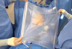
I stopped performing open power morcellation about 2 years ago and developed a new approach to enclosed morcellation. By creating an artificial pneumoperitoneum in a bag, a good working space is developed within it. This meets the needs of containment while significantly lowering the risk of tissue dissemination and also reducing the risk of bowel injury, vascular injury, and other types of morcellator-related mechanical injuries. This approach to morcellation is safer on all fronts.
My method of enclosed morcellation has been utilized in both single-port and multiport hysterectomy (total and supracervical) and myomectomy performed with either traditional laparoscopy or the robotic platform.
The morcellation process is begun after surgery is complete except for tissue removal, and the patient is hemostatic. The specimen has been placed in a location where it can be easily retrieved; I prefer the right upper quadrant.
In a single-port approach, the bag is inserted into the abdomen through an open single-port cannula at the umbilicus. I use a Steri-Drape Isolation Bag (3M), 50 cm by 50 cm, with drawstrings. (See Figure 1.) Standing across from my assistant, I tightly fan-fold the bag and wring it to purge it of air. Using an atraumatic grasper, I position the bag linearly with the opening up; it is inserted into the pelvis first, with the superior end guided upward into the mid- and then upper-abdomen.

After the bag is placed, the cannula is recapped with the multiport cap. I use the Olympus TriPort 15 (Advanced Surgical Concepts), and the abdomen is reinsufflated. A camera is inserted into one of the 5-mm ports. I use the Olympus 5-mm articulating laparoscope.
Through the other 5-mm port, I maneuver the bag to form a pocket opening by pushing the upper corners of the bag against the respective abdominal sidewalls, and the body of the bag into the pelvis. I then elevate the specimen and, with a rotational movement, I guide the specimen along the right abdominal sidewall and into the pocket opening. I then elevate the sides of the bag and ensure that the specimen is well contained. (See Figure 2.)
The drawstrings are then brought up into the port hub. The cap is removed, and the opening of the bag is pulled up through the port and out approximately 15-20 cm, as symmetrically as possible.
The port cap is then replaced within the exteriorized neck of the bag, and the bag is insufflated with traditional laparoscopic pressure of 15-18 mm, creating an artificial pneumoperitoneum. (See Figure 3.) The camera is reinserted, followed by the morcellator, into the 15-mm port of the single-site tri-port. The morcellator must be lubricated so it does not catch the bag on the way in. Morcellation is carried out under direct vision.
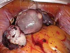
After the morcellation process is complete, the morcellator is removed, followed by the camera and the tri-port cap. The bag is desufflated and removed through the open cannula, and the abdomen is closed as usual.
In a multiport approach, the umbilical incision is extended to 15 mm after completion of the hysterectomy or myomectomy. The bag is inserted and placed in a manner similar to the single-port approach, except that the lateral ports may be utilized to further position the bag once it is placed about halfway in. The bag is similarly opened, the specimen contained, and the neck of the bag exteriorized. A 15-mm port is similarly lubricated and placed inside the neck of the bag and through the umbilical incision.
As the bag is insufflated, the lateral ports are backed slightly out so that they're flush with the abdominal wall, and the trocars are opened to allow the release of any residual gas in the abdomen. The laparoscope is then placed into the 15-mm umbilical trocar.
Visualization will ultimately occur from another site, however. One of the lateral 5-mm trocars is advanced at a right angle to the insufflated bag, and an introducer is placed to puncture the bag. I recommend using a blunt plastic trocar; a balloon-tip trocar also can be used. Contrary to what one might think, the bag will not leak any gas. The laparoscope is now transferred to this 5-mm lateral port, and the umbilical port is removed. A lubricated morcellator is inserted directly through the umbilical incision, and morcellation is performed under direct visualization from the side port.
These techniques for single-port or multiport enclosed power morcellation have been shown to be reproducible and successful – with all bags intact and specimens contained – in a multicenter analysis that is being prepared for publication. The largest uterus in the study was 1,481 g.
To make the approach less cumbersome, I have designed a morcellation device consisting of a specialized bag that will open automatically and assist in capturing the specimen. The device, which has closure tabs and a retrieval lanyard, has been developed with the input of other minimally invasive gynecologic surgeons and the design and engineering expertise of Advanced Surgical Concepts. It will be manufactured by the company pending Food and Drug Administration approval.
Concealed power morcellation: Our take
By Bernard Taylor, M.D.
The approach to the use of power morcellation within a bag in my practice was not specifically driven by the desire to prevent the dispersion of malignant leiomyosarcoma – an issue that is now front and center and indeed, is important – but by a broader desire to perform as complete an extraction as possible.
For years we have used isolation bags to conceal and contain small uteri, ovaries, and specimens that we debulk using a scalpel and remove through a small umbilical incision. However, this approach is cumbersome for removing larger uteri. Consequently, over the past year we refined our methods for performing power morcellation within a bag.
This procedure can be performed after traditional laparoscopy or robotic-assisted hysterectomy or myomectomy to extract the specimen from the abdomen. A surgical tissue bag is placed into the abdomen and is insufflated within the abdomen. Morcellation is carried out within the concealed "pseudopneumoperitoneum," and the bag is exteriorized from the abdomen, typically through the umbilical incision.
The majority of my procedures utilize a multiport approach due to the nature of my practice as a urogynecologist. After hysterectomy or myomectomy, the specimen is placed in the upper abdomen. The cuff is closed, other procedures are completed in the usual fashion, and hemostasis is reassured. In robotic procedures, we undock the robot prior to morcellation. The umbilical port is removed, and the bag is placed into the abdomen with a ring forceps or atraumatic forceps without teeth. The umbilical port is then replaced to reestablish the pneumoperitoneum for visualization.
For larger uterine specimens, I use an Isodrape isolation bag (Microtek Medical). This 18 x 18 inch bag with a drawstring opening easily accommodates uteri larger than 1,000 g. The bag is placed into the pelvis with the open end at the pelvic brim and is opened wide. I use the term "open from iliac to iliac and sacrum to bladder." Once the bag is positioned, the specimen is grasped; the patient is slowly taken out of the Trendelenburg position to allow for gravity to assist in the placement of the specimen into the bag.
The drawstrings are then grasped and taken out of the umbilical trocar with simultaneous removal of the umbilical port. At this time, the open end of the bag is exteriorized, and the specimen technically is extraperitoneal and "concealed."
To insufflate the bag at this point and create the "pseudopneumoperitoneum," a 15-mm port is lubricated and placed through the opening in the bag. The bag is then insufflated to the usual pressure (15 mm Hg). The accessory ports are opened at this time to allow for the abdominal pressure to fall and for the bag to expand and fill the abdominal cavity.
To enable visualization throughout the morcellation procedure, I use one of the previously placed lower abdomen lateral ports to place a 5-mm camera. The camera is placed after a 5-mm balloon tip trocar is introduced and advanced to perforate the bag. The balloon tip is insufflated in the usual fashion, securing the bag against the abdominal wall and preventing a gas leak.
Alternatively, I have also used a SILSport (Covidien) at the umbilicus in cases when there is a leak at the umbilical incision and the 15-mm port does not seal properly. While use of a single-port technique has been shown to be feasible for both visualization and morcellation, it can present challenges in complex cases with a larger uterus or multiple fibroids.
Either way, morcellation can take place within the concealed "pseudo-peritoneal" cavity, with the morcellator lubricated and placed either through the umbilical incision or through the Tri-Port. After morcellation is complete, the bag containing the smaller tissue fragments and blood is simply removed through the umbilicus, and closure of the abdomen is completed in the usual fashion.

I also have adopted a simplified approach to remove the smaller uteri with supracervical hysterectomy and sacral colpopexy performed laparoscopically. Despite preoperative screening with endometrial biopsy and pelvic ultrasound, several of my patients in the past have been diagnosed with either early ovarian or endometrial neoplasias on final pathology. After completing the procedure, I place the small menopausal senile uterus and adnexa into a 15-mm endoscopic bag. The bag is brought through the umbilical port, and the specimen is removed via morcellation with a scalpel or scissors.
Surgeons have asked about additional time needed to place the bag and position the specimen. Technically, specimen placement is a learned skill; once one is proficient, the case times are not any different. In fact, cases may be shorter because of the time saved by not having to retrieve uterine fragments from the abdomen and pelvis.
Theoretically, the procedure addresses concerns associated with tissue fragmentation and dissemination within the abdominal cavity. To date, there are no trials showing prevention of cancer upstaging or benign conditions such as leiomyomatosis peritonealis disseminata or endometriosis.
Enclosed vaginal morcellation for enlarged uteri
By Ceana H. Nezhat, M.D.
Removal of large uteri via minimally invasive surgery poses challenges to gynecologic surgeons. Limitations on the use of intraperitoneal electromechanical morcellation are a step forward in terms of protecting patients from undue harm, but we also must acknowledge that minimally invasive surgery has been shown to be superior to laparotomy in the majority of cases.
While safer ways to extract large specimens without risk of spreading both benign and malignant tissue are being studied and developed, gynecologic surgeons need to find alternative minimally invasive approaches for removing large specimens.
Minimally invasive approaches to extirpate uteri and myomas have been described prior to the advent of electromechanical morcellation. A natural orifice such as the vagina for gynecologic procedures is a clear choice for removing specimens. Laparoscopic-assisted myomectomy is a combination of laparoscopy and minilaparotomy for tissue extraction (JSLS 2001;5:299-303). Another alternative is extracting myoma and other tissues through a posterior colpotomy. Rates of dyspareunia and adhesions in the cul-de-sac after tissue extraction through a colpotomy are low (J. Reprod. Med. 1993;38:534-6).
Vaginal hysterectomy was chronicled and performed by the Greek physician Soranus of Ephesus in 120 A.D. (Acta Chir. Iugosl. 2011:58:9-14), and it is still the preferred route of hysterectomy when it can be performed. Large uteri can be transvaginally morcellated and vaginally extracted; however, this technique still poses the risk, although low, of spilling fragments of uterine tissue in the abdomen.
By combining total laparoscopic hysterectomy and vaginal morcellation of enlarged uteri within an enclosed specimen bag, the risk of spilling uterine fragments is reduced, in addition to avoiding potential visceral and vascular complications associated with intraperitoneal electromechanical morcellation.
Enclosed vaginal morcellation is the technique of placing the enlarged hysterectomy specimen in an endoscopic specimen retrieval bag and then transvaginally morcellating the tissue.
In the development of this technique, I have experimented with different endoscopic sacs and retractors. In my experience, the optimal laparoscopic bag is the LapSac Surgical Tissue Pouch (Cook Medical). This endoscopic, one-time use, specimen retrieval bag is flexible, durable nylon with an integral polyurethane inner coating and polypropylene drawstring (Figure 4). The bag comes in four sizes, the largest (size 8 x 10 inch, volume 1,500 mL) of which I used to assist in removing the 18-week size uterus in the accompanying video.
The other key piece of equipment for vaginal extraction of large uteri is a transvaginally-placed wound retractor. As I have previously described, the self-retaining retractor provides optimal exposure in a narrow field and minimizes trauma to surrounding tissue (J. Minim. Inv. Gynecol. 2009:16:616-7). Two wound retractors are suitable for this surgical technique: the Alexis Wound Protector/Retractor (Applied Medical) (Figure 5) and the Mobius Elastic Abdominal Retractor (Cooper Surgical) (Figure 6).
After complete laparoscopic detachment of the uterus and cervix (and adnexa), the wound retractor is placed transvaginally. One semirigid ring is pushed superiorly through the vagina and into the peritoneal cavity, where is it placed in the cul-de-sac. In this manner, a uniform, circumferential orifice is created. In narrower vaginal vaults, the ring can be compressed between the thumb and index finger to reduce its diameter and thus be introduced into the peritoneal cavity atraumatically. With the small-size retraction systems, an orifice of 2.5 to 6 cm diameter can be created.
The LapSac is folded and then inserted with ring forceps transvaginally through the wound retractor. (See Figure 7.) The bag is unfolded intraperitoneally using the integral tabs to demarcate the edges of the bag. The bag should be unfolded in the pelvis with the opening of the bag cephalad. The fundus of the hysterectomy specimen is grasped with a tenaculum and placed inside the bag. (See Figure 8.)
It is an important technical point to grab the fundus or the heaviest point of the specimen to allow weight and gravity to facilitate placing the specimen securely in the bag. If the cervix or a lighter part of the specimen is grasped, placement in the bag is more difficult and the specimen will likely slip out of the bag.

Once the entire hysterectomy specimen is in the LapSac, the bag is cinched by grasping the blue drawstrings. The opening of the bag is pulled through the vagina. The opening of the bag is externalized and the edges are attached with Allis clamps to the external edge of the wound retractor. The specimen (typically the cervix first) is grasped with a tenaculum and vaginally morcellated and cored with a scalpel (Figure 9).
Metal vaginal retractors are placed within the bag to protect against sharp injury during morcellation. I prefer to amputate the cervix and adnexa separately and en bloc to preserve as much original architecture as possible so the pathologists can properly orient and examine the tissue. After completion of the morcellation, the wound retractor is removed, the cuff closed, and the procedure completed per surgeon preference.
Dr. Shibley has been in full-time practice with Ob.Gyn. Specialists, Fairview Health Services in the Minneapolis area (Edina), for 20 years. He reported that he has a financial interest in the artificial pneumoperitoneum device under development, and that he is a consultant for Olympus.
Dr. Taylor is a urogynecologist who is a female pelvic medicine and reconstructive surgeon practicing at the Carolinas Medical Center–Advanced Surgical Specialties for Women in Charlotte, N.C. He reported that he has no disclosures relevant to this Master Class.
Dr. Nezhat is the current president of the AAGL, adjunct professor of obstetrics and gynecology at Emory University and program director of minimally invasive surgery at Northside Hospital, both in Atlanta, and adjunct clinical professor of obstetrics and gynecology at Stanford (Calif.) University. He reported that he is a consultant for Karl Storz Endoscopy.
Videos of these experts' individual techniques of electromechanical power morcellation within the confines of a bag, as well as that of Dr. Douglas Brown, director of the Center for Minimally Invasive Gynecologic Surgery at Massachusetts General Hospital, Boston, can be viewed at the SurgeryU website.
Power morcellation within an insufflated bag
By Tony Shibley, M.D.
Morcellating safely in laparoscopic hysterectomy or myomectomy involves not only the use of a bag, but also the creation of an artificial pneumoperitoneum inside the bag. Inflation of the bag allows us to create a safe and completely contained working environment in the abdomen, with good visualization and adequate working space.
In the past, specimen containment bags were used for morcellation, but the bags would adhere to tissue and would be easily cut or would tear or rupture. Without proper space and visualization, surgeons could not break tissue into smaller fragments without endangering the bag or the structures behind the bag. We were safely and precisely disconnecting tissue in our surgeries, but falling short with tissue removal. There were calls for more durable bags, but the problems of visualization and safe distance were not addressed. Eventually, the goal of morcellating within a bag was largely abandoned in favor of open power morcellation.
I stopped performing open power morcellation about 2 years ago and developed a new approach to enclosed morcellation. By creating an artificial pneumoperitoneum in a bag, a good working space is developed within it. This meets the needs of containment while significantly lowering the risk of tissue dissemination and also reducing the risk of bowel injury, vascular injury, and other types of morcellator-related mechanical injuries. This approach to morcellation is safer on all fronts.
My method of enclosed morcellation has been utilized in both single-port and multiport hysterectomy (total and supracervical) and myomectomy performed with either traditional laparoscopy or the robotic platform.
The morcellation process is begun after surgery is complete except for tissue removal, and the patient is hemostatic. The specimen has been placed in a location where it can be easily retrieved; I prefer the right upper quadrant.
In a single-port approach, the bag is inserted into the abdomen through an open single-port cannula at the umbilicus. I use a Steri-Drape Isolation Bag (3M), 50 cm by 50 cm, with drawstrings. (See Figure 1.) Standing across from my assistant, I tightly fan-fold the bag and wring it to purge it of air. Using an atraumatic grasper, I position the bag linearly with the opening up; it is inserted into the pelvis first, with the superior end guided upward into the mid- and then upper-abdomen.
After the bag is placed, the cannula is recapped with the multiport cap. I use the Olympus TriPort 15 (Advanced Surgical Concepts), and the abdomen is reinsufflated. A camera is inserted into one of the 5-mm ports. I use the Olympus 5-mm articulating laparoscope.
Through the other 5-mm port, I maneuver the bag to form a pocket opening by pushing the upper corners of the bag against the respective abdominal sidewalls, and the body of the bag into the pelvis. I then elevate the specimen and, with a rotational movement, I guide the specimen along the right abdominal sidewall and into the pocket opening. I then elevate the sides of the bag and ensure that the specimen is well contained. (See Figure 2.)
The drawstrings are then brought up into the port hub. The cap is removed, and the opening of the bag is pulled up through the port and out approximately 15-20 cm, as symmetrically as possible.
The port cap is then replaced within the exteriorized neck of the bag, and the bag is insufflated with traditional laparoscopic pressure of 15-18 mm, creating an artificial pneumoperitoneum. (See Figure 3.) The camera is reinserted, followed by the morcellator, into the 15-mm port of the single-site tri-port. The morcellator must be lubricated so it does not catch the bag on the way in. Morcellation is carried out under direct vision.
After the morcellation process is complete, the morcellator is removed, followed by the camera and the tri-port cap. The bag is desufflated and removed through the open cannula, and the abdomen is closed as usual.
In a multiport approach, the umbilical incision is extended to 15 mm after completion of the hysterectomy or myomectomy. The bag is inserted and placed in a manner similar to the single-port approach, except that the lateral ports may be utilized to further position the bag once it is placed about halfway in. The bag is similarly opened, the specimen contained, and the neck of the bag exteriorized. A 15-mm port is similarly lubricated and placed inside the neck of the bag and through the umbilical incision.
As the bag is insufflated, the lateral ports are backed slightly out so that they're flush with the abdominal wall, and the trocars are opened to allow the release of any residual gas in the abdomen. The laparoscope is then placed into the 15-mm umbilical trocar.
Visualization will ultimately occur from another site, however. One of the lateral 5-mm trocars is advanced at a right angle to the insufflated bag, and an introducer is placed to puncture the bag. I recommend using a blunt plastic trocar; a balloon-tip trocar also can be used. Contrary to what one might think, the bag will not leak any gas. The laparoscope is now transferred to this 5-mm lateral port, and the umbilical port is removed. A lubricated morcellator is inserted directly through the umbilical incision, and morcellation is performed under direct visualization from the side port.
These techniques for single-port or multiport enclosed power morcellation have been shown to be reproducible and successful – with all bags intact and specimens contained – in a multicenter analysis that is being prepared for publication. The largest uterus in the study was 1,481 g.
To make the approach less cumbersome, I have designed a morcellation device consisting of a specialized bag that will open automatically and assist in capturing the specimen. The device, which has closure tabs and a retrieval lanyard, has been developed with the input of other minimally invasive gynecologic surgeons and the design and engineering expertise of Advanced Surgical Concepts. It will be manufactured by the company pending Food and Drug Administration approval.
Concealed power morcellation: Our take
By Bernard Taylor, M.D.
The approach to the use of power morcellation within a bag in my practice was not specifically driven by the desire to prevent the dispersion of malignant leiomyosarcoma – an issue that is now front and center and indeed, is important – but by a broader desire to perform as complete an extraction as possible.
For years we have used isolation bags to conceal and contain small uteri, ovaries, and specimens that we debulk using a scalpel and remove through a small umbilical incision. However, this approach is cumbersome for removing larger uteri. Consequently, over the past year we refined our methods for performing power morcellation within a bag.
This procedure can be performed after traditional laparoscopy or robotic-assisted hysterectomy or myomectomy to extract the specimen from the abdomen. A surgical tissue bag is placed into the abdomen and is insufflated within the abdomen. Morcellation is carried out within the concealed "pseudopneumoperitoneum," and the bag is exteriorized from the abdomen, typically through the umbilical incision.
The majority of my procedures utilize a multiport approach due to the nature of my practice as a urogynecologist. After hysterectomy or myomectomy, the specimen is placed in the upper abdomen. The cuff is closed, other procedures are completed in the usual fashion, and hemostasis is reassured. In robotic procedures, we undock the robot prior to morcellation. The umbilical port is removed, and the bag is placed into the abdomen with a ring forceps or atraumatic forceps without teeth. The umbilical port is then replaced to reestablish the pneumoperitoneum for visualization.
For larger uterine specimens, I use an Isodrape isolation bag (Microtek Medical). This 18 x 18 inch bag with a drawstring opening easily accommodates uteri larger than 1,000 g. The bag is placed into the pelvis with the open end at the pelvic brim and is opened wide. I use the term "open from iliac to iliac and sacrum to bladder." Once the bag is positioned, the specimen is grasped; the patient is slowly taken out of the Trendelenburg position to allow for gravity to assist in the placement of the specimen into the bag.
The drawstrings are then grasped and taken out of the umbilical trocar with simultaneous removal of the umbilical port. At this time, the open end of the bag is exteriorized, and the specimen technically is extraperitoneal and "concealed."
To insufflate the bag at this point and create the "pseudopneumoperitoneum," a 15-mm port is lubricated and placed through the opening in the bag. The bag is then insufflated to the usual pressure (15 mm Hg). The accessory ports are opened at this time to allow for the abdominal pressure to fall and for the bag to expand and fill the abdominal cavity.
To enable visualization throughout the morcellation procedure, I use one of the previously placed lower abdomen lateral ports to place a 5-mm camera. The camera is placed after a 5-mm balloon tip trocar is introduced and advanced to perforate the bag. The balloon tip is insufflated in the usual fashion, securing the bag against the abdominal wall and preventing a gas leak.
Alternatively, I have also used a SILSport (Covidien) at the umbilicus in cases when there is a leak at the umbilical incision and the 15-mm port does not seal properly. While use of a single-port technique has been shown to be feasible for both visualization and morcellation, it can present challenges in complex cases with a larger uterus or multiple fibroids.
Either way, morcellation can take place within the concealed "pseudo-peritoneal" cavity, with the morcellator lubricated and placed either through the umbilical incision or through the Tri-Port. After morcellation is complete, the bag containing the smaller tissue fragments and blood is simply removed through the umbilicus, and closure of the abdomen is completed in the usual fashion.
I also have adopted a simplified approach to remove the smaller uteri with supracervical hysterectomy and sacral colpopexy performed laparoscopically. Despite preoperative screening with endometrial biopsy and pelvic ultrasound, several of my patients in the past have been diagnosed with either early ovarian or endometrial neoplasias on final pathology. After completing the procedure, I place the small menopausal senile uterus and adnexa into a 15-mm endoscopic bag. The bag is brought through the umbilical port, and the specimen is removed via morcellation with a scalpel or scissors.
Surgeons have asked about additional time needed to place the bag and position the specimen. Technically, specimen placement is a learned skill; once one is proficient, the case times are not any different. In fact, cases may be shorter because of the time saved by not having to retrieve uterine fragments from the abdomen and pelvis.
Theoretically, the procedure addresses concerns associated with tissue fragmentation and dissemination within the abdominal cavity. To date, there are no trials showing prevention of cancer upstaging or benign conditions such as leiomyomatosis peritonealis disseminata or endometriosis.
Enclosed vaginal morcellation for enlarged uteri
By Ceana H. Nezhat, M.D.
Removal of large uteri via minimally invasive surgery poses challenges to gynecologic surgeons. Limitations on the use of intraperitoneal electromechanical morcellation are a step forward in terms of protecting patients from undue harm, but we also must acknowledge that minimally invasive surgery has been shown to be superior to laparotomy in the majority of cases.
While safer ways to extract large specimens without risk of spreading both benign and malignant tissue are being studied and developed, gynecologic surgeons need to find alternative minimally invasive approaches for removing large specimens.
Minimally invasive approaches to extirpate uteri and myomas have been described prior to the advent of electromechanical morcellation. A natural orifice such as the vagina for gynecologic procedures is a clear choice for removing specimens. Laparoscopic-assisted myomectomy is a combination of laparoscopy and minilaparotomy for tissue extraction (JSLS 2001;5:299-303). Another alternative is extracting myoma and other tissues through a posterior colpotomy. Rates of dyspareunia and adhesions in the cul-de-sac after tissue extraction through a colpotomy are low (J. Reprod. Med. 1993;38:534-6).
Vaginal hysterectomy was chronicled and performed by the Greek physician Soranus of Ephesus in 120 A.D. (Acta Chir. Iugosl. 2011:58:9-14), and it is still the preferred route of hysterectomy when it can be performed. Large uteri can be transvaginally morcellated and vaginally extracted; however, this technique still poses the risk, although low, of spilling fragments of uterine tissue in the abdomen.
By combining total laparoscopic hysterectomy and vaginal morcellation of enlarged uteri within an enclosed specimen bag, the risk of spilling uterine fragments is reduced, in addition to avoiding potential visceral and vascular complications associated with intraperitoneal electromechanical morcellation.
Enclosed vaginal morcellation is the technique of placing the enlarged hysterectomy specimen in an endoscopic specimen retrieval bag and then transvaginally morcellating the tissue.
In the development of this technique, I have experimented with different endoscopic sacs and retractors. In my experience, the optimal laparoscopic bag is the LapSac Surgical Tissue Pouch (Cook Medical). This endoscopic, one-time use, specimen retrieval bag is flexible, durable nylon with an integral polyurethane inner coating and polypropylene drawstring (Figure 4). The bag comes in four sizes, the largest (size 8 x 10 inch, volume 1,500 mL) of which I used to assist in removing the 18-week size uterus in the accompanying video.
The other key piece of equipment for vaginal extraction of large uteri is a transvaginally-placed wound retractor. As I have previously described, the self-retaining retractor provides optimal exposure in a narrow field and minimizes trauma to surrounding tissue (J. Minim. Inv. Gynecol. 2009:16:616-7). Two wound retractors are suitable for this surgical technique: the Alexis Wound Protector/Retractor (Applied Medical) (Figure 5) and the Mobius Elastic Abdominal Retractor (Cooper Surgical) (Figure 6).
After complete laparoscopic detachment of the uterus and cervix (and adnexa), the wound retractor is placed transvaginally. One semirigid ring is pushed superiorly through the vagina and into the peritoneal cavity, where is it placed in the cul-de-sac. In this manner, a uniform, circumferential orifice is created. In narrower vaginal vaults, the ring can be compressed between the thumb and index finger to reduce its diameter and thus be introduced into the peritoneal cavity atraumatically. With the small-size retraction systems, an orifice of 2.5 to 6 cm diameter can be created.
The LapSac is folded and then inserted with ring forceps transvaginally through the wound retractor. (See Figure 7.) The bag is unfolded intraperitoneally using the integral tabs to demarcate the edges of the bag. The bag should be unfolded in the pelvis with the opening of the bag cephalad. The fundus of the hysterectomy specimen is grasped with a tenaculum and placed inside the bag. (See Figure 8.)
It is an important technical point to grab the fundus or the heaviest point of the specimen to allow weight and gravity to facilitate placing the specimen securely in the bag. If the cervix or a lighter part of the specimen is grasped, placement in the bag is more difficult and the specimen will likely slip out of the bag.
Once the entire hysterectomy specimen is in the LapSac, the bag is cinched by grasping the blue drawstrings. The opening of the bag is pulled through the vagina. The opening of the bag is externalized and the edges are attached with Allis clamps to the external edge of the wound retractor. The specimen (typically the cervix first) is grasped with a tenaculum and vaginally morcellated and cored with a scalpel (Figure 9).
Metal vaginal retractors are placed within the bag to protect against sharp injury during morcellation. I prefer to amputate the cervix and adnexa separately and en bloc to preserve as much original architecture as possible so the pathologists can properly orient and examine the tissue. After completion of the morcellation, the wound retractor is removed, the cuff closed, and the procedure completed per surgeon preference.
Dr. Shibley has been in full-time practice with Ob.Gyn. Specialists, Fairview Health Services in the Minneapolis area (Edina), for 20 years. He reported that he has a financial interest in the artificial pneumoperitoneum device under development, and that he is a consultant for Olympus.
Dr. Taylor is a urogynecologist who is a female pelvic medicine and reconstructive surgeon practicing at the Carolinas Medical Center–Advanced Surgical Specialties for Women in Charlotte, N.C. He reported that he has no disclosures relevant to this Master Class.
Dr. Nezhat is the current president of the AAGL, adjunct professor of obstetrics and gynecology at Emory University and program director of minimally invasive surgery at Northside Hospital, both in Atlanta, and adjunct clinical professor of obstetrics and gynecology at Stanford (Calif.) University. He reported that he is a consultant for Karl Storz Endoscopy.
Videos of these experts' individual techniques of electromechanical power morcellation within the confines of a bag, as well as that of Dr. Douglas Brown, director of the Center for Minimally Invasive Gynecologic Surgery at Massachusetts General Hospital, Boston, can be viewed at the SurgeryU website.
