User login
More data suggest preexisting statin use improves COVID outcomes
Compared with patients who didn’t take statins, statin users had better health outcomes. For those who used these medications, the researchers saw lower mortality, lower clinical severity, and shorter hospital stays, aligning with previous observational studies, said lead author Ettore Crimi, MD, of the University of Central Florida, Orlando, and colleagues in their abstract, which was part of the agenda for the Anesthesiology annual meeting.
They attributed these clinical improvements to the pleiotropic – non–cholesterol lowering – effects of statins.
“[These] benefits of statins have been reported since the 1990s,” Dr. Crimi said in an interview. “Statin treatment has been associated with a marked reduction of markers of inflammation, such as erythrocyte sedimentation rate (ESR) and C-reactive protein (CRP), interleukin-6 (IL-6), ferritin, and white blood cell count, among others.”
He noted that these effects have been studied in an array of conditions, including cancer, autoimmune diseases, chronic inflammatory disease, and in the perioperative setting, and with infectious diseases, including COVID-19.
In those previous studies, “preexisting statin use was protective among hospitalized COVID-19 patients, but a large, multicenter cohort study has not been reported in the United States,” Dr. Crimi and his colleagues wrote in their abstract.
To address this knowledge gap, they turned to electronic medical records from 38,875 patients hospitalized with COVID-19 from January to September 2020. Almost one-third of the population (n = 11,533) were using statins prior to hospitalization, while the remainder (n = 27,342) were nonusers.
The primary outcome was all-cause mortality. Secondary outcomes included death from COVID-19, along with a variety of severe complications. While the analysis did account for a range of potentially confounding variables, the effects of different SARS-CoV-2 variants and new therapeutics were not considered. Vaccines were not yet available at the time the data were collected.
Statin users had a 31% lower rate of all-cause mortality (odds ratio, 0.69; 95% confidence interval, 0.64-0.75; P = .001) and a 37% reduced rate of death from COVID-19 (OR, 0.63; 95% CI, 0.58-0.69; P = .001).
A litany of other secondary variables also favored statin users, including reduced rates of discharge to hospice (OR, 0.79), ICU admission (OR, 0.69), severe acute respiratory distress syndrome (ARDs; OR, 0.72), critical ARDs (OR, 0.57), mechanical ventilation (OR, 0.60), severe sepsis with septic shock (OR, 0.66), and thrombosis (OR, 0.46). Statin users also had, on average, shorter hospital stays and briefer mechanical ventilation.
“Our study showed a strong association between preexisting statin use and reduced mortality and morbidity rates in hospitalized COVID-19 patients,” the investigators concluded. “Pleiotropic benefits of statins could be repurposed for COVID-19 illness.”
Prospective studies needed before practice changes
How to best use statins against COVID-19, if at all, remains unclear, Dr. Crimi said, as initiation upon infection has generated mixed results in other studies, possibly because of statin pharmacodynamics. Cholesterol normalization can take about 6 weeks, so other benefits may track a similar timeline.
“The delayed onset of statins’ pleiotropic effects may likely fail to keep pace with the rapidly progressive, devastating COVID-19 disease,” Dr. Crimi said. “Therefore, initiating statins for an acute disease may not be an ideal first-line treatment.”
Stronger data are on the horizon, he added, noting that 19 federally funded prospective trials are underway to better understand the relationship between statins and COVID-19.
Daniel Rader, MD, of the University of Pennsylvania, Philadelphia, said the present findings are “not especially notable” because they “mostly confirm previous studies, but in a large U.S. cohort.”
Dr. Rader, who wrote about the potential repurposing of statins for COVID-19 back in the first year of the pandemic (Cell Metab. 2020 Aug 4;32[2]:145-7), agreed with the investigators that recommending changes to clinical practice would be imprudent until randomized controlled data confirm the benefits of initiating statins in patients with active COVID-19.
“More research on the impact of cellular cholesterol metabolism on SARS-CoV-2 infection of cells and generation of inflammation would also be of interest,” he added.
The investigators disclosed no competing interests. Dr. Rader disclosed relationships with Novartis, Pfizer, Verve, and others.
FROM ANESTHESIOLOGY 2022
COVID lawsuits have arrived: Which doctors are at risk?
A pregnant patient who had COVID-19 showed up at a hospital with respiratory difficulty caused by her illness. Physicians had to perform an emergency delivery of her near-term baby.
The infant survived, but the woman lost oxygen during the ordeal and suffered hypoxic brain damage. She is now suing an obstetrician, a pulmonologist, and an intensive care unit physician for medical malpractice.
The plaintiff contends there was a failure “to adequately recognize and treat her condition,” said Peter Kolbert, senior vice president for claim and litigation services for Healthcare Risk Advisors, part of TDC Group, which includes national medical liability insurer The Doctors Company.
“The physicians involved vehemently disagree and believe they treated her appropriately,” Mr. Kolbert said. “In fact, we believe their actions were heroic.”
In another case, a patient with COVID-19 and multiple comorbidities was admitted to a hospital. Physicians sedated and intubated the patient to maintain her airway. She recovered, but the patient now alleges doctors were negligent because she developed ulcers during her hospital stay. The case occurred during the height of the pandemic. In addition to the hospital, a pulmonologist, an ICU physician, and an acute care physician are named in the suit.
Both of these lawsuits are being defined as COVID claims because at the time, the plaintiffs either had COVID and needed care because of COVID, or because the care that physicians provided was affected by COVID in some way.
In the second case, the patient had COVID and needed treatment. During her recovery, ulcers developed. A significant aspect of this case is that it occurred during the height of the pandemic. Hospitals were overcrowded, the staff was swamped, and resources were limited. One factor may be that physicians were doing the best they could at the time but that the pandemic affected the extent of care they could provide.
Now, new data reflect the grim news: COVID claims have arrived. These cases from the claims database of The Doctors Company are just two examples of many COVID-related claims that have been levied since the pandemic started.
Currently, there are 162 open COVID-related claims in The Doctors Company database, according to Mr. Kolbert. A September 2022 benchmark report from Aon and the American Society for Health Care Risk Management indicates that 245 claims that pertain to patients with confirmed or suspected COVID-19 have been filed since the pandemic began. The findings in this report stem from an analysis of 95,600 hospital and physician liability claims that occurred between 2012 and 2021.
Of the 245 cases, 89 claims have been closed. The average cost was $43,000 per claim, said Kanika Vats, a director and actuary for Aon, a global firm that provides risk, reinsurance, and health solutions. Six of the claims cost $300,000 or more; the highest settlement was for $700,000.
“Most of the allegations in these claims revolve around delay in treatment or delay in diagnosis,” Ms. Vats said.
Which specialties are involved in legal actions?
Physicians working in acute care settings such as emergency departments and urgent care centers are the primary targets in COVID-related lawsuits involving doctors, say legal analysts. However, other specialties are also being affected. Physicians being sued include some who practiced telemedicine during the pandemic.
In one case, a primary care physician saw a patient via telemedicine because the physical medical office was closed. The patient was evaluated virtually and was sent for bloodwork and an x-ray.
The patient is now suing the primary care physician, alleging that failure to immediately send her to a hospital resulted in tuberculosis going untreated and that the failure led to a bad outcome. The allegation is that the physician underevaluated the case during the telemedicine visit, Mr. Kolbert said.
Drew Graham, an attorney at Hall Booth Smith PC, which is based in New York, said that most of the COVID-related liability claims he has seen involve facilities that provide postacute care, such as nursing homes and assisted living facilities. His firm has also seen a small number of COVID-related claims against physicians.
At least two of the claims involved allegations of improper treatment of COVID during hospitalizations, he said. Another involved a telehealth visit in which the patient claimed the virtual care that was provided was improper and that their condition required an in-person examination. Mr. Graham declined to specify the specialties of the physicians sued.
The Medical Professional Liability Association reports similar trends in COVID-related claims. Long-term facilities and hospitals are the most common focus of COVID-19 claims, followed by emergency medicine, primary care, and ob/gyn medical specialties, according to Kwon Miller, manager of data and analytics for MPL Association, a national trade association for medical liability insurers that operates a large claims database.
Between January 2020 and June 2022, the MPL Association Data Sharing Project recorded 280 COVID-19 events. “Events” refers to notifications, licensing board inquiries, and claims involving COVID. Of these events, 180 were closed with no indemnity payment, and 13 were closed with an average indemnity payment of $3,816, Mr. Miller said.
Complaints of delayed care associated with the pandemic are also on the rise. For example, one patient is suing a gastroenterologist for delaying his colonoscopy, alleging the postponement led to a delayed colon cancer diagnosis and worse prognosis, Mr. Kolbert said.
“It was delayed because all elective procedures at the time were being put off,” he said. “The patient claims that had they received the scheduled screening, the cancer would have been diagnosed at stage I as opposed to stage III.”
Why isn’t federal immunity shielding physicians?
A pressing question about the growing number of COVID claims is why state and federal immunity isn’t preventing such lawsuits.
In 2020, the U.S. Department of Health & Human Services published a declaration under the Public Readiness and Emergency Preparedness Act (PREP Act) that provided liability immunity to health care professionals for any activity related to medical countermeasures against COVID-19. The act allows an exception for negligence claims associated with death or serious injury caused by willful misconduct.
At the same time, most states implemented laws or executive orders shielding physicians from liability claims related to the prevention and treatment of COVID-19, unless gross negligence or willful misconduct is proven.
Mr. Graham said some COVID-related claims against physicians have included allegations of gross negligence to avoid the application of state immunity, while others combine allegations of deviations from standard of care unrelated to the pandemic.
Some plaintiffs are attempting to skirt the protections by making complaints sound as if they’re not related to COVID-19, Mr. Kolbert said. That way, they don’t have to prove gross negligence or willful misconduct at all.
“The filings at first blush may not tell you it’s a COVID case, but it may be a COVID case,” he said. “Plaintiffs’ attorneys are trying to assert that COVID defenses do not apply and that these cases are ‘traditional physician negligence’ claims. They’re trying to plead around the protections.”
The federal and state immunities are likely keeping the volume of COVID claims down overall and are discouraging some complaints from moving forward, attorneys say.
But because some plaintiffs are downplaying or ignoring the COVID association, it’s likely that more COVID lawsuits exist than anyone realizes, according to Mr. Kolbert.
“I expect there’s an underestimation of how many COVID claims are really out there,” he said.
What does the future hold for COVID claims?
Currently, the frequency and the severity of COVID claims are low, Ms. Vats said. She believes the cost of such claims will continue to remain at low levels.
“But again, there is a lot of uncertainty,” she said. “This year, states have started to roll back their immunity protections, and in a lot of states, there is no cap in awarding [noneconomic] damages. There could well be a scenario where they allege wrongful death, and in a state with no cap on the pain and suffering component, if juries continue to behave the way they have been behaving, we could see aberration verdicts.”
Another lingering issue concerns which court systems have jurisdiction in cases involving COVID-related claims. Because of the nationwide response to the pandemic, Mr. Graham thinks it makes sense that federal courts handle the cases, but the plaintiffs’ bar has generally been opposed to federal jurisdiction.
“A second issue is the long-term impact of COVID litigation on our providers,” he said. “If the protections in place to limit liability are determined to be ineffective, our state and federal leaders must act aggressively and in a bipartisan way to make sure our health care providers are protected when we face the next crisis.”
A version of this article first appeared on Medscape.com.
Hair straighteners’ risk too small for docs to advise against their use
Clarissa Ghazi gets lye relaxers, which contain the chemical sodium hydroxide, applied to her hair two to three times a year.
A recent study that made headlines over a potential link between hair straighteners and uterine cancer is not going to make her stop.
“This study is not enough to cause me to say I’ll stay away from this because [the researchers] don’t prove that using relaxers causes cancer,” Ms. Ghazi said.
Indeed, primary care doctors are unlikely to address the increased risk of uterine cancer in women who frequently use hair straighteners that the study reported.
In the recently published paper on this research, the authors said that they found an 80% higher adjusted risk of uterine cancer among women who had ever “straightened,” “relaxed,” or used “hair pressing products” in the 12 months before enrolling in their study.
This finding is “real, but small,” says internist Douglas S. Paauw, MD, professor of medicine at the University of Washington in Seattle.
Dr. Paauw is among several primary care doctors interviewed for this story who expressed little concern about the implications of this research for their patients.
“Since we have hundreds of things we are supposed to discuss at our 20-minute clinic visits, this would not make the cut,” Dr. Paauw said.
While it’s good to be able to answer questions a patient might ask about this new research, the study does not prove anything, he said.
Alan Nelson, MD, an internist-endocrinologist and former special adviser to the CEO of the American College of Physicians, said while the study is well done, the number of actual cases of uterine cancer found was small.
One of the reasons he would not recommend discussing the study with patients is that the brands of hair products used to straighten hair in the study were not identified.
Alexandra White, PhD, lead author of the study, said participants were simply asked, “In the past 12 months, how frequently have you or someone else straightened or relaxed your hair, or used hair pressing products?”
The terms “straightened,” “relaxed,” and “hair pressing products” were not defined, and “some women may have interpreted the term ‘pressing products’ to mean nonchemical products” such as flat irons, Dr. White, head of the National Institute of Environmental Health Sciences’ Environment and Cancer Epidemiology group, said in an email.
Dermatologist Crystal Aguh, MD, associate professor of dermatology at Johns Hopkins University, Baltimore, tweeted the following advice in light of the new findings: “The overall risk of uterine cancer is quite low so it’s important to remember that. For now, if you want to change your routine, there’s no downside to decreasing your frequency of hair straightening to every 12 weeks or more, as that may lessen your risk.”
She also noted that “styles like relaxer, silk pressing, and keratin treatments should only be done by a professional, as this will decrease the likelihood of hair damage and scalp irritation.
“I also encourage women to look for hair products free of parabens and phthalates (which are generically listed as “fragrance”) on products to minimize exposure to hormone disrupting chemicals.”
Not ready to go curly
Ms. Ghazi said she decided to stop using keratin straighteners years ago after she learned they are made with several added ingredients. That includes the chemical formaldehyde, a known carcinogen, according to the American Cancer Society.
“People have been relaxing their hair for a very long time, and I feel more comfortable using [a relaxer] to straighten my hair than any of the others out there,” Ms. Ghazi said.
Janaki Ram, who has had her hair chemically straightened several times, said the findings have not made her worried that straightening will cause her to get uterine cancer specifically, but that they are a reminder that the chemicals in these products could harm her in some other way.
She said the new study findings, her knowledge of the damage straightening causes to hair, and the lengthy amount of time receiving a keratin treatment takes will lead her to reduce the frequency with which she gets her hair straightened.
“Going forward, I will have this done once a year instead of twice a year,” she said.
Dr. White, the author of the paper, said in an interview that the takeaway for consumers is that women who reported frequent use of hair straighteners/relaxers and pressing products were more than twice as likely to go on to develop uterine cancer compared to women who reported no use of these products in the previous year.
“However, uterine cancer is relatively rare, so these increases in risks are small,” she said. “Less frequent use of these products was not as strongly associated with risk, suggesting that decreasing use may be an option to reduce harmful exposure. Black women were the most frequent users of these products and therefore these findings are more relevant for Black women.”
In a statement, Dr. White noted, “We estimated that 1.64% of women who never used hair straighteners would go on to develop uterine cancer by the age of 70; but for frequent users, that risk goes up to 4.05%.”
The findings were based on the Sister Study, which enrolled women living in the United States, including Puerto Rico, between 2003 and 2009. Participants needed to have at least one sister who had been diagnosed with breast cancer, been breast cancer-free themselves, and aged 35-74 years. Women who reported a diagnosis of uterine cancer before enrollment, had an uncertain uterine cancer history, or had a hysterectomy were excluded from the study.
The researchers examined hair product usage and uterine cancer incidence during an 11-year period among 33 ,947 women. The analysis controlled for variables such as age, race, and risk factors. At baseline, participants were asked to complete a questionnaire on hair products use in the previous 12 months.
“One of the original aims of the study was to better understand the environmental and genetic causes of breast cancer, but we are also interested in studying ovarian cancer, uterine cancer, and many other cancers and chronic diseases,” Dr. White said.
A version of this article first appeared on WebMD.com.
Vaccine adherence hinges on improving science communication
“I’m not getting the vaccine. Nobody knows the long-term effects, and I heard that people are getting clots.”
We were screening patients at a low-cost clinic in Philadelphia for concerns surrounding social determinants of health. During one patient visit, in addition to concerns including housing, medication affordability, and transportation, we found that she had not received the COVID-19 vaccine, and we asked if she was interested in being immunized.
News reports have endlessly covered antivaccine sentiment, but this personal encounter hit home. From simple face masks to groundbreaking vaccines, we failed as physicians to encourage widespread uptake of health-protective measures despite strong scientific backing.
Large swaths of the public deny these tools’ importance or question their safety. This is ultimately rooted in the inability of community leaders and health care professionals to communicate with the public.
Science communication is inherently difficult. Scientists use complex language, and it is hard to evaluate the lay public’s baseline knowledge. Moreover, we are trained to speak with qualifications, encourage doubt, and accept change and evolution of fact. These qualities contrast the definitive messaging necessary in public settings. COVID-19 highlighted these gaps, where regardless of novel scientific solutions, poor communication led to a resistance to accept the tested scientific solution, which ultimately was the rate-limiting factor for overcoming the virus.
As directors of Physician Executive Leadership, an organization that trains future physicians at Thomas Jefferson University to tackle emerging health care issues, we hosted Paul Offit, MD, a national media figure and vaccine advocate. Dr. Offit shared his personal growth during the pandemic, from being abruptly thrown into the spotlight to eventually honing his communication skills. Dr. Offit discussed the challenges of sharing medical knowledge with laypeople and adaptations that are necessary. We found this transformative, realizing the importance of science communication training early in medical education.
Emphasizing the humanities and building soft skills will improve outcomes and benefit broader society by producing physician-leaders in public health and policy. We hope to improve our own communication skills and work in medical education to incorporate similar training into education paradigms for future students.
As seen in our patient interaction, strong science alone will not drive patient adherence; instead, we must work at personal and system levels to induce change. Physicians have a unique opportunity to generate trust and guide evidence-based policy. We must communicate, whether one-on-one with patients, or to millions of viewers via media or policymaker settings. We hope to not only be doctors, but to be advocates, leaders, and trusted advisers for the public.
Mr. Kieran and Mr. Shah are second-year medical students at Sidney Kimmel Medical College, Philadelphia. Neither disclosed any relevant conflicts of interest. A version of this article first appeared on Medscape.com.
Mucus unplugged
Just uttering the word “mucus” is often sufficient to elicit amusement from those within earshot, but to patients with chronic inflammatory airway diseases, mucus is no laughing matter.
, which are then moved by cilia out of the airways for expulsion through coughing.
But in cystic fibrosis (CF), for example, mucus hypersecretion can be deadly. The underlying pathology of CF – a mutation in the CFTR gene, which codes for the protein CF transmembrane conductance regulator – leads to buildup in the lungs of abnormally viscous and sticky mucus, resulting in frequent, severe infections (particularly with Pseudomonas aeruginosa), progressive lung damage, and prior to the development of effective disease management, significantly premature death.
Mucus hypersecretion is also a feature of chronic obstructive pulmonary disease (COPD), noted Victor Kim, MD, from Temple University, Philadelphia, Christopher M. Evans, PhD, from the University of Colorado at Denver, Aurora, and Burton F. Dickey, MD, from the University of Texas MD Anderson Cancer Center, Houston.
In COPD, “mucus dysfunction arises from several mechanisms, including excess production due to inflammation, decreased elimination due to impaired ciliary clearance and reduced cough efficiency, and excessive concentration due to smoke-induced dysfunction of transepithelial anion transport resembling CF,” they wrote in an editorial published in the American Journal of Respiratory and Critical Care Medicine.
In patients with idiopathic pulmonary fibrosis, a polymorphism in the enhancer region of MUC5B, a gene that encodes for mucin glycoproteins, results in a 20-fold overexpression of the gene and prominent mucus production that has been shown to parallel lung inflammation and decline in forced vital capacity (FVC).
In patients with asthma, up-regulation of MUC5AC and stimulated mucus secretion conspire to obstruct airways, which can in extreme cases lead to death.
‘Short shrift’
Yet until recently, the role of mucus hypersecretion in diseases such as COPD has been largely overlooked, or as Dr. Kim and colleagues put it, “airway mucus often receives short shrift from clinicians.”
“It’s a pretty hot topic in pulmonary medicine today because it has been so neglected for so long,” Dr. Dickey said in an interview with CHEST Physician. “As clinicians we haven’t had a way to identify who needs treatment, which is ridiculous, because many of the people who expectorate a lot, like those with chronic bronchitis, don’t actually have small airway obstruction, and conversely, a lot of asthmatics, who have very serious small airway obstruction, don’t expectorate, so you can’t really tell from symptoms.”
What has changed in recent years is the use of chest CT to image muco-obstructive pathology, commonly called “mucus plugging” in the peripheral airways of patients with COPD and asthma.
“In the last decade or so, we’ve seen the emergence in obstructive lung diseases such as asthma and COPD the use of more objective measures on CT scans, including the problem of mucus plugging, which is unfortunately very common,” Dr. Kim said in an interview.
The discovery of the extent and severity of mucus in obstructive lung diseases has led to new strategies to combat mucus overconcentration, such as hydration, mucolytics, and an intriguing investigational approach to decrease calcium-induced hypersecretion with designer peptides.
Mighty mucins
Under normal physiologic conditions mucus is composed largely of water (97%) and salts (2%), with the remainder consisting of entrapped globular proteins (0.7%) and mucins (0.3%), Dr. Dickey explains.
Yet those meager mucins pack a real punch, with the ability to absorb 300 times their mass of water after secretion, creating mucus of optimal consistency and viscoelasticity.
“Personally, I’ve never understood – maybe I should have paid more attention in physics – how a compound can absorb 300-fold its mass, but it does,” he said.
In a recent review article in the journal Clinical and Translational Medicine, Dr. Dickey and colleagues described how good mucus can go bad.
“[H]igh levels of mucin production from inflammatory stimulation (termed ‘mucous metaplasia’), followed by rapid release (together, termed ‘mucus hypersecretion’), can plug airways due to mucus volume expansion. In addition, if available lumenal liquid is insufficient, concentrated mucus of excessive viscoelasticity and adhesivity can cause mucus stasis,” they wrote.
Therapeutic strategies
In patients with CF, CFTR modulator therapy has markedly reduced but not eliminated the need for some patients to have mucolytic therapy, which may include dornase alfa, a recombinant human deoxyribonuclease that reduces the viscosity of lung secretions, hypertonic saline inhaled twice daily (for patients 12 and older), mannitol, and physical manipulations to help patients clear mucus. The manipulations can include both manual percussion and the use of devices for high-frequency chest wall oscillation.
Unlike in CF, where treating the underlying genetic pathology can help to resolve the thick, sticky mucus problems and thereby significantly reduce risk of infections and progressive lung damage, treatment of mucus metaplasia or hypersecretion in other diseases is aimed at symptomatic relief; it is still unclear whether symptomatic improvement of mucus overproduction would correlate with other disease-related outcomes, Dr. Kim and Dr. Dickey noted.
Potential therapeutic strategies to reduce excess mucus in the lungs include the use of mucolytic agents to thin secretions for more effective expulsion, decreasing mucus production through the use of an interleukin-13 (IL-13) inhibitor such as the anti-asthma agent dupilumab (Dupixent), and a novel strategy, still in the experimental phase, aimed at “disrupting the fusion of mucin storage granules with the cell membrane, thereby blocking secretion,” wrote Irina Gitlin, PhD, and John Fahy, MD, from the University of California, San Francisco, in Nature.
They were referring to research by Dr. Dickey and colleagues described in the same issue of Nature focusing on the inhibition of calcium-triggered mucus secretion by the use of hydrocarbon-stapled peptides, short chains of amino acids stabilized with a chemical bridge to a hydrocarbon molecule.
Knocking secretion down, but not out
The work has centered on decreasing overproduction of mucins with a focus on the signals for mucin production, including IL-13 and interleukin-1 beta, and on the signals for rapid release of mucins, including adenosine 5’-triphosphate (ATP), best known as an intracellular energy-storage module.
“But ATP is also steadily released by ciliated cells in response to the shear stress of tidal breathing, and it tells the neighboring secretory cells to slowly and steadily release mucin. But if the ciliated cells get stressed by any of a number of mechanisms, it can release a lot of ATP, and then the secretory cell can explosively release essentially all of its mucin content,” Dr. Dickey explained.
Other important signals for rapid release of mucins are acetylcholine and histamine, and all three of these agonists – ATP, acetylcholine, and histamine – cause a rise in intracellular calcium, which triggers calcium sensors that then lead to calcium-triggered membrane fusion and secretion.
Working as a postdoc in the Dickey laboratory, Dr. Evans had previously shown that deleting MUC5B in mice led to early development of serious lung abnormalities, some of which were fatal, indicating that MUC5B, a gene that is highly preserved in evolution, is essential for respiratory health.
This observation was later supported by a study of a family with a pattern of hereditary mucin deficiency caused by a homozygous loss-of-function mutation in MUC5B. The main subject in this study was an adult woman with unexplained bronchiectasis, impaired pulmonary function, and repeated Staphylococcus aureus infections. Her sibling, who also had the biallelic mutation, had extensive sinus disease with nasal polyps. Other siblings who were heterozygous for the mutation were asymptomatic but had mild functional lung impairment.
The trick for the investigators, then, was to figure out how to reduce stimulated release of stored mucins while still preserving normal release of mucins to allow for ciliary clearance of mucus, and Dr. Dickey and colleagues appear to have accomplished this, at least in mice.
They first validated as a potential therapeutic target a protein labeled synaptotagmin-2 (Syt2). Syt2 is a calcium sensor that is an essential part of the system that triggers calcium-triggered secretion. In a model for allergic asthma, mice with Syt2 deleted from airway epithelia had marked reductions in both stimulated mucin secretion and in mucus occlusion in airway lumens, but remained otherwise healthy with normal lung function.
Working with structural biologist Axel Brunger, PhD, from Stanford (Calif.) University, Dr. Dickey and coinvestigators developed and validated a peptide that could specifically inhibit Syt2, and found that it mimicked the action of the Syt2 deletion, preventing mucus occlusion in the allergic asthma model without adversely effecting normal production.
Not ready for prime time
Dr. Dickey and colleagues are now working to translate the therapy into a form that can be used in humans, most likely as an aerosol that could be used for acute treatment of patients with mucus plugging from asthma and COPD, and also as a therapy for patients with chronic disease.
“In the chronic situation, what we would hope to do is identify patients with muco-obstructive lung disease – asthma, COPD, cystic fibrosis – who have airway mucus obstruction and then use the inhaled peptide on a regular basis as one part of a program to try to prevent this chronic mucus occlusion,” Dr. Dickey said.
As Dr. Gitlin and Dr. Fahy wrote in their editorial, “by confirming that it is possible to block calcium-regulated mucin secretion, Lai and colleagues have shown the potential of such an approach as a new therapeutic strategy for lung illnesses associated with mucus pathology, including diseases such as asthma and COPD, for which there is a large unmet medical need.”
The study by Dr. Dickey and colleagues was supported by grants from the German Research Foundation, National Institutes of Health and the Cystic Fibrosis Foundation. Dr. Dickey disclosed consulting for Arrowhead Pharmaceuticals. Dr. Kim disclosed personal fees from Medscape and others. Dr. Evans reported no relevant disclosures. Dr. Fahy and Dr. Gitlin are named inventors on patents for mucolytic drugs, and shareholders in Aer Therapeutics.
This brain surgery was BYOS: Bring your own saxophone
Tumor vs. saxophone: The surgical grudge match
Brain surgery is a notoriously difficult task. There’s a reason we say, “Well, at least it’s not brain surgery” when we’re trying to convince someone that a task isn’t that tough. Make one wrong incision, cut the wrong neuron, and it’s goodbye higher cognitive function. And most people appreciate thinking. Crazy, right?
One would imagine that the act of brain surgery would become even more difficult when the patient brings his saxophone and plays it randomly throughout the operation. It’s a hospital, after all, not a jazz club. Patients don’t get to play musical instruments during other surgeries. Why should brain surgery patients get special treatment?
As it turns out, the musical performance was actually quite helpful. A man in Italy had a brain tumor in a particularly complex area, and he’s left-handed, which apparently makes the brain’s neural pathways much more complicated. Plus, he insisted that he retain his musical ability after the surgery. So he and his medical team had a crazy thought: Why not play the saxophone throughout the surgery? After all, according to head surgeon Christian Brogna, MD, playing an instrument means you understand music, which tests many higher cognitive functions such as coordination, mathematics, and memory.
And so, at various points throughout the 9-hour surgery, the patient played his saxophone for his doctors. Doing so allowed the surgeons to map the patient’s brain in a more complete and personalized fashion. With that extra knowledge, they were able to successfully remove the tumor while maintaining the patient’s musical ability, and the patient was discharged on Oct. 13, just 3 days after his operation.
While we’re happy the patient recovered, we do have to question his choice of music. During the surgery, he played the theme to the 1970 movie “Love Story” and the Italian national anthem. Perfectly fine pieces, no doubt, but the saxophone solo in “Jungleland” exists. And we could listen to that for 9 hours straight. In fact, we do that every Friday in the LOTME office.
Basketball has the Big Dance. Mosquitoes get the Big Sniff
In this week’s installment of our seemingly never-ending series, “Mosquitoes and the scientists who love them,” we visit The Rockefeller University in New York, where the olfactory capabilities of Aedes Aegypti – the primary vector species for Zika, dengue, yellow fever, and chikungunya – became the subject of a round robin–style tournament.
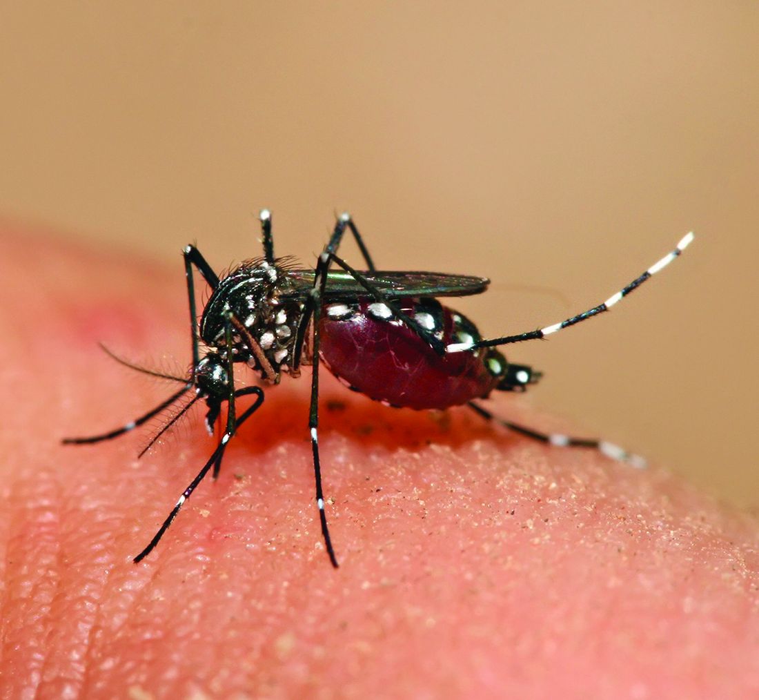
First things first, though. If you’re going to test mosquito noses, you have to give them something to smell. The researchers enrolled eight humans who were willing to wear nylon stockings on their forearms for 6 hours a day for multiple days. “Over the next few years, the researchers tested the nylons against each other in all possible pairings,” Leslie B. Vosshall, PhD, and associates said in a statement from the university. In other words, mosquito March Madness.
Nylons from different participants were hooked up in pairs to an olfactometer assay consisting of a plexiglass chamber divided into two tubes, each ending in a box that held a stocking. The mosquitoes were placed in the main chamber and observed as they flew down the tubes toward one stocking or the other.
Eventually, the “winner” of the “tournament” was Subject 33. And no, we don’t know why there was a Subject 33 since the study involved only eight participants. We do know that the nylons worn by Subject 33 were “four times more attractive to the mosquitoes than the next most-attractive study participant, and an astonishing 100 times more appealing than the least attractive, Subject 19,” according to the written statement.
Chemical analysis identified 50 molecular compounds that were elevated in the sebum of the high-attracting participants, and eventually the investigators discovered that mosquito magnets produced carboxylic acids at much higher levels than the less-attractive volunteers.
We could go on about the research team genetically engineering mosquitoes without odor receptors, but we have to save something for later. Tune in again next week for another exciting episode of “Mosquitoes and the scientists who love them.”
Are women better with words?
Men vs. Women is probably the oldest argument in the book, but there may now be movement. Researchers have been able not only to shift the advantage toward women, but also to use that knowledge to medical advantage.

When it comes to the matter of words and remembering them, women apparently have men beat. The margin is small, said lead author Marco Hirnstein, PhD, of the University of Bergen, Norway, but, after performing a meta-analysis of 168 published studies and PhD theses involving more than 350,000 participants, it’s pretty clear. The research supports women’s advantage over men in recall, verbal fluency (categorical and phonemic), and recognition.
So how is this information useful from a medical standpoint?
Dr. Hirnstein and colleagues suggested that this information can help in interpreting diagnostic assessment results. The example given was dementia diagnosis. Since women are underdiagnosed because their baseline exceeds average while men are overdiagnosed, taking gender and performance into account could clear up or catch cases that might otherwise slip through the cracks.
Now, let’s just put this part of the debate to rest and take this not only as a win for women but for science as well.
Tumor vs. saxophone: The surgical grudge match
Brain surgery is a notoriously difficult task. There’s a reason we say, “Well, at least it’s not brain surgery” when we’re trying to convince someone that a task isn’t that tough. Make one wrong incision, cut the wrong neuron, and it’s goodbye higher cognitive function. And most people appreciate thinking. Crazy, right?
One would imagine that the act of brain surgery would become even more difficult when the patient brings his saxophone and plays it randomly throughout the operation. It’s a hospital, after all, not a jazz club. Patients don’t get to play musical instruments during other surgeries. Why should brain surgery patients get special treatment?
As it turns out, the musical performance was actually quite helpful. A man in Italy had a brain tumor in a particularly complex area, and he’s left-handed, which apparently makes the brain’s neural pathways much more complicated. Plus, he insisted that he retain his musical ability after the surgery. So he and his medical team had a crazy thought: Why not play the saxophone throughout the surgery? After all, according to head surgeon Christian Brogna, MD, playing an instrument means you understand music, which tests many higher cognitive functions such as coordination, mathematics, and memory.
And so, at various points throughout the 9-hour surgery, the patient played his saxophone for his doctors. Doing so allowed the surgeons to map the patient’s brain in a more complete and personalized fashion. With that extra knowledge, they were able to successfully remove the tumor while maintaining the patient’s musical ability, and the patient was discharged on Oct. 13, just 3 days after his operation.
While we’re happy the patient recovered, we do have to question his choice of music. During the surgery, he played the theme to the 1970 movie “Love Story” and the Italian national anthem. Perfectly fine pieces, no doubt, but the saxophone solo in “Jungleland” exists. And we could listen to that for 9 hours straight. In fact, we do that every Friday in the LOTME office.
Basketball has the Big Dance. Mosquitoes get the Big Sniff
In this week’s installment of our seemingly never-ending series, “Mosquitoes and the scientists who love them,” we visit The Rockefeller University in New York, where the olfactory capabilities of Aedes Aegypti – the primary vector species for Zika, dengue, yellow fever, and chikungunya – became the subject of a round robin–style tournament.
First things first, though. If you’re going to test mosquito noses, you have to give them something to smell. The researchers enrolled eight humans who were willing to wear nylon stockings on their forearms for 6 hours a day for multiple days. “Over the next few years, the researchers tested the nylons against each other in all possible pairings,” Leslie B. Vosshall, PhD, and associates said in a statement from the university. In other words, mosquito March Madness.
Nylons from different participants were hooked up in pairs to an olfactometer assay consisting of a plexiglass chamber divided into two tubes, each ending in a box that held a stocking. The mosquitoes were placed in the main chamber and observed as they flew down the tubes toward one stocking or the other.
Eventually, the “winner” of the “tournament” was Subject 33. And no, we don’t know why there was a Subject 33 since the study involved only eight participants. We do know that the nylons worn by Subject 33 were “four times more attractive to the mosquitoes than the next most-attractive study participant, and an astonishing 100 times more appealing than the least attractive, Subject 19,” according to the written statement.
Chemical analysis identified 50 molecular compounds that were elevated in the sebum of the high-attracting participants, and eventually the investigators discovered that mosquito magnets produced carboxylic acids at much higher levels than the less-attractive volunteers.
We could go on about the research team genetically engineering mosquitoes without odor receptors, but we have to save something for later. Tune in again next week for another exciting episode of “Mosquitoes and the scientists who love them.”
Are women better with words?
Men vs. Women is probably the oldest argument in the book, but there may now be movement. Researchers have been able not only to shift the advantage toward women, but also to use that knowledge to medical advantage.
When it comes to the matter of words and remembering them, women apparently have men beat. The margin is small, said lead author Marco Hirnstein, PhD, of the University of Bergen, Norway, but, after performing a meta-analysis of 168 published studies and PhD theses involving more than 350,000 participants, it’s pretty clear. The research supports women’s advantage over men in recall, verbal fluency (categorical and phonemic), and recognition.
So how is this information useful from a medical standpoint?
Dr. Hirnstein and colleagues suggested that this information can help in interpreting diagnostic assessment results. The example given was dementia diagnosis. Since women are underdiagnosed because their baseline exceeds average while men are overdiagnosed, taking gender and performance into account could clear up or catch cases that might otherwise slip through the cracks.
Now, let’s just put this part of the debate to rest and take this not only as a win for women but for science as well.
Rapid point-of-care test could help avoid inappropriate antibiotic prescribing
The fingerstick test, FebriDx, works by detecting myxovirus resistance protein A, which the body generates in response to viral infections, and C-reactive protein (CRP), which is associated with systemic bacterial or viral infection.
In a study of 520 adults and children with symptoms of acute respiratory illness who were treated in outpatient settings, the test correctly classified bacterial infections 93.2% of the time (95% confidence interval [CI], 84.9-97.0). The negative predictive value (NPV), or probability that a person with a negative test result was truly free of a bacterial infection, was 98.7% (95% CI, 96.9-99.4).
The findings of the study, which was sponsored by the test’s manufacturer, were published in JAMA Network Open).
The ability to rule out a bacterial cause “may provide clinicians with reassurance to withhold antibiotics when supported by the clinical assessment,” the researchers wrote.
They added that the ability to identify infections that may benefit from antibiotics and confidently rule out those that will not “is essential to optimizing clinical management and addressing global antimicrobial resistance.”
FDA concerned about false negative viral infection results
FebriDx has been cleared for sale in the United Kingdom, Europe, Canada, United Arab Emirates, Brazil, and Australia, according to the manufacturer, Australia-based Lumos Diagnostics.
However, the product is not available in the United States, where the Food and Drug Administration denied marketing clearance in July. In a news release, Lumos said the FDA determined that FebriDx did not demonstrate “substantial equivalence” to a predicate device and expressed concern that false negative viral infection results could lead to missed cases of COVID-19.
In the newly published study, FebriDx identified individuals with viral infections 70.3% of the time (95% CI, 64.8-75.2). The probability that a person who tested negative for a viral infection was truly negative was 66.7% (95%CI, 60.8-72.1).
The study included patients with respiratory symptoms and recent fever who were enrolled from October 2019 to April 2021 at nine emergency departments, six urgent care clinics, and five primary care clinics in the United States. All patients were tested with FebriDx and underwent separate laboratory testing to determine a final diagnosis.
In addition, researchers recruited a control group of 120 individuals without symptoms.
Among 496 symptomatic individuals who had a final diagnosis, 73 (14.7%) were classified as having a response associated with a bacterial infection, 296 (59.7%) as having a viral-associated response, and 127 (25.6%) as negative.
FebriDx correctly ruled out a bacterial infection 88.4% of the time (95% CI, 85.0-91.1). The probability that a patient with a positive result for bacterial infection actually had a bacterial infection was 58.1% (95%CI, 49.1-66.7).
The findings bolster those of a previous study on the same test. This research included 220 patients who reported having a fever within the prior 3 days or had a measurable fever at the time of enrollment. In that study, the test correctly identified bacterial infections 85% of the time and correctly ruled out bacterial infection 93% of the time, with a NPV of 97%.
Too early to say test will be useful in practice
The idea of a test to guide the prescribing of antibiotics isn’t new, according to an expert who was not involved in FebriDx research.
Noah Ivers, MD, PhD, a family physician and associate professor at the University of Toronto who studies strategies to optimize primary care delivery, said, “many such point-of-care tests have been tried” to detect biomarkers such as CRP or procalcitonin, which is associated with bacterial infections.

Such tests have looked good in initial studies, he said, but when trialed in urgent care clinics, primary care clinics, or emergency departments, “they tend run into implementation challenges or simply lack of effects, or both.
“So, while I am happy at the news of this result, it’s too early to say with any certainty that it will prove useful in practice,” he added.
Meanwhile, Dr. Ivers said it’s “crucial that people understand that most illnesses are likely to be viral” and therefore not helped by antibiotics. When antibiotics are needed for outpatients, he said, “5 days is usually ample.”
The study was funded by Lumos Diagnostics. Among the 15 study authors, 6 had conflicts of interest disclosures, reporting ties to Inflammatix, Medical College of Wisconsin, Siemens, Technomics Research, and Lumos Diagnostics. Dr. Ivers reported no relevant financial interests.
The fingerstick test, FebriDx, works by detecting myxovirus resistance protein A, which the body generates in response to viral infections, and C-reactive protein (CRP), which is associated with systemic bacterial or viral infection.
In a study of 520 adults and children with symptoms of acute respiratory illness who were treated in outpatient settings, the test correctly classified bacterial infections 93.2% of the time (95% confidence interval [CI], 84.9-97.0). The negative predictive value (NPV), or probability that a person with a negative test result was truly free of a bacterial infection, was 98.7% (95% CI, 96.9-99.4).
The findings of the study, which was sponsored by the test’s manufacturer, were published in JAMA Network Open).
The ability to rule out a bacterial cause “may provide clinicians with reassurance to withhold antibiotics when supported by the clinical assessment,” the researchers wrote.
They added that the ability to identify infections that may benefit from antibiotics and confidently rule out those that will not “is essential to optimizing clinical management and addressing global antimicrobial resistance.”
FDA concerned about false negative viral infection results
FebriDx has been cleared for sale in the United Kingdom, Europe, Canada, United Arab Emirates, Brazil, and Australia, according to the manufacturer, Australia-based Lumos Diagnostics.
However, the product is not available in the United States, where the Food and Drug Administration denied marketing clearance in July. In a news release, Lumos said the FDA determined that FebriDx did not demonstrate “substantial equivalence” to a predicate device and expressed concern that false negative viral infection results could lead to missed cases of COVID-19.
In the newly published study, FebriDx identified individuals with viral infections 70.3% of the time (95% CI, 64.8-75.2). The probability that a person who tested negative for a viral infection was truly negative was 66.7% (95%CI, 60.8-72.1).
The study included patients with respiratory symptoms and recent fever who were enrolled from October 2019 to April 2021 at nine emergency departments, six urgent care clinics, and five primary care clinics in the United States. All patients were tested with FebriDx and underwent separate laboratory testing to determine a final diagnosis.
In addition, researchers recruited a control group of 120 individuals without symptoms.
Among 496 symptomatic individuals who had a final diagnosis, 73 (14.7%) were classified as having a response associated with a bacterial infection, 296 (59.7%) as having a viral-associated response, and 127 (25.6%) as negative.
FebriDx correctly ruled out a bacterial infection 88.4% of the time (95% CI, 85.0-91.1). The probability that a patient with a positive result for bacterial infection actually had a bacterial infection was 58.1% (95%CI, 49.1-66.7).
The findings bolster those of a previous study on the same test. This research included 220 patients who reported having a fever within the prior 3 days or had a measurable fever at the time of enrollment. In that study, the test correctly identified bacterial infections 85% of the time and correctly ruled out bacterial infection 93% of the time, with a NPV of 97%.
Too early to say test will be useful in practice
The idea of a test to guide the prescribing of antibiotics isn’t new, according to an expert who was not involved in FebriDx research.
Noah Ivers, MD, PhD, a family physician and associate professor at the University of Toronto who studies strategies to optimize primary care delivery, said, “many such point-of-care tests have been tried” to detect biomarkers such as CRP or procalcitonin, which is associated with bacterial infections.
Such tests have looked good in initial studies, he said, but when trialed in urgent care clinics, primary care clinics, or emergency departments, “they tend run into implementation challenges or simply lack of effects, or both.
“So, while I am happy at the news of this result, it’s too early to say with any certainty that it will prove useful in practice,” he added.
Meanwhile, Dr. Ivers said it’s “crucial that people understand that most illnesses are likely to be viral” and therefore not helped by antibiotics. When antibiotics are needed for outpatients, he said, “5 days is usually ample.”
The study was funded by Lumos Diagnostics. Among the 15 study authors, 6 had conflicts of interest disclosures, reporting ties to Inflammatix, Medical College of Wisconsin, Siemens, Technomics Research, and Lumos Diagnostics. Dr. Ivers reported no relevant financial interests.
FROM JAMA NETWORK OPEN
Apixaban outmatches rivaroxaban in patients with AFib and valvular heart disease
Compared with rivaroxaban, apixaban cut risks nearly in half, suggesting that clinicians should consider these new data when choosing an anticoagulant, reported lead author Ghadeer K. Dawwas, PhD, of the University of Pennsylvania, Philadelphia, and colleagues.

In the new retrospective study involving almost 20,000 patients, Dr. Dawwas and her colleagues “emulated a target trial” using private insurance claims from Optum’s deidentified Clinformatics Data Mart Database. The cohort was narrowed from a screened population of 58,210 patients with concurrent AFib and VHD to 9,947 new apixaban users who could be closely matched with 9,947 new rivaroxaban users. Covariates included provider specialty, type of VHD, demographic characteristics, measures of health care use, baseline use of medications, and baseline comorbidities.
The primary effectiveness outcome was a composite of systemic embolism and ischemic stroke, while the primary safety outcome was a composite of intracranial or gastrointestinal bleeding.
“Although several ongoing trials aim to compare apixaban with warfarin in patients with AFib and VHD, none of these trials will directly compare apixaban and rivaroxaban,” the investigators wrote. Their report is in Annals of Internal Medicine.
Dr. Dawwas and colleagues previously showed that direct oral anticoagulants (DOACs) were safer and more effective than warfarin in the same patient population. Comparing apixaban and rivaroxaban – the two most common DOACs – was the next logical step, Dr. Dawwas said in an interview.
Study results
Compared with rivaroxaban, patients who received apixaban had a 43% reduced risk of stroke or embolism (hazard ratio [HR], 0.57; 95% confidence interval [CI], 0.40-0.80). Apixaban’s ability to protect against bleeding appeared even more pronounced, with a 49% reduced risk over rivaroxaban (HR, 0.51; 95% CI, 0.41-0.62).
Comparing the two agents on an absolute basis, apixaban reduced risk of embolism or stroke by 0.2% within the first 6 months of treatment initiation, and 1.1% within the first year of initiation. At the same time points, absolute risk reductions for bleeding were 1.2% and 1.9%, respectively.
The investigators noted that their results held consistent in an alternative analysis that considered separate types of VHD.
“Based on the results from our analysis, we showed that apixaban is effective and safe in patients with atrial fibrillation and valvular heart diseases,” Dr. Dawwas said.
Head-to-head trial needed to change practice
Christopher M. Bianco, DO, associate professor of medicine at West Virginia University Heart and Vascular Institute, Morgantown, said the findings “add to the growing body of literature,” but “a head-to-head trial would be necessary to make a definitive change to clinical practice.”
Dr. Bianco, who recently conducted a retrospective analysis of apixaban and rivaroxaban that found no difference in safety and efficacy among a different patient population, said these kinds of studies are helpful in generating hypotheses, but they can’t account for all relevant clinical factors.
“There are just so many things that go into the decision-making process of [prescribing] apixaban and rivaroxaban,” he said. “Even though [Dr. Dawwas and colleagues] used propensity matching, you’re never going to be able to sort that out with a retrospective analysis.”
Specifically, Dr. Bianco noted that the findings did not include dose data. This is a key gap, he said, considering how often real-world datasets have shown that providers underdose DOACs for a number of unaccountable reasons, and how frequently patients exhibit poor adherence.
The study also lacked detail concerning the degree of renal dysfunction, which can determine drug eligibility, Dr. Bianco said. Furthermore, attempts to stratify patients based on thrombosis and bleeding risk were likely “insufficient,” he added.
Dr. Bianco also cautioned that the investigators defined valvular heart disease as any valve-related disease of any severity. In contrast, previous studies have generally restricted valvular heart disease to patients with mitral stenosis or prosthetic valves.
“This is definitely not the traditional definition of valvular heart disease, so the title is a little bit misleading in that sense, although they certainly do disclose that in the methods,” Dr. Bianco said.
On a more positive note, he highlighted the size of the patient population, and the real-world data, which included many patients who would be excluded from clinical trials.
More broadly, the study helps drive research forward, Dr. Bianco concluded; namely, by attracting financial support for a more powerful head-to-head trial that drug makers are unlikely to fund due to inherent market risk.
This study was supported by the National Institutes of Health. The investigators disclosed additional relationships with Takeda, Spark, Sanofi, and others. Dr. Bianco disclosed no conflicts of interest.
Compared with rivaroxaban, apixaban cut risks nearly in half, suggesting that clinicians should consider these new data when choosing an anticoagulant, reported lead author Ghadeer K. Dawwas, PhD, of the University of Pennsylvania, Philadelphia, and colleagues.
In the new retrospective study involving almost 20,000 patients, Dr. Dawwas and her colleagues “emulated a target trial” using private insurance claims from Optum’s deidentified Clinformatics Data Mart Database. The cohort was narrowed from a screened population of 58,210 patients with concurrent AFib and VHD to 9,947 new apixaban users who could be closely matched with 9,947 new rivaroxaban users. Covariates included provider specialty, type of VHD, demographic characteristics, measures of health care use, baseline use of medications, and baseline comorbidities.
The primary effectiveness outcome was a composite of systemic embolism and ischemic stroke, while the primary safety outcome was a composite of intracranial or gastrointestinal bleeding.
“Although several ongoing trials aim to compare apixaban with warfarin in patients with AFib and VHD, none of these trials will directly compare apixaban and rivaroxaban,” the investigators wrote. Their report is in Annals of Internal Medicine.
Dr. Dawwas and colleagues previously showed that direct oral anticoagulants (DOACs) were safer and more effective than warfarin in the same patient population. Comparing apixaban and rivaroxaban – the two most common DOACs – was the next logical step, Dr. Dawwas said in an interview.
Study results
Compared with rivaroxaban, patients who received apixaban had a 43% reduced risk of stroke or embolism (hazard ratio [HR], 0.57; 95% confidence interval [CI], 0.40-0.80). Apixaban’s ability to protect against bleeding appeared even more pronounced, with a 49% reduced risk over rivaroxaban (HR, 0.51; 95% CI, 0.41-0.62).
Comparing the two agents on an absolute basis, apixaban reduced risk of embolism or stroke by 0.2% within the first 6 months of treatment initiation, and 1.1% within the first year of initiation. At the same time points, absolute risk reductions for bleeding were 1.2% and 1.9%, respectively.
The investigators noted that their results held consistent in an alternative analysis that considered separate types of VHD.
“Based on the results from our analysis, we showed that apixaban is effective and safe in patients with atrial fibrillation and valvular heart diseases,” Dr. Dawwas said.
Head-to-head trial needed to change practice
Christopher M. Bianco, DO, associate professor of medicine at West Virginia University Heart and Vascular Institute, Morgantown, said the findings “add to the growing body of literature,” but “a head-to-head trial would be necessary to make a definitive change to clinical practice.”
Dr. Bianco, who recently conducted a retrospective analysis of apixaban and rivaroxaban that found no difference in safety and efficacy among a different patient population, said these kinds of studies are helpful in generating hypotheses, but they can’t account for all relevant clinical factors.
“There are just so many things that go into the decision-making process of [prescribing] apixaban and rivaroxaban,” he said. “Even though [Dr. Dawwas and colleagues] used propensity matching, you’re never going to be able to sort that out with a retrospective analysis.”
Specifically, Dr. Bianco noted that the findings did not include dose data. This is a key gap, he said, considering how often real-world datasets have shown that providers underdose DOACs for a number of unaccountable reasons, and how frequently patients exhibit poor adherence.
The study also lacked detail concerning the degree of renal dysfunction, which can determine drug eligibility, Dr. Bianco said. Furthermore, attempts to stratify patients based on thrombosis and bleeding risk were likely “insufficient,” he added.
Dr. Bianco also cautioned that the investigators defined valvular heart disease as any valve-related disease of any severity. In contrast, previous studies have generally restricted valvular heart disease to patients with mitral stenosis or prosthetic valves.
“This is definitely not the traditional definition of valvular heart disease, so the title is a little bit misleading in that sense, although they certainly do disclose that in the methods,” Dr. Bianco said.
On a more positive note, he highlighted the size of the patient population, and the real-world data, which included many patients who would be excluded from clinical trials.
More broadly, the study helps drive research forward, Dr. Bianco concluded; namely, by attracting financial support for a more powerful head-to-head trial that drug makers are unlikely to fund due to inherent market risk.
This study was supported by the National Institutes of Health. The investigators disclosed additional relationships with Takeda, Spark, Sanofi, and others. Dr. Bianco disclosed no conflicts of interest.
FROM ANNALS OF INTERNAL MEDICINE
Instagram may make new moms feel inadequate: Study
Does Instagram make new moms feel inadequate? Yes, suggests a new study that warns images of new mothers on social media may drive body dissatisfaction and feelings of not being good enough.
Lead researcher Megan Gow, PhD, a National Health and Medical Research Council early career fellow at the University of Sydney Children’s Hospital Westmead Clinical School, says she wanted to find out if Instagram images reflected the actual population of postpartum women.
“We were concerned images would be idealized, placing postpartum women, who are already a vulnerable group, at increased risk,” she says.
The findings, published recently in the journal Healthcare, suggest social media may not be the right platform to target health messages to new moms.
A vulnerable time
The months after an infant’s birth are a vulnerable time for new moms. Women contend with huge hormone shifts, sleep deprivation, and a major life change – all while caring for a new child.
A 2021 Nestle study found 32% of parents feel isolated, while a 2017 online poll in the United Kingdom found 54% of new moms felt “friendless.” And according to the American Psychological Association, up to one in seven new mothers will face postpartum depression, while 9% will have posttraumatic stress disorder, according to Postpartum Support International.
The pandemic may have worsened the isolation new mothers feel. A May 2022 study in the Journal of Psychiatric Research found U.S. rates of postpartum depression rose in the first year of the COVID-19 pandemic.
While new motherhood was stressful enough in the analog age, women today must contend with social media, which increases feelings of isolation. A June 2021 study published in Frontiers in Psychology said social media users between the ages of 26 and 35 reported higher rates of loneliness. That’s in line with Dr. Gow’s study, which noted 39% of Instagram’s monthly active users are women between the ages of 18 and 44. And nearly two-thirds of them – 63% – log onto the platform daily.
“The postpartum phase can feel very isolated, and being vocal about the postpartum shifts that all mothers go through helps set expectations and normalize the experience for those of us who are post partum,” says Catie de Montille, 36, a mother of two in Washington, D.C.
Instagram sets the wrong expectations
Instagram sets unreasonable expectations for new mothers, Dr. Gow and her colleagues found in their study.
She and her fellow researchers analyzed 600 posts that used #postpartumbody, a hashtag that had been posted on Instagram more than 2 million times by October 2022. Other hashtags like #mombod and #postbabybody have been used 1.9 million and 320,000 times, respectively.
Of the 600 posts, 409 (68%) focused on a woman as the central image. The researchers analyzed those 409 posts to find out if they reflected women’s post-childbirth reality.
They found that more than 9 in 10 posts (91%) showed women who appeared to have low body fat (37%) or average body fat (54%). Only 9% showed women who seemed to be overweight. And the researchers also found just 5% of images showed features commonly associated with a postpartum body, like stretch marks or scars from cesarean sections.
Women need to be aware that “what is posted on Instagram may not be realistic and is not representative of the vast majority of women in the postpartum period,” Dr. Gow says.
The images also did not portray women as physically strong.
Dr. Gow’s team examined 250 images for signs of muscularity. More than half, 52%, showed few or no defined muscles. That finding came even though more than half of the original 409 images showed women in fitness attire (40%), underwear (8%), or a bathing suit (5%).
According to Emily Fortney, PsyD, a licensed clinical psychologist in Sacramento, Calif., the study shows that health care workers must work harder to set expectations for new moms.
“This is a deeper issue of how women are overall portrayed in the media and the pressure we face to return to some unrealistic size,” she says. “We need to be encouraging women to not focus on photos, but to focus on the postpartum experience in an all-encompassing way that includes both physical and mental health.”
Childbirth as an illness to overcome?
While retail brands from Nike to Versace have begun to show a wider range of female shapes in advertisements and on the runway, postpartum women seem to be left out of this movement. Dr. Gow and her fellow researchers referred to a 2012 study that examined images in popular Australian magazines and concluded these photos likened the pregnant body to an illness from which women needed to recover.
The images posted on Instagram indicate that belief is still pervasive. The images of postpartum women in fitness clothes suggest “that women want to be seen to be exercising as a means of breaking the ‘hold’ that pregnancy had on them or ‘repairing’ their postpartum body,” Dr. Gow and her fellow researchers say.
New Orleans resident Sydney Neal, 32, a mother of two who gave birth to her youngest child in November 2021, said social media helped shape her view of what “recovery” would be like.
While Ms. Neal said some celebrities like Chrissy Teigen, a mother of two, have “kept it very real” on Instagram, she also “saw a lot of women on social media drop [their weight] quickly and post as if they were back to normal much faster than 6 months.”
Body-positive tools for new moms
Dr. Gow is continuing to study this topic. Her team is currently doing a study that will ask women about social media use, how they feel about their bodies, and how their beliefs change after viewing images tagged with #postpartumbody. (Women with children under the age of 2 can access the survey here.)
Because of the unrealistic images, Dr. Gow and her team said Instagram may not be a good tool for sharing health information with new moms.
But there are other options.
Ms. de Montille, whose children were born in 2020 and 2022, used apps like Back to You and Expectful, and she follows Karrie Locher, a postpartum and neonatal nurse and certified lactation counselor, on Instagram. She said these tools focus on the mind/body connection, which “is better than focusing on the size of your jeans.”
Women also should be able to turn to trusted health care professionals.
“Providers can start speaking about the romanticization of pregnancy and motherhood starting in prenatal care, and they can start speaking more about social media use and the pros and cons of use specifically in the perinatal period,” says Dr. Fortney. “This opens the door to a discussion on a wide range of issues that can actually help assess, prevent, and treat perinatal mood and anxiety disorders.”
Ms. Neal, the mother of two in New Orleans, said she wished her doctor had talked to her more about what to expect after giving birth.
“I don’t really know how to crack the body image nut, but I think starting in a medical setting might be helpful,” she says.
A version of this article first appeared on WebMD.com.
FROM HEALTHCARE
Connected: Preterm infant program makes progress
Martha Welch, MD, spent the better part of three decades in private practice treating children with emotional, behavioral, and developmental disorders before accepting a job on the faculty of Columbia University, New York, in 1997.
She took the position, she said, with a mission: to find evidence to support what she’d observed in her practice – that parents could, by making stronger emotional connections, change the trajectory of development for preemie infants.
With that understanding, Dr. Welch created Family Nurture Intervention (FNI), which has been shown to improve the development of premature babies.
“We saw that no matter what happened to the baby, no matter how avoidant the baby might be, we’re able to overcome this with emotional expression,” Dr. Welch said.
Over the course of the intervention, families work with a specialist who helps bring mother and baby together – both physically and emotionally – until both are calm, which can initially take several hours and over time, minutes.
FNI appears to help families – especially mothers – re-establish an emotional connection often interrupted by their babies’ stressful and uncertain stay in a neonatal intensive care unit (NICU). In turn, both the infant and maternal nervous systems become better regulated, according to researchers.
Early challenges
Babies born preterm can face a range of short-term and long-term challenges, such as breathing problems due to an underdeveloped respiratory system, an increased risk of infection from an underdeveloped immune system, and learning difficulties, according to the Mayo Clinic.
Many aspects of FNI are not new: The neonatal intensive care unit has long incorporated activities such as scent cloth exchanges, talking to the baby, and skin-to-skin contact. But the approach Dr. Welch and her colleagues advocate emphasizes building a bond between the mother and the infant.
Mounting evidence shows that FNI can improve a wide range of outcomes for premature babies. In a 2021 study, for example, Dr. Welch’s group showed that FNI was associated with lower heart rates among preemies in the NICU. A 2016 study linked the intervention to reduced depression and anxiety symptoms in mothers of preterm infants. And a 2015 randomized controlled trial showed FNI improved development and behavioral outcomes in infants up to 18 months.
A new study published in Science Translational Medicine showed that the intervention led to a greater likelihood that babies had improved cognitive development later on, narrowing the developmental gap between healthy, full-term babies.
Dr. Welch and her colleagues tested to see if FNI measurably changed brain development in preterm infants who were born at 26-34 weeks of a pregnancy.
“We were blown away by the strength of the effect,” said Pauliina Yrjölä, MSc, a doctoral student and medical physicist at the University of Helsinki, who led the study on which Dr. Welch is a co-author.
Mothers in the intervention group made as much eye contact with the infants as possible and spoke with infants about their feelings.
Intimate sensory interactions between mothers and infants physically altered infants’ cortical networks in the brain and was later correlated to improved neurocognitive performance, according to the researchers.
“I was convinced there were physiological changes; I knew that from my clinical work,” Dr. Welch said. “I wanted to show it in this concrete, scientific way.”
Preterm babies face many hurdles
“If we can prevent problems in brain network organization to the extent that’s shown in this study and improve their outcomes, this is worth millions of dollars in terms of cost to society, schooling, health care, especially education, and families,” said Ruth Grunau, PhD, a professor in the Division of Neonatology in the department of pediatrics at the University of British Columbia, Vancouver, who was not involved with the most recent study but has worked with Dr. Welch previously.
Babies born too early, especially before 32 weeks, have higher rates of death and disability, according to the Centers for Disease Control and Prevention.
And preterm babies overall may experience breathing problems and feeding difficulties almost immediately following birth. They may also experience long-term problems such as developmental delays, vision problems, and hearing problems.
Dr. Grunau said that while many other programs and interventions have been used in the neonatal intensive care unit to help infants and mothers, the results from FNI stand out.
Ms. Yrjölä said she was surprised by the strength of the correlation as the infants continued to develop. The infants receiving the Family Nurture Intervention showed brain development close to the control group, which was infants born at full-term.
“Emotional connection is a state, not a trait – and a state can be changed,” said Dr. Welch. “And in this case, it can be changed by the parent through emotional expression.”
Steps clinicians can take
Dr. Welch said the approach is highly scalable, and two NICUs that participated in the FNI studies have implemented the program as standard care.
The approach is also gaining interest outside of the clinical setting, as preschool partners have expressed interest in implementing some of the methods to promote development.
Parents, family members, and teachers can use many of the FNI techniques – such as eye contact and emotional expression – to continue to develop and strengthen connection.
For clinicians who want to implement parts of the intervention on their own, Dr. Welch said doctors can observe if the baby looks at or turns toward their mother.
Clinicians can encourage mothers to express deep, emotional feelings toward the infant. Dr. Welch stressed that feelings don’t have to be positive, as many mothers with babies in the NICU have a hard time expressing positive emotions. Crying or talking about the difficulties of their childbirth experience count as expressing emotion. The important part is that the baby hears emotion, of any kind, in the mother’s voice, Dr. Welch said.
As the connection develops, it will eventually take less time for the mother and the baby to form a bond, and eventually the pair will become autonomically regulated.
“This is what gives us hope,” she said. “We affect each other in our autonomic nervous systems. It’s why this treatment works.”
The study was funded by the Finnish Pediatric Foundation, The Finnish Academy, the Juselius Foundation, Aivosäätiö, Neuroscience Center at University of Helsinki and Helsinki University Central Hospital, gifts from the Einhorn Family Charitable Trust, the Fleur Fairman Family, M. D. Stephenson, and The National Health and Medical Research Council of Australia. The authors have disclosed no relevant financial relationships.
A version of this article first appeared on Medscape.com.