User login
Daily sunscreen slowed skin aging in middle-aged adults
Daily sunscreen users were significantly less likely than discretionary sunscreen users to show signs of skin aging after 4.5 years, according to a study of young and middle-aged adults.
However, beta-carotene supplements appeared to have no effect on skin aging.
The findings from the randomized controlled trial were published online June 3 in Annals of Internal Medicine (2013;158:781-90).
"No known randomized studies in humans have evaluated the effect of sunscreen on surface changes associated with skin aging," wrote Maria Celia B. Hughes, MMedSci., of the Queensland Institute of Medical Research, Australia, and her colleagues.
The investigators used data from the Nambour (Australia) Skin Cancer Prevention Trial, in which 1,621 adults were studied from 1992 to 1996 to test the effect of sunscreen use or dietary supplements on skin cancer risk, photoaging, and actinic keratosis development.
To determine whether consistent, daily sunscreen use could prevent progression of skin aging, researchers randomized Nambour study participants under age 55 into four groups: daily use of broad-spectrum sunscreen plus 30 mg of beta-carotene; daily sunscreen use plus a placebo supplement; discretionary sunscreen use plus 30 mg of beta-carotene; and discretionary sunscreen use plus a placebo supplement.
The investigators focused on 903 adults younger than 55 to exclude the potential effects of growing old on participants’ skin aging.
Skin aging was assessed by comparing skin microtopography based on impressions taken of the backs of participants’ hands at baseline in 1992 and 4.5 years later in 1996. Assessors were blinded to the treatment groups.
"Most of the study participants were fair skinned, and more than 90% burned on acute sun exposure," the researchers noted. The groups were similar in terms of phenotype, sun exposure, and pretrial sunscreen use. All groups reported similar amounts of sun exposure during the study period; 78% of daily sunscreen users and 76% of discretionary sunscreen users reported being outdoors for less than 50% of their weekend time. In addition, the use of other sun protection measures, including seeking shade and wearing a hat, was similar among the groups.
By the end of the study, 77% of the daily sunscreen users applied sunscreen at least 3-4 days per week, vs. 33% of the discretionary users.
Overall, 58% of the participants in the current study met criteria for moderate photoaging at baseline in 1992, and 49% met those criteria in 1996. However, at the end of the 4.5-year period, daily sunscreen users were 24% less likely to show signs of skin aging than were discretionary users, a statistically significant difference.
When the odds of having a higher microtopography grade in 1996 than in 1992 were adjusted for sunburns and photoaging of the neck, the researchers noted, "only the daily sunscreen intervention group showed no detectable increase in microtopography grade."
No significant differences in skin aging were seen in participants randomized to beta-carotene vs. placebo.
The study was limited by several factors, including limited outcome data, which reduced the power to detect moderate treatment effects. In addition, the data were insufficient to rule out an effect of beta carotene on skin aging, the researchers noted.
However, "these results have important clinical implications," the researchers said. "A unit increase in microtopography significantly correlates with risk for actinic keratoses and skin cancer." Thus, the cosmetic benefits of reducing skin changes in middle age also may reduce cancer risk.
The National Health and Medical Research Council of Australia funded the study.
FROM ANNALS OF INTERNAL MEDICINE
Major finding: After 4.5 years, daily sunscreen users were 24% less likely to show signs of skin aging than were discretionary sunscreen users.
Data source: A randomized, controlled, community-based trial of 903 adults younger than 55 years.
Disclosures: The National Health and Medical Research Council of Australia funded the study.
MelaFind: Invaluable or Inane?
JAMA Dermatology (formerly Archives of Dermatology) reported a study (Arch Dermatol. 2012;148:1083-1084) on the diagnosis of pigmented lesions by dermatologists compared with the MelaFind device (Mela Sciences), a handheld light unit that produces a 3-dimensional computer image of a pigmented lesion with subsequent recommendation of “high disorganization” versus “low disorganization.” Of 47 pigmented lesions (23 melanoma; 24 nonmelanoma), MelaFind recommended biopsy in 44 cases and no biopsy in 3 cases, with 1 lesion that was truly melanoma (96% sensitivity; 8% specificity), compared with a range of 48% to 100% (mean, 80%) sensitivity and 4% to 71% (mean, 43%) specificity among the study dermatologists. The authors quote in their conclusion paragraph that MelaFind is a “very sensitive tool to guide dermatologists.”
What’s the issue?
A recent patient forcefully suggested to me that our department should “keep up with the times” and invest in MelaFind. MelaFind’s Web site states that it is “not a screening device.” The physician fact sheet says that it is “the world’s first and only multi-spectral, non-invasive, painless, and 100% objective and automated computer vision technology that evaluates clinically atypical pigmented skin lesions and classifies them unambiguously and clearly based upon their level of 3-dimensional morphological disorganization.” Given these claims and its US Food and Drug Administration approval, how can we not own one? The word unambiguous has never been used to describe the clinical decision making and diagnosis of pigmented lesions, but we all hope for it. Quite well-known leaders in our field have spoken highly of this technology, though I await better-powered studies and more poignant data, or perhaps just a better grasp of how it works. Can someone aid us in understanding its true niche in clinical practice?
Nivolumab activity is durable in advanced melanoma
CHICAGO – Nivolumab, an investigational PD-1 inhibitor, shrank tumors by at least 30% in 33 of 107 pretreated patients with metastatic melanoma, based on results from an expanded phase I trial reported at the annual meeting of the American Society of Clinical Oncology.
An additional 11% of patients had prolonged stable disease or non-conventional, immune-related response profiles.
The findings build on favorable initial results reported at last year’s annual meeting for nivolumab in melanoma, renal cell carcinoma, and nonsmall cell lung cancer. The latest results in melanoma patients at follow-ups as long as 2 years indicate no new safety signals with nivolumab, which was associated with a 21% rate of grade 3-4 events. The rate of severe immune-related adverse events was 5%, and there were no cases of grade 3 or higher pneumonitis.
Median overall survival was nearly 17 months with nivolumab, with a 2-year survival rate of 43%. Median overall survival with vemurafenib (Zelboraf) is 16 months and with ipilimumab (Yervoy) is 10 months, with 2-year survival rates of 24% to 33% with ipilimumab, according to Dr. Mario Sznol, who presented the results from the phase I trial.
"We’re very excited that there is potential for even more activity [with nivolumab] in combination with other drugs," said Dr. Sznol, professor of medicine (medical oncology) at the Yale Cancer Center in New Haven, Conn.
Responses were seen at all five dose levels tested (0.1, 0.3, 1, 3, and 10 mg/kg), with a 41% objective response rate at the 3 mg/kg dose, which has been selected for evaluation in phase III studies.
Median overall survival across all doses of nivolumab was 16.8 months. It reached 20.3 months for the 3 mg/kg dose. The 1-year survival rate was 62% and the 2-year survival rate, 43%.
Patients in the nivolumab trial were representative of typical patients with advanced melanoma, Dr. Sznol said. All patients in the study had disease that worsened despite prior standard systemic therapies, 25% of them had three or more prior therapies and 63% had two or more prior therapies.
All had ECOG performance standards of 0 or 1. Patients received up to 12 cycles of treatment, with four doses of nivolumab per cycle, until discontinuation criteria were met.
Response has persisted after stopping treatment in 17 of 33 patients, with 12 of the 17 continuing to respond for at least 4 months, Dr. Sznol said.
The overall objective response rate included partial and complete responses. Dr. Sznol acknowledged that just one patient had a verified complete response to nivolumab and four others had near complete responses at 2 years. Historical response rates to immunotherapy drugs are 5%-10% in advanced melanoma, he noted, which is lower than the 30% response seen in these pretreated patients.
"I have seen a few relapses after 2 years of response, but some patients continue to do well at 4 years. One patient who has been off nivolumab for 2 years continues to do well at over 4 years," Dr. Sznol commented during a question and answer session.
To define the best candidates for nivolumab, molecular markers need to be identified to predict probable response, he added. As nivolumab is a PD-1 inhibitor, one potential marker is the protein PD-L1 on the surface of tumor cells, which is being studied in several other clinical trials.
The research was supported by Bristol-Myers Squibb. Dr. Sznol disclosed that he serves in a consultant or advisory role with Bristol-Myers Squibb.
AT THE ASCO ANNUAL MEETING 2013
Major finding: Median overall survival across all doses of nivolumab was 16.8 months. It reached 20.3 months for the 3 mg/kg dose that will be used in phase III studies.
Data source: An expanded phase I study of 107 pretreated patients with metastatic melanoma.
Disclosures: The research was supported by Bristol-Myers Squibb. Dr. Sznol disclosed that he serves in a consultant or advisory role with Bristol-Myers Squibb.
Adding GM-CSF to ipilimumab extends survival in metastatic melanoma
CHICAGO – Combining two approved therapies – GM-CSF and ipilimumab – extended overall survival rates by 35% and resulted in fewer grade 3-5 adverse events when compared with ipilimumab alone in a randomized study of 245 patients with metastatic melanoma.
At 1 year, the overall survival rate in the combination therapy group was 69%, with a median follow-up of nearly 18 months. At 1 year, survival in the ipilimumab-only group was 53% with a median follow-up of nearly 13 months, Dr. F. Stephen Hodi, Jr. reported at the annual meeting of the American Society of Clinical Oncology.
This is the first phase II trial to look at ipilimumab (Yervoy, Bristol-Myers Squibb) and the granulocyte macrophage colony-stimulating factor (GM-CSF) sargramostim (Leukine, Sanofi) in combination in any cancer, said Dr. Hodi, the principal investigator for the trial, which was conducted by the ECOG-ACRIN Cancer Research Group (formerly the Eastern Cooperative Oncology Group) trial. In this study, ipilimumab was used at a dose of 10 mg/kg, which is higher than the FDA-approved dose of 3 mg/kg.
"We are waiting for the data to mature in ongoing studies examining the relative efficacy of 3 mg/kg and 10 mg/kg dosing," said Dr. Hodi, director of the melanoma center at Dana-Farber Cancer Institute in Boston.
The results in the current trial are another indication of the impact that immunotherapy can have for patients with advanced melanoma. Since both GM-CSF and ipilimumab are commercially available, oncologists need to determine the best way to apply these findings in everyday practice. The next step will then be to define the role of GM-CSF in combination with other immune checkpoint targeting drugs, such as therapies that target the PD-1 and PD-L1 pathway, he said.
"We have been using GM-CSF in melanoma as a stand-alone therapy," Dr. Lynn M. Schuchter, the C. Willard Robinson Professor of Hematology-Oncology at the University of Pennsylvania, Philadelphia, and an expert on melanoma, said at a press conference where the study results were announced. "It will be interesting to see if payers will cover this (combination treatment)."
Ipilimumab targets CTLA-4, a protein that keeps immune T-cells in an inactive state. GM-CSF is a growth factor commonly used to boost white blood cell counts after chemotherapy or stem cell transplantation.
For this study, 245 patients were randomized to receive ipilimumab plus GM-CSF or ipilimumab alone. All study participants were in otherwise good health, with an ECOG performance status of 0-1 and adequate end-organ function, no autoimmune disease, and no prior use of CTLA-4 blockade or CD137 agonists. All had radiographically measurable metastatic melanoma, but with no CNS metastases, and had received up to one prior treatment over 4 weeks before starting in the trial.
The 123 patients randomized to the combination therapy were given sargramostim at 250 micrograms injected subcutaneously on day 1-14 of a 21 day cycle. For induction therapy, ipilimumab was given at a dose of 10 mg/kg intravenously once every 3 weeks for four cycles as induction therapy and once every 12 weeks as maintenance therapy. The 122 patients randomized to ipilimumab alone received the drug on the same schedule.
In both study arms, tumor shrinkage rates were comparable at 11% and 14%, and progression-free survival was similar at about 3 months. But the overall survival rate was longer in the combination treatment arm. One year after the start of therapy and with a median follow-up of 13.3 months, 69% of patients given the combination therapy and 53% of those who got ipilimumab alone were still alive, for a 35% lower risk of death with the combination therapy (hazard ratio, 0.64; P = .014).
Additionally, the combination treatment was associated with fewer serious side effects, compared with ipilimumab alone. The most significant differences were in lung and gastrointestinal toxicities.
Grade 3-5 adverse events occurred in 45% of patients given the combination therapy and in 57% given ipilimumab alone (p2 = 0.078). There were two possible treatment-related deaths in the combination arm (one colonic perforation and one cardiac arrest) and 7 possible treatment-related deaths in the ipilimumab-only arm: (two cases of multi-organ failure, two colonic perforations, one case of liver failure, and two cases of respiratory failure).
The research was supported in part by the National Cancer Institute (Cancer Therapy Evaluation Program), Sanofi, and Bristol-Myers Squibb. Dr. Hodi disclosed receiving research funding and a being a consultant or in an advisory role with Bristol-Myers Squibb.
AT THE ASCO ANNUAL MEETING 2013
Major finding: One year after the start of therapy, 69% of patients given the combination therapy and 53% of those who got ipilimumab alone were still alive, for a 35% lower risk of death with the combination therapy.
Data source: A phase-II randomized trial of 245 patients who received ipilimumab plus GM-CSF or ipilimumab alone.
Disclosures: The research was supported in part by the National Cancer Institute (Cancer Therapy Evaluation Program), Sanofi, and Bristol-Myers Squibb. Dr. Hodi disclosed receiving research funding and being a consultant or in an advisory role with Bristol-Myers Squibb.
Melanoma advances make chemo third-tier therapy
CHICAGO – With new targeted therapies and immunotherapies for metastatic melanoma, chemotherapy is now a thirdtier option for these patients, according to melanoma expert Dr. Lynn M. Schuchter, of the University of Pennsylvania, Philadelphia.
Dr. Schuchter discusses new practice-changing approaches in the treatment of melanoma in an interview at the annual meeting of the American Society of Clinical Oncology.
AT THE ASCO ANNUAL MEETING 2013
A severe case of cutaneous squamous cell carcinoma keratoacanthoma type in a 55-year-old man
Cutaneous squamous cell carcinoma (SCC) is the second most common nonmelanoma skin cancer. The clinical features of SCC typically include scaly, crusted, nonhealing, ulcerative lesions in sun-exposed areas of the body. We present here the interesting case of a patient who was diagnosed with extremely severe SCC, keratoacanthoma-type (KA; SCC-KA type) with multiple annular, crusted, papular lesions (8-20 mm) on the dorsal aspect of his hands and forearms. The patient was successfully treated with cetuximab over 78 days, with complete resolution.
*Click on the link to the left for a PDF of the full article.
FDA approves two more drugs for advanced melanoma
Two drugs that target mutations on the BRAF gene have been approved for treating metastatic or unresectable melanomas that express the mutations – along with a companion test to detect the mutations, the Food and Drug Administration announced on May 29.
Dabrafenib and trametinib are the third and fourth drugs to be approved by the FDA for metastatic or unresectable melanoma since 2011, when vemurafenib (Zelboraf) and ipilimumab (Yervoy) were approved.
Dabrafenib, a BRAF inhibitor, was approved for patients with unresectable or metastatic melanoma with the BRAF V600E mutation as detected by an FDA-approved test. It will be marketed as Tafinlar and is taken twice a day, by mouth.
Trametinib, a MEK inhibitor, was approved to treat patients with unresectable or metastatic melanomas that express the BRAF V600E or V600K mutations, as detected by the test. It will be marketed as Mekinist, and is taken by mouth once a day.
The two drugs are not approved as combination treatment, according to a statement from the FDA.
The just-approved THxID BRAF test will be used to determine if the mutations are present. About half of melanomas have a BRAF mutation, according to the FDA.
"Advancements in our understanding of the biological pathways of a disease have allowed for the development" of these two drugs, Dr. Richard Pazdur, director of the Office of Hematology and Oncology Products in the FDA Center for Drug Evaluation and Research (CDER), said in the statement. The approval of the drugs and the diagnostic test, the second that tests for the BRAF mutation, "demonstrates the commitment of pharmaceutical and diagnostic partners to develop products that detect and target the molecular drivers of cancer," added Alberto Gutierrez, Ph.D., director of the CDER Office of In Vitro Diagnostic Devices and Radiological Health.
In a study of 250 people with previously untreated BRAF V600E mutation–positive, unresectable or metastatic melanoma, the median progression-free survival (PFS) was 5.1 months among those randomized to treatment with dabrafenib vs. 2.7 months among those randomized to chemotherapy with dacarbazine, a statistically significant difference (hazard ratio, 0.33).
The most common adverse events associated with dabrafenib were hyperkeratosis, headache, fever, joint pain, noncancerous skin tumors, alopecia, and hand-foot syndrome. The most serious adverse events associated with dabrafenib treatment were an increased risk of cutaneous squamous cell carcinoma, fevers complicated by hypotension, severe rigors, dehydration, kidney failure, and hyperglycemia requiring an increased dose or initiation of treatment with glucose-lowering drugs, according to the FDA.
In a study of 322 patients with metastatic or unresectable melanoma positive for the BRAF V600E or V600K mutation, the median PFS was 4.8 months among those on trametinib vs. 1.5 months among those on chemotherapy (paclitaxel or dacarbazine), a significant difference (HR, 0.47). Those patients who had been treated with dabrafenib or another BRAF inhibitor "did not appear to benefit" from trametinib treatment, according to the FDA.
Rash, diarrhea, peripheral edema, and acneiform skin breakouts were the most common adverse effects associated with trametinib. Among the most serious adverse events associated with trametinib were heart failure, lung inflammation, skin infections, and loss of vision.
The labels for both drugs warn that the drugs can cause fetal harm.
Dabrafenib and trametinib are marketed by GlaxoSmithKline (GSK), and the THxID BRAF Kit is manufactured by bioMérieux, a French company.
The two drugs are expected to be available no later than the "early third quarter" of 2013, according to a GSK statement. The company worked with bioMérieux to develop the test, and it is the only FDA-approved test that detects the V600K mutation.
Serious adverse events associated with these drugs should be reported to the FDA’s MedWatch program at 800-332-1088 or www.fda.gov/medwatch/.
Laser-assisted PDT cleared majority of AKs in immunocompromised patients
BOSTON – Fractional laser-assisted photodynamic therapy can effectively treat actinic keratoses in organ transplant recipients, but at a cost of more intense postoperative skin reactions than with laser therapy alone, based on data from a randomized clinical trial.
Intensified ablative fractional laser-assisted photodynamic therapy (AFXL-PDT) was significantly more effective at clearing actinic keratoses (AKs) and warty lesions on the dorsal aspect of the hands of organ transplant recipients than AFXL alone, reported Dr. Merete Haedersdal of the University of Copenhagen and a visiting scientist at the Wellman Center for Photomedicine at Massachusetts General Hospital, Boston.
The treatment was painful, and 3 of 10 patients reported intense inflammation a week afterward, Dr. Haedersdal noted.
However, "this new treatment modality also works well for immunosuppressed patients, and I do think it works very well for immunosuppressed patients with multiple AKs," she said at the annual meeting of the American Society for Laser Medicine and Surgery.
High-risk population
Organ transplant recipients are at a 100-fold or greater risk for developing squamous cell carcinomas, which can arise from AKs, because of their chronic immunosuppressive treatment regimens. Cancers that develop in these patients tend to be quite aggressive, with a high rate of metastases and an attributable morality rate of 6%-8%, said Dr. Haedersdal.
"Therefore, there is a huge motivation to come up with intensified treatment regimens for these patients," she said.
PDT is a well-established therapy in transplant recipients, but it is less effective in these patients than in patients with intact immune responses. It is also less effective for thick lesions or lesions on the extremities.
Dr. Haedersdal and her colleagues previously demonstrated the efficacy of AFXL-PDT for treating AKs with a carbon dioxide laser in immunocompetent patients. In the current study, they compared the therapy with ablative fractional CO2 laser alone in 10 organ transplant recipients with a total of 680 AKs and 409 wartlike lesions on the dorsal hands, and a collective history of 21 previous squamous cell carcinomas.
In a randomized intrapatient trial, the participants first underwent targeted ablation of localized keratotic lesions, and then were randomly assigned to receive one field treatment with AFXL-PDT on one hand, and AFXL alone on the other.
For AFXL, energy was delivered with the laser set to 30 W, a 0.12-mm spot size, a 1.32- to 2.06-millisecond pulse duration, and 40-60 mJ with the target of 4.3%-5.2% coverage; settings were based on the severity of skin atrophy.
After laser exposure, lesions randomized to receive PDT were treated with methyl aminolevulinate applied under occlusion for 3 hours, and were then exposed to red diode light at 37 J/cm2.
‘Really impressive’ cure rate
The combined modality completely cleared 73% of all AKs, compared with 31% cleared by AFXL alone (P = .002), and 37% of all wartlike lesions, compared with 14% for AFXL (P = .02) at 4 months’ follow-up. The patients were clinically assessed by raters blinded to treatment type.
"Normally, when we deliver PDT for these patients, we have – from just a single treatment – cure rates on the acral lesions of about 30%-40%, so this was really impressive to get a complete cure rate for AKs at a level of 73%," Dr. Haedersdal said.
Overall, AKs treated with AFXL-PDT were rated as improved by a median of 83%, 15% as unchanged, and 3% as worsened, compared with AFXL-only rates of 52%, 47%, and 4%, respectively.
Thinner AKs responded better to treatment than thick lesions, with an odds ratio (OR) for grade 2 vs. grade 1 lesions of 0.34 (P = .001) and an OR for grade 3 vs. grade 1 of 0.21 (P = .001).
Safety data showed that AFXL treatment was generally not painful, with a mean visual analog scale (VAS) pain score of 1, and the PDT was more painful, with a mean VAS of 4.5 during LED illumination. Seven of 10 patients requested anesthesia during the procedure, Dr. Haedersdal noted.
In addition, at 1 week post treatment, 3 of 10 patients reported intense inflammation of the treated sites, 3 had pigment changes with AFXL-PDT, and 1 had a pigment change with AFXL alone.
The take-home message, Dr. Haedersdal said, is: "Do not deliver laser-assisted PDT to large areas; you have to deliver it to refined areas."
However, eight patients gave a favorable overall assessment to the combined treatments, and the remaining two rated the combined therapy and laser-only therapy as being equally effective.
The study was supported by a research grant to Dr. Haedersdal from Galderma. She also disclosed serving on a Galderma advisory board.
AT LASER 2013
Major finding: Laser-assisted photodynamic therapy completely cleared 73% of actinic keratoses on the hands of organ transplant recipients.
Data source: A randomized clinical trial comparing laser-assisted PDT with laser treatment alone in 10 patients.
Disclosures: The study was supported by a research grant to Dr. Haedersdal from Galderma. She also disclosed serving on a Galderma advisory board.
Environmental Working Group deems 25% of sunscreens safe, effective
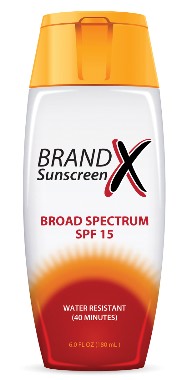
How do you advise your patients about sunscreen? Some fodder for discussion (or possibly more confusion) comes from the Environmental Working Group, which has released its annual list of which sunscreens it deems both safe and effective – according to its criteria.
The Environmental Working Group (EWG), a nonprofit environmental health research and advocacy organization based in Washington, has issued the list for the past 7 years, just before Memorial Day weekend – the date most Americans view as the official start of summer.
This year, the EWG said that only 25% of the 1,400 sunscreens, lotions, lip products, and makeups that claim sun protection properties met its standards.
"The vast majority of sunscreens available to the consumer aren’t as good as most people think they are, but there are a handful of products that rise above the rest," Sonya Lunder, senior research analyst at EWG and lead author of the report, said in a statement. "The best advice for concerned consumers is to use sun-protective clothing, stay in the shade to reduce intense sun exposure, and schedule regular skin examinations by a doctor," she said.
The EWG’s pronouncements are not without controversy. The group claimed in its press release that part of the reason that melanoma has been on the rise may be caused by "the decades of deceptive marketing claims by sunscreen manufacturers."
But your patients need not panic if their favorite sunscreen didn’t make the EWG list, according to the American Academy of Dermatology. The AAD’s position on sunscreens is practical: "The best type of sunscreen is the one you will use again and again," said Dr. Henry W. Lim, chairman of the dermatology department at Henry Ford Hospital in Detroit, in a general AAD statement on sunscreen issued on May 20.
"Just be sure to choose one that offers broad-spectrum protection, has an SPF of 30 or greater, and is water resistant," said Dr. Lim.
However, the AAD emphasized in the statement that seeking shade and wearing protective clothing are also important actions to avoid excessive sun exposure and reduce the risk of skin cancer.
The Food and Drug Administration does regulate sunscreens, having issued a final rule governing much about the products in June 2011. Most of the regulations went into effect in June 2012, but products did not start appearing with the new labels until January.
Manufacturers cannot claim that their products are waterproof or sweatproof. Products can be labeled broad spectrum if they prove to the agency that they protect against both ultraviolet B radiation (UVB) and ultraviolet A radiation (UVA) and have a sun protection factor of 15 or higher.
The EWG also says that the FDA should stop manufacturers from selling products with an SPF of 50 or higher, saying they lead consumers to spend more time in the sun than is advisable. This year, the agency will examine whether those products provide any better protection than an SPF 50 sunscreen. The agency proposed in June 2011 that sunscreens with an SPF greater than 50 be labeled "SPF 50–plus," but nothing further has been issued.
According to the EWG, its analysis of 750 beach and sport sunscreens found that the new FDA rules have not led to sunscreens that are better than those sold in the years before the ruling. It gave only 184 of them its thumbs up. Similarly, for other products that claimed to have sun protection, the EWG only recommended 22 moisturizers, 18 lip balms, and 16 kinds of makeup.
The EWG also said that consumers should not use spray sunscreens.
The AAD agreed that sprays may not be as effective because the individual might not use enough to cover all sun-exposed areas of the body.
The EWG also said that Americans should not use products that contain retinyl palmitate or oxybenzone. The group claims that retinyl palmitate is carcinogenic, and that oxybenzone is an endocrine disrupter. About half of the beach and sport sunscreens in the EWG 2013 list contain oxybenzone, the group said.
The AAD has refuted the EWG’s claims about retinyl palmitate and oxybenzone in the past.
On Twitter @aliciaault
Dual immunotherapy scores rapid response in metastatic melanoma
Hitting metastatic melanoma with two immunotherapies, each targeted to a different metabolic pathway, has shown some preliminary success in an early study.
Ipilimumab (Yervoy) used in combination with the investigational drug nivolumab shrank stage-III and -IV melanomas in approximately half of 37 evaluable patients in a phase-I study. Tumor reductions of at least 80% were seen at 12 weeks in 11 of 37 (30%) of patients and 90% of all patients who have responded continue to respond to therapy, Dr. Jedd D. Wolchok said at a press briefing sponsored by the American Society of Clinical Oncology highlighting research to be presented at its annual meeting.
Phase-I trials are conducted to examine drug safety and give indications of dose ranges; they do not allow conclusions about efficacy. A randomized phase-III trial of the nivolumab/ipilimumab combination as first-line therapy for patients with advanced melanoma is scheduled to begin in June.
Ipilimumab is a CTLA-4 inhibitor approved for the treatment of metastatic melanoma. Nivolumab is an investigational PD-1 inhibitor that has shown activity when used alone against melanoma and other cancers.
"The rapidity and magnitude of the responses are what’s so striking," Dr. Wolchok said. "We were particularly impressed that the drugs work together so well" and show that two immunotherapies offer a promising strategy for advanced melanoma.
For the trial, patients who had inoperable stage-III and stage-IV melanoma and had undergone up to three prior therapies were assigned to one of six treatment arms. Dr. Wolchok of Memorial Sloan-Kettering Cancer Center, New York, presented results on 37 evaluable patients in three completed treatment arms, in which patients received concurrent treatment with the two drugs.
In the phase-I study, nivolumab and ipilimumab were given every 3 weeks for four doses, followed by nivolumab alone every 3 weeks for four doses. At week 24, patients began to receive one concurrent combination treatment every 3 months.
The first arm of 14 patients received 3 mg/kg of ipilimumab plus 0.3 mg/kg of nivolumab, the second arm of 17 patients received 3 mg/kg of ipilimumab plus 1 mg/kg of nivolumab, and the third arm of 6 patients received 3 mg/kg of ipilimumab plus 3 mg/kg of nivolumab. In those three arms, tumor shrinkage rates were 21%, 47%, and 50%, respectively. Tumor reductions of at least 80% were seen in 29%, 41%, and 0%, respectively.
The responses to therapy were rapid for an immunotherapy – three out of four patients who responded to the concurrent treatment experienced tumor reduction within the first 3 months, which is faster than with single-agent ipilimumab, according to Dr. Wolchok.
Two of the remaining three arms of the study enrolled patients who had undergone prior ipilimumab treatment. Those patients received only nivolumab every 2 weeks for up to 48 treatments. Although the data are still preliminary, 4 of 16 evaluable patients in the study arm given nivolumab at 1 mg/kg had at least 80% tumor shrinkage after 8 weeks, even though they initially had little benefit from ipilimumab.
In the concurrently treated patients, grade 3-4 side effects – primarily due to asymptomatic lipase, AST, and ALT abnormalities – occurred in 28 of 53 patients (53%).
With sequenced treatment, grade 3-4 side effects occurred in 6 of 33 patients (18%).
Side effects were managed using standard protocols and no treatment-related deaths were reported.
The nivolumab/ipilimumab combination also is being investigated in non–small cell lung cancer and renal cell carcinoma.
The research was supported by Bristol-Myers Squibb. Dr. Wolchok serves in consultant or advisory roles with, and receives research funding from, Bristol-Meyers Squibb.
"The further exploration of immunotherapy as standalone therapy without chemotherapy for a devastating disease such as advanced melanoma is a tremendous advance. We are identifying therapies that appear better together than alone and look promising for more than just a small subset of patients. The extensive tumor shrinkage that was observed in this study is exactly the type of benefit patients and doctors have been hoping for," said Dr. Sandra M. Swain.
Dr. Swain is president of ASCO. She also is the medical director of the Washington Cancer Institute MedStar Washington Hospital Center and professor of medicine at Georgetown University, Washington. She made her remarks in a press statement.
Hitting metastatic melanoma with two immunotherapies, each targeted to a different metabolic pathway, has shown some preliminary success in an early study.
Ipilimumab (Yervoy) used in combination with the investigational drug nivolumab shrank stage-III and -IV melanomas in approximately half of 37 evaluable patients in a phase-I study. Tumor reductions of at least 80% were seen at 12 weeks in 11 of 37 (30%) of patients and 90% of all patients who have responded continue to respond to therapy, Dr. Jedd D. Wolchok said at a press briefing sponsored by the American Society of Clinical Oncology highlighting research to be presented at its annual meeting.
Phase-I trials are conducted to examine drug safety and give indications of dose ranges; they do not allow conclusions about efficacy. A randomized phase-III trial of the nivolumab/ipilimumab combination as first-line therapy for patients with advanced melanoma is scheduled to begin in June.
Ipilimumab is a CTLA-4 inhibitor approved for the treatment of metastatic melanoma. Nivolumab is an investigational PD-1 inhibitor that has shown activity when used alone against melanoma and other cancers.
"The rapidity and magnitude of the responses are what’s so striking," Dr. Wolchok said. "We were particularly impressed that the drugs work together so well" and show that two immunotherapies offer a promising strategy for advanced melanoma.
For the trial, patients who had inoperable stage-III and stage-IV melanoma and had undergone up to three prior therapies were assigned to one of six treatment arms. Dr. Wolchok of Memorial Sloan-Kettering Cancer Center, New York, presented results on 37 evaluable patients in three completed treatment arms, in which patients received concurrent treatment with the two drugs.
In the phase-I study, nivolumab and ipilimumab were given every 3 weeks for four doses, followed by nivolumab alone every 3 weeks for four doses. At week 24, patients began to receive one concurrent combination treatment every 3 months.
The first arm of 14 patients received 3 mg/kg of ipilimumab plus 0.3 mg/kg of nivolumab, the second arm of 17 patients received 3 mg/kg of ipilimumab plus 1 mg/kg of nivolumab, and the third arm of 6 patients received 3 mg/kg of ipilimumab plus 3 mg/kg of nivolumab. In those three arms, tumor shrinkage rates were 21%, 47%, and 50%, respectively. Tumor reductions of at least 80% were seen in 29%, 41%, and 0%, respectively.
The responses to therapy were rapid for an immunotherapy – three out of four patients who responded to the concurrent treatment experienced tumor reduction within the first 3 months, which is faster than with single-agent ipilimumab, according to Dr. Wolchok.
Two of the remaining three arms of the study enrolled patients who had undergone prior ipilimumab treatment. Those patients received only nivolumab every 2 weeks for up to 48 treatments. Although the data are still preliminary, 4 of 16 evaluable patients in the study arm given nivolumab at 1 mg/kg had at least 80% tumor shrinkage after 8 weeks, even though they initially had little benefit from ipilimumab.
In the concurrently treated patients, grade 3-4 side effects – primarily due to asymptomatic lipase, AST, and ALT abnormalities – occurred in 28 of 53 patients (53%).
With sequenced treatment, grade 3-4 side effects occurred in 6 of 33 patients (18%).
Side effects were managed using standard protocols and no treatment-related deaths were reported.
The nivolumab/ipilimumab combination also is being investigated in non–small cell lung cancer and renal cell carcinoma.
The research was supported by Bristol-Myers Squibb. Dr. Wolchok serves in consultant or advisory roles with, and receives research funding from, Bristol-Meyers Squibb.
AT THE ASCO 2013 PRESSCAST
Major finding: Tumor reductions of at least 80% were seen at 12 weeks in 11 of 37 (30%) of patients.
Data source: A phase-I, dose-ranging study of six cohorts and an analysis of 37 evaluable patients.
Disclosures: The research was supported by Bristol-Myers Squibb. Dr. Wolchok serves in consultant or advisory roles with, and receives research funding from, Bristol-Meyers Squibb.


