User login
Management of adnexal masses in pregnancy
Roughly 1%-2% of pregnancies are complicated by an adnexal mass, and prenatal ultrasound for fetal evaluation has detected more asymptomatic ovarian masses as a result.
The differential diagnosis for adnexal mass is broad and includes follicular or corpus luteum cysts, mature teratoma, theca lutein cyst, hydrosalpinx, endometrioma, cystadenoma, pedunculated leiomyoma, luteoma, as well as malignant neoplasms of epithelial, germ cell, and sex cord–stromal origin (J Ultrasound Med. 2004 Jun;23[6]:805-19). Most masses will be benign neoplasms, with a fraction identified as malignancies.

In 2013, Baser et al. performed a retrospective study of 151 women who underwent surgery of an adnexal mass at time of cesarean delivery. Of the 151 cases reviewed, 148 (98%) of the masses were benign (Int J Gynaecol Obstet. 2013 Nov;123[2]:124-6). Additionally, if the patient presents with pain, diagnoses such as ectopic pregnancy, heterotopic pregnancy, degenerating fibroid, and torsion should also be considered.
Diagnostic evaluation and management
The majority of adnexal masses identified in pregnancy are benign simple cysts measuring less than 5 cm in diameter. Approximately 70% of cystic masses detected in the first trimester will spontaneously resolve by the second trimester (Clin Obstet Gynecol. 2006 Sep;49[3]:492-505). However, for some masses, surgical resection is warranted.
Masses present after the first trimester and that are (1) greater than 10cm in diameter or (2) are solid or contain solid and cystic areas or have septated or papillary areas, are generally managed surgically as these features increase the risk of malignancy or complications such as adnexal torsion, rupture, or labor dystocia (Gynecol Oncol. 2006 May;101(2):315-21).

Adnexal masses without these features often resolve during pregnancy and can be expectantly managed (Obstet Gynecol. 2005 May;105[5 Pt 1]:1098-103). The optimal time for surgical intervention is after the first trimester as organogenesis is largely complete, therefore minimizing the risk of drug-induced teratogenesis, and any necessary cystectomy or oophorectomy will not disrupt the required progesterone production of the corpus luteum as this has been replaced by the placenta.
Preoperative assessment
For most cases, imaging with ultrasound is adequate for preoperative evaluation; however, in some cases, further imaging is needed for appropriate characterization of the mass. In this situation, further imaging with MRI is preferred as this modality has good resolution for visualization of soft tissue pathology and does not expose the patient and fetus to ionizing radiation. Of note, Gadolinium-based contrast should be avoided as effects have not been well established in pregnancy (AJR Am J Roentgenol. 2008 Aug;191[2]:364-70).
If there is concern for malignancy during pregnancy, drawing serum tumor markers preoperatively is typically not suggested. Oncofetal antigens, including alpha fetoprotein (AFP), human chorionic gonadotropin (hCG), carcinoembryonic antigen (CEA), and cancer antigen 125 (CA-125), are elevated during gestation, making them poor markers for malignancy. If malignancy is ultimately diagnosed, then tumor markers can be obtained immediately postoperatively.
Surgical approach and prognosis
If there is low suspicion for malignancy, a laparoscopic approach is preferable and reasonable at all stages of pregnancy, although early second trimester is ideal. Entry at Palmer’s point in the left upper quadrant versus the umbilicus is preferable in order to minimize risk of uterine injury.
If malignancy is suspected, maximum exposure should be obtained with a midline vertical incision. Peritoneal washings should be obtained on immediate entry of the peritoneal cavity, and the contralateral ovary should also be adequately examined along with a general abdominopelvic survey. If the mass demonstrates concerning features, such as solid features or presence of ascites, then the specimen should be sent for intraoperative frozen pathology, and the pathologist should be made aware of the concurrent pregnancy. If malignancy is confirmed on frozen pathology, a full staging procedure should be performed and a gynecologic oncologist consulted.
Roughly three-quarters of invasive ovarian cancers diagnosed in pregnancy are early stage disease, and the 5-year survival of ovarian cancers associated with pregnancy is between 72% and 90% (Int J Gynecol Cancer. 2006 Jan-Feb;16[1]:8-15).
In a retrospective cohort study of 101 pregnant women, 31% of adnexal masses resected in pregnant women greater than 14 weeks gestation were teratomas. In total, 23% of masses were luteal cysts. Less commonly, patients were diagnosed with serous cystadenoma (14%), endometrioma (8%), mucinous cystadenoma (7%), benign cyst (6%), tumor of low malignant potential (5%), and paratubal cyst (3%).
In this study, approximately half of the women underwent minimally invasive surgery and half had surgery via laparotomy. There were more complications in the women undergoing laparotomy (ileus) and there were no differences between the groups with regards to pregnancy and neonatal outcomes (J Minim Invasive Gynecol. 2011 Nov-Dec;18[6]:720-5).
In general, characteristics that are favorable for spontaneous resolution include masses that are simple in nature by ultrasound and less than 5 cm to 6 cm in diameter.
For women with simple-appearing masses on ultrasound, reimaging can occur during the remainder of the pregnancy at the discretion of the physician or during the postpartum period. All women should be provided with torsion and rupture precautions during the pregnancy (Am J Obstet Gynecol. 2011 Aug;205[2]:97-102). For women with more concerning features on ultrasound, referral to a gynecologic oncologist is warranted. If the decision for surgical management is made, minimally invasive surgery should be strongly considered due to minimal maternal and perinatal morbidity.
Dr. Staley is a resident physician in the department of obstetrics and gynecology at the University of North Carolina at Chapel Hill. Dr. Gehrig is professor and director of gynecologic oncology at the university. They reported having no relevant financial disclosures.
Roughly 1%-2% of pregnancies are complicated by an adnexal mass, and prenatal ultrasound for fetal evaluation has detected more asymptomatic ovarian masses as a result.
The differential diagnosis for adnexal mass is broad and includes follicular or corpus luteum cysts, mature teratoma, theca lutein cyst, hydrosalpinx, endometrioma, cystadenoma, pedunculated leiomyoma, luteoma, as well as malignant neoplasms of epithelial, germ cell, and sex cord–stromal origin (J Ultrasound Med. 2004 Jun;23[6]:805-19). Most masses will be benign neoplasms, with a fraction identified as malignancies.
In 2013, Baser et al. performed a retrospective study of 151 women who underwent surgery of an adnexal mass at time of cesarean delivery. Of the 151 cases reviewed, 148 (98%) of the masses were benign (Int J Gynaecol Obstet. 2013 Nov;123[2]:124-6). Additionally, if the patient presents with pain, diagnoses such as ectopic pregnancy, heterotopic pregnancy, degenerating fibroid, and torsion should also be considered.
Diagnostic evaluation and management
The majority of adnexal masses identified in pregnancy are benign simple cysts measuring less than 5 cm in diameter. Approximately 70% of cystic masses detected in the first trimester will spontaneously resolve by the second trimester (Clin Obstet Gynecol. 2006 Sep;49[3]:492-505). However, for some masses, surgical resection is warranted.
Masses present after the first trimester and that are (1) greater than 10cm in diameter or (2) are solid or contain solid and cystic areas or have septated or papillary areas, are generally managed surgically as these features increase the risk of malignancy or complications such as adnexal torsion, rupture, or labor dystocia (Gynecol Oncol. 2006 May;101(2):315-21).
Adnexal masses without these features often resolve during pregnancy and can be expectantly managed (Obstet Gynecol. 2005 May;105[5 Pt 1]:1098-103). The optimal time for surgical intervention is after the first trimester as organogenesis is largely complete, therefore minimizing the risk of drug-induced teratogenesis, and any necessary cystectomy or oophorectomy will not disrupt the required progesterone production of the corpus luteum as this has been replaced by the placenta.
Preoperative assessment
For most cases, imaging with ultrasound is adequate for preoperative evaluation; however, in some cases, further imaging is needed for appropriate characterization of the mass. In this situation, further imaging with MRI is preferred as this modality has good resolution for visualization of soft tissue pathology and does not expose the patient and fetus to ionizing radiation. Of note, Gadolinium-based contrast should be avoided as effects have not been well established in pregnancy (AJR Am J Roentgenol. 2008 Aug;191[2]:364-70).
If there is concern for malignancy during pregnancy, drawing serum tumor markers preoperatively is typically not suggested. Oncofetal antigens, including alpha fetoprotein (AFP), human chorionic gonadotropin (hCG), carcinoembryonic antigen (CEA), and cancer antigen 125 (CA-125), are elevated during gestation, making them poor markers for malignancy. If malignancy is ultimately diagnosed, then tumor markers can be obtained immediately postoperatively.
Surgical approach and prognosis
If there is low suspicion for malignancy, a laparoscopic approach is preferable and reasonable at all stages of pregnancy, although early second trimester is ideal. Entry at Palmer’s point in the left upper quadrant versus the umbilicus is preferable in order to minimize risk of uterine injury.
If malignancy is suspected, maximum exposure should be obtained with a midline vertical incision. Peritoneal washings should be obtained on immediate entry of the peritoneal cavity, and the contralateral ovary should also be adequately examined along with a general abdominopelvic survey. If the mass demonstrates concerning features, such as solid features or presence of ascites, then the specimen should be sent for intraoperative frozen pathology, and the pathologist should be made aware of the concurrent pregnancy. If malignancy is confirmed on frozen pathology, a full staging procedure should be performed and a gynecologic oncologist consulted.
Roughly three-quarters of invasive ovarian cancers diagnosed in pregnancy are early stage disease, and the 5-year survival of ovarian cancers associated with pregnancy is between 72% and 90% (Int J Gynecol Cancer. 2006 Jan-Feb;16[1]:8-15).
In a retrospective cohort study of 101 pregnant women, 31% of adnexal masses resected in pregnant women greater than 14 weeks gestation were teratomas. In total, 23% of masses were luteal cysts. Less commonly, patients were diagnosed with serous cystadenoma (14%), endometrioma (8%), mucinous cystadenoma (7%), benign cyst (6%), tumor of low malignant potential (5%), and paratubal cyst (3%).
In this study, approximately half of the women underwent minimally invasive surgery and half had surgery via laparotomy. There were more complications in the women undergoing laparotomy (ileus) and there were no differences between the groups with regards to pregnancy and neonatal outcomes (J Minim Invasive Gynecol. 2011 Nov-Dec;18[6]:720-5).
In general, characteristics that are favorable for spontaneous resolution include masses that are simple in nature by ultrasound and less than 5 cm to 6 cm in diameter.
For women with simple-appearing masses on ultrasound, reimaging can occur during the remainder of the pregnancy at the discretion of the physician or during the postpartum period. All women should be provided with torsion and rupture precautions during the pregnancy (Am J Obstet Gynecol. 2011 Aug;205[2]:97-102). For women with more concerning features on ultrasound, referral to a gynecologic oncologist is warranted. If the decision for surgical management is made, minimally invasive surgery should be strongly considered due to minimal maternal and perinatal morbidity.
Dr. Staley is a resident physician in the department of obstetrics and gynecology at the University of North Carolina at Chapel Hill. Dr. Gehrig is professor and director of gynecologic oncology at the university. They reported having no relevant financial disclosures.

Maternal flu shot offers far-reaching protection
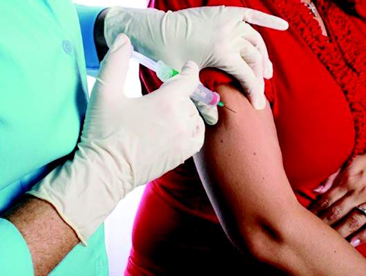
WASHINGTON – The influenza vaccine is highly recommended for pregnant women, protecting the woman, the newborn, and even the fetus, according to Dr. Sonja Rasmussen.
Multiple prospective and retrospective studies have shown that the inactivated influenza shot is safe and effective for pregnant women. And not only does it confer a transient passive immunity upon the newborn, the vaccine also guards against the dangers the flu poses to fetuses, she said at the annual meeting of the American College of Obstetricians and Gynecologists.
Among the benefits seen with the flu shot are healthier birth weights, less preterm birth, and a much lower risk of birth defects that are associated with maternal fever in the first trimester, said Dr. Rasmussen, director of public health information dissemination at the Centers for Disease Control and Prevention.
“Fever in the first trimester doubles the risk of neural tube defects in the fetus,” said Dr. Rasmussen. She cited a 2014 meta-analysis that found increased risks of other fetal anomalies associated with first trimester flu: hydrocephaly (odds ratio, 5.74), congenital heart defects (OR, 1.56), cleft lip (OR, 3.12), limb abnormalities (OR, 2.03) and digestive system defects (OR, 1.72) (Hum Reprod. 2014 Apr;29[4]:809-23).
“We don’t know if these are more due to the hyperthermia of fever or to the flu virus crossing the placenta,” she said. “But we do know these are real risks.”
Newborns come with an immature immune system that takes a while to get ramped up. In the first 6 months of life, they’re especially vulnerable to communicable illnesses, and they can become severely ill and even die from flu, Dr. Rasmussen said. A new study of nearly 250,000 mother-child pairs confirmed that maternal vaccination reduces the risks of flu hospitalization by more than 80% in infants younger than 6 months (Pediatrics. 2016 May 2. doi: 10.1542/peds.2015-2360).
For most women, the understanding that a flu shot protects their baby before and after birth is good enough reason to get one, Dr. Rasmussen said, but the benefits for the woman are extremely important. “Pregnant women experience a lot of changes in immune function and heart and lung function that increase the risk for complications from influenza. In prior pandemics, we have seen pregnant women at an increased risk of severe illness, hospitalization, and death.”
She cited data from the 2009 H1N1 epidemic that showed that 5% of those who died from H1N1 flu were pregnant women, despite the fact that they represented only 1% of the U.S. population (JAMA. 2010;303[15]:1517-25).
Despite the well-documented benefits and reassuring safety data, flu vaccine coverage remains suboptimal for pregnant women, Dr. Rasmussen said. The most recent CDC data suggest that just half of pregnant women were immunized in 2014-2015, a rate that has remained steady since 2012.
The most effective way to boost this number, Dr. Rasmussen suggested, is to stress to women that the vaccine protects both the fetus and newborn from a potentially fatal illness. “We know that a large number of babies and children die of influenza every year,” she said. “The biggest motivator for moms is their desire to protect their baby – much more so than protecting themselves.”
Dr. Rasmussen reported having no relevant financial disclosures.
EXPERT ANALYSIS FROM ACOG 2016

Vaginal delivery found safe for monoamniotic-monochorionic twins
WASHINGTON – Monoamniotic-monochorionic twins can be safely delivered vaginally, with low rates of adverse fetal or maternal outcomes.
Despite seeing cord entanglement in almost every case, a small retrospective study found no safety signals that support pre-emptive cesarean deliveries for this class of twins, Dr. Meena Khandelwal said at the annual meeting of the American College of Obstetricians and Gynecologists. In fact, vaginal delivery was associated with a significantly lower rate of intracranial hemorrhage, which occurred in 27% of the cesarean-delivered infants and none of the infants delivered vaginally.

Dr. Khandelwal and her colleagues won third prize for the study among the 2016 ACOG research awards.
In 2014, an ACOG technical bulletin advised cesarean delivery for all monoamniotic twins, a recommendation in line with those in Canada and France. But the ACOG advice was based on level C evidence, said Dr. Khandelwal of Cooper University Hospital, Camden, N.J.
“These recommendations are fear based, not fact based,” she said. “It is felt that the risk of entrapment is too great. But cord entanglement is seen in virtually all of these cases, and a large study recently found that it contributed nothing to morbidity or mortality in the fetuses.”
That study, published in 2013, reviewed 114 monoamniotic twin pregnancies (228 fetuses). There were 26 perinatal fetal deaths, only two of which were related to cord entanglement. The author found no significant difference in mortality between the fetuses with entanglement (82) and those without (Ultrasound Obstet Gynecol. 2013 Feb;41[2]:131-5).
Dr. Khandelwal conducted her own retrospective study of 29 sets of monoamniotic-monochorionic twins that were born vaginally or by cesarean delivery at two tertiary care centers in New Jersey, from 1997 to 2014. Her cohort comprised 10 pregnancies at Thomas Jefferson Hospital, Philadelphia, which only offers cesarean delivery for these cases, and 19 at Cooper University Hospital, which offers the option of vaginal delivery if the first twin is in cephalic position, the fetal heart rate is reactive at the onset of labor, and there are no contraindications to vaginal delivery.
Cooper also manages these pregnancies on an outpatient basis with vaginal delivery offered from 32 to 36 weeks. Thomas Jefferson admits them to the antepartum unit from 24 to 28 weeks and offers cesarean delivery from 32 to36 weeks.
The mean age of the women in the series was 29 years; median parity was one. Almost half of the women having a planned cesarean had a history of a prior cesarean; none of the women who planned a vaginal delivery had a prior cesarean delivery.
Of the 19 women offered vaginal delivery, 14 accepted. Of these, 10 (71%) delivered both twins vaginally. The mean time between the twin deliveries was 3 minutes. Cesarean delivery was necessary for four neonates in the planned vaginal delivery group: three due to concerning heart rate tracings and one because of a failed internal version of a transverse presentation of a second twin.
Three fetuses died before birth – one in the planned vaginal delivery group (7%) and two in the planned cesarean delivery group (13%). There was one post partum hemorrhage, which occurred in the planned cesarean group. There were no cases of chorioamnionitis. Birth weight was similar (about 1,800 grams). Cords were entangled in 100% of the vaginal delivery group and 93% of the cesarean group. Apgar at 5 minutes was 6.6 in the vaginal delivery group and 8.3 in the cesarean group.
Respiratory distress syndrome was significantly more common among the planned cesarean group (86% vs. 65%). There were eight cases of intracerebral hemorrhage in that group and none in the planned vaginal delivery group (28% vs. 0%). These were all grade 1 or 2 bleeds. There were no neonatal deaths.
The length of stay was shorter in the planned vaginal delivery group, but not significantly so (median 18 vs. 25 days).
“This is a small study but it does add valuable data on the safety of vaginal delivery in [monoamniotic-monochorionic] twins,” Dr. Khandelwal said. “Vaginal delivery can be considered a safe option in tertiary care centers.
She reported having no financial disclosures.
WASHINGTON – Monoamniotic-monochorionic twins can be safely delivered vaginally, with low rates of adverse fetal or maternal outcomes.
Despite seeing cord entanglement in almost every case, a small retrospective study found no safety signals that support pre-emptive cesarean deliveries for this class of twins, Dr. Meena Khandelwal said at the annual meeting of the American College of Obstetricians and Gynecologists. In fact, vaginal delivery was associated with a significantly lower rate of intracranial hemorrhage, which occurred in 27% of the cesarean-delivered infants and none of the infants delivered vaginally.
Dr. Khandelwal and her colleagues won third prize for the study among the 2016 ACOG research awards.
In 2014, an ACOG technical bulletin advised cesarean delivery for all monoamniotic twins, a recommendation in line with those in Canada and France. But the ACOG advice was based on level C evidence, said Dr. Khandelwal of Cooper University Hospital, Camden, N.J.
“These recommendations are fear based, not fact based,” she said. “It is felt that the risk of entrapment is too great. But cord entanglement is seen in virtually all of these cases, and a large study recently found that it contributed nothing to morbidity or mortality in the fetuses.”
That study, published in 2013, reviewed 114 monoamniotic twin pregnancies (228 fetuses). There were 26 perinatal fetal deaths, only two of which were related to cord entanglement. The author found no significant difference in mortality between the fetuses with entanglement (82) and those without (Ultrasound Obstet Gynecol. 2013 Feb;41[2]:131-5).
Dr. Khandelwal conducted her own retrospective study of 29 sets of monoamniotic-monochorionic twins that were born vaginally or by cesarean delivery at two tertiary care centers in New Jersey, from 1997 to 2014. Her cohort comprised 10 pregnancies at Thomas Jefferson Hospital, Philadelphia, which only offers cesarean delivery for these cases, and 19 at Cooper University Hospital, which offers the option of vaginal delivery if the first twin is in cephalic position, the fetal heart rate is reactive at the onset of labor, and there are no contraindications to vaginal delivery.
Cooper also manages these pregnancies on an outpatient basis with vaginal delivery offered from 32 to 36 weeks. Thomas Jefferson admits them to the antepartum unit from 24 to 28 weeks and offers cesarean delivery from 32 to36 weeks.
The mean age of the women in the series was 29 years; median parity was one. Almost half of the women having a planned cesarean had a history of a prior cesarean; none of the women who planned a vaginal delivery had a prior cesarean delivery.
Of the 19 women offered vaginal delivery, 14 accepted. Of these, 10 (71%) delivered both twins vaginally. The mean time between the twin deliveries was 3 minutes. Cesarean delivery was necessary for four neonates in the planned vaginal delivery group: three due to concerning heart rate tracings and one because of a failed internal version of a transverse presentation of a second twin.
Three fetuses died before birth – one in the planned vaginal delivery group (7%) and two in the planned cesarean delivery group (13%). There was one post partum hemorrhage, which occurred in the planned cesarean group. There were no cases of chorioamnionitis. Birth weight was similar (about 1,800 grams). Cords were entangled in 100% of the vaginal delivery group and 93% of the cesarean group. Apgar at 5 minutes was 6.6 in the vaginal delivery group and 8.3 in the cesarean group.
Respiratory distress syndrome was significantly more common among the planned cesarean group (86% vs. 65%). There were eight cases of intracerebral hemorrhage in that group and none in the planned vaginal delivery group (28% vs. 0%). These were all grade 1 or 2 bleeds. There were no neonatal deaths.
The length of stay was shorter in the planned vaginal delivery group, but not significantly so (median 18 vs. 25 days).
“This is a small study but it does add valuable data on the safety of vaginal delivery in [monoamniotic-monochorionic] twins,” Dr. Khandelwal said. “Vaginal delivery can be considered a safe option in tertiary care centers.
She reported having no financial disclosures.
AT ACOG 2016
Key clinical point: Vaginal delivery is a safe option for monoamniotic-monochorionic twins.
Major finding: Successful vaginal delivery of both twins occurred in 71% of those who attempted it.
Data source: A retrospective study comprising 29 sets of monoamniotic-monochorionic twins.
Disclosures: Dr. Khandelwal reported having no financial disclosures.

Pregnancy alters pharmacodynamics of infliximab, adalimumab in women with IBD
SAN DIEGO – Blood levels of infliximab rose during pregnancy, while adalimumab levels remained stable, even after researchers accounted for changes in albumin, body mass index, and C-reactive protein levels, according to a novel single-center study of 25 women with inflammatory bowel disease (IBD).
Furthermore, blood levels of both anti–tumor necrosis factor agents varied considerably among patients, reported Dr. Cynthia Seow, a gastroenterologist at the University of Calgary (Alta.). “We should consider therapeutic drug monitoring during the prepregnancy period in order to optimize the dose during pregnancy,” she said. “Therapeutic drug monitoring may also be considered for pregnant women receiving infliximab in the second trimester to guide third-trimester dosing.
Active IBD during pregnancy increases the risk of relapse and preterm birth, Dr. Seow noted at the annual Digestive Diseases Week. Thus, infliximab and adalimumab are used to keep IBD in check during pregnancy, even though they cross the placenta and reach higher levels in the cord blood and newborn (Clin Gastroenterol Hepatol. 2013 March;11[3]:286-92) than in the mother. “However, it is not known how pregnancy itself influences the pharmacokinetics of anti-TNF agents, or the implications of this on prescribed dosing,” said Dr. Seow.
Therefore, she and her colleagues analyzed blood samples from 25 women receiving stable maintenance anti-TNF therapy for IBD, who attended a median of three prenatal visits at the University of Calgary IBD Pregnancy Clinic. Fifteen women received infliximab during 15 pregnancies, and 10 women received adalimumab during 11 pregnancies. Infliximab levels were drawn at trough times, while adalimumab levels were usually drawn 3 days before the next injection. Blood samples were tested only after delivery, and anti-TNF doses were not adjusted during pregnancy.
The infliximab group included eight women with Crohn’s disease and seven women with ulcerative colitis, and the adalimumab group included nine women with Crohn’s disease and one with ulcerative colitis, said Dr. Seow. The treatment groups were similar in terms of age at diagnosis and pregnancy, time on anti-TNF agents, and average gestational age at delivery, which was 39.2 weeks (range, 38.1-40.2 weeks) for the infliximab group and 38.4 weeks (range, 37.2-39.6 weeks) for the adalimumab patients.
Median infliximab concentrations rose from 8.5 mcg/mL in the first trimester to a peak of 21 mcg/mL during the middle of the third trimester (P = .04), and then dropped to nearly preconception levels after delivery, Dr. Seow reported. “This change persisted irrespective of disease phenotype,” she added. Albumin levels correlated inversely with infliximab levels. In contrast, median adalimumab levels ranged between 8.6 and 12.2 mcg/mL during pregnancy, dropped to 6.8 mcg/mL after birth, and were unrelated to albumin levels.
Body mass index and C-reactive protein levels did not affect blood levels of either drug, and the researchers found no differences in pharmacokinetics in subgroups of patients who had only two blood draws, subtherapeutic drug levels, or consistently absent drug levels, Dr. Seow said. Three patients had detectable antibodies during pregnancy, all of whom had a stable clinical course, she said. “The antibody levels appeared to decrease as the pregnancy progressed, and then appeared to increase again after delivery.” A third of infliximab patients and nearly half of adalimumab patients were receiving combination treatments for IBD, and their anti-TNF blood levels resembled those of patients on monotherapy, she also noted.
The researchers did not test cord blood or blood sample from the newborns, but based on past evidence, fetal anti-TNF exposure has implications for current live vaccination recommendations in infants, Dr. Seow emphasized. “The long-term consequences of anti-TNF exposure remain unknown,” she concluded.
Dr. Seow disclosed ties with Janssen, AbbVie, Takeda, Shire, and Actavis.
AT DDW® 2016
Key clinical point: Blood levels of infliximab rose during pregnancy, while adalimumab levels remained stable, even after researchers accounted for changes in albumin, body mass index, and C-reactive protein levels.
Major finding: Median infliximab concentrations rose from 8.5 mcg/mL in the first trimester to a peak of 21 mcg/mL during the middle of the third trimester (P = .04). Median adalimumab levels ranged between 8.6 and 12.2 mcg/mL during pregnancy.
Data source: A prospective study of 25 pregnant women with ulcerative colitis or Crohn’s disease.
Disclosures: Dr. Seow disclosed ties with Janssen, AbbVie, Takeda, Shire, and Actavis.

VIDEO: Children exposed to marijuana at risk for long-term neurocognitive issues
BALTIMORE – The legalization of recreational marijuana in certain states has brought with it a host of new challenges to health care professionals, particularly ob.gyns. and pediatricians, who may be faced with parents who use the drug but are unaware – or unwilling to recognize – the dangers of marijuana exposure to their children.
“A longitudinal, very good study that [showed] regular use [of marijuana] by adolescents [is] associated with about a 6-8 point reduction in adult IQ, and persistent neurocognitive deficits [that] may not even be fully reversible,” said Dr. Paula D. Riggs of the University of Colorado in Aurora.
In an interview at the annual meeting of the Pediatric Academic Societies, Dr. Riggs discussed this and other key studies that point to the dangers of marijuana exposure to children, both before and after birth, and how important it is that all health care professionals ask parents the right questions to ensure that children aren’t being put in danger of long-term cognitive repercussions of marijuana use.
Dr. Riggs did not report any relevant financial disclosures.
The video associated with this article is no longer available on this site. Please view all of our videos on the MDedge YouTube channel
AT THE PAS ANNUAL MEETING

ECLS May Save Mother and Fetus
In pregnant women with acute respiratory distress syndrome, extracorporeal life support can be effective and safe for both the mother and fetus, according to a meta-analysis of 332 articles published in the April issue of the Journal of Thoracic and Cardiovascular Surgery (2016;151:1154-60).
Dr. Sarah A. Moore and her coauthors at the University of New Mexico, Albuquerque, reported that their literature search yielded a total of 45 patients treated with extracorporeal life support (ECLS) or extracorporeal membrane oxygenation (ECMO). The reports were published from 1991-2015.
Dr. Moore and her colleagues also reported on the first successful use of ECLS in a pregnant patient at their own institution with life-threatening hantavirus cardiopulmonary syndrome.
The researchers extrapolated from the literature were case reports and small case series. In the 45-patient study cohort, the survival rate was 77.8% after ECLS for mothers and 65.1% for the fetuses. The average gestational age was 26.5 weeks, ranging from 28 to 43 weeks, and the patients were on ECLS for an average of 12.2 days, with a range of one to 57 days.
The most common reason for ECLS in this cohort was severe H1N1 influenza, otherwise known as swine flu, complicated with acute respiratory distress syndrome (ARDS). The largest series, from France, involved 11 pregnant women treated with ECMO for severe ARDS secondary to severe H1N1 influenza (PLoS One. 2010;5:e13112). Unlike other reports, the New Mexico meta-analysis did not include postpartum patients.
The mitigating case for the study was a previously healthy 25-year-old pregnant woman who was in respiratory failure with hantavirus cardiopulmonary syndrome (HCPS) when she arrived at University of New Mexico Health Sciences Center. Despite mechanical ventilation, the patient remained severely hypoxic and developed worsening hypertension. “The patient was placed on venoarterial ECMO for 72 hours, recovered without complications, and delivered a healthy infant,” Dr. Moore and her colleagues said. “The mother and son remain asymptomatic 6 years later.”
Dr. Moore and her colleagues said strategies used in the nonpregnant population with ARDS might not be appropriate in pregnant mothers for two reasons: permissive hypercapnia may harm the fetus; and prone positioning can be difficult for women in late-term pregnancy. Also, corticosteroids for H1N1 influenza have been controversial.
That doesn’t mean ECLS in pregnant women is not without its complications; the most common was major bleeding, reported in seven of the reviewed articles. Other complications included hemolysis, cannula dislodgement, uterine compression causing ineffective flow rate that improved after emergency cesarean section, and nosocomial infections, including urinary tract and line-related infections.
The study also took a closer look at the use of ECMO in pregnant women during the 2009 H1N1 pandemic; 8 of 33 pregnant women placed on ECMO died, compared with two maternal deaths among the 12 pregnant women placed on ECLS for other reasons.
Dr. Moore and her coauthors acknowledged several limitations of their study, namely the likelihood of selection bias, “given that centers are less inclined to publish their bad outcomes.” Other limits the researchers noted are: the small cohort obviated a proper statistical analysis; there was no control group; and the survival rate in pregnant women with ARDS who do not have ECLS is unknown. Dr. Moore and her coauthors had no relationships to disclose.
The meta-analysis by Dr. Moore and her colleagues provides strong support for ECMO in pregnant women with ARDS. What is lacking, but was not the authors’ focus, is how to maximize survival.
When managing pregnant women with severe cardiopulmonary dysfunction, the decision matrix for extracorporeal support requires rapid assessment of cardiopulmonary function and involves multidisciplinary collaboration, including critical care teams, maternal-fetal medicine physicians, perfusion services, and cardiothoracic surgery.

|
| Mitchel L. Zoler/Frontline Medical News Dr. Nicholas Smedira |
Initially, a pulmonary artery catheter and transthoracic echocardiography are needed to determine cardiac output to direct the decision-making on whether venoarterial or venovenous support is indicated. An experienced perfusionist should be brought in to assess the ECMO cannula and circuit capabilities.
Lower-extremity venoarterial ECMO can cause cerebral and cardiac hypoxia in patients with mild cardiac dysfunction, usually of the right ventricle, secondary to hypoxia, acidosis, and hypercarbia. In the cohort Dr. Moore and her colleagues included in their study, venovenous ECMO is safest and most effective.
With the successful use of ECMO during pregnancy, the rewards can be spectacular: How often can we save two lives with one operation?
Dr. Nicholas G. Smedira is with the Cleveland Clinic. He made his remarks in an invited commentary (J Thorac Cardiovasc Surg. 2016;151:1161-2). Dr. Smedira had no disclosures.
The meta-analysis by Dr. Moore and her colleagues provides strong support for ECMO in pregnant women with ARDS. What is lacking, but was not the authors’ focus, is how to maximize survival.
When managing pregnant women with severe cardiopulmonary dysfunction, the decision matrix for extracorporeal support requires rapid assessment of cardiopulmonary function and involves multidisciplinary collaboration, including critical care teams, maternal-fetal medicine physicians, perfusion services, and cardiothoracic surgery.
|
|
| Mitchel L. Zoler/Frontline Medical News Dr. Nicholas Smedira |
Initially, a pulmonary artery catheter and transthoracic echocardiography are needed to determine cardiac output to direct the decision-making on whether venoarterial or venovenous support is indicated. An experienced perfusionist should be brought in to assess the ECMO cannula and circuit capabilities.
Lower-extremity venoarterial ECMO can cause cerebral and cardiac hypoxia in patients with mild cardiac dysfunction, usually of the right ventricle, secondary to hypoxia, acidosis, and hypercarbia. In the cohort Dr. Moore and her colleagues included in their study, venovenous ECMO is safest and most effective.
With the successful use of ECMO during pregnancy, the rewards can be spectacular: How often can we save two lives with one operation?
Dr. Nicholas G. Smedira is with the Cleveland Clinic. He made his remarks in an invited commentary (J Thorac Cardiovasc Surg. 2016;151:1161-2). Dr. Smedira had no disclosures.
In pregnant women with acute respiratory distress syndrome, extracorporeal life support can be effective and safe for both the mother and fetus, according to a meta-analysis of 332 articles published in the April issue of the Journal of Thoracic and Cardiovascular Surgery (2016;151:1154-60).
Dr. Sarah A. Moore and her coauthors at the University of New Mexico, Albuquerque, reported that their literature search yielded a total of 45 patients treated with extracorporeal life support (ECLS) or extracorporeal membrane oxygenation (ECMO). The reports were published from 1991-2015.
Dr. Moore and her colleagues also reported on the first successful use of ECLS in a pregnant patient at their own institution with life-threatening hantavirus cardiopulmonary syndrome.
The researchers extrapolated from the literature were case reports and small case series. In the 45-patient study cohort, the survival rate was 77.8% after ECLS for mothers and 65.1% for the fetuses. The average gestational age was 26.5 weeks, ranging from 28 to 43 weeks, and the patients were on ECLS for an average of 12.2 days, with a range of one to 57 days.
The most common reason for ECLS in this cohort was severe H1N1 influenza, otherwise known as swine flu, complicated with acute respiratory distress syndrome (ARDS). The largest series, from France, involved 11 pregnant women treated with ECMO for severe ARDS secondary to severe H1N1 influenza (PLoS One. 2010;5:e13112). Unlike other reports, the New Mexico meta-analysis did not include postpartum patients.
The mitigating case for the study was a previously healthy 25-year-old pregnant woman who was in respiratory failure with hantavirus cardiopulmonary syndrome (HCPS) when she arrived at University of New Mexico Health Sciences Center. Despite mechanical ventilation, the patient remained severely hypoxic and developed worsening hypertension. “The patient was placed on venoarterial ECMO for 72 hours, recovered without complications, and delivered a healthy infant,” Dr. Moore and her colleagues said. “The mother and son remain asymptomatic 6 years later.”
Dr. Moore and her colleagues said strategies used in the nonpregnant population with ARDS might not be appropriate in pregnant mothers for two reasons: permissive hypercapnia may harm the fetus; and prone positioning can be difficult for women in late-term pregnancy. Also, corticosteroids for H1N1 influenza have been controversial.
That doesn’t mean ECLS in pregnant women is not without its complications; the most common was major bleeding, reported in seven of the reviewed articles. Other complications included hemolysis, cannula dislodgement, uterine compression causing ineffective flow rate that improved after emergency cesarean section, and nosocomial infections, including urinary tract and line-related infections.
The study also took a closer look at the use of ECMO in pregnant women during the 2009 H1N1 pandemic; 8 of 33 pregnant women placed on ECMO died, compared with two maternal deaths among the 12 pregnant women placed on ECLS for other reasons.
Dr. Moore and her coauthors acknowledged several limitations of their study, namely the likelihood of selection bias, “given that centers are less inclined to publish their bad outcomes.” Other limits the researchers noted are: the small cohort obviated a proper statistical analysis; there was no control group; and the survival rate in pregnant women with ARDS who do not have ECLS is unknown. Dr. Moore and her coauthors had no relationships to disclose.
Key clinical point: Extracorporeal life support (ECLS) during pregnancy is effective and relatively safe for both mother and fetus.
Major finding: Survival rates with ECLS were 77.8% for mothers and 65.1% for fetuses.
Data source: Meta-analysis of 332 articles and a total of 45 patients treated with ECLS during pregnancy.
Disclosures: The study investigators had no relationships to disclose.

Methadone programs allowed pregnancy outcomes similar to nonusers
Methadone users under professional programs have similar pregnancy outcomes as nonmethadone users, according to a study of more than 34,000 deliveries.
Dr. Conisha Holloman of Winnie Palmer Hospital Orlando, Fla., analyzed 34,482 deliveries from 2010 to 2013, comparing women enrolled in methadone programs, those with cocaine or heroine use but no treatment or self medication with methadone, and a large group of controls.
“What we found was a significant difference between preterm births in the groups,” Dr. Holloman said at the annual meeting of the American College of Obstetricians and Gynecologists.
The preterm birth rate was 15% among the 55 patients enrolled in methadone programs. The preterm birth rate rose to 36% among the 34,408 patients in the control group, who had no addiction issues. Comparatively, the preterm birth rate was 53% among the 19 patients who tested positive for cocaine, heroine, or methadone use and were either self treating or not enrolled in a treatment program.
The study also noted neonatal intensive care unit (NICU) admission rates were higher for infants of drug-using patients who were self treating or who were not enrolled in methadone programs. The NICU admission rate for infants of those patients was 26%, compared with 16% for the women in the methadone program and 7% for the control group.
The demographics in the groups were comparable, with no significant difference between age, primigravida, and class III obesity status.
“We encourage women to get into a methadone assisted program, because a lot of these women end up getting earlier prenatal care and better social situations as far as high-risk behavior and improved nutrition for maternal health,” said Dr. Holloman.
Dr. Holloman reported having no relevant financial disclosures.
At ACOG 2016
Key clinical point: Women enrolled in supervised methadone programs had similar preterm birth rates to healthy controls.
Major finding: Preterm birth rates were 15% among methadone users and 36% among controls.
Data source: A retrospective study of 34,482 deliveries from 2010 to 2013.
Disclosures: Dr. Holloman reported having no relevant financial disclosures.
CDC monitoring 279 pregnant women in U.S. with Zika infection
More pregnant women in the United States and its territories may be at risk for Zika virus–related birth complications than previously reported, according to the Centers for Disease Control and Prevention.
As of May 12, CDC officials were monitoring 279 pregnant women with laboratory evidence of possible Zika virus infection – 157 in U.S. states and 122 in U.S. territories, according to a report published in the Morbidity and Mortality Weekly Report (2016 May 20. doi: 10.15585/mmwr.mm6520e1).

“We’ve learned a lot in the last 4 months, and now we know of reports of asymptomatic Zika infections linked to microcephaly, brain defects, and miscarriage,” Margaret Honein, Ph.D., chief of the birth defects branch at the CDC’s National Center for Birth Defects and Developmental Disabilities, said during a May 20 media briefing to announce enhanced tracking of the virus.
Recently published data indicating congenital defects in children born to mothers who had no recollection of ever having Zika-like symptoms spurred the CDC to widen its scope of data capture, Dr. Honein said.
Since February 2016, the CDC has been relying on data from the national arboviral disease surveillance system (ArboNET) to track Zika in pregnant women with symptoms of the Zika virus infection, or those with pregnancy complications consistent with the virus. Although ArboNET describes a subset of pregnancies at risk from Zika virus, it does not track actual congenital outcomes.
Now, federal health officials have created two registries solely dedicated to collecting data on birth outcomes among both symptomatic and asymptomatic women with any laboratory evidence of Zika virus infections. In the continental United States, there is the U.S. Zika Pregnancy Registry (USZPR). In Puerto Rico, there is the Zika Active Pregnancy Surveillance System (ZAPSS).
The 279 pregnant women currently being monitored reflect a broader group of pregnant women than numbers previously reported since it includes pregnant women who have any laboratory evidence of possible Zika virus infection, regardless of them being asymptomatic or whether they recalled ever having symptoms.
New figures on Zika infection in pregnancy will be posted on the CDC website each Thursday at noon EDT, and will reflect the previous week’s numbers.
“This reporting is in line with our recommendations for ongoing monitoring of pregnancies at risk for poor outcomes associated with Zika, based on our current understanding [of the virus],” Dr. Honein said. She recommended that pregnant women be tested for the virus at the start of prenatal care and again during the third trimester.The registry data will be used to determine those most at risk, characterize adverse Zika-related outcomes, and help officials plan for services needed by affected families, said Dr. Honein, encouraging all jurisdictions covered by the surveillance systems to participate in the data collection as quickly as possible.
Meanwhile, Dr. Honein said that the CDC is withholding specific information on known adverse birth outcomes in order to respect the affected families’ privacy, but did say that as of May 20, the CDC was aware of “less than a dozen adverse outcomes, including miscarriages and birth defects.”
Dr. Honein also declined to say how many of the cases reported to the registry involve sexual transmission of the virus versus actual contact with the Aedes aegypti mosquito.
On Twitter @whitneymcknight
More pregnant women in the United States and its territories may be at risk for Zika virus–related birth complications than previously reported, according to the Centers for Disease Control and Prevention.
As of May 12, CDC officials were monitoring 279 pregnant women with laboratory evidence of possible Zika virus infection – 157 in U.S. states and 122 in U.S. territories, according to a report published in the Morbidity and Mortality Weekly Report (2016 May 20. doi: 10.15585/mmwr.mm6520e1).
“We’ve learned a lot in the last 4 months, and now we know of reports of asymptomatic Zika infections linked to microcephaly, brain defects, and miscarriage,” Margaret Honein, Ph.D., chief of the birth defects branch at the CDC’s National Center for Birth Defects and Developmental Disabilities, said during a May 20 media briefing to announce enhanced tracking of the virus.
Recently published data indicating congenital defects in children born to mothers who had no recollection of ever having Zika-like symptoms spurred the CDC to widen its scope of data capture, Dr. Honein said.
Since February 2016, the CDC has been relying on data from the national arboviral disease surveillance system (ArboNET) to track Zika in pregnant women with symptoms of the Zika virus infection, or those with pregnancy complications consistent with the virus. Although ArboNET describes a subset of pregnancies at risk from Zika virus, it does not track actual congenital outcomes.
Now, federal health officials have created two registries solely dedicated to collecting data on birth outcomes among both symptomatic and asymptomatic women with any laboratory evidence of Zika virus infections. In the continental United States, there is the U.S. Zika Pregnancy Registry (USZPR). In Puerto Rico, there is the Zika Active Pregnancy Surveillance System (ZAPSS).
The 279 pregnant women currently being monitored reflect a broader group of pregnant women than numbers previously reported since it includes pregnant women who have any laboratory evidence of possible Zika virus infection, regardless of them being asymptomatic or whether they recalled ever having symptoms.
New figures on Zika infection in pregnancy will be posted on the CDC website each Thursday at noon EDT, and will reflect the previous week’s numbers.
“This reporting is in line with our recommendations for ongoing monitoring of pregnancies at risk for poor outcomes associated with Zika, based on our current understanding [of the virus],” Dr. Honein said. She recommended that pregnant women be tested for the virus at the start of prenatal care and again during the third trimester.The registry data will be used to determine those most at risk, characterize adverse Zika-related outcomes, and help officials plan for services needed by affected families, said Dr. Honein, encouraging all jurisdictions covered by the surveillance systems to participate in the data collection as quickly as possible.
Meanwhile, Dr. Honein said that the CDC is withholding specific information on known adverse birth outcomes in order to respect the affected families’ privacy, but did say that as of May 20, the CDC was aware of “less than a dozen adverse outcomes, including miscarriages and birth defects.”
Dr. Honein also declined to say how many of the cases reported to the registry involve sexual transmission of the virus versus actual contact with the Aedes aegypti mosquito.
On Twitter @whitneymcknight

Care bundle reduces cesarean surgical site infections
WASHINGTON – The rate of cesarean delivery surgical site infections fell significantly at Yale New Haven (Conn.) Hospital after implementation of a multidisciplinary care bundle with protocols covering preoperative, intraoperative, and postoperative care.
An analysis of two 3-month sampling periods – one before implementation of the bundle of care and one after – showed a drop in the surgical site infection (SSI) rate among total cesarean sections from 3.4% to 2.2%.
At the annual meeting of the American College of Obstetricians and Gynecologists, Dr. Ashley Pritchard of the hospital described the care bundle and urged obstetricians to consider the impact of even small reductions in SSIs after a cesarean.
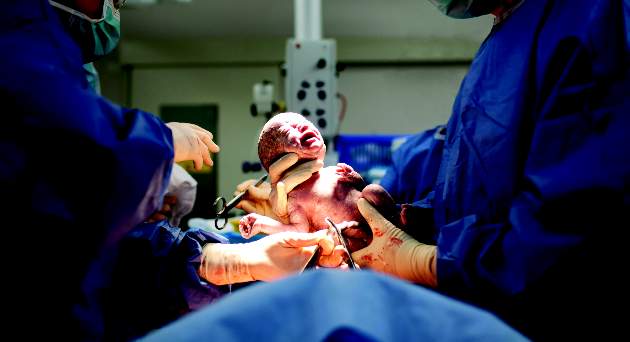
“Infection is the most common complication following cesarean delivery … 2.5%-16% of all cesarean deliveries will result in a surgical site infection, and there is significant underestimation as between 15% to 80% of infections are diagnosed after patients leave the hospital,” she said.
A cesarean delivery SSI task force created by the hospital’s obstetric patient safety program developed the care bundle after reviewing best practices, guidelines, and evidence-based reviews.
Preoperative protocols for planned cesareans focused on patient education and included a preoperative appointment and instructions for showering the night before surgery, not shaving for more than 24 hours prior to scheduled surgery, using 2% chlorhexidine wipes both the night prior to surgery and the day of surgery, and other hygiene processes.
“We know from numerous studies that chlorhexidine is superior to iodine, but we also have found that with these wipes you get a level of antibiosis on the skin surface that decreases surgical site infections at the time of incisions,” Dr. Pritchard said.
For the operative care part of the bundle, staff were reeducated about the scrubbing protocol, proper attire and limits on operating room traffic, and the correct and timely use of antibiotics (for example, a cephalosporin administered within 30 minutes of incision). Staff also watched a video and were quizzed on the proper technique and timing for preoperative skin preparation.
Increased attention was paid to normothermia and included preoperative use of warming blankets and proper temperature in the operating room and post–anesthesia care unit.
“We’ve learned from colorectal and trauma surgery that normothermia and patient warming lead to reduced SSI,” Dr. Pritchard said. “This hasn’t been proven with cesarean delivery, but we know there’s improved maternal and fetal well-being with preoperative warming.”
Postoperatively, the use of supplemental oxygen was discontinued unless clinically indicated “since it’s been shown to have no positive effect on SSI,” she said. Incision dressing application and removal were also standardized, with sterile dressings maintained for at least 24 hours – with a tag labeling the date and time of application – and no more than 48 hours. At discharge, patients were given clear discharge instructions and a postpartum appointment for an incision check.
During the 3-month sampling period prior to implementation of the care bundle, there were 382 cesarean deliveries, and 147 patients presented for a postpartum appointment (either the prescribed visit or a later “issue visit”) within 30 days (38%). Of these patients, 8.6% were diagnosed with an SSI.
In the postimplementation sampling period, which began 6 months after rollout, there were 361 cesarean deliveries at the hospital, and 297 patients (77%) presented for postpartum care. Of these patients, 2.9% were diagnosed with an SSI.
An analysis based on the total number of cesarean deliveries performed at the hospital (planned and unplanned) during the two 3-month periods showed a decline in the cesarean delivery SSI rate from 3.4% to 2.2%. “This is statistically significant. It shows a dramatic decline in the SSI rate in our patient population … a clear impact of the bundle of care,” Dr. Pritchard said.
The Yale team attributes the significant increase in postoperative visit attendance to the preoperative protocol for planned cesareans. “We think it had something to do with our creating better relationships by having [patients] present preoperatively and starting their care prior to incision,” she said.
The preoperative visit also provided an opportunity to identify and treat any active skin infections, upper respiratory infections, or chronic colonizations (without evidence of completion of treatment) before delivery. “If necessary and if possible, [we could] push back their cesarean section date to ensure adequate treatment had been achieved,” Dr. Pritchard said.
All aspects of the care bundle were rolled out simultaneously. Next steps for the New Haven team include further analysis of provider and patient views, a look at the sustainability of the bundle of care and its impact, and a cost analysis. “We reduced our SSIs, but we’ve also added a number of elements to our care spectrum, pre- and postoperatively,” Dr. Pritchard said.
For now, one thing seems clear: “We learned from cardiac and colorectal surgery that there really is strength in the bundle, that the sum of the parts is greater than the individual aspects,” she said.
Dr. Pritchard reported that she and her coinvestigators have no relevant financial disclosures.
WASHINGTON – The rate of cesarean delivery surgical site infections fell significantly at Yale New Haven (Conn.) Hospital after implementation of a multidisciplinary care bundle with protocols covering preoperative, intraoperative, and postoperative care.
An analysis of two 3-month sampling periods – one before implementation of the bundle of care and one after – showed a drop in the surgical site infection (SSI) rate among total cesarean sections from 3.4% to 2.2%.
At the annual meeting of the American College of Obstetricians and Gynecologists, Dr. Ashley Pritchard of the hospital described the care bundle and urged obstetricians to consider the impact of even small reductions in SSIs after a cesarean.
“Infection is the most common complication following cesarean delivery … 2.5%-16% of all cesarean deliveries will result in a surgical site infection, and there is significant underestimation as between 15% to 80% of infections are diagnosed after patients leave the hospital,” she said.
A cesarean delivery SSI task force created by the hospital’s obstetric patient safety program developed the care bundle after reviewing best practices, guidelines, and evidence-based reviews.
Preoperative protocols for planned cesareans focused on patient education and included a preoperative appointment and instructions for showering the night before surgery, not shaving for more than 24 hours prior to scheduled surgery, using 2% chlorhexidine wipes both the night prior to surgery and the day of surgery, and other hygiene processes.
“We know from numerous studies that chlorhexidine is superior to iodine, but we also have found that with these wipes you get a level of antibiosis on the skin surface that decreases surgical site infections at the time of incisions,” Dr. Pritchard said.
For the operative care part of the bundle, staff were reeducated about the scrubbing protocol, proper attire and limits on operating room traffic, and the correct and timely use of antibiotics (for example, a cephalosporin administered within 30 minutes of incision). Staff also watched a video and were quizzed on the proper technique and timing for preoperative skin preparation.
Increased attention was paid to normothermia and included preoperative use of warming blankets and proper temperature in the operating room and post–anesthesia care unit.
“We’ve learned from colorectal and trauma surgery that normothermia and patient warming lead to reduced SSI,” Dr. Pritchard said. “This hasn’t been proven with cesarean delivery, but we know there’s improved maternal and fetal well-being with preoperative warming.”
Postoperatively, the use of supplemental oxygen was discontinued unless clinically indicated “since it’s been shown to have no positive effect on SSI,” she said. Incision dressing application and removal were also standardized, with sterile dressings maintained for at least 24 hours – with a tag labeling the date and time of application – and no more than 48 hours. At discharge, patients were given clear discharge instructions and a postpartum appointment for an incision check.
During the 3-month sampling period prior to implementation of the care bundle, there were 382 cesarean deliveries, and 147 patients presented for a postpartum appointment (either the prescribed visit or a later “issue visit”) within 30 days (38%). Of these patients, 8.6% were diagnosed with an SSI.
In the postimplementation sampling period, which began 6 months after rollout, there were 361 cesarean deliveries at the hospital, and 297 patients (77%) presented for postpartum care. Of these patients, 2.9% were diagnosed with an SSI.
An analysis based on the total number of cesarean deliveries performed at the hospital (planned and unplanned) during the two 3-month periods showed a decline in the cesarean delivery SSI rate from 3.4% to 2.2%. “This is statistically significant. It shows a dramatic decline in the SSI rate in our patient population … a clear impact of the bundle of care,” Dr. Pritchard said.
The Yale team attributes the significant increase in postoperative visit attendance to the preoperative protocol for planned cesareans. “We think it had something to do with our creating better relationships by having [patients] present preoperatively and starting their care prior to incision,” she said.
The preoperative visit also provided an opportunity to identify and treat any active skin infections, upper respiratory infections, or chronic colonizations (without evidence of completion of treatment) before delivery. “If necessary and if possible, [we could] push back their cesarean section date to ensure adequate treatment had been achieved,” Dr. Pritchard said.
All aspects of the care bundle were rolled out simultaneously. Next steps for the New Haven team include further analysis of provider and patient views, a look at the sustainability of the bundle of care and its impact, and a cost analysis. “We reduced our SSIs, but we’ve also added a number of elements to our care spectrum, pre- and postoperatively,” Dr. Pritchard said.
For now, one thing seems clear: “We learned from cardiac and colorectal surgery that there really is strength in the bundle, that the sum of the parts is greater than the individual aspects,” she said.
Dr. Pritchard reported that she and her coinvestigators have no relevant financial disclosures.
AT ACOG 2016
Key clinical point: A multidisciplinary bundle of care was effective in reducing the rate of surgical site infections with cesarean delivery.
Major finding: The rate of cesarean delivery SSIs decreased from 3.4% to 2.2% after implementation of a care bundle.
Data source: An analysis of a quality improvement project at Yale New Haven Hospital.
Disclosures: Dr. Pritchard reported that she and her coinvestigators have no relevant financial disclosures.

Elective induction of labor at 39 (vs 41) weeks: Caveats and considerations
Tasked with tackling the literature on the subject and debating the question of whether or not it is best to electively induce labor in women with low-risk pregnancies at 39 weeks (vs at 41 weeks after expectant management), Errol Norwitz, MD, PhD, Chairman of the Department of Obstetrics and Gynecology and Professor at Tufts University School of Medicine in Boston, Massachusetts, and Charles Lockwood, MD, Senior Vice President at the University of South Florida (USF) and Dean of the USF Health Morsani College of Medicine in Tampa came to the same conclusion: Elective induction of labor (eIOL) at 39 weeks is superior to expectant management when it comes to fetal outcomes.
In addition, they both agreed that complication rates to the mother (ie, number of cesarean deliveries [CDs]) would not be increased, and possibly even reduced, with eIOL at 39 weeks versus 41 weeks. Dr. Norwitz postulated that, with IOL there is no increase in CD rate in multiparous women and nulliparous women with a favorable cervical exam but that there likely could be an increase in the CD rate for nulliparous women with an unfavorable cervical exam.
The finding that eIOL at 39 weeks is better than at 41 weeks for the infant is likely due to a bigger baby size past 39 weeks (with more traumatic deliveries) and higher rates of postmaturity complications, said Dr. Lockwood. And for the mother, eIOL at 39 weeks can reduce risks—of preeclampsia, abruption, sepsis, and others—the presenters pointed out.
Arriving at their conclusions: The dataDr. Norwitz explained the challenge before them in this unusual “debate.” “In ObGyn we all read the same literature but we often come away with very different takes as to what the implications are and how we incorporate this into our management algorithms. Instead of taking a pro/con approach, with one assigned to ‘yes’ and the other assigned to ‘no,’ and selectively picking out the literature to support our positions, what we did was we each went away, read the literature, synthesized it, and tried to answer this question for ourselves.”
Dr. Norwitz, who is widely published and known for his research on the causes and prevention of preeclampsia and preterm labor, examined and presented the published literature for benefit and harms to the fetus and mother in continuing pregnancy past 39 weeks.
Dr. Lockwood also examined the literature, including a large population cohort of about 1.27 million women that examined CD rates, perinatal mortality, and neonatal and maternal outcomes of eIOL at 39 weeks versus expectant management.1 He presented, however, that the best evidence to compare the question at hand would be a randomized clinical trial comparing specifically eIOL at 39 weeks versus expectant management, with IOL at 41 weeks. To be powered to detect a difference in perinatal and maternal mortality, this trial would need to include 2.2 to 12.6 million women, he maintained. “When empirical evidence doesn’t exist, the only alternative is some kind of other modeling,” he said. Therefore, he and a team of researchers conducted a Monte Carlo microsimulation modeling decision analysis, taking into account “all outcomes and all preferences that we possibly could cull from the literature.”
Which women actually could benefit from eIOL at 39 weeks?Women with high-risk pregnancies were not included in this debate or considered. Dr. Norwitz clearly defined his case patient at the outset as a 22-year-old G1 at 39 0/7 weeks who has had an uncomplicated pregnancy but is now complaining of decreased fetal movement and tells you that she is worried because her sister lost her baby at 40 weeks to stillbirth. She specifically asks, “Doctor, why can’t you induce my labor now?”
What counseling a patient about the risks/benefits of eIOL at 39 weeks would requireTwo fundamentals would need to be ensured. The first: precise gestational dating. Dr. Norwitz pointed out that menstrual history can be inaccurate, especially in women with irregular cycles, who are taking hormonal contraception, or who have intermenstrual bleeding. “Early dating ultrasound is the best way to date pregnancies and probably should be done routinely,” although it is not currently standard of care in the United States, he said.
The second fundamental: a true induction of labor process, not one that involves “stopping at 5 PM,” said Dr. Lockwood.
“Although Dr. Lockwood’s 5-PM statement was made tongue-in-cheek,” said Dr. Norwitz after the debate, “he certainly was implying that a genuine effort should be made to effect a vaginal delivery—that is, giving the most effective cervical ripening agents, allowing enough time to pass, and defining clearly the criteria for failed IOL. You shouldn’t just throw the towel in at 5 PM because you want to go home.”
Cost implicationsElectively inducing labor in all women with low-risk pregnancies at 39 weeks is a strategy with unknown cost implications, and the cost difference between this strategy and expectant management up to 41 weeks is not known.
“We need to be sure that we understand the cost implications of these strategies, which of course would need to be balanced against the potential perinatal and maternal morbidity and fetal death,” said Dr. Lockwood.
The meaning of “elective”Dr. Norwitz also made the point postdebate that the American College of Obstetricians and Gynecologists (ACOG) does not support elective IOL prior to 39 weeks’ gestation. “The key term here is ‘elective,’" he said, "which refers to a delivery without a clear medical or obstetric indication. ACOG does support delivery prior to 39 weeks’ gestation, there just needs to be an appropriate indication.”
1. Stock SJ, Ferguson E, Duffy A, Ford I, Chalmers J, Norman JE. Outcomes of elective induction of labour compared with expectant management: population based study. BMJ. 2012;344:e2838.
Tasked with tackling the literature on the subject and debating the question of whether or not it is best to electively induce labor in women with low-risk pregnancies at 39 weeks (vs at 41 weeks after expectant management), Errol Norwitz, MD, PhD, Chairman of the Department of Obstetrics and Gynecology and Professor at Tufts University School of Medicine in Boston, Massachusetts, and Charles Lockwood, MD, Senior Vice President at the University of South Florida (USF) and Dean of the USF Health Morsani College of Medicine in Tampa came to the same conclusion: Elective induction of labor (eIOL) at 39 weeks is superior to expectant management when it comes to fetal outcomes.
In addition, they both agreed that complication rates to the mother (ie, number of cesarean deliveries [CDs]) would not be increased, and possibly even reduced, with eIOL at 39 weeks versus 41 weeks. Dr. Norwitz postulated that, with IOL there is no increase in CD rate in multiparous women and nulliparous women with a favorable cervical exam but that there likely could be an increase in the CD rate for nulliparous women with an unfavorable cervical exam.
The finding that eIOL at 39 weeks is better than at 41 weeks for the infant is likely due to a bigger baby size past 39 weeks (with more traumatic deliveries) and higher rates of postmaturity complications, said Dr. Lockwood. And for the mother, eIOL at 39 weeks can reduce risks—of preeclampsia, abruption, sepsis, and others—the presenters pointed out.
Arriving at their conclusions: The dataDr. Norwitz explained the challenge before them in this unusual “debate.” “In ObGyn we all read the same literature but we often come away with very different takes as to what the implications are and how we incorporate this into our management algorithms. Instead of taking a pro/con approach, with one assigned to ‘yes’ and the other assigned to ‘no,’ and selectively picking out the literature to support our positions, what we did was we each went away, read the literature, synthesized it, and tried to answer this question for ourselves.”
Dr. Norwitz, who is widely published and known for his research on the causes and prevention of preeclampsia and preterm labor, examined and presented the published literature for benefit and harms to the fetus and mother in continuing pregnancy past 39 weeks.
Dr. Lockwood also examined the literature, including a large population cohort of about 1.27 million women that examined CD rates, perinatal mortality, and neonatal and maternal outcomes of eIOL at 39 weeks versus expectant management.1 He presented, however, that the best evidence to compare the question at hand would be a randomized clinical trial comparing specifically eIOL at 39 weeks versus expectant management, with IOL at 41 weeks. To be powered to detect a difference in perinatal and maternal mortality, this trial would need to include 2.2 to 12.6 million women, he maintained. “When empirical evidence doesn’t exist, the only alternative is some kind of other modeling,” he said. Therefore, he and a team of researchers conducted a Monte Carlo microsimulation modeling decision analysis, taking into account “all outcomes and all preferences that we possibly could cull from the literature.”
Which women actually could benefit from eIOL at 39 weeks?Women with high-risk pregnancies were not included in this debate or considered. Dr. Norwitz clearly defined his case patient at the outset as a 22-year-old G1 at 39 0/7 weeks who has had an uncomplicated pregnancy but is now complaining of decreased fetal movement and tells you that she is worried because her sister lost her baby at 40 weeks to stillbirth. She specifically asks, “Doctor, why can’t you induce my labor now?”
What counseling a patient about the risks/benefits of eIOL at 39 weeks would requireTwo fundamentals would need to be ensured. The first: precise gestational dating. Dr. Norwitz pointed out that menstrual history can be inaccurate, especially in women with irregular cycles, who are taking hormonal contraception, or who have intermenstrual bleeding. “Early dating ultrasound is the best way to date pregnancies and probably should be done routinely,” although it is not currently standard of care in the United States, he said.
The second fundamental: a true induction of labor process, not one that involves “stopping at 5 PM,” said Dr. Lockwood.
“Although Dr. Lockwood’s 5-PM statement was made tongue-in-cheek,” said Dr. Norwitz after the debate, “he certainly was implying that a genuine effort should be made to effect a vaginal delivery—that is, giving the most effective cervical ripening agents, allowing enough time to pass, and defining clearly the criteria for failed IOL. You shouldn’t just throw the towel in at 5 PM because you want to go home.”
Cost implicationsElectively inducing labor in all women with low-risk pregnancies at 39 weeks is a strategy with unknown cost implications, and the cost difference between this strategy and expectant management up to 41 weeks is not known.
“We need to be sure that we understand the cost implications of these strategies, which of course would need to be balanced against the potential perinatal and maternal morbidity and fetal death,” said Dr. Lockwood.
The meaning of “elective”Dr. Norwitz also made the point postdebate that the American College of Obstetricians and Gynecologists (ACOG) does not support elective IOL prior to 39 weeks’ gestation. “The key term here is ‘elective,’" he said, "which refers to a delivery without a clear medical or obstetric indication. ACOG does support delivery prior to 39 weeks’ gestation, there just needs to be an appropriate indication.”
1. Stock SJ, Ferguson E, Duffy A, Ford I, Chalmers J, Norman JE. Outcomes of elective induction of labour compared with expectant management: population based study. BMJ. 2012;344:e2838.
1. Stock SJ, Ferguson E, Duffy A, Ford I, Chalmers J, Norman JE. Outcomes of elective induction of labour compared with expectant management: population based study. BMJ. 2012;344:e2838.