User login
Neuropathic Pain in Cancer: A Personalised Approach
Neuropathic pain in individuals with cancer remains a challenging clinical problem that requires advanced pharmacologic skills and a clear understanding of the molecular mechanisms that drive it.
At the 32nd Annual Congress of the Italian Society of Palliative Care, 3 experts outlined an updated framework that begins with basic biology and leads to the most appropriate therapeutic choice.
Fibre Reprogramming
Diego Maria Michele Fornasari, professor of pharmacology and director of the Postgraduate School of Specialization in Pharmacology and Clinical Toxicology at the University of Milan, Milan, Italy, outlined how a healthy nerve fibre becomes chronically hyperexcitable.
“The pathogenic mechanisms of pain, even though we experience hundreds of different pains, can be counted on the fingers of one hand,” he said.
In oncology, nerve injury caused by tumour compression, chemotherapy, or radiotherapy initiates a molecular cascade that reshapes the nerve fibre.
“The injured area is reprogrammed because the neuron attempts to regenerate, and to regenerate it must express proteins seen during embryonic life,” Fornasari said.
This process leads to the appearance of embryonic sodium channels (particularly Nav1.3), which impart abnormally high electrical activity to neurones.
The second mechanism involves the loss of a protective physiologic feature, namely, intermittent failure of signal conduction. Under normal conditions, occasional conduction failures reduce pain intensity. In neuropathic pain, overexpression of hyperpolarisation–activated cyclic nucleotide-gated channels, the same channels that govern sinoatrial node automaticity, makes the neuron hyperexcitable and unable to modulate transmission. The result is that “all stimuli reach the spinal cord and the central nervous system.”
Fornasari illustrated the concerns regarding pain in bone metastases. “The tumour cell is an intelligent cell, capable of producing substances that can interfere with nociceptive mechanisms,” he said.
He also highlighted the mechanisms of pain in patients with bone metastases. Prostate cancer cells that metastasised to the bone that produce tumour cells produce large amounts of nerve growth factor, which does not promote growth in adult neurones but causes hypertrophy and sprouting of nerve fibres. “Each of these ‘spikes’ is actually a neuropathic lesion of the fibre,” Fornasari explained, clarifying how this mechanism contributes to the often-devastating intensity of pain from bone metastases.
Diagnostic Precision
Francesca Dina Ricchini, oncologist and palliative care specialist working in the Palliative Care Unit at the National Cancer Institute in Milan, Italy, addressed the translation of molecular knowledge into clinical diagnosis. “It is very important to be able to translate these concepts into diagnostic tools,” she said.
Neuropathic involvement affects approximately 40% of individuals with cancer, with 20% presenting with pure neuropathic pain and another 20% with mixed pain.
Approximately 75% of cases are related to the disease itself, and the remainder are related to oncologic treatments. Despite its prevalence, neuropathic pain remains underdiagnosed, contributing to suboptimal analgesia and an impaired quality of life.
Screening tools such as PainDETECT, DN4, and the LANSS scale can support assessment, but Ricchini cautioned that “they are tools that can be used as aids but do not make a diagnosis on their own and have not been validated in oncology patients.”
She noted that quantitative sensory testing (QST) complements clinical judgement by quantifying sensory thresholds for thermal, mechanical, and pain stimuli, aiding in the identification of neuropathic pain mechanisms, particularly in oncology settings. QST can complement clinical judgement and diagnostic algorithms developed by the International Association for the Study of Pain to help clinicians systematically identify neuropathic components.
“The key is to start from symptoms, what the patient tells us,” Ricchini added.
However, bedside assessments remain essential. Simple tool needles, cotton, and the clinician’s thumb allow the evaluation of hyperalgesia, allodynia, and other neuropathic features of the affected area. With careful history taking and physical examination, clinicians can often reach an accurate diagnosis without complex testing.
Opioid Use
Sebastiano Mercadante, director of the Anesthesia & Intensive Care and Pain Relief and Supportive Care Unit in La Maddalena Cancer Center Palermo at the University of Palermo in Palermo, Italy, discussed opioid therapy in neuropathic pain. “Responsiveness to opioid drugs varies from patient to patient,” he said.
Opioids develop tolerance and have a ceiling effect that is driven by adverse events. Mercadante emphasised the need to “optimise their use” by staying within the therapeutic window where the benefits outweigh the harm.
Responsiveness depends on genetic polymorphisms affecting receptors and metabolism, pain patterns, age, sex, and psychological distress. “Those with the worst response are patients with significant psychological distress,” he noted.
The opioid escalation index helps to stratify patients. Slow escalation predicts a better prognosis, whereas rapid escalation should prompt specialist involvement and evaluation of factors such as delirium, anxiety, and depression in patients.
Drug Strategies
Switching opioids can be helpful when they become less effective. Mercadante explained that switching opioids “can facilitate receptor internalisation, excluding from continuous stimulation,” allowing “receptor refreshing” and allowing an overstimulated system to be cleared and restoring analgesia. Switching to methadone, when conducted by experienced clinicians, often yields substantial benefits for patients.
Fornasari clarified the mechanisms of action of gabapentinoids, which act on the alpha-2-delta subunit of calcium channels by restoring normal channel numbers rather than by closing them. This distinction is important because it is a slow mechanism that requires approximately 2 weeks for full effect, which is crucial information for clinicians who often discontinue treatment prematurely after a few days of treatment.
Antidepressants, including amitriptyline and serotonin-norepinephrine reuptake inhibitors such as duloxetine, strengthen descending inhibitory pathways by increasing serotonin and noradrenaline levels.
Fornasari also mentioned desvenlafaxine, the active metabolite of venlafaxine, which “has greater activity on descending noradrenergic pathways” and could be particularly useful when the predominant component is noradrenergic.
Topical Options
Capsaicin is an option for chemotherapy-induced peripheral neuropathy.
Chemotherapy-induced neuropathy, the expert explained, is related to the overexpression of transient receptor potential cation channel subfamily V member 1, also known as the capsaicin receptor. Topical capsaicin abnormally stimulates these receptors, causing a massive influx of calcium that “defunctionalises the fibre” for periods ranging from a week to 2 months.
Clinical Takeaways
Therefore, neuropathic pain, particularly in patients with cancer, requires a multimodal approach that begins with an understanding of the molecular mechanisms to develop personalised treatment options. As Fornasari concluded, “Often, a lack of knowledge of a drug’s mechanism of action leads to its inappropriate, and ultimately ineffective, use.” The challenge for clinicians is to translate this knowledge into tangible improvements for patients, remembering that each patient is unique and requires personalised care that goes beyond standardised protocols.
Clinicians aim to translate these insights into meaningful relief, recognising that each patient requires tailored care.
Practical Guidance
- Accurate diagnosis: Screening tools should only be used as support, and decisions should be on the basis of a detailed history and physical examination. In individuals with cancer, the interview always began with the patient’s account.
- Recognising negative prognostic factors: Neuropathic pain, incident pain, psychological distress, delirium, and unrealistic expectations indicate a more difficult course and require a more intensive approach.
- Opioids should be used wisely: Dose escalation should be monitored, rotation should be considered when needed, and psychological factors should be assessed throughout care. As experts have noted, clinicians should try at least 2 or 3 opioids and at least 2 routes of administration before considering invasive procedures.
- Integrating adjuvant medicines early: Gabapentin and other antidepressants are not third-line treatments for individuals with cancer. Adequately dosed for at least 2 weeks to allow therapeutic benefit.
- Consider topical options: In localised peripheral neuropathies, capsaicin can be a valid addition to treatment.
- Use of non-opioid medicines: Some individuals respond exceptionally well to anti-inflammatory agents, with effects that can match those of high morphine doses.
This story was translated from Univadis Italy, part of the this news organization Professional Network.
A version of this article appeared on Medscape.com.
FDA Okays CAR T-Cell Therapy for Marginal Zone Lymphoma
The FDA has approved lisocabtagene maraleucel (Breyanzi, Bristol Myers Squibb) for relapsed or refractory marginal zone lymphoma (MZL) in adults after at least two prior lines of systemic therapy.
Lisocabtagene maraleucel (liso–cel) is now the only CD19-directed chimeric antigen receptor (CAR) T–cell therapy approved for MZL. The approval marks liso-cel’s fifth indication, the most of any CD19-directed CAR T–cell therapy, BMS said in a press release.
Prior approvals are also in the relapsed or refractory setting and include large B-cell lymphoma, follicular lymphoma, mantle cell lymphoma, and chronic lymphocytic leukemia/small lymphocytic lymphoma.
MZL is a slow-growing subtype of non-Hodgkin lymphoma (NHL), accounting for about 7% of all NHL cases and typically diagnosed in older adults. Prognosis is generally favorable, but in patients who relapse or become refractory, NHL can transform into diffuse large B-cell lymphoma.
Basis for Approval
Liso-cel’s new approval was based on the MZL cohort of the single arm TRANSFORM FL trial, which included 66 patients in the third or later lines; 95.5% responded to the one-time treatment, with 62.1% having a complete response. Responses were durable in 90.1% of patients at 2 years, according to the BMS press release.
In terms of safety, 76% of MZL patients developed cytokine release syndrome, which was grade 3 or worse in 4.5%. Nervous system disorders included headache (21%, grade ≥ 3 in 1.5%), encephalopathy (21%, grade ≥ 3 in 1.5%), tremor (21%), dizziness (16%), and aphasia (10%).
Labeling also warns of hypersensitivity reactions, serious infections, prolonged cytopenias, hypogammaglobulinemia, and secondary malignancies.
Cost of Treatment
One-time treatment costs $567,237.18, according to drugs.com. BMS noted liso-cel is broadly covered by commercial and government insurance programs.
M. Alexander Otto is a physician assistant with a master’s degree in medical science and a journalism degree from Newhouse. He is an award–winning medical journalist who worked for several major news outlets before joining this news organization. Alex is also an MIT Knight Science Journalism fellow. Email: [email protected]
A version of this article first appeared on Medscape.com.
RSV Vaccine Effective in Older Veterans, But Protection Declines Over Time
RSV Vaccine Effective in Older Veterans, But Protection Declines Over Time
TOPLINE:
A single dose of respiratory syncytial virus (RSV) vaccine provided protection against RSV illness and associated health care use in nearly 290,000 older US veterans over 2 respiratory illness seasons compared with unvaccinated individuals; however, protection declined over time, particularly among immunocompromised individuals.
METHODOLOGY:
- Researchers emulated a target trial to assess the long-term effectiveness of a single does of RSV vaccine, administered between September 2023 and March 2024, to prevent RSV infection and associated health care use among older US veterans.
- The primary outcome was any positive RSV test from 14 days after vaccination; secondary outcomes included RSV-associated emergency department or urgent care visits, hospitalizations, and ICU admissions.
- The median follow-up duration, measured from 14 days after vaccination, was 15.8 months, with a maximum of 19.0 months.
TAKEAWAY:
- The estimated vaccines effectiveness against RSV infection decreased from 82.5% (95% CI, 77.5%-86.9%) over 0 to 1 month to 59.4% (95% CI, 55.6%-63.5%) over 0 to 18 months of follow-up.
- Protection against RSV-associated emergency department and urgent care visits fell from 84.9% over 0 to 1 month to 60.6% over 0 to 18 months, and the estimated effectiveness against hospitalizations decreased from 88.9% to 57.3% over the same interval.
- The estimated effectiveness against RSV-associated ICU admissions reduced from 92.5% (95% CI, 61.1%-100%) over 0 to 1 month to 71.9% (95% CI, 42.8%-90.0%) over 0 to 18 months.
- Among immunocompromised individuals, protection against RSV infection showed the largest decline from 75.2% at 0 to 1 month to 39.7% over 18 months.
IN PRACTICE:
"Boosters may be needed, but for now, our efforts should be focused on saving lives and decreasing disease by encouraging vaccination of persons 75 years and older and those 60 years and older with underlying health issues," experts wrote in an accompanying editorial.
SOURCE:
The study was led by Kristina L. Bajema, MD, Veterans Affairs Portland Health Care System, Portland, Oregon. It was published online on November 24, 2025, in JAMA Internal Medicine.
LIMITATIONS:
RSV documentation may have been incomplete for veterans who sought care outside the Veterans Health Administration. The cohort was predominantly White men, limiting generalizability. Residual confounding could not be excluded. Estimates of long-term effectiveness should be interpreted cautiously because they reflect patients who remained in care and may differ from the original matched cohort.
DISCLOSURES:
The study was supported by the US Department of Veterans Affairs Cooperative Studies Program, US Department of Health and Human Services, Biomedical Advanced Research and Development Authority, and FDA. Two authors reported receiving grants from the study funder and/or the Patient-Centered Outcomes Research Institute; one of these authors also reported co-ownership of van Breemen & Hynes, LLC, unrelated to the submitted work.
This article was created using several editorial tools, including AI, as part of the process. Human editors reviewed this content before publication.
A version of this article first appeared on Medscape.com.
RSV Vaccine Effective in Older Veterans, But Protection Declines Over Time
RSV Vaccine Effective in Older Veterans, But Protection Declines Over Time
Single-Incision Robotic Surgery Exhibits Safety, Feasibility in Colorectal Cases
Single-Incision Robotic Surgery Exhibits Safety, Feasibility in Colorectal Cases
TOPLINE: A novel single-incision robotic surgery technique for colorectal procedures demonstrated feasibility with 0% conversion to open surgery rate; only 1 case required additional ports. The technique achieved a 30-day all-severity morbidity rate of 20% and major morbidity of 6%.
METHODOLOGY:
- Researchers conducted a retrospective review to report a unique, single-incision robotic surgery technique that uses a fascial wound protector device and multiport robotic surgical system in colorectal surgery.
- Analysis included 50 patients (60% women) with mean ages of 53.5 years and median BMI of 27.2 kg/m2.
- Study was performed at a single quaternary, urban, academic institution from December 2023 to April 2025.
- Patients aged ≥ 18 years with colorectal indications who underwent robotic single-incision surgery using a Da Vinci multiport robotic platform were included.
TAKEAWAY:
- Conversion to open surgery rate was 0%; 1 case required additional robotic ports.
- The 30-day all-severity morbidity rate was 20%; 30-day major morbidity was 6%.
- Pathologies treated included Crohn's disease (26%), diverticulitis (22%), colon cancer (16%), colostomy status (8%), and rectal cancer (4%).
- Successful procedures included right-sided colectomies (14%), left-sided colectomies (28%), total colectomy (4%), rectal resection (4%), small bowel procedures (22%), and ostomy creation/reversal (18%).
IN PRACTICE: "Our rSIS technique utilizing a multiport robotic system is safe and feasible across a wide spectrum of colorectal procedures," wrote the study authors.
LIMITATIONS: According to the authors, reproducible successful completion of surgeries using this technique may be challenging in populations requiring deep pelvic dissections, especially in narrow male pelvis cases, and in patients with very high BMI and significant intra-abdominal adipose tissue.
DISCLOSURES: The authors report no financial support was received for this study and declare no competing interests.
This article was created using several editorial tools, including AI, as part of the process. Human editors reviewed this content before publication.
Single-Incision Robotic Surgery Exhibits Safety, Feasibility in Colorectal Cases
Single-Incision Robotic Surgery Exhibits Safety, Feasibility in Colorectal Cases
Beta-Blockers May Reduce Mortality in Patients with Gastric Varices
, according to investigators.
In a real-world dataset of patients with gastric varices, NSBBs were associated with a 38% reduced mortality rate, supporting the common belief that protective effects extend across different types of varices, lead author Rebecca H. Moon, MD, of Kaiser Permanente Los Angeles Medical Center, and colleagues, reported.
“Overall, numerous randomized trials have established NSBB efficacy in primary and secondary prevention of esophageal variceal hemorrhage,” the investigators wrote in Gastro Hep Advances. “While these benefits are presumed to extend to gastric varices, empirical data on NSBB efficacy in gastric varices management remain scarce.”
To address this knowledge gap, Moon and colleagues conducted a retrospective cohort study of 1,276 adults (aged 18-75 years) diagnosed with gastric varices between 2015 and 2021 within the Kaiser Permanente Southern California system.
Patients were followed through February 2022. Those with splenectomy, transjugular intrahepatic portosystemic shunt (TIPS) performed outside the system, or use of more than one type of NSBB were excluded. NSBB exposure was defined as therapy initiated within 1 year before or after variceal diagnosis.
Outcomes included gastric and esophageal variceal hemorrhage, TIPS, liver transplantation, and mortality. Multivariable logistic regression was used to compare NSBB users with nonusers and to assess individual effects of different NSBBs while adjusting for baseline characteristics.
The study population had a mean age of 58 years with a male predominance (63%). Approximately half (48%) of the patients were Hispanic. Common comorbidities included hypertension (66%), obesity (49%), and type 2 diabetes (45%). More than half of the patients (52.6%) had coexisting esophageal varices, 38% had ascites, and 22% had a history of hepatic encephalopathy.
In total, 767 patients (62%) received an NSBB. Propranolol and nadolol were most commonly prescribed, while carvedilol use was rare. Median follow-up was 1.1 years.
Overall, 40% of patients died during the study period. Mortality was significantly lower among NSBB users compared with nonusers (39.2% vs 50.9%), corresponding to a 38% reduced risk (odds ratio [OR], 0.62; 95% CI, 0.46–0.84). Nadolol was associated with the lowest mortality risk (OR, 0.55; 95% CI, 0.38–0.79), followed by propranolol (OR, 0.71; 95% CI, 0.50–1.00). Carvedilol use was too infrequent for meaningful analysis.
Rates of gastric variceal hemorrhage (7%), esophageal variceal hemorrhage (22%), TIPS (4%), and liver transplantation (5%) did not differ significantly between NSBB and non-NSBB groups.
“The observed reduction in mortality among NSBB users, particularly those on nadolol, suggests a potential survival benefit,” the investigators wrote. “However, the lack of statistically significant differences in other clinical outcomes, including gastric variceal hemorrhage, esophageal variceal hemorrhage, TIPS, and liver transplantation, indicates that the primary benefit of NSBBs in gastric varices management may be limited to mortality reduction rather than prevention of other complications.”
Moon and colleagues went on to call for additional research.
“Further prospective studies are needed to elucidate the effects of NSBBs on gastric varices and to refine treatment strategies for this high-risk population,” they wrote. “Given the substantial mortality associated with gastric variceal hemorrhage, continued research into novel therapeutic approaches is essential to improving outcomes for patients with gastric varices.”
Publication costs were covered by Kaiser Permanente Los Angeles Medical Education and Research. The investigators disclosed no conflicts of interest.
FROM GASTRO HEP ADVANCES
UK Approves Targeted Therapy for Cervical Cancer
UK Approves Targeted Therapy for Cervical Cancer
The Medicines and Healthcare products Regulatory Agency (MHRA) has approved tisotumab vedotin (Genmab AS) for adults with recurrent or metastatic cervical cancer.
The decision, made via the International Recognition Procedure, applies to patients whose disease has progressed after prior systemic therapy. It provides a new treatment option for a high-risk group with limited alternatives.
How the Treatment Works
Tisotumab vedotin is an antibody-drug conjugate that combines a tissue factor-directed human monoclonal antibody with monomethyl auristatin E, a microtubule-disrupting agent. The therapy targets tissue factor, which is overexpressed in a several solid tumours, including recurrent cervical cancer.
It is administered as a 30-minute intravenous infusion once every 3 weeks.
What Trials Showed
The approval is based on evidence from multiple clinical studies demonstrating tisotumab vedotin's efficacy in previously treated patients.
In the phase 2 innovaTV 204 study, 102 patients were enrolled and 101 received at least 1 dose of tisotumab vedotin. The confirmed objective response rate was 24%, including seven complete responses and 17 partial responses, demonstrating clinically meaningful activity in a heavily pretreated population.
Further evidence came from the phase 3 innovaTV-301 trial, which randomly assigned 502 patients to receive either tisotumab vedotin or investigator's-choice chemotherapy.
Median overall survival was 11.5 months with the new therapy compared with 9.5 months in the chemotherapy arm, translating to roughly a 30% reduction in the risk for death. The confirmed objective response rate was also significantly higher with tisotumab vedotin—17.8% vs 5.2%—underscoring its advantage over standard treatment options.
Safety and Tolerability
Ocular toxicity and peripheral neuropathy were the most notable adverse reactions.
Common treatment-related events in the phase 2 study included alopecia (38%), epistaxis (30%), nausea, conjunctivitis (26%), and fatigue (26%).
Grade 3 or higher treatment-related adverse events occurred in about 28% of patients. Clinicians should be alert to conjunctivitis and keratitis as well as sensory neuropathic symptoms (numbness, tingling, or a burning sensation in the hands and feet).
Julian Beach, interim executive director of healthcare quality and access at the MHRA, said that patient safety is the agency's "top priority." "We will continue to monitor its safety closely as it becomes more widely used," he added.
The Summary of Product Characteristics and Patient Information Leaflets will be published on the MHRA website within 7 days of approval.
A version of this article first appeared on Medscape.com.
UK Approves Targeted Therapy for Cervical Cancer
UK Approves Targeted Therapy for Cervical Cancer
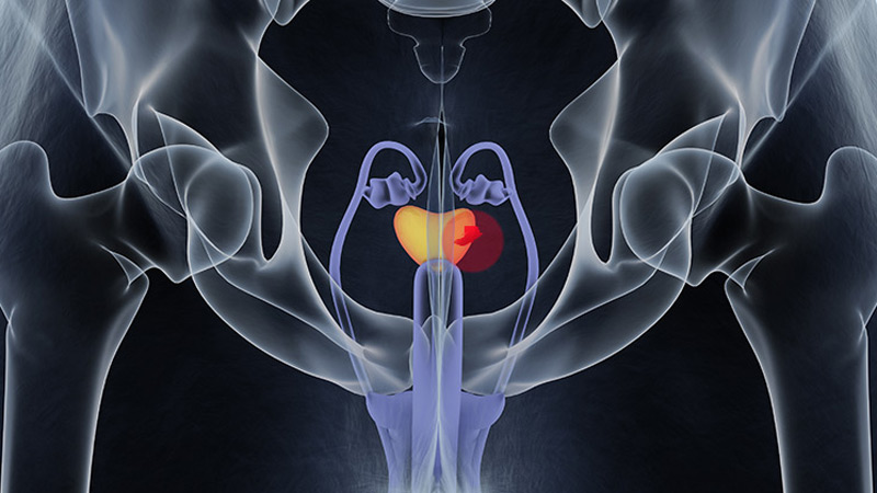
FDA OKs Blood-Based Test to Help Diagnose Prostate Cancer
FDA OKs Blood-Based Test to Help Diagnose Prostate Cancer
The FDA has granted Cleveland Diagnostics' IsoPSA test premarket approval (PMA) to help detect prostate cancer in men aged ≥ 50 years with elevated PSA levels.
IsoPSA is a blood assay that detects variations of the PSA protein that signal a higher likelihood of high-grade tumors. It is one of several biomarker tests included in the National Comprehensive Cancer Network's guidelines on early detection of prostate cancer.
Cleveland Diagnostics noted that 75% of prostate biopsies are negative for high-grade disease. IsoPSA and similar tests aim to help identify men who need a biopsy while allowing others avoid an unnecessary procedure.
IsoPSA has been available since 2020 under the FDA's Laboratory-Developed Test rubric, meaning that blood samples had to be shipped for analysis to Cleveland Diagnostics' lab. With the PMA, testing can now be done at CLIA-certified labs across the country.
The company expects the approval should increase access to IsoPSA and reduce turnaround time. "We remain focused on executing our commercial strategy and expanding access to IsoPSA," company President and CEO Arnon Chait, PhD, said in a press release.
The approval was based, in part, on a prospective validation study of 888 men scheduled for prostate biopsy. IsoPSA demonstrated an AUC of 0.783 for high-grade tumors, with a sensitivity of 90.2% and a specificity of 45.5%. In a real-world clinical utility study with 900 patients, IsoPSA testing led to a 55% decrease in biopsy recommendations.
The test is covered by Medicare and a growing number of commercial payers, Cleveland Diagnostics said.
M. Alexander Otto is a physician assistant with a master's degree in medical science and a journalism degree from Newhouse. He is an award-winning medical journalist who worked for several major news outlets before joining Medscape Medical News. Alex is also an MIT Knight Science Journalism Fellow. Email: [email protected].
A version of this article first appeared on Medscape.com.
FDA OKs Blood-Based Test to Help Diagnose Prostate Cancer
FDA OKs Blood-Based Test to Help Diagnose Prostate Cancer
Expert Advice for Difficult GERD Cases
PHOENIX – , said Kristle L. Lynch, MD, in a presentation at the American College of Gastroenterology (ACG) 2025 Annual Scientific Meeting.
Patients with persistent GERD who have cough and asthma, as well as those with hoarseness, globus, or other ear, nose, and throat concerns, may first undergo workup by other clinicians, said Lynch, who is a professor of clinical medicine and director of the Physiology and Motility Laboratory at the Perelman School of Medicine at the University of Pennsylvania in Philadelphia.
With these patients, “given the low pretest probability of an acid reflux source, it makes sense to bypass a trial of acid-suppressing medication if symptoms persist and instead move on to consideration of upper endoscopy,” she said.
“If negative for damage or sequelae of reflux, this test is often followed by advanced reflux testing and high-resolution manometry, to help diagnose the cause of reflux and swallowing problems,” she noted.
For patients with heartburn, regurgitation, or noncardiac chest pain (and no other concerning symptoms), moving to an empiric trial of antisecretory therapy before endoscopy can be reasonable, Lynch said. But if symptoms persist, an endoscopy should be done before any advanced pH testing.
Advanced pH Testing Options
Advanced reflux testing options for persistent GERD after an endoscopy include catheter-based pH-impedance testing and wireless pH testing, said Lynch.
“Wireless pH monitoring offers improved patient comfort by eliminating the need for a nasal catheter, allowing individuals to maintain normal daily activities and diet during testing,” Lynch told GI & Hepatology News.
“It also enables extended monitoring periods of up to 96 hours, enhancing the detection of intermittent acid reflux episodes,” she said. “However, unlike pH-impedance catheter-based testing, wireless pH measures only acid exposure and cannot identify nonacid reflux events.”
In addition, the wireless capsule may detach prematurely or cause mild chest discomfort, and its endoscopic placement adds procedural complexity with the need for anesthesia, as well as additional cost, she noted.
Catheter-based pH-impedance testing involves sedation-free catheter placement while the patient is awake, followed by monitoring for 24 hours, and involves assessment of bolus movement in tandem with pH measurements, Lynch said. The catheter monitors levels of acid refluxing into the esophagus and transmits this information to the smartphone-sized computer worn by the patient over the 24-hour period.
Unlike wireless testing, impedance testing allows for adjunctive measures of mean nocturnal baseline impedance (MNBI) and post-reflux swallow-induced peristaltic waves (PSPW), the latter of which is shown to predict response to proton pump inhibitor (PPI) therapy in patients with reflux.
Logistics include a concurrent manometry for the lower esophageal sphincter to optimize location, followed by a transnasal placement. Challenges associated with the catheter-based test include potential catheter migration and patient discomfort, she added.
Assessing pH Testing Results
After pH testing of either type, abnormal acid exposure time (AET) when the pH in the lower esophagus is greater than 6% supports a diagnosis of GERD; AET between 4% and 6% is considered borderline GERD; and AET less than 4% is considered normal and outside the bounds of pathologic reflux, as per the Lyon Consensus, Lynch explained.
Treatment for patients meeting the GERD criteria based on AET includes not only diet and lifestyle changes but also potential pharmacologics, including acid suppressants, alginates, gamma-aminobutyric acid agonists, and prokinetics. In some cases, anti-reflux surgery may be considered, such as magnetic sphincter augmentation, transoral incisionless fundoplication, or Roux-en-Y gastric bypass, Lynch said.
But not all heartburn in patients with persistent GERD is acid-related, she said. Functional heartburn and reflux hypersensitivity can be seen in these cases.
Patients not meeting the criteria for GERD on advanced pH testing and diagnosed with reflux hypersensitivity or functional heartburn may be treated with neuromodulators or nonpharmacologic therapies such as hypnosis, diaphragmatic breathing, cognitive-behavioral therapy, or acupuncture, said Lynch in her presentation.
Overall, “advanced pH testing should be assessed within the framework of other investigations and the patient as a whole,” she emphasized.
Challenges in the Clinic
One of the biggest mistakes while diagnosing a patient with persistent GERD symptoms despite PPI therapy is always assuming their symptoms are driven by ongoing pathologic acid exposure, said Michael Kingsley, MD, a gastroenterologist specializing in gastrointestinal motility disorders and a clinical assistant professor of medicine at the University of Pittsburgh, Pittsburgh, in an interview.
He agreed with Lynch that patients with reflux hypersensitivity or functional heartburn are more likely to benefit from interventions other than a PPI, such as a neuromodulator.
But managing GERD in the clinic also requires making time to assess whether patients are taking PPI correctly and to counsel them on lifestyle modifications, he noted.
One of the current biggest challenges in GERD is how best to manage a patient who falls into the inconclusive range of not definitely normal or abnormal on pH testing, Kingsley told GI & Hepatology News.
“As alluded to in this talk, MNBI and PSPW are emerging metrics, both of which require a pH-impedance study (as opposed to a wireless pH study),” he said. “Further elucidating the ideal application and cutoffs of these metrics may provide additional tools for best treating patients who fall into the “inconclusive” range with standard metrics.”
Lynch and Kingsley disclosed no financial conflicts of interest.
A version of this article appeared on Medscape.com .
FROM ACG 2025
GI Side Effects of Immune Checkpoint Inhibitors Linked to Colon Adenoma Risk
PHOENIX — — potentially compounding their cancer burden to include a risk for colon cancer, new research showed.
“The cancer population is already at a higher baseline adenoma risk, and our findings show that ICI-mediated diarrhea and colitis compounds this,” said first author Tanvi Gupta, MD, Department of Internal Medicine, The University of Texas Health Science Center, Houston, in presenting the findings at American College of Gastroenterology (ACG) 2025 Annual Scientific Meeting.
“The study supports a risk-stratified approach to surveillance colonoscopy perhaps within 1 year of ICI-mediated diarrhea and colitis, or earlier if risk factors present,” she said.
ICIs, such as PD-1 and PD-L1 inhibitors, have emerged as highly effective front-line treatments for various cancers, however, diarrhea and colitis are among their most notorious off-target adverse effects, believed to be caused by excessive inflammatory activity from the therapy.
Interestingly, prolonged courses of colitis of more than 3 months have in fact been associated with improved long-term outcomes and survival related to the cancer being treated.
However, prolonged inflammation of the intestine is not without key risks, including compromised colonic mucosa, and such complications are particularly concerning considering indications that the colitis can persist for years even after ICIs are discontinued, the authors noted.
With a previous small study from the researchers at MD Anderson suggesting a higher risk for adenomas associated with ICI-mediated diarrhea and colitis within 1 year of diagnosis, Gupta and colleagues investigated the effects in a larger retrospective study, enrolling 248 patients treated for cancer at MD Anderson from 2010 to 2025.
All patients had developed ICI-mediated diarrhea and/or colitis, confirmed by colonoscopy within 90 days of onset, and all underwent a subsequent colonoscopy at any time point.
The patients, who had a median age of about 63.1 years, were 57.9% men and 90.3% White individuals. Of the patients, 43.7% had been treated with either PD-1 or PD-L1 inhibitors, whereas 42.5% received combination therapy including CTLA-4 inhibitors.
Patients’ predominant cancer types were melanoma and genitourinary malignancies. About 65% had cancer stage IV.
They were compared with a group of historical control individuals who had been treated with ICIs but did not develop diarrhea/colitis, and who had normal baseline and follow-up colonoscopies.
Overall, 71, or nearly 30% of the patients, developed adenomas on follow-up colonoscopies at least 6 months later, with more than 50% of the adenomas developing rapidly, within 7.5 months of ICI-mediated diarrhea and colitis onset. Rates subsequently declined over the following 6 years.
Of 210 who developed ICI-mediated diarrhea and colitis and had baseline and 1-year colonoscopies, those with baseline polyps had an increased risk for detection of adenomas on follow-up endoscopy compared with those with no baseline polyps (33.9% vs 15.9%; P = .004).
However, compared with the patients who were treated with ICIs but did not develop diarrhea and colitis (n = 31), those who did develop the side effects had a higher risk of developing adenomas, even if they had no prior history of polyps (n = 139; P = .002).
Likewise, among those with active histological inflammation at baseline, the risk for adenoma development was higher among those with baseline adenomas vs no adenomas (32.6% vs 15.8%; P = .014), but the risk was higher even among those with no baseline polyps but who did have active inflammation compared with those with ICI treatment but none of the gastrointestinal (GI) side effects (P = .003).
Of those who did develop adenomas, tubular adenomas were the most common types of adenomas, increasing from a median of 46.4% of polyps per patient at baseline to 87.5% at a follow-up endoscopy.
And the size of adenomas increased, with the rate of those under 5 mm at baseline of 100% dropping to 83.3% at the follow-up colonoscopy.
Overall, the findings underscore the role of ICI-mediated diarrhea and colitis in adenoma development.
“We know the cancer population is at a higher baseline risk of adenomas, and ICI-mediated diarrhea and colitis compounds this,” Gupta said.
Importantly, the findings indicate that “histological inflammation drives mucosal injury and adenoma development, regardless of baseline polyps.”
Findings Raise Questions of Surveillance
Danny Issa, MD, an interventional endoscopist and assistant professor of medicine of David Geffen School of Medicine at UCLA, who co-moderated the study, noted that, while longer-term studies are needed, “it’s an interesting study and eye-opening for the many patients on these medications now — but we do need more data.”
“The key question raised is are these patients at a higher risk for colon cancer? It’s possible, and it’s important for clinicians to keep that in mind,” he told GI & Hepatology News.
Further commenting, session co-moderator Sita S. Chokhavatia, MD, AGAF, a gastroenterologist with Valley Medical Group in Paramus, New Jersey, noted the alarming context of ICIs already being used for an existing cancer in the first place.
“So they have one type of cancer and now these patients may be at a higher risk for getting adenomas linked to colon cancer, and so an important question is how often to follow-up on these patients,” she said.
The study’s senior author, Yinghong Wang, MD, PhD, a professor and director of the Oncology-GI Toxicity program at MD Anderson, noted that at MD Anderson, “we routinely offer the first surveillance colonoscopy 1 year after the index ICI-mediated diarrhea and colitis.”
“The following surveillance interval will be based on the finding of this first surveillance colonoscopy,” she told GI & Hepatology News.
Wang recommended that others treating ICI-mediated diarrhea and colitis should follow suit, “given the higher incidence and rapid development of colonic adenomas during this time frame.”
Gupta, Wang, and Chokhavatia had no disclosures to report. Issa reported relationships with Boston Scientific and Eli Lilly.
A version of this article appeared on Medscape.com.
FROM ACG 2025
Higher Epilepsy Mortality in Posttraumatic Cases, VA Study Finds
The risk of death in patients with posttraumatic epilepsy (PTE) varies dramatically by type of brain injury, with some facing twice the mortality rate as those with other forms of epilepsy, according to a new study of Veterans Health Administration data.
Of 210,182 veterans with epilepsy followed for a median of 6 years, those who developed PTE after diffuse cerebral injury, focal cerebral injury, or skull/facial fractures had 16% to 18% higher mortality rates than veterans with nontraumatic epilepsy (NTE) the study found. Published in Neurology, the analysis was completed by Zulfi Haneef, MBBS, MD, of Baylor College of Medicine Medical Center, and colleagues.
Young patients who developed PTE after extracerebral hemorrhage faced the highest risk — double the mortality rate of those with NTE.
“These numbers are striking considering that the group against which these rates are compared — other causes of epilepsy — itself suffers from a high mortality rate,” Haneef said in an interview with Federal Practitioner. “Our findings argue for risk-stratified follow-up in PTE based on the underlying TBI [traumatic brain injury] mechanism and age at epilepsy onset.”
How Common is PTE?
PTE is defined as “long-term predisposition to developing recurrent and unprovoked seizures caused by a traumatic brain injury,” according to neurologist Edilberto Amorim, MD, of University of California at San Francisco Weill Institute for Neurosciences, who was not involved with the study but is familiar with its findings. “We do not fully understand why some people with a traumatic brain injury develop epilepsy and others do not, but the risk is higher with more severe types of TBI.”
PTE accounts for about 5% of all epilepsy cases, Amorim said. The study cites research linking PTE to mortality risk that’s 1.75 to 2.30 higher than in people without epilepsy.
Haneef said the study aimed to shed light on mortality in PTE. “Although epilepsy and TBI are each linked to higher mortality, it had never been conclusively shown that PTE specifically carries higher mortality than nontraumatic epilepsy,” he said. “We set out to answer that question in a large national veterans cohort and to see whether mortality differs by the type of antecedent TBI.”
Methodology and Findings
Researchers tracked 210,182 veterans diagnosed with epilepsy from 2005 to 2022 through the end of 2024: 28,832 with PTE (mean onset age 52.6 years, 7.4% female, 74.2% White, 16.2% Black) and 181,350 with NTE (mean onset age 60.9 years, 8.5% female, 71.0% White, 21.4% Black).
Patients with PTE were defined as having had documentation of TBI within 5 years previous to receiving an epilepsy diagnosis.
Among those with NTE (median follow-up, 6.0 years), 51.1% died. In the PTE group (median follow-up, 6.4 years), 37.3% died.
After adjustment for differences in age, sex, and comorbidities, the risk of mortality in PTE was slightly higher than in NTE (adjusted hazard ratio [aHR], 1.02); the risk was lower for the concussive TBI subtype (aHR, 0.91, both P < .05). “The underlying injury in concussion
is likely to be less severe compared with structural TBI, which may have led to the lower relative mortality observed,” the authors wrote.
However, risk of mortality in PTE was higher than in NTE for cases with underlying TBI subtypes of skull/facial fracture (aHR, 1.18), diffuse cerebral injury (aHR, 1.17), and focal cerebral injury (aHR, 1.16).
“These injuries are associated with greater structural brain damage and sustained neuroinflammation, which are factors linked to harder-to-treat (drug-resistant) epilepsy, which carries higher mortality,” Haneef said. “They may also coexist with extracranial trauma and medical comorbidity that compound long-term risk.”
Among various age groups, there was a notably higher risk of mortality linked to patients aged 18 to 39 years at onset with extracerebral PTE (aHR, 2.02, vs NTE): “In younger patients, extracerebral bleeds (eg, subdural, epidural, and subarachnoid) may reflect higher-energy trauma and more aggressive secondary cascades, amplifying epilepsy severity and longer lifetime exposure to risk. Mechanistic differences in hemorrhage types across ages may also contribute,” Haneef said.
Perspective on Findings
Amorim said the new research is “very useful,” although it has limitations that are common in large database studies. “A key point that this study highlights is the variability in the impact of TBI type on mortality and the differential risk across different age groups,” he said.
As for the higher risk in younger people, Amorim said this may be related to severity of injury: “Older patients often have TBI after falls, while younger patients are more frequently involved in traffic accidents or victims of violence,” he said
In the big picture, Amorim said, “studies like this highlight the importance of moving beyond a one-size-fits-all approach in epilepsy care. Understanding the nuances of posttraumatic epilepsy—how the type of injury, age, and other factors affect outcomes—can help us personalize treatment and counseling and maybe even guide future research into preventing or mitigating epilepsy after brain injury. New methods to automate review of medical records with higher resolution, such as large language models and natural language processing, may make this type of study with large databases even more comprehensive and impactful.”
Haneef said the findings highlight the importance of recognizing PTE as a higher-risk epilepsy and prioritizing early specialty care, especially after focal/diffuse brain injury or fracture. “Screen proactively for drug resistance and fast-track definitive therapies—surgery and device-based therapies—when indicated,” Haneef said. “Management should also include optimized antiseizure therapy, comorbidity control, and safety counseling, since many deaths may be preventable with coordinated multidisciplinary care.”
Haneef added that clinicians should “pay particular attention to younger PTE patients with extracerebral hemorrhage, who showed the greatest relative mortality.”
He also noted that the US Department of Veterans Affairs has comprehensive Epilepsy Centers of Excellence across the country.
The US Department of Defense (DoD) funded the study. Haneef discloses DoD funding, and another author discloses DoD and VA funding. Other authors have no disclosures.
Amorim discloses funding from DoD, NIH, American Heart Association, Regents of the University of California, Cures Within Reach, Zoll Foundation, and Hellman Foundation.