Two days ago, a 27-year-old woman noticed that her vision was blurry in her right eye. She has come to see her primary care physician for advice. This is the first time this has happened to her. She describes seeing a grayish blur over the center of her vision, but she has not noted any other symptoms except for some soreness around the right eye, which is worse with eye movements.
How should she be assessed and treated?
IMPORTANT TO RECOGNIZE
Sudden vision loss is one of the more common problems encountered in ophthalmology and neurology.
Optic neuritis, a demyelinating inflammatory condition that causes acute vision loss, is associated with multiple sclerosis (MS), and recognizing its classic clinical manifestations early is important so that appropriate diagnostic testing (magnetic resonance imaging [MRI]) and treatment (corticosteroids and immunomodulators) can be started.
Although a comprehensive review of all the optic neuropathies is beyond the scope of this paper, in the pages that follow we review some of the most common causes, which may be first seen by the general internist.
FOUR SUBTYPES OF OPTIC NEURITIS
There are four subtypes of optic neuritis:
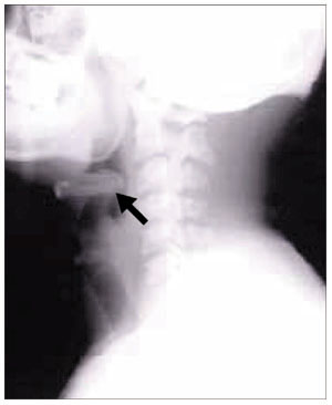
Figure 1. In retrobulbar optic neuritis, the inflammation and demyelination occur behind the globe of the eye. The optic disc appears normal with no signs of swelling or pallor.Retrobulbar neuritis (Figure 1), or inflammation of the optic nerve behind the eye, is the form most commonly associated with MS.
Papillitis (Figure 2), or inflammation of the optic disc, can also be associated with MS.
Perineuritis is inflammation of the optic nerve sheath, sparing the optic nerve itself. Usually, patients are older, and vision loss is mild to moderate. Perineuritis is commonly due to infectious or inflammatory conditions, eg, syphilis or sarcoidosis.
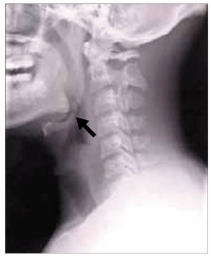
Figure 2. In papillitis, mild swelling and elevation of the optic disc can be seen. The small splinter hemorrhage seen at 10 o’clock is not typical of optic neuritis associated with multiple sclerosis.Neuroretinitis may occur at any age. There is concomitant swelling of the optic nerve and macula. Exudates that form around the macula give the appearance of a star.
Perineuritis and neuroretinitis are not associated with MS, and if they are found they suggest another etiology. In the rest of this review, “optic neuritis” means retrobulbar optic neuritis, the form most commonly seen in clinical practice.
MOST COMMON IN YOUNG WOMEN
Acute demyelinating optic neuritis most often affects women in their 20s and 30s.1–3 Studies in the United States have estimated its annual incidence to be 5.1 to 6.4 per 100,000.4,5 The incidence is higher in populations at higher latitudes and lower near the equator. It is less common in blacks than in whites.6
In children, optic neuritis is not as strongly associated with MS, especially when there is optic disc swelling or bilateral involvement. Most children have a good visual outcome, although approximately 20% may be visually disabled.7–9
FEATURES: VISION LOSS AND EYE PAIN
Most of our current knowledge of the clinical features of optic neuritis comes from the Optic Neuritis Treatment Trial (ONTT),10 conducted in the 1990s. This trial enrolled 457 patients 18 to 46 years old who had acute unilateral optic neuritis. The patients had to have symptoms consistent with acute unilateral optic neuritis for 8 days or less. They could not have evidence of any systemic disease (except for MS) or have received prior treatment for MS. Therefore, this study was quite representative of the patients with optic neuritis that one might encounter in the clinic and is highly important in both characterizing optic neuritis and defining its treatment.
The study found that the two most common symptoms are vision loss and eye pain.
Vision loss in optic neuritis typically occurs over several hours to days, and vision reaches a nadir within 1 to 2 weeks. Typically, patients begin to recover 2 to 4 weeks after the onset of the vision loss. The optic nerve may take up to 6 to 12 months to heal completely, but most patients recover as much vision as they are going to within the first few months.11 More than two-thirds of patients have at least 20/20 vision once they have fully recovered from the optic neuritis. Only 3% of patients become completely blind.
Eye pain is very common in optic neuritis (it affected 87% of patients in the ONTT) and typically worsens with eye movement. The eye is also sore to touch. The pain generally begins at the same time as the visual loss and improves along with it. Eye movements also may bring about photopsia (flickering or flashes of light), a symptom reported by 30% of the ONTT participants.
Loss of color vision out of proportion to the loss of visual acuity is characteristic of optic neuropathies. In the ONTT, 88% of the involved eyes had abnormal color vision as assessed by the Ishihara test (using pseudoisochromatic plates), and 94% as assessed by the Farnsworth-Munsell 100 hue test, which is more sensitive but cumbersome. The most common patterns of color vision loss in optic nerve disease are loss of red (protanopia) and green (deutranopia).
A good way to screen for loss of color vision is to test for color desaturation. First, ask the patient to fixate with the normal eye on a bright red object (for example, the cap from a bottle of ophthalmic mydriatic drops or a pen cap). Then ask the patient to compare the intensity of the redness between the good eye and the affected eye. The patient can quantify the color desaturation by rating what percentage of red is lost in the affected eye compared with the normal eye.
Temporary exacerbations of visual problems during fever (the Uhthoff phenomenon) can occur in patients who have had optic neuritis. These transient pseudoexacerbations are not new episodes of optic neuritis and should resolve after the body temperature returns to normal.
A relative afferent pupillary defect should be seen in the involved eye in all patients with optic neuritis if the other eye is uninvolved and healthy.12 The best way to elicit this sign is to perform the swinging light test in a dark room with the patient fixating at a distant target, which prevents miosis due to accommodation. When the light is swung to the involved eye, the pupil dilates because less light stimulus reaches the midbrain through the affected optic nerve. As the optic nerve heals and recovers, this sign may become subtle, but it persists in more than 90% of cases.12
Findings on funduscopy
Examination of the fundus is helpful in the clinical diagnosis of optic neuritis.
Two-thirds of the ONTT patients had retrobulbar neuritis, and one-third had papillitis. If optic nerve swelling is present, it is typically mild.
Peripapillary hemorrhages were exceedingly rare in the cases of papillitis (only 6%) and were associated with a very low to zero risk of developing MS. If peripapillary hemorrhages are found on examination, one should consider another diagnosis, such as anterior ischemic optic neuropathy.11
CASE CONTINUED
Our patient undergoes a neurologic examination, which reveals an afferent pupillary defect in the right eye and visual acuity of 20/100 in the right eye and 20/20 in the left. Visual fields are normal in the left eye as assessed by confrontation, but there is a central scotoma in the right.
OTHER TYPES OF NEUROPATHY
Optic neuritis is a clinical diagnosis based on the history and findings on examination. If the patient does not have its typical features (Table 1), then other diagnoses should be pursued with serologic and cerebrospinal fluid studies.
The following should be included in the differential diagnosis of optic neuritis:
Ischemic optic neuropathy
Ischemic optic neuropathy is more common in patients age 50 and older, whereas optic neuritis is more common in younger patients. Most patients with ischemic optic neuropathy have hypertension, hypercholesterolemia, diabetes mellitus, obstructive sleep apnea, or other vascular risk factors. The disease has several important subtypes, as discussed below.
Figure 3. Left, fundus photo several weeks after an attack of nonarteritic anterior ischemic optic neuropathy demonstrating pallor of the superior half of the disc. Middle, an associated inferior altitudinal defect. Right, sectoral swelling of the disc with flame or splinter hemorrhages is commonly seen. This is not typical of demyelinating optic neuritis.Nonarteritic anterior ischemic optic neuropathy is the most common form. Typically, there is acute onset of painless vision loss in one eye and an associated altitudinal field defect (Figure 3). For example, if the superior rim of the optic nerve acutely develops swelling and then becomes pale, a corresponding altitudinal cut would develop in the inferior visual field, respecting the horizontal meridian. Many patients first notice the vision loss upon waking up in the morning.13
Although patients with nonarteritic anterior ischemic optic neuropathy typically have vasculopathic risk factors such as hypertension, diabetes mellitus, peripheral vascular disease, or hypercholesterolemia, there is no proven causation between the two. The age of these patients ranges from 50 to 70, with an average age of 66.
The disc appears swollen and may have flame or splinter hemorrhages (Figure 3). The cup of the involved disc is typically small. The visual loss is believed to be the result of poor perfusion in the circulation of the posterior ciliary artery, which supplies the optic nerve head.1 If the other eye also has a small cup, it is considered to be at risk of future ischemic events. In one study,14 the opposite eye became involved within the next 5 years in 14.7% of all cases. The risk of recurrent disease in the same eye is low (6.4% in another study15).
Arteritic anterior ischemic optic neuropathy is more common in patients over age 70 and is usually due to giant cell arteritis, which has a significant association with polymyalgia rheumatica. Patients may have jaw claudication, proximal myalgia and arthralgia, scalp tenderness, headache, fatigue, and a significantly elevated erythrocyte sedimentation rate and C-reactive protein level. These features should be looked for in the review of systems, although patients may not have all of them.
The funduscopic examination may reveal a pale, swollen disc, peripapillary hemorrhages, branch or central retinal artery occlusions, or cotton-wool spots.
Temporal artery biopsy is the gold standard for diagnosis, but treatment with corticosteroids should not be delayed pending biopsy or other test results.1
Thrombocytosis has been associated with a higher risk of permanent vision loss in patients with giant cell arteritis.16
Posterior ischemic optic neuropathy is the least common form of ischemic optic neuropathy. This diagnosis should be entertained in older patients who report acute, painless vision loss but have a normal funduscopic examination. Giant cell arteritis must be considered first in this setting.
Bilateral posterior ischemic optic neuropathy can occur (although rarely) in patients undergoing cardiac or spinal surgery.17 Risk factors thought to be associated with perioperative disease include anemia, hypotension, substantial blood loss during the surgery, surgeries longer than 6.5 hours, carotid atherosclerosis, and diabetes.18
There are no effective treatments for most ischemic optic neuropathies with the crucial exception of giant cell arteritis.
Neuromyelitis optica (Devic disease)
Neuromyelitis optica (Devic disease) is a combination of optic neuritis and transverse myelitis (Table 2). Clinically, the disease spares the nervous system except for the optic nerves and spinal cord. The onset of the optic neuritis may precede or follow the onset of the transverse myelitis by up to 2 to 4 years.19 Usually, the optic neuritis is bilateral and retrobulbar and results in severe vision loss, worse than that seen in patients with MS.19,20
The transverse myelitis may cause paraplegia or quadriplegia, depending on the location of the lesion in the spinal cord (cervical vs thoracic). The transverse myelitis in neuromyelitis optica is distinct from that seen in MS. In neuromyelitis optica, the transverse myelitis is longitudinally extensive, spanning more than three vertebral bodies in length. In MS, spinal cord lesions usually are more discrete and involve one or two spinal cord segments.21
Recently, serum neuromyelitis optica immunoglobulin G (IgG) antibody has been shown to be a significant biomarker of this disease. Its sensitivity ranges from approximately 60% to 70% and its specificity is greater than 90%.22 This antibody binds to aquaporin-4, an important water-channel protein in the blood-brain barrier of the central nervous system, and evidence suggests that it is involved in the pathogenesis of the disease.23
Initially, it was proposed that MRI of the brain had to be normal for neuromyelitis optica to be diagnosed.21 However, the proposed 2006 criteria allow for some abnormal T2 and fluid-attenuated inversion recovery (FLAIR) hyperintensities in the periaqueductal gray matter and diencephalon.22
The spinal fluid in neuromyelitis optica may show a pleocytosis larger than that seen in MS (> 50 white blood cells per mm3) and may have a significant neutrophilic component.21 Oligoclonal bands are not typically present.
It is still debated whether neuromyelitis optica is a separate disease from MS or a subset of it. The implications of this debate may affect its management, as discussed below.
Inflammatory optic neuropathies
Inflammatory optic neuropathies can be caused by infections (eg, syphilis, cat scratch disease) or by noninfectious conditions (eg, sarcoidosis). Lyme disease is rarely a cause of retrobulbar optic neuritis but may cause papillitis.24 West Nile virus has also been reported to cause optic neuritis.25 Lupus may cause an optic neuropathy by inflammatory or ischemic mechanisms.26
Compressive optic neuropathies
Compressive optic neuropathies may be due to mass lesions, tumors, thyroid eye disease, or other orbital processes. MRI of the brain and orbits will confirm or rule out diagnoses associated with compressive optic neuropathy.
Genetic causes
Genetic causes of optic neuropathy include the Leber and Kjer hereditary optic neuropathies.
Leber optic neuropathy involves subacute and painless vision loss, with sequential involvement of both eyes over a period of weeks to months. This disorder predominantly affects men (80%–90% of patients) and is inherited from maternal mitochondrial DNA. The three most common mutations implicated in Leber optic neuropathy (located at base pairs 11,778, 3,460, and 14,484 in the mitochondrial DNA) are involved in more than 90% of cases. The prognosis for recovery varies depending on the genotype.27 These genes encode proteins that are part of complex I of the mitochondrial respiratory chain.28 Funduscopic examination most commonly shows circumpapillary telangiectasia, although up to one-third of patients can have a normal-appearing disc initially. Central vision is affected more severely than peripheral vision.29
Kjer autosomal-dominant optic atrophy is the most common hereditary optic neuropathy. This disease primarily affects children in the first decade of life with slowly progressive loss of vision. As with other optic neuropathies, there will eventually be pallor of the optic disc, a cecocentral scotoma, and loss of color perception. The OPA1 gene located on chromosome 3q28 has been implicated in most patients with dominant optic atrophy; a test is commercially available for diagnosis.30,31
Toxic and metabolic causes
Many agents can cause optic neuropathy. Toxins strongly associated with optic neuropathy include carbon monoxide, methanol, ethylene glycol, perchloroethylene, and tobacco. Drugs linked to optic neuropathy are ethambutol (Myambutol), clioquinol (Vioform), isoniazid (Nydrazid), amiodarone (Cordarone), linezolid (Zyvox), methotrexate, sildenafil (Viagra), oxymetazoline (contained in Afrin and other nasal sprays), and infliximab (Remicade).32–37 Additionally, several chemotherapeutic agents are known to cause optic atrophy, including vincristine (Oncovin), cisplatin (Platinol), carboplatin (Paraplatin), and paclitaxel (Abraxane, Onxol).
Nutritional deficiencies are presumed to have played a significant role in the endemics of optic neuropathy that have occurred in poor countries, such as in Cuba during the 1990s.38 Most nutritional optic neuropathies appear to be exacerbated by tobacco.39
MRI ASSESSES RISK OF MS
The diagnosis of optic neuritis is clinical, based on the history and physical findings.
However, MRI of the brain and orbits with gadolinium contrast has become the cornerstone of the evaluation in patients with optic neuritis. And MRI not only helps confirm the clinical diagnosis, but it also more importantly offers very strong prognostic information about the risk of future demyelinating events and MS.
Gadolinium-enhanced fat-saturated T1-weighted MRI of the orbits is the best sequence to show the inflammation of the optic nerve in optic neuritis (fat saturation is necessary to hide the bright signal of the orbital fat tissue).
Contrast-enhanced MRI can also help differentiate optic neuritis from nonarteritic anterior ischemic optic neuropathy. MRI of the orbits with gadolinium contrast shows enhancement of the affected optic nerve in approximately 95% of cases of optic neuritis, whereas optic nerve enhancement rarely occurs in nonarteritic anterior ischemic optic neuropathy.40
Brain MRI may show other white matter lesions (either hyperintensities on T2-weighted images or enhancement of T1-weighted images postcontrast), which denote a higher risk of developing MS. In 15-year follow-up data from the ONTT, monosymptomatic patients with no white matter lesions had a 25% risk of MS (defined at the time the ONTT was conducted as a second demyelinating event), while those with one lesion or more had a 72% risk.41
An earlier, prospective study in 102 Italian patients with optic neuritis found the risk of developing MS to be about 36% at 6 years and 42% at 8 years (using the Posner diagnostic criteria). When brain MRI data were analyzed, those with one or more lesions had a 52% risk of developing MS at 8 years, whereas those with no MRI lesions did not develop MS.42
Other studies have stratified the risk of MS in patients with clinically isolated syndromes (including not only optic neuritis, but also other neurologic symptoms such as brainstem, motor, or sensory deficits). At mean follow-ups ranging from 5 to 14 years, the risk of developing MS was 8% to 24% in patients with normal findings on brain MRI compared with 56% to 88% in those with abnormal MRI findings.43,44
Optic neuritis patients with atypical white matter lesions on brain MRI may benefit from lumbar puncture to look for oligoclonal bands, to measure the IgG index and the IgG synthesis rate, and to test for myelin basic protein in the cerebrospinal fluid. Of patients with acute optic neuritis, 79% have abnormalities in their cerebrospinal fluid. Oligoclonal bands are present in 69%, and for patients with oligoclonal bands, the 5-year probability of developing MS is estimated to be 65%, compared with 10% in those without bands. If the patient has no oligoclonal bands and has normal findings on brain MRI, he or she will not have MS 5 years later.45–47
Patients with optic neuritis who have no white matter lesions on brain MRI may be further risk-stratified on the basis of their clinical findings. In the ONTT 15-year follow-up, MS did not develop in any patient who had no brain lesions on baseline MRI, no prior optic neuritis in the contralateral eye, and no prior neurologic symptoms or signs, even if the patient had severe disc swelling (eg, peripapillary hemorrhage or retinal exudates) or if vision was reduced to no light perception.41
CASE CONTINUED: FINDINGS ON MRI
Figure 4. The patient’s magnetic resonance image. Top, an axial T2 image with contrast; bottom, sagittal T1 image with contrast. The white matter lesions indicate she is at risk of developing multiple sclerosis.
Our patient undergoes MRI, which shows lesions on axial T2 and sagittal T1 imaging with contrast (Figure 4). Of note, there are significant lesions perpendicular to the corpus callosum (Dawson fingers), some of which enhance with contrast. The enhancement indicates breakdown of the blood-brain barrier and suggests that there is active inflammation in the white matter.
Patients in the ONTT were randomized to receive one of three treatments:
Oral prednisone 1 mg/kg/day for 14 days and then tapered over 4 days
Intravenous methylprednisolone (Solu-Medrol) 250 mg four times per day for 3 days followed by oral prednisone 1 mg/kg/day for 11 days and then tapered for 4 days
Oral placebo for 14 days.
The primary visual outcomes measured were visual acuity and contrast sensitivity.48
Patients who received intravenous methylprednisolone recovered their visual function faster, although the visual outcomes after 6 months were no better with methylprednisolone than with placebo or oral prednisone. Intravenous methylprednisolone also reduced the risk of MS within the first 2 years in patients with high-risk brain MRIs.
Surprisingly, patients in the oral prednisone group had a higher risk of recurrent optic neuritis in both eyes than did patients given intravenous methylprednisolone or placebo (30% at 2 years with oral prednisone vs 16% with placebo and 13% with intravenous methylprednisolone).48 At 10 years, the risk of recurrent optic neuritis was still higher in the oral prednisone group (44%) than in the intravenous methylprednisolone group (29%) (P = .03). However, the difference between the oral prednisone and placebo groups was no longer statistically significant (P = .07).49 Oral prednisone alone is therefore contraindicated in the treatment of typical unilateral demyelinating optic neuritis.
Many patients can now be treated with intravenous infusions of methylprednisolone at home for episodes of optic neuritis.
Risks vs benefits of corticosteroid therapy
When deciding whether to treat an individual patient who has optic neuritis with intravenous corticosteroids, one should consider all the benefits and risks.
Corticosteroids do not affect long-term visual outcome, although they do hasten recovery. Patients with mild vision loss (visual acuity better than 20/40), no significant visual field loss, and no enhancing lesions on brain MRI can forgo therapy with intravenous corticosteroids.
On the other hand, we strongly favor intravenous corticosteroid treatment in patients who have both acute optic neuritis and high signal abnormalities on brain MRI, since it may delay the onset of MS. In addition, patients with severe vision loss should receive intravenous corticosteroids to hasten their recovery. In the rare circumstance in which intravenous corticosteroids are not available, high-dose oral methyl-prednisolone (500 mg daily for 5 days and then tapered over 10 days) may be acceptable.50
The side effects of corticosteroids are minimal when they are given for a brief time in otherwise healthy patients. The most common side effects are mood changes, facial flushing, sleep perturbations, weight gain, and dyspepsia.48
IMMUNOGLOBUL IN: LITTLE BENEFIT
In a double-blind, randomized trial, patients were treated with intravenous immunoglobulin 0.4 g/kg or placebo on days 0, 1, 2, 30, and 60. No difference was found in the primary outcomes of contrast sensitivity, visual acuity, or color vision from 1 week up to 6 months. There was also no significant difference in MRI outcomes between the two groups. The number of relapses was similar between both groups after 6 months.51,52
PLASMA EXCHANGE: FEW DATA
Data on plasma exchange are too scarce for us to make any recommendations. In one trial in 10 patients with severe optic neuritis, 3 patients appeared to benefit from plasma exchange. All patients had already received two doses of intravenous steroids.53
IMMUNOMODULATORY THERAPY MAY PREVENT MULTIPLE SCLEROSIS IN SOME
The most important clinical decision to make in patients with optic neuritis is whether to begin immunomodulatory therapy. Patients who may benefit the most from immunomodulatory therapy are those with abnormal white matter lesions on brain MRI, as they are at higher risk of developing MS.
Collectively, data from three studies indicate that early treatment in patients with clinically isolated syndromes, such as optic neuritis, reduces the rate of MS to 35% (from 50% without treatment) and reduces the number of new active lesions on MRI by approximately 50%.54–56
In addition, the Betaferon/Betaseron in Newly Emerging Multiple Sclerosis for Initial Treatment (BENEFIT) trial57 found that at 3 years the rate of disability was 40% lower in patients who started immunomodulatory therapy (interferon beta-1b; Betaseron) early vs later. (Early treatment meant starting within 60 days of the clinically isolated syndrome; late treatment began 2 years later.) This study suggests that early treatment may reduce future disability, although these results need to be confirmed in prospective trials.
Therefore, once the diagnosis is secure, patients with optic neuritis should be referred to a neurologist with experience in treating MS to begin treatment with immunomodulatory therapy, such as glatiramer acetate (Copaxone), interferon beta-1a (Avonex, Refib), or interferon beta-1b (Betaseron).
Patients who have a normal MRI of the brain may consider deferring therapy, since they are at low risk of developing MS. These patients should undergo surveillance MRI (at least annually at first) to look for the development of white matter lesions, as the ONTT showed even this cohort has a 22% risk of developing MS.
If neuromyelitis optica is suspected (ie, in patients with severe unilateral or bilateral vision loss, recurrent optic neuritis, paraplegia, or quadriplegia), the serum neuromyelitis optic antibody can be tested, keeping in mind that 30% to 40% of patients with neuromyelitis optica will be seronegative for this antibody. Other tests supporting the diagnosis of neuromyelitis optica may include an MRI of the spine showing longitudinally extensive transverse myelitis, a polymorphonuclear pleocytosis in the cerebrospinal fluid, absent oligoclonal bands in the cerebrospinal fluid, and normal MRI of the brain (with some possible signal abnormalities in the periaqueductal gray matter and around the diencephalon).
Because neuromyelitis optica is believed to be mediated primarily by the humoral immune system, immunomodulatory therapy is not a first-line treatment. Patients with neuromyelitis optica can be treated initially with corticosteroids, intravenous immunoglobulin therapy, plasma exchange, or immunosuppressive agents such as azathioprine (Imuran), rituximab (Rituxan), or cyclophosphamide (Cytoxan). The choice of medication should be deferred to a neurologist familiar with treatment of this disorder.
The risk of MS may be lower in children than in adults. One large, retrospective study found the cumulative risk of developing MS (the study predated the McDonald criteria) was 13% at 10 years and 19% by 20 years.58 More recently, a large series from Toronto reported a higher rate of MS development in children with optic neuritis (36% at two years).59 By comparison, studies of adults with unilateral optic neuritis found a 38% to 39% risk of converting to MS at 10 years.5,41 The use of immunomodulatory therapies to reduce the risk of MS has not been well studied in children, since the prevalence is low in this age group.
The most common side effects of the beta-interferons are flulike symptoms (fatigue, myalgia), injection site reactions, and elevations of aminotransferase levels. Most patients are able to tolerate the side effects if the beta-interferon is taken with acetaminophen (Tylenol) or with over-the-counter nonsteroidal anti-inflammatory drugs.
Glatiramer acetate does not cause flulike symptoms or elevate aminotransferases, but it does require more frequent injections. Rarely, it may cause an idiosyncratic panic-like attack.
CASE CONTINUED
The best therapeutic regimen for this patient would be intravenous methylprednisolone, and subsequently a disease-modifying, immunomodulatory agent (interferon beta or glatiramer acetate). Our patient chose to start therapy with interferon beta-1a 30 μg intramuscularly once a week. She has been tolerating the medication well and has had no new neurologic or visual symptoms for the past 2 years.
References
Liu GT. Visual loss: optic neuropathies. In: Liu GT, Volpe NJ, Galetta SL, editors. Neuro–Ophthalmology: Diagnosis and Management. Philadelphia, PA: WB Saunders, 2001:103–187.
Wray SH. Optic neuritis. In: Albert DM, Jakobiec FA, editors. Principles and Practice of Ophthalmology. Philadelphia, PA: WB Saunders, 1994:2539–2568.
Optic Neuritis Study Group. The clinical profile of optic neuritis: experience of the Optic Neuritis Treatment Trial. Arch Ophthalmol1991; 109:1673–1678.
Percy AK, Nobrega FT, Kurland LT. Optic neuritis and multiple sclerosis: an epidemiologic study. Arch Ophthalmol1972; 87:135–139.
Rodriguez M, Siva A, Cross SA, O’Brien PC, Kurland LT. Optic neuritis: a population–based study in Olmsted County, Minnesota. Neurology1995; 45:244–250.
Phillips PH, Newman NJ, Lynn MJ. Optic neuritis in African Americans. Arch Neurol1998; 55:186–192.
Brady KM, Brar AS, Lee AG, Coats DK, Paysse EA, Steinkuller PG. Optic neuritis in children: clinical features and visual outcome. J AAPOS1999; 3:98–103.
Kriss A, Francis DA, Cuendet B, et al. Recovery after optic neuritis in childhood. J Neurol Neurosurg Psychiatry1988; 51:1253–1258.
Beck RW. The Optic Neuritis Treatment Trial. Arch Ophthalmol1988; 106:1051–1053.
Optic Neuritis Study Group. Visual function 15 years after optic neuritis. Ophthalmology2008; 115:1079–1082.
Cox TA, Thompson HS, Corbett JJ. Relative afferent pupillary defects in optic neuritis. Am J Ophthalmol1981; 92:685–690.
Arnold AC. Ischemic optic neuropathies. Ophthalmol Clin North Am2001; 14:83–98.
Newman NJ, Scherer R, Langenberg P, et al. The fellow eye in NAION: report from the ischemic optic neuropathy decompression trial follow–up study. Am J Ophthalmol2002; 134:317–328.
Hayreh SS, Podhajsky PA, Zimmerman B. Ipsilateral recurrence of nonarteritic anterior ischemic optic neuropathy. Am J Ophthalmol2001; 132:734–742.
Liozon E, Herrmann F, Ly K, et al. Risk factors for visual loss in giant cell (temporal) arteritis: a prospective study of 174 patients. Am J Med2001; 111:211–217.
Buono LM, Foroozan R. Perioperative posterior ischemic optic neuropathy: review of the literature. Surv Ophthalmol2005; 50:15–26.
American Society of Anesthesiologists Task Force on Perioperative Blindness. Practice advisory for perioperative visual loss associated with spine surgery: a report by the American Society of Anesthesiologists Task Force on Perioperative Blindness. Anesthesiology2006; 104:1319–1328.
Merle H, Olindo S, Bonnan M, et al. Natural history of the visual impairment of relapsing neuromyelitis optica. Ophthalmology2007; 114:810–815.
Papais-Alvarenga RM, Carellos SC, Alvarenga MP, Holander C, Bichara RP, Thuler LC. Clinical course of optic neuritis in patients with relapsing neuromyelitis optica. Arch Ophthalmol2008; 126:12–16.
Wingerchuk DM, Hogancamp WF, O'Brien PC, Weinshenker BG. The clinical course of neuromyelitis optica (Devic's syndrome). Neurology1999; 53:1107–1114.
Wingerchuk DM, Lennon VA, Pittock SJ, Lucchinetti CF, Weinshenker BG. Revised diagnostic criteria for neuromyelitis optica. Neurology2006; 66:1485–1489.
Takahashi T, Fujihara K, Nakashima I, et al. Anti–aquaporin–4 antibody is involved in the pathogenesis of NMO: a study on antibody titre. Brain2007; 130:1235–1243.
Sibony P, Halperin J, Coyle PK, Patel K. Reactive Lyme serology in optic neuritis. J Neuroophthalmol2005; 25:71–82.
Anninger WV, Lomeo MD, Dingle J, Epstein AD, Lubow M. West Nile virus–associated optic neuritis and chorioretinitis. Am J Ophthalmol2003; 136:1183–1185.
Jabs DA, Miller NR, Newman SA, Johnson MA, Stevens MB. Optic neuropathy in systemic lupus erythematosus. Arch Ophthalmol1986; 104:564–568.
Howell N. LHON and other optic nerve atrophies: the mitochondrial connection. Dev Ophthalmol2003; 37:94–108.
Newman NJ. Hereditary optic neuropathies. In: Miller NR, Newman NJ, editors. Walsh and Hoyt’s Clinical Neuro-ophthalmology. Philadelphia, PA: Lippincott Williams & Wilkins, 2005;465–501.
Smith JL, Hoyt WF, Susac JO. Ocular fundus in acute Leber optic neuropathy. Arch Ophthalmol1973; 90:349–354.
Votruba M, Thiselton D, Bhattacharya SS. Optic disc morphology of patients with OPA1 autosomal dominant optic atrophy. Br J Ophthalmol2003; 87:48–53.
Alexander C, Votruba M, Pesch UE, et al. OPA1, encoding a dynamin– related GTPase, is mutated in autosomal dominant atrophy linked to chromosome 3q28. Nat Genet2000; 26:211–215.
McKinley SH, Foroozan R. Optic neuropathy associated with linezolid treatment. J Neuroophthalmol2005; 25:18–21.
Melamud A, Kosmorsky GS, Lee MS. Ocular ethambutol toxicity. Mayo Clin Proc2003; 78:1409–1411.
Kerrison JB. Optic neuropathies caused by toxins and adverse drug reactions. Ophthalmol Clin North Am2004; 17:481–488.
Pomeranz HD, Bhavsar AR. Nonarteritic ischemic optic neuropathy developing soon after use of sildenafil (Viagra): a report of seven new cases. J Neuroophthalmol2005; 25:9–13.
Fivgas GD, Newman NJ. Anterior ischemic optic neuropathy following the use of a nasal decongestant. Am J Ophthalmol1999; 127:104–106.
The Cuba Neuropathy Field Investigation Team. Epidemic optic neuropathy in Cuba—clinical characterization and risk factors. N Engl J Med1995; 333:1176–1182.
Solberg Y, Rosner M, Belkin M. The association between cigarette smoking and ocular diseases. Surv Ophthalmol1998; 42:535–547.
Rizzo JF, Andreoli CM, Rabinov JD. Use of magnetic resonance imaging to differentiate optic neuritis and nonarteritic anterior ischemic optic neuropathy. Ophthalmology2002; 109:1679–1684.
The Optic Neuritis Study Group. Multiple sclerosis risk after optic neuritis: final Optic Neuritis Treatment Trial follow-up. Arch Neurol2008; 65:727–732.
Ghezzi A, Martinelli V, Torri V, et al. Long–term follow–up of isolated optic neuritis: the risk of developing multiple sclerosis, its outcome, and the prognostic role of paraclinical tests. J Neurol1999; 246:770– 775.
Brex PA, Ciccarelli O, O'Riordan JI, Sailer M, Thompson AJ, Miller DH. A longitudinal study of abnormalities on MRI and disability from multiple sclerosis. N Engl J Med2002; 346:158–164.
Tintore M, Rovira A, Rio J, et al. Baseline MRI predicts future attacks and disability in clinically isolated syndromes. Neurology2006; 67:968–972.
Söderström M, Ya–Ping J, Hillert J. Optic neuritis: prognosis for multiple sclerosis from MRI, CSF, and HLA findings. Neurology1998; 50:708–714.
Frederiksen JL, Madsen HO, Ryder LP, Larsson HB, Morling N, Svejgaard A. HLA typing in acute optic neuritis: relation to multiple sclerosis and magnetic resonance imaging findings. Arch Neurol1997; 54:76–80.
Frederiksen JL, Larsson HB, Oleson J. Correlation of magnetic resonance imaging and CSF findings in patients with acute monosymptomatic optic neuritis. Acta Neurol Scand1992; 86:317–322.
Beck RW, Cleary PA, Anderson MM, et al. A randomized, controlled trial of corticosteroids in the treatment of acute optic neuritis. The Optic Neuritis Study Group. N Engl J Med1992; 326:581–588.
Beck RW, Trobe JD, Moke PS, et al. High- and low-risk profiles for the development of multiple sclerosis within 10 years after optic neuritis: experience of the optic neuritis treatment trial. Arch Ophthalmol2003; 121:944–949.
Sellebjerg F, Nielsen HS, Frederiksen JL, Olesen J. A randomized, controlled trial of oral high-dose methylprednisolone in acute optic neuritis. Neurology1999; 52:1479–1484.
Noseworthy JH, O’Brien PC, Peterson TM, et al. A randomized trial of intravenous immunoglobulin in inflammatory demyelinating optic neuritis. Neurology2001; 56:1514–1522.
Roed HG, Langkilde A, Sellebjerg F, et al. A double–blind, randomized trial of IV immunoglobulin treatment in acute optic neuritis. Neurology2005; 64:804–810.
Ruprecht K, Klinker E, Dintelmann T, Rieckmann P, Gold R. Plasma exchange for severe optic neuritis: treatment of 10 patients. Neurology2004; 63:1081–1083.
CHAMPS Study Group. Interferon beta-1a for optic neuritis patients at high risk for multiple sclerosis. Am J Ophthalmol2001; 132:463– 471.
Comi G, Filippi M, Barkhof F, et al. Effect of early interferon treatment on conversion to definite multiple sclerosis: a randomised study. Lancet2001; 357:1576–1582.
Kappos L, Polman CH, Freedman MS, et al. Treatment with interferon beta-1b delays conversion to clinically definite and McDonald MS in patients with clinically isolated syndromes. Neurology2006; 67:1242–1249.
Kappos L, Freedman MS, Polman CH, et al. Effect of early versus delayed interferon beta-1b treatment on disability after a first clinical event suggestive of multiple sclerosis: a 3-year follow-up analysis of the BENEFIT study. Lancet2007; 370:389–397.
Lucchinetti CF, Kiers L, O’Duffy A, et al. Risk factors for developing multiple sclerosis after childhood optic neuritis. Neurology1997; 49:1413–1418.
Wilejto M, Shroff M, Buncic JR, Kennedy J, Goia C, Banwell B. The clinical features, MRI findings, and outcome of optic neuritis in children. Neurology2006; 67:258–262.
Benjamin J. Osborne, MD Assistant Professor of Neurology and Ophthalmology, Departments of Neurology and Ophthalmology, Georgetown University Medical Center, Washington, DC
Nicholas J. Volpe, MD Niessen Professor of Ophthalmology and Neurology, Vice Chair for Clinical Practice and Residency Program Director, Division of Neuro-Ophthalmology, University of Pennsylvania School of Medicine, Scheie Eye Institute, Philadelphia, PA
Address: Nicholas J. Volpe, MD, Scheie Eye Institute; 39th and Market Streets, Philadelphia, PA 19104; e-mail [email protected]
Author and Disclosure Information
Benjamin J. Osborne, MD Assistant Professor of Neurology and Ophthalmology, Departments of Neurology and Ophthalmology, Georgetown University Medical Center, Washington, DC
Nicholas J. Volpe, MD Niessen Professor of Ophthalmology and Neurology, Vice Chair for Clinical Practice and Residency Program Director, Division of Neuro-Ophthalmology, University of Pennsylvania School of Medicine, Scheie Eye Institute, Philadelphia, PA
Address: Nicholas J. Volpe, MD, Scheie Eye Institute; 39th and Market Streets, Philadelphia, PA 19104; e-mail [email protected]
Two days ago, a 27-year-old woman noticed that her vision was blurry in her right eye. She has come to see her primary care physician for advice. This is the first time this has happened to her. She describes seeing a grayish blur over the center of her vision, but she has not noted any other symptoms except for some soreness around the right eye, which is worse with eye movements.
How should she be assessed and treated?
IMPORTANT TO RECOGNIZE
Sudden vision loss is one of the more common problems encountered in ophthalmology and neurology.
Optic neuritis, a demyelinating inflammatory condition that causes acute vision loss, is associated with multiple sclerosis (MS), and recognizing its classic clinical manifestations early is important so that appropriate diagnostic testing (magnetic resonance imaging [MRI]) and treatment (corticosteroids and immunomodulators) can be started.
Although a comprehensive review of all the optic neuropathies is beyond the scope of this paper, in the pages that follow we review some of the most common causes, which may be first seen by the general internist.
FOUR SUBTYPES OF OPTIC NEURITIS
There are four subtypes of optic neuritis:
Figure 1. In retrobulbar optic neuritis, the inflammation and demyelination occur behind the globe of the eye. The optic disc appears normal with no signs of swelling or pallor.Retrobulbar neuritis (Figure 1), or inflammation of the optic nerve behind the eye, is the form most commonly associated with MS.
Papillitis (Figure 2), or inflammation of the optic disc, can also be associated with MS.
Perineuritis is inflammation of the optic nerve sheath, sparing the optic nerve itself. Usually, patients are older, and vision loss is mild to moderate. Perineuritis is commonly due to infectious or inflammatory conditions, eg, syphilis or sarcoidosis.
Figure 2. In papillitis, mild swelling and elevation of the optic disc can be seen. The small splinter hemorrhage seen at 10 o’clock is not typical of optic neuritis associated with multiple sclerosis.Neuroretinitis may occur at any age. There is concomitant swelling of the optic nerve and macula. Exudates that form around the macula give the appearance of a star.
Perineuritis and neuroretinitis are not associated with MS, and if they are found they suggest another etiology. In the rest of this review, “optic neuritis” means retrobulbar optic neuritis, the form most commonly seen in clinical practice.
MOST COMMON IN YOUNG WOMEN
Acute demyelinating optic neuritis most often affects women in their 20s and 30s.1–3 Studies in the United States have estimated its annual incidence to be 5.1 to 6.4 per 100,000.4,5 The incidence is higher in populations at higher latitudes and lower near the equator. It is less common in blacks than in whites.6
In children, optic neuritis is not as strongly associated with MS, especially when there is optic disc swelling or bilateral involvement. Most children have a good visual outcome, although approximately 20% may be visually disabled.7–9
FEATURES: VISION LOSS AND EYE PAIN
Most of our current knowledge of the clinical features of optic neuritis comes from the Optic Neuritis Treatment Trial (ONTT),10 conducted in the 1990s. This trial enrolled 457 patients 18 to 46 years old who had acute unilateral optic neuritis. The patients had to have symptoms consistent with acute unilateral optic neuritis for 8 days or less. They could not have evidence of any systemic disease (except for MS) or have received prior treatment for MS. Therefore, this study was quite representative of the patients with optic neuritis that one might encounter in the clinic and is highly important in both characterizing optic neuritis and defining its treatment.
The study found that the two most common symptoms are vision loss and eye pain.
Vision loss in optic neuritis typically occurs over several hours to days, and vision reaches a nadir within 1 to 2 weeks. Typically, patients begin to recover 2 to 4 weeks after the onset of the vision loss. The optic nerve may take up to 6 to 12 months to heal completely, but most patients recover as much vision as they are going to within the first few months.11 More than two-thirds of patients have at least 20/20 vision once they have fully recovered from the optic neuritis. Only 3% of patients become completely blind.
Eye pain is very common in optic neuritis (it affected 87% of patients in the ONTT) and typically worsens with eye movement. The eye is also sore to touch. The pain generally begins at the same time as the visual loss and improves along with it. Eye movements also may bring about photopsia (flickering or flashes of light), a symptom reported by 30% of the ONTT participants.
Loss of color vision out of proportion to the loss of visual acuity is characteristic of optic neuropathies. In the ONTT, 88% of the involved eyes had abnormal color vision as assessed by the Ishihara test (using pseudoisochromatic plates), and 94% as assessed by the Farnsworth-Munsell 100 hue test, which is more sensitive but cumbersome. The most common patterns of color vision loss in optic nerve disease are loss of red (protanopia) and green (deutranopia).
A good way to screen for loss of color vision is to test for color desaturation. First, ask the patient to fixate with the normal eye on a bright red object (for example, the cap from a bottle of ophthalmic mydriatic drops or a pen cap). Then ask the patient to compare the intensity of the redness between the good eye and the affected eye. The patient can quantify the color desaturation by rating what percentage of red is lost in the affected eye compared with the normal eye.
Temporary exacerbations of visual problems during fever (the Uhthoff phenomenon) can occur in patients who have had optic neuritis. These transient pseudoexacerbations are not new episodes of optic neuritis and should resolve after the body temperature returns to normal.
A relative afferent pupillary defect should be seen in the involved eye in all patients with optic neuritis if the other eye is uninvolved and healthy.12 The best way to elicit this sign is to perform the swinging light test in a dark room with the patient fixating at a distant target, which prevents miosis due to accommodation. When the light is swung to the involved eye, the pupil dilates because less light stimulus reaches the midbrain through the affected optic nerve. As the optic nerve heals and recovers, this sign may become subtle, but it persists in more than 90% of cases.12
Findings on funduscopy
Examination of the fundus is helpful in the clinical diagnosis of optic neuritis.
Two-thirds of the ONTT patients had retrobulbar neuritis, and one-third had papillitis. If optic nerve swelling is present, it is typically mild.
Peripapillary hemorrhages were exceedingly rare in the cases of papillitis (only 6%) and were associated with a very low to zero risk of developing MS. If peripapillary hemorrhages are found on examination, one should consider another diagnosis, such as anterior ischemic optic neuropathy.11
CASE CONTINUED
Our patient undergoes a neurologic examination, which reveals an afferent pupillary defect in the right eye and visual acuity of 20/100 in the right eye and 20/20 in the left. Visual fields are normal in the left eye as assessed by confrontation, but there is a central scotoma in the right.
OTHER TYPES OF NEUROPATHY
Optic neuritis is a clinical diagnosis based on the history and findings on examination. If the patient does not have its typical features (Table 1), then other diagnoses should be pursued with serologic and cerebrospinal fluid studies.
The following should be included in the differential diagnosis of optic neuritis:
Ischemic optic neuropathy
Ischemic optic neuropathy is more common in patients age 50 and older, whereas optic neuritis is more common in younger patients. Most patients with ischemic optic neuropathy have hypertension, hypercholesterolemia, diabetes mellitus, obstructive sleep apnea, or other vascular risk factors. The disease has several important subtypes, as discussed below.
Figure 3. Left, fundus photo several weeks after an attack of nonarteritic anterior ischemic optic neuropathy demonstrating pallor of the superior half of the disc. Middle, an associated inferior altitudinal defect. Right, sectoral swelling of the disc with flame or splinter hemorrhages is commonly seen. This is not typical of demyelinating optic neuritis.Nonarteritic anterior ischemic optic neuropathy is the most common form. Typically, there is acute onset of painless vision loss in one eye and an associated altitudinal field defect (Figure 3). For example, if the superior rim of the optic nerve acutely develops swelling and then becomes pale, a corresponding altitudinal cut would develop in the inferior visual field, respecting the horizontal meridian. Many patients first notice the vision loss upon waking up in the morning.13
Although patients with nonarteritic anterior ischemic optic neuropathy typically have vasculopathic risk factors such as hypertension, diabetes mellitus, peripheral vascular disease, or hypercholesterolemia, there is no proven causation between the two. The age of these patients ranges from 50 to 70, with an average age of 66.
The disc appears swollen and may have flame or splinter hemorrhages (Figure 3). The cup of the involved disc is typically small. The visual loss is believed to be the result of poor perfusion in the circulation of the posterior ciliary artery, which supplies the optic nerve head.1 If the other eye also has a small cup, it is considered to be at risk of future ischemic events. In one study,14 the opposite eye became involved within the next 5 years in 14.7% of all cases. The risk of recurrent disease in the same eye is low (6.4% in another study15).
Arteritic anterior ischemic optic neuropathy is more common in patients over age 70 and is usually due to giant cell arteritis, which has a significant association with polymyalgia rheumatica. Patients may have jaw claudication, proximal myalgia and arthralgia, scalp tenderness, headache, fatigue, and a significantly elevated erythrocyte sedimentation rate and C-reactive protein level. These features should be looked for in the review of systems, although patients may not have all of them.
The funduscopic examination may reveal a pale, swollen disc, peripapillary hemorrhages, branch or central retinal artery occlusions, or cotton-wool spots.
Temporal artery biopsy is the gold standard for diagnosis, but treatment with corticosteroids should not be delayed pending biopsy or other test results.1
Thrombocytosis has been associated with a higher risk of permanent vision loss in patients with giant cell arteritis.16
Posterior ischemic optic neuropathy is the least common form of ischemic optic neuropathy. This diagnosis should be entertained in older patients who report acute, painless vision loss but have a normal funduscopic examination. Giant cell arteritis must be considered first in this setting.
Bilateral posterior ischemic optic neuropathy can occur (although rarely) in patients undergoing cardiac or spinal surgery.17 Risk factors thought to be associated with perioperative disease include anemia, hypotension, substantial blood loss during the surgery, surgeries longer than 6.5 hours, carotid atherosclerosis, and diabetes.18
There are no effective treatments for most ischemic optic neuropathies with the crucial exception of giant cell arteritis.
Neuromyelitis optica (Devic disease)
Neuromyelitis optica (Devic disease) is a combination of optic neuritis and transverse myelitis (Table 2). Clinically, the disease spares the nervous system except for the optic nerves and spinal cord. The onset of the optic neuritis may precede or follow the onset of the transverse myelitis by up to 2 to 4 years.19 Usually, the optic neuritis is bilateral and retrobulbar and results in severe vision loss, worse than that seen in patients with MS.19,20
The transverse myelitis may cause paraplegia or quadriplegia, depending on the location of the lesion in the spinal cord (cervical vs thoracic). The transverse myelitis in neuromyelitis optica is distinct from that seen in MS. In neuromyelitis optica, the transverse myelitis is longitudinally extensive, spanning more than three vertebral bodies in length. In MS, spinal cord lesions usually are more discrete and involve one or two spinal cord segments.21
Recently, serum neuromyelitis optica immunoglobulin G (IgG) antibody has been shown to be a significant biomarker of this disease. Its sensitivity ranges from approximately 60% to 70% and its specificity is greater than 90%.22 This antibody binds to aquaporin-4, an important water-channel protein in the blood-brain barrier of the central nervous system, and evidence suggests that it is involved in the pathogenesis of the disease.23
Initially, it was proposed that MRI of the brain had to be normal for neuromyelitis optica to be diagnosed.21 However, the proposed 2006 criteria allow for some abnormal T2 and fluid-attenuated inversion recovery (FLAIR) hyperintensities in the periaqueductal gray matter and diencephalon.22
The spinal fluid in neuromyelitis optica may show a pleocytosis larger than that seen in MS (> 50 white blood cells per mm3) and may have a significant neutrophilic component.21 Oligoclonal bands are not typically present.
It is still debated whether neuromyelitis optica is a separate disease from MS or a subset of it. The implications of this debate may affect its management, as discussed below.
Inflammatory optic neuropathies
Inflammatory optic neuropathies can be caused by infections (eg, syphilis, cat scratch disease) or by noninfectious conditions (eg, sarcoidosis). Lyme disease is rarely a cause of retrobulbar optic neuritis but may cause papillitis.24 West Nile virus has also been reported to cause optic neuritis.25 Lupus may cause an optic neuropathy by inflammatory or ischemic mechanisms.26
Compressive optic neuropathies
Compressive optic neuropathies may be due to mass lesions, tumors, thyroid eye disease, or other orbital processes. MRI of the brain and orbits will confirm or rule out diagnoses associated with compressive optic neuropathy.
Genetic causes
Genetic causes of optic neuropathy include the Leber and Kjer hereditary optic neuropathies.
Leber optic neuropathy involves subacute and painless vision loss, with sequential involvement of both eyes over a period of weeks to months. This disorder predominantly affects men (80%–90% of patients) and is inherited from maternal mitochondrial DNA. The three most common mutations implicated in Leber optic neuropathy (located at base pairs 11,778, 3,460, and 14,484 in the mitochondrial DNA) are involved in more than 90% of cases. The prognosis for recovery varies depending on the genotype.27 These genes encode proteins that are part of complex I of the mitochondrial respiratory chain.28 Funduscopic examination most commonly shows circumpapillary telangiectasia, although up to one-third of patients can have a normal-appearing disc initially. Central vision is affected more severely than peripheral vision.29
Kjer autosomal-dominant optic atrophy is the most common hereditary optic neuropathy. This disease primarily affects children in the first decade of life with slowly progressive loss of vision. As with other optic neuropathies, there will eventually be pallor of the optic disc, a cecocentral scotoma, and loss of color perception. The OPA1 gene located on chromosome 3q28 has been implicated in most patients with dominant optic atrophy; a test is commercially available for diagnosis.30,31
Toxic and metabolic causes
Many agents can cause optic neuropathy. Toxins strongly associated with optic neuropathy include carbon monoxide, methanol, ethylene glycol, perchloroethylene, and tobacco. Drugs linked to optic neuropathy are ethambutol (Myambutol), clioquinol (Vioform), isoniazid (Nydrazid), amiodarone (Cordarone), linezolid (Zyvox), methotrexate, sildenafil (Viagra), oxymetazoline (contained in Afrin and other nasal sprays), and infliximab (Remicade).32–37 Additionally, several chemotherapeutic agents are known to cause optic atrophy, including vincristine (Oncovin), cisplatin (Platinol), carboplatin (Paraplatin), and paclitaxel (Abraxane, Onxol).
Nutritional deficiencies are presumed to have played a significant role in the endemics of optic neuropathy that have occurred in poor countries, such as in Cuba during the 1990s.38 Most nutritional optic neuropathies appear to be exacerbated by tobacco.39
MRI ASSESSES RISK OF MS
The diagnosis of optic neuritis is clinical, based on the history and physical findings.
However, MRI of the brain and orbits with gadolinium contrast has become the cornerstone of the evaluation in patients with optic neuritis. And MRI not only helps confirm the clinical diagnosis, but it also more importantly offers very strong prognostic information about the risk of future demyelinating events and MS.
Gadolinium-enhanced fat-saturated T1-weighted MRI of the orbits is the best sequence to show the inflammation of the optic nerve in optic neuritis (fat saturation is necessary to hide the bright signal of the orbital fat tissue).
Contrast-enhanced MRI can also help differentiate optic neuritis from nonarteritic anterior ischemic optic neuropathy. MRI of the orbits with gadolinium contrast shows enhancement of the affected optic nerve in approximately 95% of cases of optic neuritis, whereas optic nerve enhancement rarely occurs in nonarteritic anterior ischemic optic neuropathy.40
Brain MRI may show other white matter lesions (either hyperintensities on T2-weighted images or enhancement of T1-weighted images postcontrast), which denote a higher risk of developing MS. In 15-year follow-up data from the ONTT, monosymptomatic patients with no white matter lesions had a 25% risk of MS (defined at the time the ONTT was conducted as a second demyelinating event), while those with one lesion or more had a 72% risk.41
An earlier, prospective study in 102 Italian patients with optic neuritis found the risk of developing MS to be about 36% at 6 years and 42% at 8 years (using the Posner diagnostic criteria). When brain MRI data were analyzed, those with one or more lesions had a 52% risk of developing MS at 8 years, whereas those with no MRI lesions did not develop MS.42
Other studies have stratified the risk of MS in patients with clinically isolated syndromes (including not only optic neuritis, but also other neurologic symptoms such as brainstem, motor, or sensory deficits). At mean follow-ups ranging from 5 to 14 years, the risk of developing MS was 8% to 24% in patients with normal findings on brain MRI compared with 56% to 88% in those with abnormal MRI findings.43,44
Optic neuritis patients with atypical white matter lesions on brain MRI may benefit from lumbar puncture to look for oligoclonal bands, to measure the IgG index and the IgG synthesis rate, and to test for myelin basic protein in the cerebrospinal fluid. Of patients with acute optic neuritis, 79% have abnormalities in their cerebrospinal fluid. Oligoclonal bands are present in 69%, and for patients with oligoclonal bands, the 5-year probability of developing MS is estimated to be 65%, compared with 10% in those without bands. If the patient has no oligoclonal bands and has normal findings on brain MRI, he or she will not have MS 5 years later.45–47
Patients with optic neuritis who have no white matter lesions on brain MRI may be further risk-stratified on the basis of their clinical findings. In the ONTT 15-year follow-up, MS did not develop in any patient who had no brain lesions on baseline MRI, no prior optic neuritis in the contralateral eye, and no prior neurologic symptoms or signs, even if the patient had severe disc swelling (eg, peripapillary hemorrhage or retinal exudates) or if vision was reduced to no light perception.41
CASE CONTINUED: FINDINGS ON MRI
Figure 4. The patient’s magnetic resonance image. Top, an axial T2 image with contrast; bottom, sagittal T1 image with contrast. The white matter lesions indicate she is at risk of developing multiple sclerosis.
Our patient undergoes MRI, which shows lesions on axial T2 and sagittal T1 imaging with contrast (Figure 4). Of note, there are significant lesions perpendicular to the corpus callosum (Dawson fingers), some of which enhance with contrast. The enhancement indicates breakdown of the blood-brain barrier and suggests that there is active inflammation in the white matter.
Patients in the ONTT were randomized to receive one of three treatments:
Oral prednisone 1 mg/kg/day for 14 days and then tapered over 4 days
Intravenous methylprednisolone (Solu-Medrol) 250 mg four times per day for 3 days followed by oral prednisone 1 mg/kg/day for 11 days and then tapered for 4 days
Oral placebo for 14 days.
The primary visual outcomes measured were visual acuity and contrast sensitivity.48
Patients who received intravenous methylprednisolone recovered their visual function faster, although the visual outcomes after 6 months were no better with methylprednisolone than with placebo or oral prednisone. Intravenous methylprednisolone also reduced the risk of MS within the first 2 years in patients with high-risk brain MRIs.
Surprisingly, patients in the oral prednisone group had a higher risk of recurrent optic neuritis in both eyes than did patients given intravenous methylprednisolone or placebo (30% at 2 years with oral prednisone vs 16% with placebo and 13% with intravenous methylprednisolone).48 At 10 years, the risk of recurrent optic neuritis was still higher in the oral prednisone group (44%) than in the intravenous methylprednisolone group (29%) (P = .03). However, the difference between the oral prednisone and placebo groups was no longer statistically significant (P = .07).49 Oral prednisone alone is therefore contraindicated in the treatment of typical unilateral demyelinating optic neuritis.
Many patients can now be treated with intravenous infusions of methylprednisolone at home for episodes of optic neuritis.
Risks vs benefits of corticosteroid therapy
When deciding whether to treat an individual patient who has optic neuritis with intravenous corticosteroids, one should consider all the benefits and risks.
Corticosteroids do not affect long-term visual outcome, although they do hasten recovery. Patients with mild vision loss (visual acuity better than 20/40), no significant visual field loss, and no enhancing lesions on brain MRI can forgo therapy with intravenous corticosteroids.
On the other hand, we strongly favor intravenous corticosteroid treatment in patients who have both acute optic neuritis and high signal abnormalities on brain MRI, since it may delay the onset of MS. In addition, patients with severe vision loss should receive intravenous corticosteroids to hasten their recovery. In the rare circumstance in which intravenous corticosteroids are not available, high-dose oral methyl-prednisolone (500 mg daily for 5 days and then tapered over 10 days) may be acceptable.50
The side effects of corticosteroids are minimal when they are given for a brief time in otherwise healthy patients. The most common side effects are mood changes, facial flushing, sleep perturbations, weight gain, and dyspepsia.48
IMMUNOGLOBUL IN: LITTLE BENEFIT
In a double-blind, randomized trial, patients were treated with intravenous immunoglobulin 0.4 g/kg or placebo on days 0, 1, 2, 30, and 60. No difference was found in the primary outcomes of contrast sensitivity, visual acuity, or color vision from 1 week up to 6 months. There was also no significant difference in MRI outcomes between the two groups. The number of relapses was similar between both groups after 6 months.51,52
PLASMA EXCHANGE: FEW DATA
Data on plasma exchange are too scarce for us to make any recommendations. In one trial in 10 patients with severe optic neuritis, 3 patients appeared to benefit from plasma exchange. All patients had already received two doses of intravenous steroids.53
IMMUNOMODULATORY THERAPY MAY PREVENT MULTIPLE SCLEROSIS IN SOME
The most important clinical decision to make in patients with optic neuritis is whether to begin immunomodulatory therapy. Patients who may benefit the most from immunomodulatory therapy are those with abnormal white matter lesions on brain MRI, as they are at higher risk of developing MS.
Collectively, data from three studies indicate that early treatment in patients with clinically isolated syndromes, such as optic neuritis, reduces the rate of MS to 35% (from 50% without treatment) and reduces the number of new active lesions on MRI by approximately 50%.54–56
In addition, the Betaferon/Betaseron in Newly Emerging Multiple Sclerosis for Initial Treatment (BENEFIT) trial57 found that at 3 years the rate of disability was 40% lower in patients who started immunomodulatory therapy (interferon beta-1b; Betaseron) early vs later. (Early treatment meant starting within 60 days of the clinically isolated syndrome; late treatment began 2 years later.) This study suggests that early treatment may reduce future disability, although these results need to be confirmed in prospective trials.
Therefore, once the diagnosis is secure, patients with optic neuritis should be referred to a neurologist with experience in treating MS to begin treatment with immunomodulatory therapy, such as glatiramer acetate (Copaxone), interferon beta-1a (Avonex, Refib), or interferon beta-1b (Betaseron).
Patients who have a normal MRI of the brain may consider deferring therapy, since they are at low risk of developing MS. These patients should undergo surveillance MRI (at least annually at first) to look for the development of white matter lesions, as the ONTT showed even this cohort has a 22% risk of developing MS.
If neuromyelitis optica is suspected (ie, in patients with severe unilateral or bilateral vision loss, recurrent optic neuritis, paraplegia, or quadriplegia), the serum neuromyelitis optic antibody can be tested, keeping in mind that 30% to 40% of patients with neuromyelitis optica will be seronegative for this antibody. Other tests supporting the diagnosis of neuromyelitis optica may include an MRI of the spine showing longitudinally extensive transverse myelitis, a polymorphonuclear pleocytosis in the cerebrospinal fluid, absent oligoclonal bands in the cerebrospinal fluid, and normal MRI of the brain (with some possible signal abnormalities in the periaqueductal gray matter and around the diencephalon).
Because neuromyelitis optica is believed to be mediated primarily by the humoral immune system, immunomodulatory therapy is not a first-line treatment. Patients with neuromyelitis optica can be treated initially with corticosteroids, intravenous immunoglobulin therapy, plasma exchange, or immunosuppressive agents such as azathioprine (Imuran), rituximab (Rituxan), or cyclophosphamide (Cytoxan). The choice of medication should be deferred to a neurologist familiar with treatment of this disorder.
The risk of MS may be lower in children than in adults. One large, retrospective study found the cumulative risk of developing MS (the study predated the McDonald criteria) was 13% at 10 years and 19% by 20 years.58 More recently, a large series from Toronto reported a higher rate of MS development in children with optic neuritis (36% at two years).59 By comparison, studies of adults with unilateral optic neuritis found a 38% to 39% risk of converting to MS at 10 years.5,41 The use of immunomodulatory therapies to reduce the risk of MS has not been well studied in children, since the prevalence is low in this age group.
The most common side effects of the beta-interferons are flulike symptoms (fatigue, myalgia), injection site reactions, and elevations of aminotransferase levels. Most patients are able to tolerate the side effects if the beta-interferon is taken with acetaminophen (Tylenol) or with over-the-counter nonsteroidal anti-inflammatory drugs.
Glatiramer acetate does not cause flulike symptoms or elevate aminotransferases, but it does require more frequent injections. Rarely, it may cause an idiosyncratic panic-like attack.
CASE CONTINUED
The best therapeutic regimen for this patient would be intravenous methylprednisolone, and subsequently a disease-modifying, immunomodulatory agent (interferon beta or glatiramer acetate). Our patient chose to start therapy with interferon beta-1a 30 μg intramuscularly once a week. She has been tolerating the medication well and has had no new neurologic or visual symptoms for the past 2 years.
References
Liu GT. Visual loss: optic neuropathies. In: Liu GT, Volpe NJ, Galetta SL, editors. Neuro–Ophthalmology: Diagnosis and Management. Philadelphia, PA: WB Saunders, 2001:103–187.
Wray SH. Optic neuritis. In: Albert DM, Jakobiec FA, editors. Principles and Practice of Ophthalmology. Philadelphia, PA: WB Saunders, 1994:2539–2568.
Optic Neuritis Study Group. The clinical profile of optic neuritis: experience of the Optic Neuritis Treatment Trial. Arch Ophthalmol1991; 109:1673–1678.
Percy AK, Nobrega FT, Kurland LT. Optic neuritis and multiple sclerosis: an epidemiologic study. Arch Ophthalmol1972; 87:135–139.
Rodriguez M, Siva A, Cross SA, O’Brien PC, Kurland LT. Optic neuritis: a population–based study in Olmsted County, Minnesota. Neurology1995; 45:244–250.
Phillips PH, Newman NJ, Lynn MJ. Optic neuritis in African Americans. Arch Neurol1998; 55:186–192.
Brady KM, Brar AS, Lee AG, Coats DK, Paysse EA, Steinkuller PG. Optic neuritis in children: clinical features and visual outcome. J AAPOS1999; 3:98–103.
Kriss A, Francis DA, Cuendet B, et al. Recovery after optic neuritis in childhood. J Neurol Neurosurg Psychiatry1988; 51:1253–1258.
Beck RW. The Optic Neuritis Treatment Trial. Arch Ophthalmol1988; 106:1051–1053.
Optic Neuritis Study Group. Visual function 15 years after optic neuritis. Ophthalmology2008; 115:1079–1082.
Cox TA, Thompson HS, Corbett JJ. Relative afferent pupillary defects in optic neuritis. Am J Ophthalmol1981; 92:685–690.
Arnold AC. Ischemic optic neuropathies. Ophthalmol Clin North Am2001; 14:83–98.
Newman NJ, Scherer R, Langenberg P, et al. The fellow eye in NAION: report from the ischemic optic neuropathy decompression trial follow–up study. Am J Ophthalmol2002; 134:317–328.
Hayreh SS, Podhajsky PA, Zimmerman B. Ipsilateral recurrence of nonarteritic anterior ischemic optic neuropathy. Am J Ophthalmol2001; 132:734–742.
Liozon E, Herrmann F, Ly K, et al. Risk factors for visual loss in giant cell (temporal) arteritis: a prospective study of 174 patients. Am J Med2001; 111:211–217.
Buono LM, Foroozan R. Perioperative posterior ischemic optic neuropathy: review of the literature. Surv Ophthalmol2005; 50:15–26.
American Society of Anesthesiologists Task Force on Perioperative Blindness. Practice advisory for perioperative visual loss associated with spine surgery: a report by the American Society of Anesthesiologists Task Force on Perioperative Blindness. Anesthesiology2006; 104:1319–1328.
Merle H, Olindo S, Bonnan M, et al. Natural history of the visual impairment of relapsing neuromyelitis optica. Ophthalmology2007; 114:810–815.
Papais-Alvarenga RM, Carellos SC, Alvarenga MP, Holander C, Bichara RP, Thuler LC. Clinical course of optic neuritis in patients with relapsing neuromyelitis optica. Arch Ophthalmol2008; 126:12–16.
Wingerchuk DM, Hogancamp WF, O'Brien PC, Weinshenker BG. The clinical course of neuromyelitis optica (Devic's syndrome). Neurology1999; 53:1107–1114.
Wingerchuk DM, Lennon VA, Pittock SJ, Lucchinetti CF, Weinshenker BG. Revised diagnostic criteria for neuromyelitis optica. Neurology2006; 66:1485–1489.
Takahashi T, Fujihara K, Nakashima I, et al. Anti–aquaporin–4 antibody is involved in the pathogenesis of NMO: a study on antibody titre. Brain2007; 130:1235–1243.
Sibony P, Halperin J, Coyle PK, Patel K. Reactive Lyme serology in optic neuritis. J Neuroophthalmol2005; 25:71–82.
Anninger WV, Lomeo MD, Dingle J, Epstein AD, Lubow M. West Nile virus–associated optic neuritis and chorioretinitis. Am J Ophthalmol2003; 136:1183–1185.
Jabs DA, Miller NR, Newman SA, Johnson MA, Stevens MB. Optic neuropathy in systemic lupus erythematosus. Arch Ophthalmol1986; 104:564–568.
Howell N. LHON and other optic nerve atrophies: the mitochondrial connection. Dev Ophthalmol2003; 37:94–108.
Newman NJ. Hereditary optic neuropathies. In: Miller NR, Newman NJ, editors. Walsh and Hoyt’s Clinical Neuro-ophthalmology. Philadelphia, PA: Lippincott Williams & Wilkins, 2005;465–501.
Smith JL, Hoyt WF, Susac JO. Ocular fundus in acute Leber optic neuropathy. Arch Ophthalmol1973; 90:349–354.
Votruba M, Thiselton D, Bhattacharya SS. Optic disc morphology of patients with OPA1 autosomal dominant optic atrophy. Br J Ophthalmol2003; 87:48–53.
Alexander C, Votruba M, Pesch UE, et al. OPA1, encoding a dynamin– related GTPase, is mutated in autosomal dominant atrophy linked to chromosome 3q28. Nat Genet2000; 26:211–215.
McKinley SH, Foroozan R. Optic neuropathy associated with linezolid treatment. J Neuroophthalmol2005; 25:18–21.
Melamud A, Kosmorsky GS, Lee MS. Ocular ethambutol toxicity. Mayo Clin Proc2003; 78:1409–1411.
Kerrison JB. Optic neuropathies caused by toxins and adverse drug reactions. Ophthalmol Clin North Am2004; 17:481–488.
Pomeranz HD, Bhavsar AR. Nonarteritic ischemic optic neuropathy developing soon after use of sildenafil (Viagra): a report of seven new cases. J Neuroophthalmol2005; 25:9–13.
Fivgas GD, Newman NJ. Anterior ischemic optic neuropathy following the use of a nasal decongestant. Am J Ophthalmol1999; 127:104–106.
The Cuba Neuropathy Field Investigation Team. Epidemic optic neuropathy in Cuba—clinical characterization and risk factors. N Engl J Med1995; 333:1176–1182.
Solberg Y, Rosner M, Belkin M. The association between cigarette smoking and ocular diseases. Surv Ophthalmol1998; 42:535–547.
Rizzo JF, Andreoli CM, Rabinov JD. Use of magnetic resonance imaging to differentiate optic neuritis and nonarteritic anterior ischemic optic neuropathy. Ophthalmology2002; 109:1679–1684.
The Optic Neuritis Study Group. Multiple sclerosis risk after optic neuritis: final Optic Neuritis Treatment Trial follow-up. Arch Neurol2008; 65:727–732.
Ghezzi A, Martinelli V, Torri V, et al. Long–term follow–up of isolated optic neuritis: the risk of developing multiple sclerosis, its outcome, and the prognostic role of paraclinical tests. J Neurol1999; 246:770– 775.
Brex PA, Ciccarelli O, O'Riordan JI, Sailer M, Thompson AJ, Miller DH. A longitudinal study of abnormalities on MRI and disability from multiple sclerosis. N Engl J Med2002; 346:158–164.
Tintore M, Rovira A, Rio J, et al. Baseline MRI predicts future attacks and disability in clinically isolated syndromes. Neurology2006; 67:968–972.
Söderström M, Ya–Ping J, Hillert J. Optic neuritis: prognosis for multiple sclerosis from MRI, CSF, and HLA findings. Neurology1998; 50:708–714.
Frederiksen JL, Madsen HO, Ryder LP, Larsson HB, Morling N, Svejgaard A. HLA typing in acute optic neuritis: relation to multiple sclerosis and magnetic resonance imaging findings. Arch Neurol1997; 54:76–80.
Frederiksen JL, Larsson HB, Oleson J. Correlation of magnetic resonance imaging and CSF findings in patients with acute monosymptomatic optic neuritis. Acta Neurol Scand1992; 86:317–322.
Beck RW, Cleary PA, Anderson MM, et al. A randomized, controlled trial of corticosteroids in the treatment of acute optic neuritis. The Optic Neuritis Study Group. N Engl J Med1992; 326:581–588.
Beck RW, Trobe JD, Moke PS, et al. High- and low-risk profiles for the development of multiple sclerosis within 10 years after optic neuritis: experience of the optic neuritis treatment trial. Arch Ophthalmol2003; 121:944–949.
Sellebjerg F, Nielsen HS, Frederiksen JL, Olesen J. A randomized, controlled trial of oral high-dose methylprednisolone in acute optic neuritis. Neurology1999; 52:1479–1484.
Noseworthy JH, O’Brien PC, Peterson TM, et al. A randomized trial of intravenous immunoglobulin in inflammatory demyelinating optic neuritis. Neurology2001; 56:1514–1522.
Roed HG, Langkilde A, Sellebjerg F, et al. A double–blind, randomized trial of IV immunoglobulin treatment in acute optic neuritis. Neurology2005; 64:804–810.
Ruprecht K, Klinker E, Dintelmann T, Rieckmann P, Gold R. Plasma exchange for severe optic neuritis: treatment of 10 patients. Neurology2004; 63:1081–1083.
CHAMPS Study Group. Interferon beta-1a for optic neuritis patients at high risk for multiple sclerosis. Am J Ophthalmol2001; 132:463– 471.
Comi G, Filippi M, Barkhof F, et al. Effect of early interferon treatment on conversion to definite multiple sclerosis: a randomised study. Lancet2001; 357:1576–1582.
Kappos L, Polman CH, Freedman MS, et al. Treatment with interferon beta-1b delays conversion to clinically definite and McDonald MS in patients with clinically isolated syndromes. Neurology2006; 67:1242–1249.
Kappos L, Freedman MS, Polman CH, et al. Effect of early versus delayed interferon beta-1b treatment on disability after a first clinical event suggestive of multiple sclerosis: a 3-year follow-up analysis of the BENEFIT study. Lancet2007; 370:389–397.
Lucchinetti CF, Kiers L, O’Duffy A, et al. Risk factors for developing multiple sclerosis after childhood optic neuritis. Neurology1997; 49:1413–1418.
Wilejto M, Shroff M, Buncic JR, Kennedy J, Goia C, Banwell B. The clinical features, MRI findings, and outcome of optic neuritis in children. Neurology2006; 67:258–262.
Optic neuritis is most common in women in their 20s and 30s, whereas ischemic optic neuropathy, which is more common, primarily affects older people.
The diagnosis of optic neuritis is primarily clinical, but magnetic resonance imaging confirms the diagnosis and, more importantly, assesses the risk of MS.
Intravenous methylprednisolone (Solu-Medrol) does not affect the long-term visual outcome, but it speeds visual recovery and reduces the risk of MS. Surprisingly, oral prednisone seems to increase the risk of recurrent optic neuritis and is therefore contraindicated.
Early treatment with interferon beta reduces the risk of MS and should be considered in patients at high risk.
Congenital long QT syndrome is one of a group of abnormalities of cardiac repolarization that can cause syncope and sudden death in apparently healthy people. It was once considered very rare, but current estimates of its prevalence range from 1 in 2,500 people to 1 in 7,000,1,2 and its prevalence is expected to increase with heightened awareness and screening.
Our understanding of the genetic basis of long QT syndrome is increasing, giving us the ability to classify different types of the disease. For instance, one type is triggered by exercise, especially swimming. Another is associated with sleep or inactivity, and electrocardiographic abnormalities lessen with an increased heart rate. Yet another type can be triggered by a startle, something as simple as an alarm clock going off.
Given the increasing recognition of long QT syndrome and its risks, primary care providers are likely to find themselves encountering challenging management decisions. In this review, we seek to provide a practical overview to aid in clinical decision-making. Our focus is on congenital forms of long QT syndrome rather than on those that are acquired, eg, by the use of certain drugs. Of note, although there is no cure for this condition, appropriate therapy can dramatically reduce the risk of sudden death.3–5
10 GENOTYPES OF LONG QT IDENTIFIED
First described in 1957 by Jervell and Lange-Nielsen,6 congenital long QT syndrome became an area of intensive research, and 25 years ago an international registry of patients and their families was established.7 Initially, research was limited to clinical factors such as symptoms and electrocardiographic features, but advances in molecular genetics have accelerated our understanding of this disease.7,8
Although the homozygous form of QT prolongation, Jervell and Lange-Nielsen syndrome,6 was recognized first because of its greater clinical severity, most affected patients have a heterozygous mutation pattern, termed the Romano-Ward syndrome.9,10
To date, 10 distinct genetic types of long QT syndrome have been identified, designated LQT1 through LQT10. Each is associated with an abnormality in a specific ion channel (or subunit of an ion channel) that regulates the cardiac action potential.
Even though genetic testing is becoming more accessible, a specific mutation cannot be identified in 30% or more of people with clinically confirmed long QT syndrome.11 Most patients successfully genotyped have LQT1, LQT2, or LQT3; of these, 45% to 50% have LQT1, 40% to 45% have LQT2, and 5% to 15% have LQT3.11–13 Given the overwhelming prevalence of LQT1, LQT2, and LQT3 and, hence, the relative robustness of the data on them, we will limit the rest of our discussion to these three types.
Figure 1.
With each heartbeat, cardiac cells go through a cycle of electrical depolarization and repolarization, as sodium, potassium, and calcium ions move across the cell membrane via specific channels. In the ventricles, the cycle (Figure 1) consists of five phases:
Phase 0: The cell swiftly depolarizes as sodium rapidly moves into the cell via the INa channel. This depolarization leads to the stimulus for the cell to contract.
Phase 1: The cell rapidly partially repolarizes as potassium leaves the cell via the Ito channel.
Phase 2: Repolarization reaches a plateau, with sodium continuing to enter the cell via INa channels (although the current is much slower than in phase 0) along with calcium via L-type ICa channels, somewhat balanced by outward movement of potassium (the rapid-acting current, or IKr, and later the slow-acting current, or IKs). During this phase the cell is still relatively refractory, ie, it cannot fire again.
Phase 3: The cell repolarizes further, as the outward currents (IKr, IKs, and the inward-rectifier, or IK1) increase.
Phase 4: The cell is completely repolarized and ready to go through the cycle again.
Phases 0 through 3 are of longer duration in long QT syndrome, and this longer duration is seen as prolongation of the QT interval on the electrocardiogram.
Complicating the picture, different anatomic areas of the heart have different numbers and types of ion channels, and the resulting electrical heterogeneity is important in understanding the arrhythmogenic mechanisms in long QT syndrome. The ventricle itself comprises three layers: the epicardium, the mid-myocardium (“M-cell” layer), and the endocardium. Each of these layers repolarizes at a different rate, a phenomenon referred to as “transmural dispersion of refractoriness.” The M-cell layer has a stronger late INa current and weaker IKs current than the epicardium and endocardium. A consequence of this difference has been noted during bradycardia, when the large contribution of late INa fosters relatively greater prolongation of the M-cell action potential, which increases transmural dispersion of refractoriness and the potential for reentrant arrhythmias.14
LQT1: Events occur during exercise
People with LQT1, the most common variant of long QT syndrome, are more likely to have a cardiac event during exercise than patients with LQT2 or LQT3. In particular, and for as yet unexplained reasons, many patients with LQT1 have cardiac events while swimming.15 These observations suggest a potential role for beta-blocker therapy in these patients to reduce the maximal heart rate and blunt the effects of adrenaline. The benefits of beta-blockers have been confirmed experimentally and clinically.3,16,17
LQT1 is associated with a mutation in the KvLQT1 gene (also known as KCNQ1), which codes for a protein (alpha subunit) that co-assembles with another protein (minK, or beta subunit) to form the slow component of the delayed rectifier potassium channel IKs. (Interestingly, LQT5 also results from a mutation in minK, therefore explaining some of the clinical similarities between LQT1 and LQT5.)
Under normal circumstances, IKs activity is up-regulated by beta-adrenergic stimulation.14 This, combined with its slow inactivation, leads to a greater number of channels remaining active during rapid heart rates, resulting in a commensurate abbreviation of the action potential duration. In the case of LQT1, however, a decrease in the activity of IKs hinders the normal truncation of the action potential duration, resulting in prolonged repolarization times. Not unexpectedly, this effect is more marked at higher heart rates.
Furthermore, and perhaps more importantly, the addition of beta-adrenergic input to an IKs-deficient system markedly increases the gradient of repolarization across the ventricular myocardium, thereby setting the stage for reentry.14
This heart rate dependency of transmural dispersion of refractoriness manifests clinically when one examines the factors that predispose patients to arrhythmic events in the various genetic types of long QT syndrome.
LQT2: Events triggered by startle or auditory stimuli
Although patients with LQT2 are less likely than patients with LQT1 to have episodes during exertion, they are more likely to have arrhythmic events triggered by auditory stimuli or sudden startle.18
LQT2 is caused by a loss of the rapid component of the delayed rectifying potassium current IKr. The IKr channel, like the IKs channel, is heteromeric, with two subunits labelled HERG and MiRP1. In LQT2 the HERG subunit is affected, resulting in a loss of function and, hence, less repolarizing current. This leads to prolongation of the action potential. Similar effects are seen in LQT6, in which a mutation in the MiRP1 subunit reduces IKr. Under normal conditions, the IKr current activates slightly earlier than IKs. It should also be noted that unlike IKs, the IKr current is not influenced by adrenergic tone.
LQT3: Events occur during sleep or inactivity
Patients with LQT3, unlike those with LQT1, are prone to syncope or cardiac arrest during inactive periods or sleep. In fact, their electrocardiographic abnormalities actually become less marked with increased heart rate due to increased adrenergic tone, a clinical feature that may be useful in discerning this particular genotype.19
LQT3 is caused by a mutation in SCN5A, the gene encoding the sodium channel INa. This mutation results in an increase in sodium influx into the cell during phase 2 and phase 3 and, hence, prolongation of the action potential duration. (A loss-of-function mutation—ie, the opposite change—in this protein is believed to be responsible for the Brugada syndrome.)
Beta-blockade has not been shown to confer the same protection in LQT3 as in LQT1 and LQT2, but it has also not been shown to increase events. There is some evidence to support pacemaker therapy to avoid bradycardia as a means of decreasing the event rate in this population.20 There is also evidence to suggest a benefit from drugs such as flecainide (Tambocor) or mexiletine (Mexitil), which inhibit the late sodium current, but these trials are ongoing and therapy with these agents cannot be recommended at this time.21
CONSIDER THE DIAGNOSIS IF THE QTc IS ABOVE 440 MS
When long QT syndrome is suspected, the diagnosis22 starts with the surface electrocardiogram. The QT interval runs from the onset of the QRS complex to the end of the T wave, with normal values being from 350 to 440 ms. The U-wave should be excluded from the measurement if distinct from the T wave; on the other hand, complex, multiphasic T waves or T-U complexes should be included.23,24
The QT interval is adjusted for heart rate. This corrected QT interval (QTc) equals the QT interval (in seconds) divided by the square root of the RR interval (in seconds). If the QTc is greater than 470 ms (ie, prolonged) or 440–460 ms (borderline), then long QT syndrome must be considered. After puberty, females have a QTc about 10 ms longer on average than males.
However, structural heart disease such as significant hypertrophy,25 ischemia,26 infarction,27 or heart failure28 and other factors may also affect repolarization, and if any of these is present, the prolonged QTc may not represent congenital long QT syndrome. Drug-induced or other acquired causes of a long QT interval (such as hypokalemia) should also be excluded.29
Is the prolonged QT interval ‘high normal’ or pathogenic?
As with many other variables in medicine, the QTc has a Gaussian distribution. Hence, some people who seem normal, ie, they have no identifiable gene mutation or symptoms, may have a QTc of 460 to 470 ms.11 This overlap of “high normal” QTc and true long QT syndrome presents a key diagnostic challenge, ie, how to identify patients truly at risk without incorrectly labeling and restricting normal patients.30–32
Given the relatively low prevalence of long QT syndrome in the general population (= 1 in 2,500), an asymptomatic patient with a borderline QTc (eg, 450 ms), normal T-wave morphology, and no family history of long QT syndrome or sudden death is much more likely not to have the syndrome. Conversely, a QTc that is “normal” does not mean the patient does not harbor a long QT mutation, especially when a family member has been definitively diagnosed.31
Compounding the problem of diagnosis, clinicians and some cardiac specialists often either measure the QTc incorrectly or disagree on how to measure it in actual tracings to diagnose or exclude long QT syndrome.33
Analyzing T wave morphology
After analysis of the QT interval, attention is directed to the T wave morphology. Abnormalities such as low amplitude, inversion, or notches support the diagnosis of long QT syndrome and are helpful if the QTc is borderline-long.34 Moss et al35 showed that characteristic patterns of the ST segment and T wave yield clues to the genotype in patients with long QT syndrome. In their study of patients of known genotype, they provided one of the earliest indications of genotype-specific patterns in this syndrome.35 In addition, if possible, one should look for dynamic changes in the QTc with exercise, as this too can provide insight not only to the diagnosis, but also to the particular genotype. In the absence of exercise electrocardiography, provocative testing with infusion of epinephrine (with ready availability of external defibrillation) has also proven informative.19,33,36
What is the clinical picture and family history?
Naturally, the above information needs to be analyzed in the context of the larger clinical picture (Table 1). Specifically, is there a history of syncope or ill-defined seizure disorder? Convulsive syncope due to polymorphic ventricular tachycardia from QT prolongation is sometimes misinterpreted as a seizure. Are family members similarly affected, or is there a history of sudden death in the family?
What are event triggers?
When symptoms or events are identified, it is often illuminating to discern the circumstances surrounding the events, with attention to possible triggers. Clearly, when events are associated with swimming or loud noises or startling situations, the clinical likelihood of long QT syndrome increases dramatically. In the absence of a positive genotype, the diagnosis is often measured in probabilities (Table 1). If a patient has been genotyped as positive, then he or she is called “genotypically affected”; the phenotype depends on whether the QTc is prolonged, but beta-blockade is advisable in genotype-positive patients regardless.
Could it be another repolarization abnormality?
Finally, one needs to be vigilant for other re-polarization abnormalities, such as those seen in the Brugada syndrome, arrhythmogenic right ventricular cardiomyopathy, or even short QT syndrome, as well as normal variants. While the diagnosis of these disorders is beyond the scope of this review, they are seen in a similar demographic group and have similar symptoms. Short QT syndrome is due to a gain of function of one of the potassium channels, the opposite of what is seen in long QT syndrome. Also, whereas LQT3 is caused by a gain of function of the sodium channel (SCN5A), the opposite functional change in the sodium channel (ie, a loss of function) produces Brugada syndrome (or conduction system disease).
STRATIFYING THE RISK OF AN EVENT
Once the diagnosis has been made, the next objective is to determine the patient’s risk of a serious arrhythmic event, information that helps in choosing one therapeutic alternative over another.
Several studies have analyzed the differing clinical courses of the three main phenotypes. In 1998, Zareba and colleagues37 published an analysis of cardiac events among 541 genotyped patients from the international long QT registry. Included were 112 patients with LQT1, 72 with LQT2, and 62 with LQT3. The authors evaluated several factors, including the likelihood of having an event (syncope, cardiac arrest, or sudden death) before age 40, the influence of gender and QTc on the event rate, and the lethality of events. Although the likelihood of an event was significantly higher with LQT1 and LQT2, the death rate from the events was essentially the same across all three groups, reflecting the higher likelihood of fatal events in those with LQT3. Furthermore, within each genotype, the longer the QTc, the greater the event rate.37 These findings underscore the heterogeneity of long QT syndrome and the need to consider factors such as genotype (when available) and QTc when making clinical decisions.
In 2003, Priori et al38 revisited the issue of risk stratification, this time looking at 647 patients drawn from 196 families genotyped with long QT syndrome and followed for a mean of 28 years.38 They evaluated the influence of QTc, genotype, and gender on the risk of a first long-QT-related event occurring before age 40. Without therapy, by age 40, 13% of patients had died suddenly or had had a cardiac arrest, thus defining the “natural history” of the disease. A QTc longer than 500 ms was the single most powerful predictor of events. Also, in those with LQT2, females fared worse than their male counterparts, while the opposite was true in the cohort with LQT3, and no sex bias was observed in the cohort with LQT1.38 Unlike the situation in the study of Zareba et al, events in patients with LQT3 were not more likely to be lethal.
‘Silent carriers’
Another finding of the study38 was that the cohort with LQT1 had a 36% prevalence of a “silent carrier” state, ie, having a mutation but a normal QTc. Although the risk of events was lower in silent carriers, it was not zero. This underscores the importance of genetic screening of family members of symptomatic individuals, even if the family members have normal electrocardiograms.
A risk stratification scheme
Priori et al38 proposed a risk stratification scheme to aid in clinical decision-making, emphasizing the high risk of events, including death, associated with a QTc greater than 500 ms in LQT1 or LQT2, as well as in males with LQT3. Table 2 incorporates data from these and other studies into a novel risk-stratification scheme.3,37–40
In a recent study in 812 adults ages 18 to 40 who had long QT syndrome mutations,41 predictors of life-threatening events including aborted cardiac arrest or death due to QT prolongation included female sex (males had many fewer events after age 18), a QTc interval exceeding 500 ms, and recent syncopal events. Adults with LQT2 had more events when syncope was included. Beta-blockers reduced the rate of aborted cardiac arrest or death by 60%.
THERAPEUTIC CONSIDERATIONS
Beta-blocker therapy
In 2000, Shimizu and Antzelevitch17 studied the effects of beta-adrenergic agonists and antagonists in an experimental model of LQT1, LQT2, and LQT3. The transmural dispersion of refractoriness was indeed increased by beta-agonists in LQT1 and LQT2, whereas it was actually reduced in LQT3. This finding was not entirely unexpected, based on the underlying defect in each subtype; it was also in keeping with the clinical observation of the increased event rate with activity or emotional triggers in LQT1 and LQT2, as opposed to the predisposition for events at rest in LQT3.
A retrospective analysis of the international registry3 found that beta-blockers reduced the overall rate of cardiac events by 68% in probands and 42% in affected family members. Unfortunately, patients who had an event before they started beta-blocker therapy still faced a 32% chance of another event over the next 5 years while on therapy (including a 5% risk of cardiac arrest); in patients who had a history of aborted cardiac arrest, the rate of recurrent arrest on therapy was 14% over the same period. Furthermore, only patients with LQT1 or LQT2 benefitted from beta-blockers.
A subsequent analysis that included only adults showed a 60% reduction in the event rate with beta-blockade.41 The influence of the type and the dose of beta-blocker on prognosis has not been conclusively proven, but experience is greatest with propranolol (Inderal) and nadolol (Corgard).
Implantable cardioverter-defibrillators
Given the incomplete effectiveness of beta-blockers in preventing sudden death in long QT syndrome, implanting a cardioverter-defibrillator may be appropriate in some patients.42
In 2003, Zareba et al40 published a retrospective analysis of cardioverter-defibrillator implantation in 125 patients with long QT syndrome who had an aborted cardiac arrest while taking a beta-blocker. These patients were compared with a group of patients with long QT syndrome who also experienced aborted cardiac arrest while on beta-blockers but who did not receive a cardioverter-defibrillator. In 3 years, 2% of those with cardioverter-defibrillators died, compared with 9% in the medically treated group.
Additional studies have corroborated the effectiveness of implantable cardioverter-defibrillators, including in children.43,44
Sympathetic denervation
Given the early observations of events during times of increased adrenergic tone, removal of sympathetic input to the heart via left cervical-thoracic sympathetic denervation (ganglionectomy) has been used as a means of preventing events in patients with long QT syndrome.45 However, this therapy is not widely available and is used mainly in young children, in patients with Jervell and Lange-Nielsen syndrome, and in patients who receive frequent implantable cardioverter-defibrillator shocks who are taking beta-blockers.
Flecainide, mexiletine, oral potassium
As mentioned above, flecainide and mexiletine, which inhibit the late sodium current, have been suggested as beneficial, but these trials are ongoing, and therapy with these agents is not recommended at this time.21
Potassium supplementation, either directly or via spironolactone (Aldactone), is also being studied, especially for LQT1 and LQT2.
PREGNANCY AND LONG QT SYNDROME
As we have shown, the molecular heterogeneity of long QT syndrome can make it both a diagnostic and a therapeutic challenge under the best of circumstances, and this is even more so in pregnancy.
Relatively little has been published about the natural history of long QT syndrome in pregnancy. One retrospective study22 included 422 women from the international registry who had had at least one pregnancy: 111 probands and 311 first-degree relatives. The first-degree relatives were further classified as “affected,” “borderline,” or “unaffected” on the basis of their QTc. The primary end point was the occurrence of long-QT-related death, aborted cardiac arrest, or syncope.
Events were markedly more frequent in the 40 weeks after delivery than during the 40 weeks of pregnancy or the 40 weeks immediately preceding pregnancy. Other notable findings were that beta-blockers dramatically reduced the event rate and that events were rare in first-degree relatives classified as borderline or unaffected.
The exact cause of the clustering of events in the postpartum period is unknown. While it is tempting to invoke the relative bradycardia of the postpartum period or perhaps the hormonal influence on the sympathetic drive, this remains speculative. Other recent data confirm that the postpartum period is a time of high risk, suggest that women with LQT2 are at higher risk than those with LQT1, and substantiate that beta-blocker therapy is indicated and safe during pregnancy.46–48
DRUGS TO AVOID
The list of drugs that prolong the QT interval is already quite long and seems to grow daily. Generally, drugs that block the rapid component of the delayed rectifier potassium channel (IKr) are the offenders; this is, essentially, an iatrogenic form of LQT2. Examples include macrolide antibiotics (eg, erythromycin), phenothiazine antipsychotics (including some antiemetics), and class III antiarrhythmics. Also to be avoided are sympathomimetics.
While the propensity of erythromycin or droperidol (Inapsine) to prolong the QT interval is well known, lesser-known offenders such as methadone (Dolophine) are often involved in clinically significant arrhythmic events.49 Often, a second drug delaying the metabolism or excretion of another drug is responsible.
Keeping abreast of all the drugs that prolong the QT interval can be challenging, but fortunately, several excellent resources are available, including two user-friendly databases, www.torsades.org and www.long-qt-syndrome.com. In addition, for use at the point of care, most PDA or pocket drug databases provide similar information. As a general rule, the agents listed in these sources are safe for use in the general population but greatly increase the risk of arrhythmia in patients with long QT syndrome.
When choosing an agent and weighing its arrhythmic risk, one should be mindful of its therapeutic window, its metabolism and excretion pathways, and its interactions. A narrow therapeutic window poses a potential problem in and of itself: when a drug with a narrow therapeutic window also has only one means of metabolism or elimination, the risk of adverse events is considerably magnified. Drug-drug interactions are especially relevant with antiarrhythmic agents; in such cases it is advisable to consult with a cardiologist or electrophysiologist.
EMOTIONAL AND PSYCHOLOGICAL ASPECTS AND RESOURCES
The diagnosis of long QT syndrome nearly always has a large emotional and psychologic impact on the patient and family and entails the need the need for emotional adjustment, perhaps requiring counseling. The patient’s or family’s fear of sudden death on learning of the diagnosis is obvious. If the diagnosis in the family was made after a family member died, the other members may have guilt about their survival and about not having pushed health care providers for a diagnosis earlier. Parents can feel emotional trauma and guilt about transmitting the mutation to a child.
A recommendation to quit a sport, which may have been one of the patient’s favorite activities or a source of identity, is often one of the hardest adjustments patients and families face. Patients and their physicians can find information and support from the Cardiac Arrhythmias Research and Education Foundation (www.longqt.org) and the Sudden Arrhythmia Death Syndromes Foundation (www.sads.org).
GENERAL TIPS
Congenital long QT syndrome should be suspected when the electrocardiogram shows the characteristic QT abnormalities or when there is a history of syncope or ill-defined “seizures” in the patient or in the patient’s family.
Because of the heterogeneity of the syndrome, genotyping is often useful in making therapeutic decisions. (See Table 3 for recommendations on who should undergo genetic testing.) Examples are the avoidance of alarm clocks in bedrooms of patients with LQT2 and the restriction of physical activity (particularly swimming) in those with LQT1.
As a general rule, beta-blockers are advised for probands and affected family members. When patients on beta-blocker therapy experience further syncope or aborted cardiac arrest, implantation of a cardioverter-defibrillator is appropriate. These devices carry concerns, such as infection or fracture of the leads and the lifelong need for generator changes; therefore, they should be reserved only for those patients at high risk. In a selected few, left cervical-thoracic sympathetic denervation may be appropriate as well.
References
Vincent MG. Long QT syndrome. Cardiol Clin2000; 18:309–325.
Quaglini S, Rognoni C, Spazzolini C, Priori SG, Mannarino S, Schwartz PJ. Cost-effectiveness of neonatal ECG screening for the long QT syndrome. Eur Heart J2006; 27:1824–1832.
Moss AJ, Zareba W, Hall WJ, et al. Effectiveness and limitations of beta-blocker therapy in congenital long-QT syndrome. Circulation2000; 101:616–623.
Schwartz P, Periti M, Malliani A. The long Q-T syndrome. Am Heart J1975; 89:378–390.
Priori SG, Napolitano C, Schwartz PJ, et al. Association of long QT syndrome loci and cardiac events among patients treated with beta-blockers. JAMA2004; 292:1341–1344.
Jervell A, Lange-Neilsen F. Congenital deafmutism, functional heart disease with prolongation of the Q-T interval and sudden death. Am Heart J1957; 54:59–68.
Moss AJ, Schwartz PJ. 25th anniversary of the International Long QT Syndrome Registry: an ongoing quest to uncover the secrets of long QT syndrome. Circulation2005; 111:1199–1201.
Roden DM, Viswanathan PC. Genetics of acquired long QT syndrome. J Clin Invest2005; 115:2025–2032.
Romano C, Gemme G, Pongiglione R. Aritmie cardiache rare in età pediatrica. Clin Pediatr1963; 45:656–683.
Ward O. A new familial cardiac syndrome in children. J Ir Med Assoc1964; 54:103–106.
Napolitano C, Priori SG, Schwartz PJ, et al. Genetic testing in the long QT syndrome: development and validation of an efficient approach to genotyping in clinical practice. JAMA2005; 294:2975–2980.
Zareba W. Genotype-specific ECG patterns in the long QT syndrome. J Electrocardiol2006; 39:S101–S106.
Modell SM, Lehmann MH. The long QT syndrome family of cardiac ion channelopathies: A HuGE review. Genet Med2006; 8:143–155.
Antzelevitch C, Shimizu W. Cellular mechanisms underlying the long QT syndrome. Curr Opin Cardiol2002; 17:43–51.
Schwartz PJ, Priori SG, Spazzolini C, et al. Genotype-phenotype correlations in the long QT syndrome: gene-specific triggers for life-threatening arrhythmias. Circulation2001; 103:89–95.
Zygmunt AC. Intracellular calcium activates chloride current in canine ventricular myocytes. Am J Physiol1994; 267:1984–1995.
Shimizu W, Antzelevitch C. Differential effects of beta-adrenergic agonists and antagonists in LQT1, LQT2, and LQT3 models of the long QT syndrome. J Am Coll Cardiol2000; 35:778–786.
Wilde AAM, Jongbloed RJE, Doevendans PA, et al. Auditory stimuli as a trigger for arrhythmic events differentiate HERG-related (LQT2) from KVLQT1-related patients (LQT1). J Am Coll Cardiol1999; 33:327–332.
Shimizu W, Noda T, Takaki H, et al. Diagnostic value of epinephrine test for genotyping LQT1, LQT2, and LQT3 forms of congenital long QT syndrome. Heart Rhythm2004; 3:276–283.
Van den Berg MP, Wilde AA, Viersma TJW, et al. Possible bradycardiac mode of death and successful pacemaker treatment in a large family with features of long QT syndrome type 3 and Brugada syndrome. J Cardiovasc Electrophysiol2001; 12:630–636.
Moss AJ, Windle JR, Hall WJ, et al. Safety and efficacy of flecainide in subjects with long QT-3 syndrome (delta-KPQ mutation): a randomized, double-blind, placebo-controlled clinical trial. Ann Noninvasive Electrocardiol2005; 10suppl 4:59–66.
Rashba EJ, Zareba W, Moss AJ, et al. Influence of pregnancy on the risk for cardiac events in patients with long QT syndrome. Circulation1998; 97:451–456.
Goldenberg I, Moss AJ, Zareba W. QT interval: how to measure it and what is “normal.” J Cardiovasc Electrophysiol2006; 17:333–336.
Moss AJ. Measurement of the QT interval and the risk associated with QTc interval prolongation: a review. Am J Cardiol1993; 72:23B–25B.
Jouven X, Hagege A, Charron P, et al. Relation between QT duration and maximal wall thickness in familial hypertrophic cardiomyopathy. Heart2002; 88:153–157.
Kenigsberg DN, Khanal S, Kowalski M, Krishnan SC. Prolongation of the QTc interval is seen uniformly during early transmural ischemia. J Am Coll Cardiol2007; 49:1299–1305.
Halkin A, Roth A, Lurie I, Fish R, Belhassen B, Viskin S. Pause-dependent torsade de pointes following acute myocardial infarction: a variant of the acquired long QT syndrome. J Am Coll Cardiol2001; 38:1168–1174.
Tomaselli GF, Zipes DP. What causes sudden death in heart failure?Circ Res2004; 95:754–763.
Roden DM, Viswanathan PC. Genetics of acquired long QT syndrome. J Clin Invest2005; 115:2025–2032.
Taggart NW, Haglund CM, Tester DJ, Ackerman MJ. Diagnostic miscues in congenital long-QT syndrome. Circulation2007; 115:2613–2620.
Vetter VL. Clues or miscues? How to make the right interpretation and correctly diagnose long-QT syndrome. Circulation2007; 115:2595–2598.
Mason JW, Hancock EW, Gettes LS. Recommendations for the standardization and interpretation of the electrocardiogram: part II: electrocardiography diagnostic statement list: a scientific statement from the American Heart Association Electrocardiography and Arrhythmias Committee, Council on Clinical Cardiology; the American College of Cardiology Foundation; and the Heart Rhythm Society: Endorsed by the International Society for Computerized Electrocardiology. Circulation2007; 115:1325–1332.
Viskin S. Drug challenge with epinephrine or isoproterenol for diagnosing a long QT syndrome: should we try this at home?J Cardiovasc Electrophysiol2005; 16:285–287.
Schwartz PJ. The long QT syndrome. In:Kulbertus HE, Wellens HJJ, editors. Sudden Death. The Hague: Martinus Nijhoff, 1980:358–378.
Moss AJ, Zareba W, Benhorin J, et al. ECG T-wave patterns in genetically distinct forms of the hereditary long QT syndrome. Circulation1995; 92:2929–2934.
Vyas H, Hejlik J, Ackerman MJ. Epinephrine QT stress testing in the evaluation of congenital long-QT syndrome: diagnostic accuracy of the paradoxical QT response. Circulation2006; 113:1385–1392.
Zareba W, Moss AJ, Schwartz PJ, et al. Influence of the genotype on the clinical course of the long QT syndrome. N Engl J Med1998; 339:960–965.
Priori SG, Schwartz PJ, Napolitano C, et al. Risk stratification in the long QT syndrome. N Engl J Med2003; 348:1866–1874.
Schwartz PJ, Moss AJ, Vincent GM, Crampton RS. Diagnostic criteria for the long QT syndrome. An update. Circulation1993; 88:782–784.
Zareba W, Moss AJ, Daubert JP, et al. Implantable cardioverter defibrillator in high-risk long QT syndrome patients. J Cardiovasc Electrophysiol2003; 14:337–341.
Sauer AJ, Moss AJ, McNitt S, et al. Long QT syndrome in adults. J Am Coll Cardiol2007; 49:329–337.
Daubert JP, Zareba W, Rosero SZ, Budzikowski A, Robinson JL, Moss AJ. Role of implantable cardioverter defibrillator therapy in patients with long QT syndrome. Am Heart J2007; 153:53–58.
Groh WJ, Silka MJ, Oliver RP, Halperin BD, McAnulty JH, Kron J. Use of implantable cardioverter-defibrillators in the congenital long QT syndrome. Am J Cardiol1996; 78:703–706.
Silka MJ, Kron J, Dunnigan A, Dick M. Sudden cardiac death and the use of implantable cardioverter-defibrillators in pediatric patients. The Pediatric Electrophysiology Society. Circulation1993; 87:800–807.
Moss A, McDonald J. Unilateral cervicothoracic sympathetic ganglionectomy for the treatment of long QT interval syndrome. N Engl J Med1971; 285:903–904.
Heradien MJ, Goosen A, Crotti L, et al. Does pregnancy increase cardiac risk for LQT1 patients with the KCNQ1-A341V mutation?J Am Coll Cardiol2006; 48:1410–1415.
Khositseth A, Tester DJ, Will ML, Bell CM, Ackerman MJ. Identification of a common genetic substrate underlying postpartum cardiac events in congenital long QT syndrome. Heart Rhythm2004; 1:60–64.
Seth R, Moss AJ, McNitt S, et al. Long QT syndrome and pregnancy. J Am Coll Cardiol2007; 49:1092–1098.
Krantz MJ, Lewkowiez L, Hays H, Woodroffe MA, Robertson AD, Mehler PS. Torsade de pointes associated with very-high-dose methadone. Ann Intern Med2002; 137:501–504.
Ethan Levine, DO Cardiology Division, Department of Medicine, University of Rochester Medical Center, Rochester, NY
Spencer Z. Rosero, MD Cardiology Division, Department of Medicine, University of Rochester Medical Center, Rochester, NY
Adam S. Budzikowski, MD Cardiology Division, Department of Medicine, University of Rochester Medical Center, Rochester, NY
Arthur J. Moss, MD Cardiology Division, Department of Medicine, University of Rochester Medical Center, Rochester, NY
Wojciech Zareba, MD Cardiology Division, Department of Medicine, University of Rochester Medical Center, Rochester, NY
James P. Daubert, MD Associate Professor of Medicine, and Cardiology Division, Department of Medicine, University of Rochester Medical Center, Rochester, NY
Address: James P. Daubert, MD, University of Rochester Medical Center, Box 679-Cardiology Division, 601 Elmwood Avenue, Rochester, NY 14642-8679; e-mail [email protected]
Dr. Daubert has disclosed that he has received consulting fees from Medtronic and CryoCor corporations and has ownership interest in Boston Scientific.
Author and Disclosure Information
Ethan Levine, DO Cardiology Division, Department of Medicine, University of Rochester Medical Center, Rochester, NY
Spencer Z. Rosero, MD Cardiology Division, Department of Medicine, University of Rochester Medical Center, Rochester, NY
Adam S. Budzikowski, MD Cardiology Division, Department of Medicine, University of Rochester Medical Center, Rochester, NY
Arthur J. Moss, MD Cardiology Division, Department of Medicine, University of Rochester Medical Center, Rochester, NY
Wojciech Zareba, MD Cardiology Division, Department of Medicine, University of Rochester Medical Center, Rochester, NY
James P. Daubert, MD Associate Professor of Medicine, and Cardiology Division, Department of Medicine, University of Rochester Medical Center, Rochester, NY
Address: James P. Daubert, MD, University of Rochester Medical Center, Box 679-Cardiology Division, 601 Elmwood Avenue, Rochester, NY 14642-8679; e-mail [email protected]
Dr. Daubert has disclosed that he has received consulting fees from Medtronic and CryoCor corporations and has ownership interest in Boston Scientific.
Congenital long QT syndrome is one of a group of abnormalities of cardiac repolarization that can cause syncope and sudden death in apparently healthy people. It was once considered very rare, but current estimates of its prevalence range from 1 in 2,500 people to 1 in 7,000,1,2 and its prevalence is expected to increase with heightened awareness and screening.
Our understanding of the genetic basis of long QT syndrome is increasing, giving us the ability to classify different types of the disease. For instance, one type is triggered by exercise, especially swimming. Another is associated with sleep or inactivity, and electrocardiographic abnormalities lessen with an increased heart rate. Yet another type can be triggered by a startle, something as simple as an alarm clock going off.
Given the increasing recognition of long QT syndrome and its risks, primary care providers are likely to find themselves encountering challenging management decisions. In this review, we seek to provide a practical overview to aid in clinical decision-making. Our focus is on congenital forms of long QT syndrome rather than on those that are acquired, eg, by the use of certain drugs. Of note, although there is no cure for this condition, appropriate therapy can dramatically reduce the risk of sudden death.3–5
10 GENOTYPES OF LONG QT IDENTIFIED
First described in 1957 by Jervell and Lange-Nielsen,6 congenital long QT syndrome became an area of intensive research, and 25 years ago an international registry of patients and their families was established.7 Initially, research was limited to clinical factors such as symptoms and electrocardiographic features, but advances in molecular genetics have accelerated our understanding of this disease.7,8
Although the homozygous form of QT prolongation, Jervell and Lange-Nielsen syndrome,6 was recognized first because of its greater clinical severity, most affected patients have a heterozygous mutation pattern, termed the Romano-Ward syndrome.9,10
To date, 10 distinct genetic types of long QT syndrome have been identified, designated LQT1 through LQT10. Each is associated with an abnormality in a specific ion channel (or subunit of an ion channel) that regulates the cardiac action potential.
Even though genetic testing is becoming more accessible, a specific mutation cannot be identified in 30% or more of people with clinically confirmed long QT syndrome.11 Most patients successfully genotyped have LQT1, LQT2, or LQT3; of these, 45% to 50% have LQT1, 40% to 45% have LQT2, and 5% to 15% have LQT3.11–13 Given the overwhelming prevalence of LQT1, LQT2, and LQT3 and, hence, the relative robustness of the data on them, we will limit the rest of our discussion to these three types.
Figure 1.
With each heartbeat, cardiac cells go through a cycle of electrical depolarization and repolarization, as sodium, potassium, and calcium ions move across the cell membrane via specific channels. In the ventricles, the cycle (Figure 1) consists of five phases:
Phase 0: The cell swiftly depolarizes as sodium rapidly moves into the cell via the INa channel. This depolarization leads to the stimulus for the cell to contract.
Phase 1: The cell rapidly partially repolarizes as potassium leaves the cell via the Ito channel.
Phase 2: Repolarization reaches a plateau, with sodium continuing to enter the cell via INa channels (although the current is much slower than in phase 0) along with calcium via L-type ICa channels, somewhat balanced by outward movement of potassium (the rapid-acting current, or IKr, and later the slow-acting current, or IKs). During this phase the cell is still relatively refractory, ie, it cannot fire again.
Phase 3: The cell repolarizes further, as the outward currents (IKr, IKs, and the inward-rectifier, or IK1) increase.
Phase 4: The cell is completely repolarized and ready to go through the cycle again.
Phases 0 through 3 are of longer duration in long QT syndrome, and this longer duration is seen as prolongation of the QT interval on the electrocardiogram.
Complicating the picture, different anatomic areas of the heart have different numbers and types of ion channels, and the resulting electrical heterogeneity is important in understanding the arrhythmogenic mechanisms in long QT syndrome. The ventricle itself comprises three layers: the epicardium, the mid-myocardium (“M-cell” layer), and the endocardium. Each of these layers repolarizes at a different rate, a phenomenon referred to as “transmural dispersion of refractoriness.” The M-cell layer has a stronger late INa current and weaker IKs current than the epicardium and endocardium. A consequence of this difference has been noted during bradycardia, when the large contribution of late INa fosters relatively greater prolongation of the M-cell action potential, which increases transmural dispersion of refractoriness and the potential for reentrant arrhythmias.14
LQT1: Events occur during exercise
People with LQT1, the most common variant of long QT syndrome, are more likely to have a cardiac event during exercise than patients with LQT2 or LQT3. In particular, and for as yet unexplained reasons, many patients with LQT1 have cardiac events while swimming.15 These observations suggest a potential role for beta-blocker therapy in these patients to reduce the maximal heart rate and blunt the effects of adrenaline. The benefits of beta-blockers have been confirmed experimentally and clinically.3,16,17
LQT1 is associated with a mutation in the KvLQT1 gene (also known as KCNQ1), which codes for a protein (alpha subunit) that co-assembles with another protein (minK, or beta subunit) to form the slow component of the delayed rectifier potassium channel IKs. (Interestingly, LQT5 also results from a mutation in minK, therefore explaining some of the clinical similarities between LQT1 and LQT5.)
Under normal circumstances, IKs activity is up-regulated by beta-adrenergic stimulation.14 This, combined with its slow inactivation, leads to a greater number of channels remaining active during rapid heart rates, resulting in a commensurate abbreviation of the action potential duration. In the case of LQT1, however, a decrease in the activity of IKs hinders the normal truncation of the action potential duration, resulting in prolonged repolarization times. Not unexpectedly, this effect is more marked at higher heart rates.
Furthermore, and perhaps more importantly, the addition of beta-adrenergic input to an IKs-deficient system markedly increases the gradient of repolarization across the ventricular myocardium, thereby setting the stage for reentry.14
This heart rate dependency of transmural dispersion of refractoriness manifests clinically when one examines the factors that predispose patients to arrhythmic events in the various genetic types of long QT syndrome.
LQT2: Events triggered by startle or auditory stimuli
Although patients with LQT2 are less likely than patients with LQT1 to have episodes during exertion, they are more likely to have arrhythmic events triggered by auditory stimuli or sudden startle.18
LQT2 is caused by a loss of the rapid component of the delayed rectifying potassium current IKr. The IKr channel, like the IKs channel, is heteromeric, with two subunits labelled HERG and MiRP1. In LQT2 the HERG subunit is affected, resulting in a loss of function and, hence, less repolarizing current. This leads to prolongation of the action potential. Similar effects are seen in LQT6, in which a mutation in the MiRP1 subunit reduces IKr. Under normal conditions, the IKr current activates slightly earlier than IKs. It should also be noted that unlike IKs, the IKr current is not influenced by adrenergic tone.
LQT3: Events occur during sleep or inactivity
Patients with LQT3, unlike those with LQT1, are prone to syncope or cardiac arrest during inactive periods or sleep. In fact, their electrocardiographic abnormalities actually become less marked with increased heart rate due to increased adrenergic tone, a clinical feature that may be useful in discerning this particular genotype.19
LQT3 is caused by a mutation in SCN5A, the gene encoding the sodium channel INa. This mutation results in an increase in sodium influx into the cell during phase 2 and phase 3 and, hence, prolongation of the action potential duration. (A loss-of-function mutation—ie, the opposite change—in this protein is believed to be responsible for the Brugada syndrome.)
Beta-blockade has not been shown to confer the same protection in LQT3 as in LQT1 and LQT2, but it has also not been shown to increase events. There is some evidence to support pacemaker therapy to avoid bradycardia as a means of decreasing the event rate in this population.20 There is also evidence to suggest a benefit from drugs such as flecainide (Tambocor) or mexiletine (Mexitil), which inhibit the late sodium current, but these trials are ongoing and therapy with these agents cannot be recommended at this time.21
CONSIDER THE DIAGNOSIS IF THE QTc IS ABOVE 440 MS
When long QT syndrome is suspected, the diagnosis22 starts with the surface electrocardiogram. The QT interval runs from the onset of the QRS complex to the end of the T wave, with normal values being from 350 to 440 ms. The U-wave should be excluded from the measurement if distinct from the T wave; on the other hand, complex, multiphasic T waves or T-U complexes should be included.23,24
The QT interval is adjusted for heart rate. This corrected QT interval (QTc) equals the QT interval (in seconds) divided by the square root of the RR interval (in seconds). If the QTc is greater than 470 ms (ie, prolonged) or 440–460 ms (borderline), then long QT syndrome must be considered. After puberty, females have a QTc about 10 ms longer on average than males.
However, structural heart disease such as significant hypertrophy,25 ischemia,26 infarction,27 or heart failure28 and other factors may also affect repolarization, and if any of these is present, the prolonged QTc may not represent congenital long QT syndrome. Drug-induced or other acquired causes of a long QT interval (such as hypokalemia) should also be excluded.29
Is the prolonged QT interval ‘high normal’ or pathogenic?
As with many other variables in medicine, the QTc has a Gaussian distribution. Hence, some people who seem normal, ie, they have no identifiable gene mutation or symptoms, may have a QTc of 460 to 470 ms.11 This overlap of “high normal” QTc and true long QT syndrome presents a key diagnostic challenge, ie, how to identify patients truly at risk without incorrectly labeling and restricting normal patients.30–32
Given the relatively low prevalence of long QT syndrome in the general population (= 1 in 2,500), an asymptomatic patient with a borderline QTc (eg, 450 ms), normal T-wave morphology, and no family history of long QT syndrome or sudden death is much more likely not to have the syndrome. Conversely, a QTc that is “normal” does not mean the patient does not harbor a long QT mutation, especially when a family member has been definitively diagnosed.31
Compounding the problem of diagnosis, clinicians and some cardiac specialists often either measure the QTc incorrectly or disagree on how to measure it in actual tracings to diagnose or exclude long QT syndrome.33
Analyzing T wave morphology
After analysis of the QT interval, attention is directed to the T wave morphology. Abnormalities such as low amplitude, inversion, or notches support the diagnosis of long QT syndrome and are helpful if the QTc is borderline-long.34 Moss et al35 showed that characteristic patterns of the ST segment and T wave yield clues to the genotype in patients with long QT syndrome. In their study of patients of known genotype, they provided one of the earliest indications of genotype-specific patterns in this syndrome.35 In addition, if possible, one should look for dynamic changes in the QTc with exercise, as this too can provide insight not only to the diagnosis, but also to the particular genotype. In the absence of exercise electrocardiography, provocative testing with infusion of epinephrine (with ready availability of external defibrillation) has also proven informative.19,33,36
What is the clinical picture and family history?
Naturally, the above information needs to be analyzed in the context of the larger clinical picture (Table 1). Specifically, is there a history of syncope or ill-defined seizure disorder? Convulsive syncope due to polymorphic ventricular tachycardia from QT prolongation is sometimes misinterpreted as a seizure. Are family members similarly affected, or is there a history of sudden death in the family?
What are event triggers?
When symptoms or events are identified, it is often illuminating to discern the circumstances surrounding the events, with attention to possible triggers. Clearly, when events are associated with swimming or loud noises or startling situations, the clinical likelihood of long QT syndrome increases dramatically. In the absence of a positive genotype, the diagnosis is often measured in probabilities (Table 1). If a patient has been genotyped as positive, then he or she is called “genotypically affected”; the phenotype depends on whether the QTc is prolonged, but beta-blockade is advisable in genotype-positive patients regardless.
Could it be another repolarization abnormality?
Finally, one needs to be vigilant for other re-polarization abnormalities, such as those seen in the Brugada syndrome, arrhythmogenic right ventricular cardiomyopathy, or even short QT syndrome, as well as normal variants. While the diagnosis of these disorders is beyond the scope of this review, they are seen in a similar demographic group and have similar symptoms. Short QT syndrome is due to a gain of function of one of the potassium channels, the opposite of what is seen in long QT syndrome. Also, whereas LQT3 is caused by a gain of function of the sodium channel (SCN5A), the opposite functional change in the sodium channel (ie, a loss of function) produces Brugada syndrome (or conduction system disease).
STRATIFYING THE RISK OF AN EVENT
Once the diagnosis has been made, the next objective is to determine the patient’s risk of a serious arrhythmic event, information that helps in choosing one therapeutic alternative over another.
Several studies have analyzed the differing clinical courses of the three main phenotypes. In 1998, Zareba and colleagues37 published an analysis of cardiac events among 541 genotyped patients from the international long QT registry. Included were 112 patients with LQT1, 72 with LQT2, and 62 with LQT3. The authors evaluated several factors, including the likelihood of having an event (syncope, cardiac arrest, or sudden death) before age 40, the influence of gender and QTc on the event rate, and the lethality of events. Although the likelihood of an event was significantly higher with LQT1 and LQT2, the death rate from the events was essentially the same across all three groups, reflecting the higher likelihood of fatal events in those with LQT3. Furthermore, within each genotype, the longer the QTc, the greater the event rate.37 These findings underscore the heterogeneity of long QT syndrome and the need to consider factors such as genotype (when available) and QTc when making clinical decisions.
In 2003, Priori et al38 revisited the issue of risk stratification, this time looking at 647 patients drawn from 196 families genotyped with long QT syndrome and followed for a mean of 28 years.38 They evaluated the influence of QTc, genotype, and gender on the risk of a first long-QT-related event occurring before age 40. Without therapy, by age 40, 13% of patients had died suddenly or had had a cardiac arrest, thus defining the “natural history” of the disease. A QTc longer than 500 ms was the single most powerful predictor of events. Also, in those with LQT2, females fared worse than their male counterparts, while the opposite was true in the cohort with LQT3, and no sex bias was observed in the cohort with LQT1.38 Unlike the situation in the study of Zareba et al, events in patients with LQT3 were not more likely to be lethal.
‘Silent carriers’
Another finding of the study38 was that the cohort with LQT1 had a 36% prevalence of a “silent carrier” state, ie, having a mutation but a normal QTc. Although the risk of events was lower in silent carriers, it was not zero. This underscores the importance of genetic screening of family members of symptomatic individuals, even if the family members have normal electrocardiograms.
A risk stratification scheme
Priori et al38 proposed a risk stratification scheme to aid in clinical decision-making, emphasizing the high risk of events, including death, associated with a QTc greater than 500 ms in LQT1 or LQT2, as well as in males with LQT3. Table 2 incorporates data from these and other studies into a novel risk-stratification scheme.3,37–40
In a recent study in 812 adults ages 18 to 40 who had long QT syndrome mutations,41 predictors of life-threatening events including aborted cardiac arrest or death due to QT prolongation included female sex (males had many fewer events after age 18), a QTc interval exceeding 500 ms, and recent syncopal events. Adults with LQT2 had more events when syncope was included. Beta-blockers reduced the rate of aborted cardiac arrest or death by 60%.
THERAPEUTIC CONSIDERATIONS
Beta-blocker therapy
In 2000, Shimizu and Antzelevitch17 studied the effects of beta-adrenergic agonists and antagonists in an experimental model of LQT1, LQT2, and LQT3. The transmural dispersion of refractoriness was indeed increased by beta-agonists in LQT1 and LQT2, whereas it was actually reduced in LQT3. This finding was not entirely unexpected, based on the underlying defect in each subtype; it was also in keeping with the clinical observation of the increased event rate with activity or emotional triggers in LQT1 and LQT2, as opposed to the predisposition for events at rest in LQT3.
A retrospective analysis of the international registry3 found that beta-blockers reduced the overall rate of cardiac events by 68% in probands and 42% in affected family members. Unfortunately, patients who had an event before they started beta-blocker therapy still faced a 32% chance of another event over the next 5 years while on therapy (including a 5% risk of cardiac arrest); in patients who had a history of aborted cardiac arrest, the rate of recurrent arrest on therapy was 14% over the same period. Furthermore, only patients with LQT1 or LQT2 benefitted from beta-blockers.
A subsequent analysis that included only adults showed a 60% reduction in the event rate with beta-blockade.41 The influence of the type and the dose of beta-blocker on prognosis has not been conclusively proven, but experience is greatest with propranolol (Inderal) and nadolol (Corgard).
Implantable cardioverter-defibrillators
Given the incomplete effectiveness of beta-blockers in preventing sudden death in long QT syndrome, implanting a cardioverter-defibrillator may be appropriate in some patients.42
In 2003, Zareba et al40 published a retrospective analysis of cardioverter-defibrillator implantation in 125 patients with long QT syndrome who had an aborted cardiac arrest while taking a beta-blocker. These patients were compared with a group of patients with long QT syndrome who also experienced aborted cardiac arrest while on beta-blockers but who did not receive a cardioverter-defibrillator. In 3 years, 2% of those with cardioverter-defibrillators died, compared with 9% in the medically treated group.
Additional studies have corroborated the effectiveness of implantable cardioverter-defibrillators, including in children.43,44
Sympathetic denervation
Given the early observations of events during times of increased adrenergic tone, removal of sympathetic input to the heart via left cervical-thoracic sympathetic denervation (ganglionectomy) has been used as a means of preventing events in patients with long QT syndrome.45 However, this therapy is not widely available and is used mainly in young children, in patients with Jervell and Lange-Nielsen syndrome, and in patients who receive frequent implantable cardioverter-defibrillator shocks who are taking beta-blockers.
Flecainide, mexiletine, oral potassium
As mentioned above, flecainide and mexiletine, which inhibit the late sodium current, have been suggested as beneficial, but these trials are ongoing, and therapy with these agents is not recommended at this time.21
Potassium supplementation, either directly or via spironolactone (Aldactone), is also being studied, especially for LQT1 and LQT2.
PREGNANCY AND LONG QT SYNDROME
As we have shown, the molecular heterogeneity of long QT syndrome can make it both a diagnostic and a therapeutic challenge under the best of circumstances, and this is even more so in pregnancy.
Relatively little has been published about the natural history of long QT syndrome in pregnancy. One retrospective study22 included 422 women from the international registry who had had at least one pregnancy: 111 probands and 311 first-degree relatives. The first-degree relatives were further classified as “affected,” “borderline,” or “unaffected” on the basis of their QTc. The primary end point was the occurrence of long-QT-related death, aborted cardiac arrest, or syncope.
Events were markedly more frequent in the 40 weeks after delivery than during the 40 weeks of pregnancy or the 40 weeks immediately preceding pregnancy. Other notable findings were that beta-blockers dramatically reduced the event rate and that events were rare in first-degree relatives classified as borderline or unaffected.
The exact cause of the clustering of events in the postpartum period is unknown. While it is tempting to invoke the relative bradycardia of the postpartum period or perhaps the hormonal influence on the sympathetic drive, this remains speculative. Other recent data confirm that the postpartum period is a time of high risk, suggest that women with LQT2 are at higher risk than those with LQT1, and substantiate that beta-blocker therapy is indicated and safe during pregnancy.46–48
DRUGS TO AVOID
The list of drugs that prolong the QT interval is already quite long and seems to grow daily. Generally, drugs that block the rapid component of the delayed rectifier potassium channel (IKr) are the offenders; this is, essentially, an iatrogenic form of LQT2. Examples include macrolide antibiotics (eg, erythromycin), phenothiazine antipsychotics (including some antiemetics), and class III antiarrhythmics. Also to be avoided are sympathomimetics.
While the propensity of erythromycin or droperidol (Inapsine) to prolong the QT interval is well known, lesser-known offenders such as methadone (Dolophine) are often involved in clinically significant arrhythmic events.49 Often, a second drug delaying the metabolism or excretion of another drug is responsible.
Keeping abreast of all the drugs that prolong the QT interval can be challenging, but fortunately, several excellent resources are available, including two user-friendly databases, www.torsades.org and www.long-qt-syndrome.com. In addition, for use at the point of care, most PDA or pocket drug databases provide similar information. As a general rule, the agents listed in these sources are safe for use in the general population but greatly increase the risk of arrhythmia in patients with long QT syndrome.
When choosing an agent and weighing its arrhythmic risk, one should be mindful of its therapeutic window, its metabolism and excretion pathways, and its interactions. A narrow therapeutic window poses a potential problem in and of itself: when a drug with a narrow therapeutic window also has only one means of metabolism or elimination, the risk of adverse events is considerably magnified. Drug-drug interactions are especially relevant with antiarrhythmic agents; in such cases it is advisable to consult with a cardiologist or electrophysiologist.
EMOTIONAL AND PSYCHOLOGICAL ASPECTS AND RESOURCES
The diagnosis of long QT syndrome nearly always has a large emotional and psychologic impact on the patient and family and entails the need the need for emotional adjustment, perhaps requiring counseling. The patient’s or family’s fear of sudden death on learning of the diagnosis is obvious. If the diagnosis in the family was made after a family member died, the other members may have guilt about their survival and about not having pushed health care providers for a diagnosis earlier. Parents can feel emotional trauma and guilt about transmitting the mutation to a child.
A recommendation to quit a sport, which may have been one of the patient’s favorite activities or a source of identity, is often one of the hardest adjustments patients and families face. Patients and their physicians can find information and support from the Cardiac Arrhythmias Research and Education Foundation (www.longqt.org) and the Sudden Arrhythmia Death Syndromes Foundation (www.sads.org).
GENERAL TIPS
Congenital long QT syndrome should be suspected when the electrocardiogram shows the characteristic QT abnormalities or when there is a history of syncope or ill-defined “seizures” in the patient or in the patient’s family.
Because of the heterogeneity of the syndrome, genotyping is often useful in making therapeutic decisions. (See Table 3 for recommendations on who should undergo genetic testing.) Examples are the avoidance of alarm clocks in bedrooms of patients with LQT2 and the restriction of physical activity (particularly swimming) in those with LQT1.
As a general rule, beta-blockers are advised for probands and affected family members. When patients on beta-blocker therapy experience further syncope or aborted cardiac arrest, implantation of a cardioverter-defibrillator is appropriate. These devices carry concerns, such as infection or fracture of the leads and the lifelong need for generator changes; therefore, they should be reserved only for those patients at high risk. In a selected few, left cervical-thoracic sympathetic denervation may be appropriate as well.
References
Vincent MG. Long QT syndrome. Cardiol Clin2000; 18:309–325.
Quaglini S, Rognoni C, Spazzolini C, Priori SG, Mannarino S, Schwartz PJ. Cost-effectiveness of neonatal ECG screening for the long QT syndrome. Eur Heart J2006; 27:1824–1832.
Moss AJ, Zareba W, Hall WJ, et al. Effectiveness and limitations of beta-blocker therapy in congenital long-QT syndrome. Circulation2000; 101:616–623.
Schwartz P, Periti M, Malliani A. The long Q-T syndrome. Am Heart J1975; 89:378–390.
Priori SG, Napolitano C, Schwartz PJ, et al. Association of long QT syndrome loci and cardiac events among patients treated with beta-blockers. JAMA2004; 292:1341–1344.
Jervell A, Lange-Neilsen F. Congenital deafmutism, functional heart disease with prolongation of the Q-T interval and sudden death. Am Heart J1957; 54:59–68.
Moss AJ, Schwartz PJ. 25th anniversary of the International Long QT Syndrome Registry: an ongoing quest to uncover the secrets of long QT syndrome. Circulation2005; 111:1199–1201.
Roden DM, Viswanathan PC. Genetics of acquired long QT syndrome. J Clin Invest2005; 115:2025–2032.
Romano C, Gemme G, Pongiglione R. Aritmie cardiache rare in età pediatrica. Clin Pediatr1963; 45:656–683.
Ward O. A new familial cardiac syndrome in children. J Ir Med Assoc1964; 54:103–106.
Napolitano C, Priori SG, Schwartz PJ, et al. Genetic testing in the long QT syndrome: development and validation of an efficient approach to genotyping in clinical practice. JAMA2005; 294:2975–2980.
Zareba W. Genotype-specific ECG patterns in the long QT syndrome. J Electrocardiol2006; 39:S101–S106.
Modell SM, Lehmann MH. The long QT syndrome family of cardiac ion channelopathies: A HuGE review. Genet Med2006; 8:143–155.
Antzelevitch C, Shimizu W. Cellular mechanisms underlying the long QT syndrome. Curr Opin Cardiol2002; 17:43–51.
Schwartz PJ, Priori SG, Spazzolini C, et al. Genotype-phenotype correlations in the long QT syndrome: gene-specific triggers for life-threatening arrhythmias. Circulation2001; 103:89–95.
Zygmunt AC. Intracellular calcium activates chloride current in canine ventricular myocytes. Am J Physiol1994; 267:1984–1995.
Shimizu W, Antzelevitch C. Differential effects of beta-adrenergic agonists and antagonists in LQT1, LQT2, and LQT3 models of the long QT syndrome. J Am Coll Cardiol2000; 35:778–786.
Wilde AAM, Jongbloed RJE, Doevendans PA, et al. Auditory stimuli as a trigger for arrhythmic events differentiate HERG-related (LQT2) from KVLQT1-related patients (LQT1). J Am Coll Cardiol1999; 33:327–332.
Shimizu W, Noda T, Takaki H, et al. Diagnostic value of epinephrine test for genotyping LQT1, LQT2, and LQT3 forms of congenital long QT syndrome. Heart Rhythm2004; 3:276–283.
Van den Berg MP, Wilde AA, Viersma TJW, et al. Possible bradycardiac mode of death and successful pacemaker treatment in a large family with features of long QT syndrome type 3 and Brugada syndrome. J Cardiovasc Electrophysiol2001; 12:630–636.
Moss AJ, Windle JR, Hall WJ, et al. Safety and efficacy of flecainide in subjects with long QT-3 syndrome (delta-KPQ mutation): a randomized, double-blind, placebo-controlled clinical trial. Ann Noninvasive Electrocardiol2005; 10suppl 4:59–66.
Rashba EJ, Zareba W, Moss AJ, et al. Influence of pregnancy on the risk for cardiac events in patients with long QT syndrome. Circulation1998; 97:451–456.
Goldenberg I, Moss AJ, Zareba W. QT interval: how to measure it and what is “normal.” J Cardiovasc Electrophysiol2006; 17:333–336.
Moss AJ. Measurement of the QT interval and the risk associated with QTc interval prolongation: a review. Am J Cardiol1993; 72:23B–25B.
Jouven X, Hagege A, Charron P, et al. Relation between QT duration and maximal wall thickness in familial hypertrophic cardiomyopathy. Heart2002; 88:153–157.
Kenigsberg DN, Khanal S, Kowalski M, Krishnan SC. Prolongation of the QTc interval is seen uniformly during early transmural ischemia. J Am Coll Cardiol2007; 49:1299–1305.
Halkin A, Roth A, Lurie I, Fish R, Belhassen B, Viskin S. Pause-dependent torsade de pointes following acute myocardial infarction: a variant of the acquired long QT syndrome. J Am Coll Cardiol2001; 38:1168–1174.
Tomaselli GF, Zipes DP. What causes sudden death in heart failure?Circ Res2004; 95:754–763.
Roden DM, Viswanathan PC. Genetics of acquired long QT syndrome. J Clin Invest2005; 115:2025–2032.
Taggart NW, Haglund CM, Tester DJ, Ackerman MJ. Diagnostic miscues in congenital long-QT syndrome. Circulation2007; 115:2613–2620.
Vetter VL. Clues or miscues? How to make the right interpretation and correctly diagnose long-QT syndrome. Circulation2007; 115:2595–2598.
Mason JW, Hancock EW, Gettes LS. Recommendations for the standardization and interpretation of the electrocardiogram: part II: electrocardiography diagnostic statement list: a scientific statement from the American Heart Association Electrocardiography and Arrhythmias Committee, Council on Clinical Cardiology; the American College of Cardiology Foundation; and the Heart Rhythm Society: Endorsed by the International Society for Computerized Electrocardiology. Circulation2007; 115:1325–1332.
Viskin S. Drug challenge with epinephrine or isoproterenol for diagnosing a long QT syndrome: should we try this at home?J Cardiovasc Electrophysiol2005; 16:285–287.
Schwartz PJ. The long QT syndrome. In:Kulbertus HE, Wellens HJJ, editors. Sudden Death. The Hague: Martinus Nijhoff, 1980:358–378.
Moss AJ, Zareba W, Benhorin J, et al. ECG T-wave patterns in genetically distinct forms of the hereditary long QT syndrome. Circulation1995; 92:2929–2934.
Vyas H, Hejlik J, Ackerman MJ. Epinephrine QT stress testing in the evaluation of congenital long-QT syndrome: diagnostic accuracy of the paradoxical QT response. Circulation2006; 113:1385–1392.
Zareba W, Moss AJ, Schwartz PJ, et al. Influence of the genotype on the clinical course of the long QT syndrome. N Engl J Med1998; 339:960–965.
Priori SG, Schwartz PJ, Napolitano C, et al. Risk stratification in the long QT syndrome. N Engl J Med2003; 348:1866–1874.
Schwartz PJ, Moss AJ, Vincent GM, Crampton RS. Diagnostic criteria for the long QT syndrome. An update. Circulation1993; 88:782–784.
Zareba W, Moss AJ, Daubert JP, et al. Implantable cardioverter defibrillator in high-risk long QT syndrome patients. J Cardiovasc Electrophysiol2003; 14:337–341.
Sauer AJ, Moss AJ, McNitt S, et al. Long QT syndrome in adults. J Am Coll Cardiol2007; 49:329–337.
Daubert JP, Zareba W, Rosero SZ, Budzikowski A, Robinson JL, Moss AJ. Role of implantable cardioverter defibrillator therapy in patients with long QT syndrome. Am Heart J2007; 153:53–58.
Groh WJ, Silka MJ, Oliver RP, Halperin BD, McAnulty JH, Kron J. Use of implantable cardioverter-defibrillators in the congenital long QT syndrome. Am J Cardiol1996; 78:703–706.
Silka MJ, Kron J, Dunnigan A, Dick M. Sudden cardiac death and the use of implantable cardioverter-defibrillators in pediatric patients. The Pediatric Electrophysiology Society. Circulation1993; 87:800–807.
Moss A, McDonald J. Unilateral cervicothoracic sympathetic ganglionectomy for the treatment of long QT interval syndrome. N Engl J Med1971; 285:903–904.
Heradien MJ, Goosen A, Crotti L, et al. Does pregnancy increase cardiac risk for LQT1 patients with the KCNQ1-A341V mutation?J Am Coll Cardiol2006; 48:1410–1415.
Khositseth A, Tester DJ, Will ML, Bell CM, Ackerman MJ. Identification of a common genetic substrate underlying postpartum cardiac events in congenital long QT syndrome. Heart Rhythm2004; 1:60–64.
Seth R, Moss AJ, McNitt S, et al. Long QT syndrome and pregnancy. J Am Coll Cardiol2007; 49:1092–1098.
Krantz MJ, Lewkowiez L, Hays H, Woodroffe MA, Robertson AD, Mehler PS. Torsade de pointes associated with very-high-dose methadone. Ann Intern Med2002; 137:501–504.
Because of the heterogeneity of the syndrome, genotyping is often useful in making therapeutic decisions, such as avoiding alarm clocks in bedrooms in patients with long QT genetic type 2, or restricting physical activity (particularly swimming) in patients with genetic type 1.
When patients on beta-blocker therapy experience further syncopal episodes or aborted cardiac arrest and are considered at high risk, implantation of a cardioverter-defibrillator is appropriate.
In a select few patients, left cervical-thoracic sympathetic denervation may be appropriate.
Disallow All Ads
Alternative CME
Consolidated Pubs: Do Not Show Source Publication Logo
In Reply: We appreciate Dr. Hirsch’s comments and are pleased to expand the discussion of this important point.
He is correct in his assertion that dissection and aneurysm are distinct processes. But the goal of this review was to remind practitioners to consider the aorta as a possible source of pain when it occurs acutely or in an atypical manner.
A number of aortic processes can cause back pain, and aneurysm and dissection are two of them, aneurysm being more common than aortic dissection. But the pain can also be from aortic ulceration, aortitis, contained rupture of an aneurysm, and other more esoteric problems.
Aortic dissection often presents as a tearing, severe, thoracic back pain. Pain from a progressive abdominal aneurysm is more commonly referred to the lower back or flank and can be severe and unrelenting. It is rarely described as a tearing pain like that of dissection.
It is difficult on initial physical examination to distinguish aneurysm from dissection. The key to diagnosing aneurysm is to detect the pulsatile abdominal mass. A pulsatile, tender abdominal mass with hypotension and back pain is classically associated with rupture of an abdominal aortic aneurysm. The combination of back pain, a deficit in peripheral pulses, and hypertension is more often associated with dissection.
Without imaging and appropriate consultation, it is difficult for even an experienced provider to definitively diagnose these disorders. Without a bit of suspicion, even with a careful physical examination either disorder might be overlooked entirely, with disastrous effect. The purpose of our review was to remind the reader that these conditions, while uncommon or even rare, do occur and should be sought out in patients presenting with acute, atypical lumbar and thoracic back pain. As with each of the conditions discussed in this review, the decision to linger a bit over the patient’s history and then perform a basic, focused physical examination can be life-saving.
Your documentation of a patient’s refusal to undergo a test or intervention should include: an assessment of the patient’s competence to make decisions, a statement indicating a lack of coercion; a description of your discussion with him (or her) regarding the need for the treatment, alternatives to treatment, possible risks of treatment, and potential consequences of refusal; and a summary of the patient’s reasons for refusal (strength of recommendation [SOR]: C, based on expert opinion and case series).
Clinical commentary
Keep the dialogue going (and this form may help) Timothy E. Huber, MD Oroville, Calif
We all have (or will) come across patients who refuse a clearly indicated intervention. Some are well informed, some are misinformed, and some have no desire to be informed. All, however, need education before they can make a reasoned, competent decision.
An “Against Medical Advice” sheet provides little education and sets up barriers between the 2 sides. An “Informed Refusal of Care” sheet should be used in the same manner as “Informed Consent for Care.” It can properly educate the uninformed or misinformed patient, and spark a discussion with the well-informed patient regarding the nature of their choice. The point of an “Informed Refusal of Care” sheet is to be a summary of the dialogue between 2 people about the care that one person can provide and the care that one person wishes to receive. When this occurs, both people can depart knowing that they gave—and received—relevant information about the situation.
FAST TRACK
Medical records must clearly reflect the decision-making process between doctor and patient—and any third parties
Evidence summary
The law of informed consent defines the right to informed refusal. Thus, each case must establish:
that the patient or decision maker is competent,
that the decision is voluntary, and
that the physician disclosed the risks of the choice to the patient, including a discussion of risks and alternatives to treatment, and potential consequences of treatment refusal, including jeopardy to health or life.1
The general standard of disclosure has evolved to what an ordinary, reasonable patient would wish to know.2 To understand the patient’s perspective,3 reasons for the refusal should be explored4 and documented.5
Medical records that clearly reflect the decision-making process can be pivotal in the success or failure of legal claims.6 In addition to the discussion with the patient, the medical record should describe any involvement of family or other third parties. If imminently or potentially serious consequences are likely to result from patient refusal, health care providers might consider having the refusal signed and witnessed.7
Not all AMA forms afford protection. There are samples of refusal of consent forms,8 but a study of annotated case law revealed that the “discharge against medical advice” forms used by some hospitals might provide little legal protection.9 Documenting what specific advice was given to the patient is most important.
Recommendations from others
The American College of Obstetricians and Gynecologists addresses this issue explicitly in a committee opinion on Informed Refusal.2 They advocate documenting the explanation of the need for the proposed treatment, the patient’s refusal to consent, the patient’s reasons, and the possible consequences of refusal.
FAST TRACK
An Informed Refusal of Care form can educate an uninformed or misinformed patient, or prompt a discussion with a well-informed patient
Guidelines on vaccination refusal from the Advisory Committee on Immunization Practices and the American Academy of Family Physicians encourage physicians to enter into a thorough discussion of the risks and benefits of immunization, and document such discussions clearly in the medical record.10
The American Academy of Pediatrics has published a “Refusal to Vaccinate” form,11 though they warn that it does not substitute for good communication.12
The Renal Physicians Association and the American Society of Nephrology guideline on dialysis promotes the concepts of patient autonomy, informed consent or refusal, and the necessity of documenting physician-patient discussions.13
Likewise, the American Academy of Pediatrics addresses similar issues in its guidelines on forgoing life-sustaining medical treatment.14
References
1. Moskop JC. Informed consent and refusal of treatment: challenges for emergency physicians. Emerg Med Clin North Am 2006;24:605-618.
2. ACOG, Committee on Professional Liability. Informed refusal. Obstet Gynecol 2004;104:1465-1466.
3. Carrese JA. Refusal of care: patients’ well-being and physicians’ ethical obligations. JAMA 2006;296:691-695.
4. Parker MH, Tobin B. Refusal of treatment. Med J Aust 2001;174:531-532.
5. Wettstein RM. The right to refuse psychiatric treatment. Psychiatr Clin North Am 1999;22:173-182.
6. Roach WH, Jr, Hoban RG, Broccolo BM, Roth AB, Blanchard TP. Medical Records and the Law (4th ed). American Health Information management Association. Sudbury, Mass: Jones and Bartlett Publishers, 2006: 98.
7. Siegel DM. Consent and refusal of treatment. Emerg Med Clin North Am 1993;11:833-840.
8. Johnson LJ. Malpractice Consult: documenting refusal to consent. Med Econ 2002;79:143.-
9. Devitt PJ, Devitt AC, Dewan M. An examination of whether discharging patients against medical advice protects physicians from malpractice charges. Psychiatr Serv 2000;51:899-902.
10. Kroger AT, Atkinson WL, Marcuse EK, Pickering LK. Advisory Committee on Immunization Practices (ACIP) Centers for Disease Control and Prevention (CDC). General recommendations on immunization: recommendations of the Advisory Committee on Immunization Practices (ACIP). MMWR Recomm Rep 2006;55(RR-15):1-48.Erratum in: MMWR Morb Mortal Wkly Rep.2006;55:1303.
11. CISP: Childhood Immunization Support Program Web site. American Academy of Pediatrics. Available at: www.cispimmunize.org/pro/pdf/refusaltovaccinate_revised%204-11-06.pdf. Accessed on November 8, 2007.
12. Diekema DS. American Academy of Pediatrics, Committee on Bioethics. Responding to parental refusals of immunization of children. Pediatrics 2005;115:1428-1431.
13. Galla JH. Clinical practice guideline on shared decision-making in the appropriate initiation of and withdrawal from dialysis. The Renal Physicians’ Association and the American Society of Nephrology. J Am Soc Nephrol. 2000;11:1340-1342.Corrected and republished in J Am Soc Nephrol 2000;11: 2 p. following 1788.
14. American Academy of Pediatrics, Committee on Bioethics: Guidelines on foregoing life-sustaining medical treatment. Pediatrics 1994;93:532-536.
Your documentation of a patient’s refusal to undergo a test or intervention should include: an assessment of the patient’s competence to make decisions, a statement indicating a lack of coercion; a description of your discussion with him (or her) regarding the need for the treatment, alternatives to treatment, possible risks of treatment, and potential consequences of refusal; and a summary of the patient’s reasons for refusal (strength of recommendation [SOR]: C, based on expert opinion and case series).
Clinical commentary
Keep the dialogue going (and this form may help) Timothy E. Huber, MD Oroville, Calif
We all have (or will) come across patients who refuse a clearly indicated intervention. Some are well informed, some are misinformed, and some have no desire to be informed. All, however, need education before they can make a reasoned, competent decision.
An “Against Medical Advice” sheet provides little education and sets up barriers between the 2 sides. An “Informed Refusal of Care” sheet should be used in the same manner as “Informed Consent for Care.” It can properly educate the uninformed or misinformed patient, and spark a discussion with the well-informed patient regarding the nature of their choice. The point of an “Informed Refusal of Care” sheet is to be a summary of the dialogue between 2 people about the care that one person can provide and the care that one person wishes to receive. When this occurs, both people can depart knowing that they gave—and received—relevant information about the situation.
FAST TRACK
Medical records must clearly reflect the decision-making process between doctor and patient—and any third parties
Evidence summary
The law of informed consent defines the right to informed refusal. Thus, each case must establish:
that the patient or decision maker is competent,
that the decision is voluntary, and
that the physician disclosed the risks of the choice to the patient, including a discussion of risks and alternatives to treatment, and potential consequences of treatment refusal, including jeopardy to health or life.1
The general standard of disclosure has evolved to what an ordinary, reasonable patient would wish to know.2 To understand the patient’s perspective,3 reasons for the refusal should be explored4 and documented.5
Medical records that clearly reflect the decision-making process can be pivotal in the success or failure of legal claims.6 In addition to the discussion with the patient, the medical record should describe any involvement of family or other third parties. If imminently or potentially serious consequences are likely to result from patient refusal, health care providers might consider having the refusal signed and witnessed.7
Not all AMA forms afford protection. There are samples of refusal of consent forms,8 but a study of annotated case law revealed that the “discharge against medical advice” forms used by some hospitals might provide little legal protection.9 Documenting what specific advice was given to the patient is most important.
Recommendations from others
The American College of Obstetricians and Gynecologists addresses this issue explicitly in a committee opinion on Informed Refusal.2 They advocate documenting the explanation of the need for the proposed treatment, the patient’s refusal to consent, the patient’s reasons, and the possible consequences of refusal.
FAST TRACK
An Informed Refusal of Care form can educate an uninformed or misinformed patient, or prompt a discussion with a well-informed patient
Guidelines on vaccination refusal from the Advisory Committee on Immunization Practices and the American Academy of Family Physicians encourage physicians to enter into a thorough discussion of the risks and benefits of immunization, and document such discussions clearly in the medical record.10
The American Academy of Pediatrics has published a “Refusal to Vaccinate” form,11 though they warn that it does not substitute for good communication.12
The Renal Physicians Association and the American Society of Nephrology guideline on dialysis promotes the concepts of patient autonomy, informed consent or refusal, and the necessity of documenting physician-patient discussions.13
Likewise, the American Academy of Pediatrics addresses similar issues in its guidelines on forgoing life-sustaining medical treatment.14
EVIDENCE-BASED ANSWER
Your documentation of a patient’s refusal to undergo a test or intervention should include: an assessment of the patient’s competence to make decisions, a statement indicating a lack of coercion; a description of your discussion with him (or her) regarding the need for the treatment, alternatives to treatment, possible risks of treatment, and potential consequences of refusal; and a summary of the patient’s reasons for refusal (strength of recommendation [SOR]: C, based on expert opinion and case series).
Clinical commentary
Keep the dialogue going (and this form may help) Timothy E. Huber, MD Oroville, Calif
We all have (or will) come across patients who refuse a clearly indicated intervention. Some are well informed, some are misinformed, and some have no desire to be informed. All, however, need education before they can make a reasoned, competent decision.
An “Against Medical Advice” sheet provides little education and sets up barriers between the 2 sides. An “Informed Refusal of Care” sheet should be used in the same manner as “Informed Consent for Care.” It can properly educate the uninformed or misinformed patient, and spark a discussion with the well-informed patient regarding the nature of their choice. The point of an “Informed Refusal of Care” sheet is to be a summary of the dialogue between 2 people about the care that one person can provide and the care that one person wishes to receive. When this occurs, both people can depart knowing that they gave—and received—relevant information about the situation.
FAST TRACK
Medical records must clearly reflect the decision-making process between doctor and patient—and any third parties
Evidence summary
The law of informed consent defines the right to informed refusal. Thus, each case must establish:
that the patient or decision maker is competent,
that the decision is voluntary, and
that the physician disclosed the risks of the choice to the patient, including a discussion of risks and alternatives to treatment, and potential consequences of treatment refusal, including jeopardy to health or life.1
The general standard of disclosure has evolved to what an ordinary, reasonable patient would wish to know.2 To understand the patient’s perspective,3 reasons for the refusal should be explored4 and documented.5
Medical records that clearly reflect the decision-making process can be pivotal in the success or failure of legal claims.6 In addition to the discussion with the patient, the medical record should describe any involvement of family or other third parties. If imminently or potentially serious consequences are likely to result from patient refusal, health care providers might consider having the refusal signed and witnessed.7
Not all AMA forms afford protection. There are samples of refusal of consent forms,8 but a study of annotated case law revealed that the “discharge against medical advice” forms used by some hospitals might provide little legal protection.9 Documenting what specific advice was given to the patient is most important.
Recommendations from others
The American College of Obstetricians and Gynecologists addresses this issue explicitly in a committee opinion on Informed Refusal.2 They advocate documenting the explanation of the need for the proposed treatment, the patient’s refusal to consent, the patient’s reasons, and the possible consequences of refusal.
FAST TRACK
An Informed Refusal of Care form can educate an uninformed or misinformed patient, or prompt a discussion with a well-informed patient
Guidelines on vaccination refusal from the Advisory Committee on Immunization Practices and the American Academy of Family Physicians encourage physicians to enter into a thorough discussion of the risks and benefits of immunization, and document such discussions clearly in the medical record.10
The American Academy of Pediatrics has published a “Refusal to Vaccinate” form,11 though they warn that it does not substitute for good communication.12
The Renal Physicians Association and the American Society of Nephrology guideline on dialysis promotes the concepts of patient autonomy, informed consent or refusal, and the necessity of documenting physician-patient discussions.13
Likewise, the American Academy of Pediatrics addresses similar issues in its guidelines on forgoing life-sustaining medical treatment.14
References
1. Moskop JC. Informed consent and refusal of treatment: challenges for emergency physicians. Emerg Med Clin North Am 2006;24:605-618.
2. ACOG, Committee on Professional Liability. Informed refusal. Obstet Gynecol 2004;104:1465-1466.
3. Carrese JA. Refusal of care: patients’ well-being and physicians’ ethical obligations. JAMA 2006;296:691-695.
4. Parker MH, Tobin B. Refusal of treatment. Med J Aust 2001;174:531-532.
5. Wettstein RM. The right to refuse psychiatric treatment. Psychiatr Clin North Am 1999;22:173-182.
6. Roach WH, Jr, Hoban RG, Broccolo BM, Roth AB, Blanchard TP. Medical Records and the Law (4th ed). American Health Information management Association. Sudbury, Mass: Jones and Bartlett Publishers, 2006: 98.
7. Siegel DM. Consent and refusal of treatment. Emerg Med Clin North Am 1993;11:833-840.
8. Johnson LJ. Malpractice Consult: documenting refusal to consent. Med Econ 2002;79:143.-
9. Devitt PJ, Devitt AC, Dewan M. An examination of whether discharging patients against medical advice protects physicians from malpractice charges. Psychiatr Serv 2000;51:899-902.
10. Kroger AT, Atkinson WL, Marcuse EK, Pickering LK. Advisory Committee on Immunization Practices (ACIP) Centers for Disease Control and Prevention (CDC). General recommendations on immunization: recommendations of the Advisory Committee on Immunization Practices (ACIP). MMWR Recomm Rep 2006;55(RR-15):1-48.Erratum in: MMWR Morb Mortal Wkly Rep.2006;55:1303.
11. CISP: Childhood Immunization Support Program Web site. American Academy of Pediatrics. Available at: www.cispimmunize.org/pro/pdf/refusaltovaccinate_revised%204-11-06.pdf. Accessed on November 8, 2007.
12. Diekema DS. American Academy of Pediatrics, Committee on Bioethics. Responding to parental refusals of immunization of children. Pediatrics 2005;115:1428-1431.
13. Galla JH. Clinical practice guideline on shared decision-making in the appropriate initiation of and withdrawal from dialysis. The Renal Physicians’ Association and the American Society of Nephrology. J Am Soc Nephrol. 2000;11:1340-1342.Corrected and republished in J Am Soc Nephrol 2000;11: 2 p. following 1788.
14. American Academy of Pediatrics, Committee on Bioethics: Guidelines on foregoing life-sustaining medical treatment. Pediatrics 1994;93:532-536.
Humphrey O. Atiemo, MD Glickman Urological Institute, Cleveland Clinic
Louis Moy, MD University of Pennsylvania, Philadelphia
Sandip Vasavada, MD Glickman Urological Institute, Cleveland Clinic,
Raymond Rackley, MD Glickman Urological Institute, Cleveland Clinic
Address: Raymond Rackley, MD, Glickman Urological Institute, A100, Cleveland Clinic, 9500 Euclid Avenue, Cleveland, OH 44195; e-mail: [email protected]
Dr. Atiemo has indicated that he is on the speaker's bureau of the Pfizer corporation.
Dr. Vasavada has indicated that he serves as a consultant or on the speaker's bureaus of the Novartis and Pfizer corporations and owns stock in the Neurostim Design and Innovation Medical corporation.
Dr. Rackley has indicated that he has received grant or research support from the Allergan and Novartis corporations and the Interstitial Cystitis Association; serves as a consultant or on the speaker's bureau of the Allergan, American Medical Systems, Astellas, Boston Scientific, Novartis, and Pfizer corporations; and owns stock in the Neurostim Design and Innovation Medical corporation.
Author and Disclosure Information
Humphrey O. Atiemo, MD Glickman Urological Institute, Cleveland Clinic
Louis Moy, MD University of Pennsylvania, Philadelphia
Sandip Vasavada, MD Glickman Urological Institute, Cleveland Clinic,
Raymond Rackley, MD Glickman Urological Institute, Cleveland Clinic
Address: Raymond Rackley, MD, Glickman Urological Institute, A100, Cleveland Clinic, 9500 Euclid Avenue, Cleveland, OH 44195; e-mail: [email protected]
Dr. Atiemo has indicated that he is on the speaker's bureau of the Pfizer corporation.
Dr. Vasavada has indicated that he serves as a consultant or on the speaker's bureaus of the Novartis and Pfizer corporations and owns stock in the Neurostim Design and Innovation Medical corporation.
Dr. Rackley has indicated that he has received grant or research support from the Allergan and Novartis corporations and the Interstitial Cystitis Association; serves as a consultant or on the speaker's bureau of the Allergan, American Medical Systems, Astellas, Boston Scientific, Novartis, and Pfizer corporations; and owns stock in the Neurostim Design and Innovation Medical corporation.
Robert J. Fox, MD Medical Director, Mellen Center for Multiple Sclerosis Treatment and Research, Department of Neurology, The Cleveland Clinic Foundation; Cleveland Clinic Lerner School of Medicine, Case Western Reserve University, Cleveland
Francois Bethoux, MD Mellen Center for Multiple Sclerosis Treatment and Research, Department of Neurology, The Cleveland Clinic Foundation
Myla D. Goldman, MD Mellen Center for Multiple Sclerosis Treatment and Research, Department of Neurology, The Cleveland Clinic Foundation
Jeffrey A. Cohen, MD Mellen Center for Multiple Sclerosis Treatment and Research, Department of Neurology, The Cleveland Clinic Foundation
Address: Robert J. Fox, MD, Mellen Center, U10, The Cleveland Clinic Foundation, 9500 Euclid Avenue, Cleveland, OH 44195; e-mail [email protected]
Dr. Fox has indicated that he has served as a consultant or lecturer for the Accorda Therapeutics, Biogen Idec, Genentech, Merck, Questor Pharmaceuticals, Serono, and Teva Neuroscience corporations.
Dr. Bethoux has indicated that he has served as a lecturer for the Biogen Idec corporation.
Dr. Cohen has indicated that he has served as a consultant for the Biogen Idec and Teva Neuroscience corporations and has received research support from the Biogen Idec corporation.
This work was supported by grants from the National Institutes of Health (NINDS K23 NS 47211 to RJF), National MS Society (RG 33548A2 to RJF, FP 1521-A-1 to MDG), the Nancy Davis Center Without Walls (RJF and JAC), and Potiker Fellowship (MDG).
Author and Disclosure Information
Robert J. Fox, MD Medical Director, Mellen Center for Multiple Sclerosis Treatment and Research, Department of Neurology, The Cleveland Clinic Foundation; Cleveland Clinic Lerner School of Medicine, Case Western Reserve University, Cleveland
Francois Bethoux, MD Mellen Center for Multiple Sclerosis Treatment and Research, Department of Neurology, The Cleveland Clinic Foundation
Myla D. Goldman, MD Mellen Center for Multiple Sclerosis Treatment and Research, Department of Neurology, The Cleveland Clinic Foundation
Jeffrey A. Cohen, MD Mellen Center for Multiple Sclerosis Treatment and Research, Department of Neurology, The Cleveland Clinic Foundation
Address: Robert J. Fox, MD, Mellen Center, U10, The Cleveland Clinic Foundation, 9500 Euclid Avenue, Cleveland, OH 44195; e-mail [email protected]
Dr. Fox has indicated that he has served as a consultant or lecturer for the Accorda Therapeutics, Biogen Idec, Genentech, Merck, Questor Pharmaceuticals, Serono, and Teva Neuroscience corporations.
Dr. Bethoux has indicated that he has served as a lecturer for the Biogen Idec corporation.
Dr. Cohen has indicated that he has served as a consultant for the Biogen Idec and Teva Neuroscience corporations and has received research support from the Biogen Idec corporation.
This work was supported by grants from the National Institutes of Health (NINDS K23 NS 47211 to RJF), National MS Society (RG 33548A2 to RJF, FP 1521-A-1 to MDG), the Nancy Davis Center Without Walls (RJF and JAC), and Potiker Fellowship (MDG).
Hair loss has various etiologies. Correct diagnosis of hair disorders is complex and requires the evaluation of clinical presentation, history, physical examination, and laboratory test results. In the patient with a sexually transmitted disease (STD), alopecia may be an important associated finding and can provide clues to diagnosis. This review focuses on the relationship between hair loss and STDs. Specifically, we review alopecia in association with syphilis and human immunodeficiency virus (HIV) infection and the medications used to treat these infections. In addition, we review the literature regarding the putative association between alopecia areata and cytomegalovirus (CMV). There are multiple mechanisms involved in hair loss in these diseases, including the diseases themselves, systemic sequelae of these infections, autoimmune phenomena, and side effects of medications.
Syphilis
When considering the STDs associated with hair loss, syphilis is usually the first STD described because of the large incidence of the disease and its many reported cases of associated hair loss. This is especially important due to the increasing number of current cases of syphilis. Hair loss does not occur in primary syphilis except when associated with a primary chancre of scalp. Hair loss in secondary syphilis, also known as latent syphilis, occurs infrequently; various series report an incidence of 2.9% to 7%.1,2 There are 2 types of secondary syphilitic alopecia. The first is an uncommon symptomatic type found in association with an actual secondary lesion (usually papulosquamous) on the scalp. The second is termed essential syphilitic alopecia, which designates hair loss in the absence of visible syphilitic scalp lesions. Essential syphilitic alopecia has been divided into 3 types: the classic patchy "moth-eaten" alopecia (Figure), a generalized thinning of the hair, and the moth-eaten type in combination with general thinning of the hair. Of these, patchy moth-eaten alopecia occurs most frequently. The diffuse hair loss of essential syphilitic alopecia as the only manifestation of syphilis is uncommon. Cuozzo et al3 described 2 patients in whom the first sign of disease was alopecia.
Moth-eaten alopecia of syphilis is a characteristic manifestation of secondary syphilis that usually affects the scalp and occasionally other areas such as the eyebrows, beard, and pubic area.4 This form of alopecia may be confused with trichotillomania, traction alopecia, and alopecia areata.5 Pareek4 described a case of an unusual location of patchy moth-eaten alopecia that presented on the anterior side of the lower legs of a 30-year-old man in conjunction with patchy alopecia on the scalp and thinning of the eyebrows. With penicillin administration, hair of the legs, scalp, and eyebrows started to grow; the hair was fully regrown within 6 months, which suggests good prognosis with treatment instigation for syphilitic alopecia of all areas.
Jordaan and Louw5 systematically documented the histopathologic features of 12 patients with moth-eaten alopecia. Characteristic features included follicular plugging; a sparse, perivascular and perifollicular lymphocytic infiltrate; telogenization; and follicle-oriented melanin clumping.5 van der Willigen et al6 conducted a study of hair roots in 11 and 8 patients with primary and secondary syphilis, respectively. A decreased number of anagen hair roots; an increased number of catagen hair roots, dysplastic/dystrophic hair roots, and anagen hair roots with sheaths; and more than 20% angulation were observed in both groups.6 In addition, Lee and Hsu7 noted the histopathologic similarity between alopecia syphilitica and alopecia areata. They reported the histopathologic findings of alopecia syphilitica from 9 patients with secondary syphilis and acute hair loss. The alopecia was moth-eaten in 4 patients and was diffuse but slightly moth-eaten in 5. Microscopically, the dermoepidermal interface was not involved. The number of hair follicles was diminished, with increased numbers of catagens and telogens. Lymphocytic infiltration was present around the hair bulbs and fibrous tracts in 8 patients, and plasma cells were present in 4 biopsy specimens. Except for the follicular changes, the findings resembled those of macular/maculopapular syphilides outside the scalp. With the follicular changes, the overall patterns closely resembled alopecia areata. Results of the modified Steiner stain did not reveal spirochetes in any of the patients and failed to differentiate between alopecia syphilitica and alopecia areata. Comparing the alopecia syphilitica patients with 13 patients with alopecia areata, the authors found only a few differentiating features. They concluded that the presence of peribulbar eosinophils strongly suggests alopecia areata.7 Without peribulbar eosinophils, the presence of plasma cells, abundant lymphocytes in the isthmus, or peribulbar lymphoid aggregates suggests alopecia syphilitica. Elston et al8 observed several cases of syphilis with numerous eosinophils in the peribulbar infiltrate and noted that it can be indistinguishable from alopecia areata.
When an associated skin rash or lymphadenopathy is present, the diagnosis of syphilis may be suggested and confirmed by positive serology test results. If such findings are not present, a biopsy specimen to differentiate from other forms of alopecia should be obtained. Because moth-eaten alopecia and alopecia areata have similar resemblance microscopically, syphilis serologic tests are needed.
The treatment of syphilis also has been shown to be a cause of alopecia. Pareek9 described the association of syphilitic alopecia and Herxheimer reaction. A 25-year-old man presented with syphilis with widespread thinning of the scalp hair, eyebrows, and pubic area; the scalp showed patchy moth-eaten alopecia. He was treated with 1 to 2 megaunits of procaine penicillin daily for 10 days. Six hours after the first injection, the patient's temperature rose to 103°F; in addition to malaise, headache, flush, and sore throat, he had a transient skin rash and marked loss of hair. All the symptoms disappeared by the next day. Two to 3 weeks later, the lymphadenopathy had disappeared, and the patient's eyebrows and pubic hair started to regrow. The scalp hair was fully regrown 10 weeks from the onset of treatment. The author concluded that diffuse and extensive hair loss after the first injection of penicillin was part of the Herxheimer reaction.9
HIV
Hair loss is common in patients with HIV; in black patients, this loss may be associated with hair straightening.10 Possible causes of hair loss frequently are present in patients with HIV, including chronic HIV infection itself, acute and chronic systemic infections, local infections, nutritional deficits, immune and endocrine dysregulation, and exposure to multiple drugs.10 Alopecia areata and alopecia universalis also have been reported in patients with HIV.11-14
Smith et al10 studied and reviewed the clinical and histopathologic features of hair loss in 10 patients with HIV. They noted that the most characteristic change in the hair of patients with HIV was hair loss with straightening, sometimes associated with fine hair texture and an increased tendency for broken hairs. These changes are seen in late-stage disease, most commonly in black patients. Each patient had telogen effluvium, and it was observed that any chronic or acute infection (including HIV) can lead to this condition. Nutritional deficits, often prominent in HIV patients, may lead to or potentiate telogen effluvium. Secondary infections and changes in bowel mucosa may lead to specific nutritional deficiencies even before evidence of clinical wasting is seen. In addition to caloric and protein malnutrition that may affect hair growth, minerals such as copper, zinc, and selenium are decreased in patients with HIV. Elevated levels of interleukin 6 and tumor necrosis factor α, which increase epidermal proliferation, may predispose patients to abnormal keratinization by increasing the proliferative rate and nutritional requirements.10
Endocrine regulation is another important factor in hair growth. In late-stage HIV disease, androgen levels decrease while estradiol levels increase. Although thyroid hormone levels are normal in advanced HIV, thyroid functions are elevated to more than expected for the amount of wasting and may contribute to the change of hair texture,10 autoimmune mechanism, associated diseases, and HIV medication side effects.
In the Smith et al10 study, scanning electron microscopy was performed on plucked and pulled hairs of 10 patients with late-stage HIV-1 infection. In addition, scalp biopsy specimens were examined in both vertical and transverse sections. All patients had telogen effluvium. Numerous apoptotic or necrotic keratinocytes were seen in the upper external root sheath follicular epithelium; a mild to moderate perifollicular mononuclear cell infiltrate, often containing eosinophils, also was seen. Additionally, the mononuclear infiltrate was seen surrounding and within the basaloid cells of the follicles in telogen phase; the midfollicular area had the most marked inflammatory infiltrate. Variable dystrophy of the hair shafts also was a consistent feature. Although telogen effluvium is a common response to a wide spectrum of biologic stresses, the presence of apoptotic or necrotic keratinocytes within the upper end of the external root sheath epithelium, as well as dystrophy of hairs, may be markers of hair loss in patients with HIV-1 infection.10
Autoimmune alopecia, including alopecia areata and alopecia universalis, can be seen in association with HIV.11-15 Ostlere et al11 first reported a case of alopecia universalis that developed in a patient 2 years after HIV antibody was detected. The patient showed loss of all scalp hair, eyelashes, eyebrows, and body hair. Two possible mechanisms for the development of alopecia were suggested. The first was that HIV induced nonspecific polyclonal B-cell activation with production of autoantibody either directly or via activated T cells; this supports a humoral theory of alopecia areata pathogenesis. Alternatively, the authors postulated that HIV induced a change in the balance between helper and suppressor cells, which resulted in aberrant cell-mediated immune effect at the hair follicles.11 Werninghaus and Kaminer12 described a similar patient with alopecia universalis; a biopsy specimen revealed perifollicular fibrosis without inflammation.
Stewart and Smoller13 described an HIV-positive patient with altered T-lymphocyte subsets in whom alopecia universalis developed. Results of a skin biopsy of the patient's scalp demonstrated a classic perifollicular lymphocytic infiltrate; results of immunophenotyping of the same specimen revealed that most cells were CD4+ lymphocytes. During the active loss of hair, the patient's ratio of CD4/CD8 cells was decreased; however, the ratio normalized during the period of hair regrowth. Their data suggested that systemic immune dysfunction, as seen in HIV infection, may be more important in mediating alopecia areata than localized immune responses. Because of the proposed mechanism of alopecia areata developing in this patient (ie, influx of CD4+ lymphocytes to the perifollicular regions of skin when the CD4/CD8 cells ratio is low), the authors were surprised that alopecia areata is not more common in patients with HIV infection.13
Cho et al14 described the association of vitiligo and alopecia areata in patients with HIV. They noted that the development of autoimmune diseases, though not life threatening, is an interesting phenomenon that may result from immune dysfunction or from B-cell infection by HIV, Epstein-Barr virus, or other unknown viruses. They described a 47-year-old man who had vitiligo and alopecia areata approximately 2 years after testing positive for HIV antibodies.14 Grossman et al15 described an HIV-seropositive man with acquired eyelash trichomegaly and alopecia areata. They noted that this combination of clinical manifestations is intriguing because the new onset of elongated eyelashes in patients with acquired immunodeficiency syndrome usually has been associated with severe immunosuppression, and alopecia areata has a presumed autoimmune etiology that requires T-cell activation. They concluded that the occurrence of these dichotomous conditions illustrates the potential selective pathogenesis of progressive HIV infection.15
Medications used in the treatment of HIV can play a role in hair loss. Geletko et al16 reported a 33-year-old HIV-infected man who developed alopecia areata after beginning therapy with zidovudine, a nucleoside analogue reverse transcriptase inhibitor. The alopecia reversed after the drug was discontinued. The authors proposed that patients with lower CD4+ counts may be more predisposed to zidovudine-induced alopecia than those in the earlier stages of HIV with higher CD4+ counts.16
Indinavir-related alopecia was described by d'Arminio Monforte et al.17 Of 337 patients given indinavir in combination with nucleoside analogues, 5 patients with HIV developed severe alopecia, which was evident clinically after a mean of 50 days of treatment. All patients were receiving triple therapy that included indinavir. Three patients had diffuse shedding of hair involving the entire scalp, and 2 had circumscribed circular areas of alopecia resulting in complete severe hair loss.17 Bouscarat et al18 reported 10 more cases of hair loss associated with indinavir therapy in patients receiving triple antiviral treatment that included indinavir. Hair loss developed during the first 6 months of indinavir therapy and initially involved the lower limbs. Progressive hair regrowth occurred within 4 months after indinavir was replaced by other treatments.18
Ginarte et al19 described significant alopecia induced by indinavir plus ritonavir therapy in 3 patients a few weeks after beginning treatment. The authors noted that patients receiving indinavir often experience retinoidlike effects such as alopecia, xerosis, and cheilitis. Nonscarring alopecia can develop in patients receiving indinavir, with or without retinoid effects.19 Hair loss also has been noted with the use of crixivan.20
CMV
CMV is a prevalent viral pathogen.21 Most people with acute CMV experience an inapparent infection. The virus usually is spread through close personal contact, including sexual transmission. There has been debate over the link of alopecia areata with CMV. In 1995, Skinner et al22 described using polymerase chain reaction (PCR) techniques to find evidence of CMV DNA in paraffin block sections of lesions of alopecia areata. Of 21 patient biopsy specimens, 10 had alopecia areata and 11 had other hair loss conditions. Of the 10 alopecia areata samples, 9 were positive for CMV; no other hair loss samples were positive for CMV.22 Skinner et al23 theorized that CMV may achieve latency in the hair root. Reactivation of CMV was thought to be one of the pathogenic mechanisms in alopecia areata; the authors argued that a lymphocytic surveillance of not-quite-latent CMV would explain much of the behavior of alopecia areata, which has a tendency for intermittent relapses and remissions.23
The association between alopecia areata and CMV was refuted by Garcia-Hernandez et al,24 who used 3 different PCR assays to detect CMV DNA in skin punch biopsy specimens of 3 patient groups: 40 patients with alopecia areata, 3 patients with HIV and alopecia areata, and 12 patients with other types of alopecia. PCR assays are known to be the most sensitive assay for CMV detection; this study used different PCR assays to achieve maximum sensitivity for CMV. No CMV DNA amplification was found in any of the specimens.24
Offidani et al25 further contradicted this association. The purpose of their study was to clarify the role of CMV infection and to demonstrate the absence of replication of other autoimmune disease–related herpesviruses (eg, Epstein-Barr virus) in the pathogenesis of alopecia areata. After extraction of mRNA from tissue samples of 4 patients with active patchy alopecia areata, reverse transcriptase PCR was carried out using primers specific for some viral members of the β Herpesviridae subfamily of the Herpesviridae family (eg, CMV, Epstein-Barr virus, herpes simplex virus). The authors could not detect any replication of the CMV or other β Herpesviridae in the samples collected, which supports the hypothesis that CMV is not the triggering factor in alopecia areata, neither as a reactivator of the immune response nor as a trigger of the autoimmunity.25
Conclusion
Although many etiologies exist for hair loss, STDs should not be overlooked in a sexually active patient presenting with an otherwise unexplainable cause of this condition. A full workup, including clinical history, physical examination, and laboratory tests, should include STDs in the differential diagnosis (Table).
Chapel TA. The signs and symptoms of secondary syphilis. Sex Transm Dis. 1980;7:161-164.
Mindel A, Tovey SJ, Timmins DJ, et al. Primary and secondary syphilis, 20 years' experience. 2. clinical features. Genitourin Med. 1989;65:1-3.
Cuozzo DW, Benson PM, Sperling LC, et al. Essential syphilitic alopecia revisited. J Am Acad Dermatol. 1995;32:840-844.
Pareek SS. Unusual location of syphilitic alopecia: a case report. Sex Transm Dis. 1982;9:43-44.
Jordaan HF, Louw M. The moth-eaten alopecia of secondary syphilis. a histopathological study of 12 patients. Am J Dermatopathol. 1995;17:158-162.
van der Willigen AH, Peereboom-Wynia JD, van der Hoek JC, et al. Hair root studies in patients suffering from primary and secondary syphilis. Acta Derm Venereol. 1987;67:250-254.
Lee JY, Hsu ML. Alopecia syphilitica, a simulator of alopecia areata: histopathology and differential diagnosis. J Cutan Pathol. 1991;18:87-92.
Elston DM, McCollough ML, Bergfeld WF, et al. Eosinophils in fibrous tracts and near hair bulbs: a helpful diagnostic feature of alopecia areata. J Am Acad Dermatol. 1997;37:101-106.
Smith KJ, Skelton HG, DeRusso D, et al. Clinical and histopathologic features of hair loss in patients with HIV-1 infection. J Am Acad Dermatol. 1996;34:63-68.
Ostlere LS, Langtry JA, Staughton RC, et al. Alopecia universalis in a patient seropositive for the human immunodeficiency virus. J Am Acad Dermatol. 1992;27:630-631.
Werninghaus K, Kaminer MS. HIV and alopecia universalis [letter]. J Am Acad Dermatol. 1993;29:667.
Stewart MI, Smoller BR. Alopecia universalis in an HIV-positive patient: possible insight into pathogenesis. J Cutan Pathol. 1993;20:180-183.
Cho M, Cohen PR, Duvic M. Vitiligo and alopecia areata in patients with human immunodeficiency virus infection. South Med J. 1995;88:489-491.
Grossman MC, Cohen PR, Grossman ME. Acquired eyelash trichomegaly and alopecia areata in a human immunodeficiency virus–infected patient. Dermatology. 1996;193:52-53.
Geletko SM, Segarra M, Mikolich DJ. Alopecia associated with zidovudine therapy. Pharmacotherapy. 1996;16:79-81.
d'Arminio Monforte A, Testa L, Gianotto M, et al. Indinavir-related alopecia [letter]. AIDS. 1998;12:328.
Bouscarat F, Prevot MH, Matheron S. Alopecia associated with indinavir therapy [letter]. N Engl J Med. 1999;341:618.
Ginarte M, Losada E, Prieto A, et al. Generalized hair loss induced by indinavir plus ritonavir therapy [letter]. AIDS. 2002;16:1695-1696.
Fornataro K, Jefferys R. Crixivan side effect update—hair loss and ingrown toenails. Body Posit. 1999;12:12.
Taylor GH. Cytomegalovirus. Am Fam Physician. 2003;67:519-524.
Skinner RB, Light WH, Bale GF, et al. Alopecia areata and presence of cytomegalovirus DNA [letter]. JAMA. 1995;273:1419-1420.
Skinner RB, Light WH, Leonardi C, et al. A molecular approach to alopecia areata. J Invest Dermatol. 1995;104(suppl 5):3S-4S.
Garcia-Hernandez MJ, Torres MJ, Palomares JC, et al. No evidence of cytomegalovirus DNA in alopecia areata [letter]. J Invest Dermatol. 1998;110:185.
Offidani A, Amerio P, Bernardini ML, et al. Role of cytomegalovirus replication in alopecia areata pathogenesis. J Cutan Med Surg. 2000;4:63-65.
Dr. Vafaie is a dermatology resident, New York Medical College, New York. Drs. Weinberg and Smith are Assistant Clinical Professors of Dermatology, Columbia University College of Physicians and Surgeons, New York. Dr. Mizuguchi is a Clinical Professor and the Director of the Hair Restoration Surgery Center, St. Luke's-Roosevelt Hospital Center and Beth Israel Medical Center, New York.
Drs. Vafaie and Smith report no conflict of interest. Dr. Weinberg has received grants/research support from Abbott Laboratories and Genentech, Inc, and is on the speakers bureau for Abbott Laboratories; Amgen Inc; Connetics Corporation; Dermik Laboratories; Genentech, Inc; and Stiefel Laboratories, Inc. Dr. Mizuguchi is a consultant for Hill Top Research, Inc, and on the speakers bureau for Novartis Pharmaceuticals Corporation.
Author and Disclosure Information
Dr. Vafaie is a dermatology resident, New York Medical College, New York. Drs. Weinberg and Smith are Assistant Clinical Professors of Dermatology, Columbia University College of Physicians and Surgeons, New York. Dr. Mizuguchi is a Clinical Professor and the Director of the Hair Restoration Surgery Center, St. Luke's-Roosevelt Hospital Center and Beth Israel Medical Center, New York.
Drs. Vafaie and Smith report no conflict of interest. Dr. Weinberg has received grants/research support from Abbott Laboratories and Genentech, Inc, and is on the speakers bureau for Abbott Laboratories; Amgen Inc; Connetics Corporation; Dermik Laboratories; Genentech, Inc; and Stiefel Laboratories, Inc. Dr. Mizuguchi is a consultant for Hill Top Research, Inc, and on the speakers bureau for Novartis Pharmaceuticals Corporation.
Hair loss has various etiologies. Correct diagnosis of hair disorders is complex and requires the evaluation of clinical presentation, history, physical examination, and laboratory test results. In the patient with a sexually transmitted disease (STD), alopecia may be an important associated finding and can provide clues to diagnosis. This review focuses on the relationship between hair loss and STDs. Specifically, we review alopecia in association with syphilis and human immunodeficiency virus (HIV) infection and the medications used to treat these infections. In addition, we review the literature regarding the putative association between alopecia areata and cytomegalovirus (CMV). There are multiple mechanisms involved in hair loss in these diseases, including the diseases themselves, systemic sequelae of these infections, autoimmune phenomena, and side effects of medications.
Syphilis
When considering the STDs associated with hair loss, syphilis is usually the first STD described because of the large incidence of the disease and its many reported cases of associated hair loss. This is especially important due to the increasing number of current cases of syphilis. Hair loss does not occur in primary syphilis except when associated with a primary chancre of scalp. Hair loss in secondary syphilis, also known as latent syphilis, occurs infrequently; various series report an incidence of 2.9% to 7%.1,2 There are 2 types of secondary syphilitic alopecia. The first is an uncommon symptomatic type found in association with an actual secondary lesion (usually papulosquamous) on the scalp. The second is termed essential syphilitic alopecia, which designates hair loss in the absence of visible syphilitic scalp lesions. Essential syphilitic alopecia has been divided into 3 types: the classic patchy "moth-eaten" alopecia (Figure), a generalized thinning of the hair, and the moth-eaten type in combination with general thinning of the hair. Of these, patchy moth-eaten alopecia occurs most frequently. The diffuse hair loss of essential syphilitic alopecia as the only manifestation of syphilis is uncommon. Cuozzo et al3 described 2 patients in whom the first sign of disease was alopecia.
Moth-eaten alopecia of syphilis is a characteristic manifestation of secondary syphilis that usually affects the scalp and occasionally other areas such as the eyebrows, beard, and pubic area.4 This form of alopecia may be confused with trichotillomania, traction alopecia, and alopecia areata.5 Pareek4 described a case of an unusual location of patchy moth-eaten alopecia that presented on the anterior side of the lower legs of a 30-year-old man in conjunction with patchy alopecia on the scalp and thinning of the eyebrows. With penicillin administration, hair of the legs, scalp, and eyebrows started to grow; the hair was fully regrown within 6 months, which suggests good prognosis with treatment instigation for syphilitic alopecia of all areas.
Jordaan and Louw5 systematically documented the histopathologic features of 12 patients with moth-eaten alopecia. Characteristic features included follicular plugging; a sparse, perivascular and perifollicular lymphocytic infiltrate; telogenization; and follicle-oriented melanin clumping.5 van der Willigen et al6 conducted a study of hair roots in 11 and 8 patients with primary and secondary syphilis, respectively. A decreased number of anagen hair roots; an increased number of catagen hair roots, dysplastic/dystrophic hair roots, and anagen hair roots with sheaths; and more than 20% angulation were observed in both groups.6 In addition, Lee and Hsu7 noted the histopathologic similarity between alopecia syphilitica and alopecia areata. They reported the histopathologic findings of alopecia syphilitica from 9 patients with secondary syphilis and acute hair loss. The alopecia was moth-eaten in 4 patients and was diffuse but slightly moth-eaten in 5. Microscopically, the dermoepidermal interface was not involved. The number of hair follicles was diminished, with increased numbers of catagens and telogens. Lymphocytic infiltration was present around the hair bulbs and fibrous tracts in 8 patients, and plasma cells were present in 4 biopsy specimens. Except for the follicular changes, the findings resembled those of macular/maculopapular syphilides outside the scalp. With the follicular changes, the overall patterns closely resembled alopecia areata. Results of the modified Steiner stain did not reveal spirochetes in any of the patients and failed to differentiate between alopecia syphilitica and alopecia areata. Comparing the alopecia syphilitica patients with 13 patients with alopecia areata, the authors found only a few differentiating features. They concluded that the presence of peribulbar eosinophils strongly suggests alopecia areata.7 Without peribulbar eosinophils, the presence of plasma cells, abundant lymphocytes in the isthmus, or peribulbar lymphoid aggregates suggests alopecia syphilitica. Elston et al8 observed several cases of syphilis with numerous eosinophils in the peribulbar infiltrate and noted that it can be indistinguishable from alopecia areata.
When an associated skin rash or lymphadenopathy is present, the diagnosis of syphilis may be suggested and confirmed by positive serology test results. If such findings are not present, a biopsy specimen to differentiate from other forms of alopecia should be obtained. Because moth-eaten alopecia and alopecia areata have similar resemblance microscopically, syphilis serologic tests are needed.
The treatment of syphilis also has been shown to be a cause of alopecia. Pareek9 described the association of syphilitic alopecia and Herxheimer reaction. A 25-year-old man presented with syphilis with widespread thinning of the scalp hair, eyebrows, and pubic area; the scalp showed patchy moth-eaten alopecia. He was treated with 1 to 2 megaunits of procaine penicillin daily for 10 days. Six hours after the first injection, the patient's temperature rose to 103°F; in addition to malaise, headache, flush, and sore throat, he had a transient skin rash and marked loss of hair. All the symptoms disappeared by the next day. Two to 3 weeks later, the lymphadenopathy had disappeared, and the patient's eyebrows and pubic hair started to regrow. The scalp hair was fully regrown 10 weeks from the onset of treatment. The author concluded that diffuse and extensive hair loss after the first injection of penicillin was part of the Herxheimer reaction.9
HIV
Hair loss is common in patients with HIV; in black patients, this loss may be associated with hair straightening.10 Possible causes of hair loss frequently are present in patients with HIV, including chronic HIV infection itself, acute and chronic systemic infections, local infections, nutritional deficits, immune and endocrine dysregulation, and exposure to multiple drugs.10 Alopecia areata and alopecia universalis also have been reported in patients with HIV.11-14
Smith et al10 studied and reviewed the clinical and histopathologic features of hair loss in 10 patients with HIV. They noted that the most characteristic change in the hair of patients with HIV was hair loss with straightening, sometimes associated with fine hair texture and an increased tendency for broken hairs. These changes are seen in late-stage disease, most commonly in black patients. Each patient had telogen effluvium, and it was observed that any chronic or acute infection (including HIV) can lead to this condition. Nutritional deficits, often prominent in HIV patients, may lead to or potentiate telogen effluvium. Secondary infections and changes in bowel mucosa may lead to specific nutritional deficiencies even before evidence of clinical wasting is seen. In addition to caloric and protein malnutrition that may affect hair growth, minerals such as copper, zinc, and selenium are decreased in patients with HIV. Elevated levels of interleukin 6 and tumor necrosis factor α, which increase epidermal proliferation, may predispose patients to abnormal keratinization by increasing the proliferative rate and nutritional requirements.10
Endocrine regulation is another important factor in hair growth. In late-stage HIV disease, androgen levels decrease while estradiol levels increase. Although thyroid hormone levels are normal in advanced HIV, thyroid functions are elevated to more than expected for the amount of wasting and may contribute to the change of hair texture,10 autoimmune mechanism, associated diseases, and HIV medication side effects.
In the Smith et al10 study, scanning electron microscopy was performed on plucked and pulled hairs of 10 patients with late-stage HIV-1 infection. In addition, scalp biopsy specimens were examined in both vertical and transverse sections. All patients had telogen effluvium. Numerous apoptotic or necrotic keratinocytes were seen in the upper external root sheath follicular epithelium; a mild to moderate perifollicular mononuclear cell infiltrate, often containing eosinophils, also was seen. Additionally, the mononuclear infiltrate was seen surrounding and within the basaloid cells of the follicles in telogen phase; the midfollicular area had the most marked inflammatory infiltrate. Variable dystrophy of the hair shafts also was a consistent feature. Although telogen effluvium is a common response to a wide spectrum of biologic stresses, the presence of apoptotic or necrotic keratinocytes within the upper end of the external root sheath epithelium, as well as dystrophy of hairs, may be markers of hair loss in patients with HIV-1 infection.10
Autoimmune alopecia, including alopecia areata and alopecia universalis, can be seen in association with HIV.11-15 Ostlere et al11 first reported a case of alopecia universalis that developed in a patient 2 years after HIV antibody was detected. The patient showed loss of all scalp hair, eyelashes, eyebrows, and body hair. Two possible mechanisms for the development of alopecia were suggested. The first was that HIV induced nonspecific polyclonal B-cell activation with production of autoantibody either directly or via activated T cells; this supports a humoral theory of alopecia areata pathogenesis. Alternatively, the authors postulated that HIV induced a change in the balance between helper and suppressor cells, which resulted in aberrant cell-mediated immune effect at the hair follicles.11 Werninghaus and Kaminer12 described a similar patient with alopecia universalis; a biopsy specimen revealed perifollicular fibrosis without inflammation.
Stewart and Smoller13 described an HIV-positive patient with altered T-lymphocyte subsets in whom alopecia universalis developed. Results of a skin biopsy of the patient's scalp demonstrated a classic perifollicular lymphocytic infiltrate; results of immunophenotyping of the same specimen revealed that most cells were CD4+ lymphocytes. During the active loss of hair, the patient's ratio of CD4/CD8 cells was decreased; however, the ratio normalized during the period of hair regrowth. Their data suggested that systemic immune dysfunction, as seen in HIV infection, may be more important in mediating alopecia areata than localized immune responses. Because of the proposed mechanism of alopecia areata developing in this patient (ie, influx of CD4+ lymphocytes to the perifollicular regions of skin when the CD4/CD8 cells ratio is low), the authors were surprised that alopecia areata is not more common in patients with HIV infection.13
Cho et al14 described the association of vitiligo and alopecia areata in patients with HIV. They noted that the development of autoimmune diseases, though not life threatening, is an interesting phenomenon that may result from immune dysfunction or from B-cell infection by HIV, Epstein-Barr virus, or other unknown viruses. They described a 47-year-old man who had vitiligo and alopecia areata approximately 2 years after testing positive for HIV antibodies.14 Grossman et al15 described an HIV-seropositive man with acquired eyelash trichomegaly and alopecia areata. They noted that this combination of clinical manifestations is intriguing because the new onset of elongated eyelashes in patients with acquired immunodeficiency syndrome usually has been associated with severe immunosuppression, and alopecia areata has a presumed autoimmune etiology that requires T-cell activation. They concluded that the occurrence of these dichotomous conditions illustrates the potential selective pathogenesis of progressive HIV infection.15
Medications used in the treatment of HIV can play a role in hair loss. Geletko et al16 reported a 33-year-old HIV-infected man who developed alopecia areata after beginning therapy with zidovudine, a nucleoside analogue reverse transcriptase inhibitor. The alopecia reversed after the drug was discontinued. The authors proposed that patients with lower CD4+ counts may be more predisposed to zidovudine-induced alopecia than those in the earlier stages of HIV with higher CD4+ counts.16
Indinavir-related alopecia was described by d'Arminio Monforte et al.17 Of 337 patients given indinavir in combination with nucleoside analogues, 5 patients with HIV developed severe alopecia, which was evident clinically after a mean of 50 days of treatment. All patients were receiving triple therapy that included indinavir. Three patients had diffuse shedding of hair involving the entire scalp, and 2 had circumscribed circular areas of alopecia resulting in complete severe hair loss.17 Bouscarat et al18 reported 10 more cases of hair loss associated with indinavir therapy in patients receiving triple antiviral treatment that included indinavir. Hair loss developed during the first 6 months of indinavir therapy and initially involved the lower limbs. Progressive hair regrowth occurred within 4 months after indinavir was replaced by other treatments.18
Ginarte et al19 described significant alopecia induced by indinavir plus ritonavir therapy in 3 patients a few weeks after beginning treatment. The authors noted that patients receiving indinavir often experience retinoidlike effects such as alopecia, xerosis, and cheilitis. Nonscarring alopecia can develop in patients receiving indinavir, with or without retinoid effects.19 Hair loss also has been noted with the use of crixivan.20
CMV
CMV is a prevalent viral pathogen.21 Most people with acute CMV experience an inapparent infection. The virus usually is spread through close personal contact, including sexual transmission. There has been debate over the link of alopecia areata with CMV. In 1995, Skinner et al22 described using polymerase chain reaction (PCR) techniques to find evidence of CMV DNA in paraffin block sections of lesions of alopecia areata. Of 21 patient biopsy specimens, 10 had alopecia areata and 11 had other hair loss conditions. Of the 10 alopecia areata samples, 9 were positive for CMV; no other hair loss samples were positive for CMV.22 Skinner et al23 theorized that CMV may achieve latency in the hair root. Reactivation of CMV was thought to be one of the pathogenic mechanisms in alopecia areata; the authors argued that a lymphocytic surveillance of not-quite-latent CMV would explain much of the behavior of alopecia areata, which has a tendency for intermittent relapses and remissions.23
The association between alopecia areata and CMV was refuted by Garcia-Hernandez et al,24 who used 3 different PCR assays to detect CMV DNA in skin punch biopsy specimens of 3 patient groups: 40 patients with alopecia areata, 3 patients with HIV and alopecia areata, and 12 patients with other types of alopecia. PCR assays are known to be the most sensitive assay for CMV detection; this study used different PCR assays to achieve maximum sensitivity for CMV. No CMV DNA amplification was found in any of the specimens.24
Offidani et al25 further contradicted this association. The purpose of their study was to clarify the role of CMV infection and to demonstrate the absence of replication of other autoimmune disease–related herpesviruses (eg, Epstein-Barr virus) in the pathogenesis of alopecia areata. After extraction of mRNA from tissue samples of 4 patients with active patchy alopecia areata, reverse transcriptase PCR was carried out using primers specific for some viral members of the β Herpesviridae subfamily of the Herpesviridae family (eg, CMV, Epstein-Barr virus, herpes simplex virus). The authors could not detect any replication of the CMV or other β Herpesviridae in the samples collected, which supports the hypothesis that CMV is not the triggering factor in alopecia areata, neither as a reactivator of the immune response nor as a trigger of the autoimmunity.25
Conclusion
Although many etiologies exist for hair loss, STDs should not be overlooked in a sexually active patient presenting with an otherwise unexplainable cause of this condition. A full workup, including clinical history, physical examination, and laboratory tests, should include STDs in the differential diagnosis (Table).
Chapel TA. The signs and symptoms of secondary syphilis. Sex Transm Dis. 1980;7:161-164.
Mindel A, Tovey SJ, Timmins DJ, et al. Primary and secondary syphilis, 20 years' experience. 2. clinical features. Genitourin Med. 1989;65:1-3.
Cuozzo DW, Benson PM, Sperling LC, et al. Essential syphilitic alopecia revisited. J Am Acad Dermatol. 1995;32:840-844.
Pareek SS. Unusual location of syphilitic alopecia: a case report. Sex Transm Dis. 1982;9:43-44.
Jordaan HF, Louw M. The moth-eaten alopecia of secondary syphilis. a histopathological study of 12 patients. Am J Dermatopathol. 1995;17:158-162.
van der Willigen AH, Peereboom-Wynia JD, van der Hoek JC, et al. Hair root studies in patients suffering from primary and secondary syphilis. Acta Derm Venereol. 1987;67:250-254.
Lee JY, Hsu ML. Alopecia syphilitica, a simulator of alopecia areata: histopathology and differential diagnosis. J Cutan Pathol. 1991;18:87-92.
Elston DM, McCollough ML, Bergfeld WF, et al. Eosinophils in fibrous tracts and near hair bulbs: a helpful diagnostic feature of alopecia areata. J Am Acad Dermatol. 1997;37:101-106.
Smith KJ, Skelton HG, DeRusso D, et al. Clinical and histopathologic features of hair loss in patients with HIV-1 infection. J Am Acad Dermatol. 1996;34:63-68.
Ostlere LS, Langtry JA, Staughton RC, et al. Alopecia universalis in a patient seropositive for the human immunodeficiency virus. J Am Acad Dermatol. 1992;27:630-631.
Werninghaus K, Kaminer MS. HIV and alopecia universalis [letter]. J Am Acad Dermatol. 1993;29:667.
Stewart MI, Smoller BR. Alopecia universalis in an HIV-positive patient: possible insight into pathogenesis. J Cutan Pathol. 1993;20:180-183.
Cho M, Cohen PR, Duvic M. Vitiligo and alopecia areata in patients with human immunodeficiency virus infection. South Med J. 1995;88:489-491.
Grossman MC, Cohen PR, Grossman ME. Acquired eyelash trichomegaly and alopecia areata in a human immunodeficiency virus–infected patient. Dermatology. 1996;193:52-53.
Geletko SM, Segarra M, Mikolich DJ. Alopecia associated with zidovudine therapy. Pharmacotherapy. 1996;16:79-81.
d'Arminio Monforte A, Testa L, Gianotto M, et al. Indinavir-related alopecia [letter]. AIDS. 1998;12:328.
Bouscarat F, Prevot MH, Matheron S. Alopecia associated with indinavir therapy [letter]. N Engl J Med. 1999;341:618.
Ginarte M, Losada E, Prieto A, et al. Generalized hair loss induced by indinavir plus ritonavir therapy [letter]. AIDS. 2002;16:1695-1696.
Fornataro K, Jefferys R. Crixivan side effect update—hair loss and ingrown toenails. Body Posit. 1999;12:12.
Taylor GH. Cytomegalovirus. Am Fam Physician. 2003;67:519-524.
Skinner RB, Light WH, Bale GF, et al. Alopecia areata and presence of cytomegalovirus DNA [letter]. JAMA. 1995;273:1419-1420.
Skinner RB, Light WH, Leonardi C, et al. A molecular approach to alopecia areata. J Invest Dermatol. 1995;104(suppl 5):3S-4S.
Garcia-Hernandez MJ, Torres MJ, Palomares JC, et al. No evidence of cytomegalovirus DNA in alopecia areata [letter]. J Invest Dermatol. 1998;110:185.
Offidani A, Amerio P, Bernardini ML, et al. Role of cytomegalovirus replication in alopecia areata pathogenesis. J Cutan Med Surg. 2000;4:63-65.
References
Chapel TA. The signs and symptoms of secondary syphilis. Sex Transm Dis. 1980;7:161-164.
Mindel A, Tovey SJ, Timmins DJ, et al. Primary and secondary syphilis, 20 years' experience. 2. clinical features. Genitourin Med. 1989;65:1-3.
Cuozzo DW, Benson PM, Sperling LC, et al. Essential syphilitic alopecia revisited. J Am Acad Dermatol. 1995;32:840-844.
Pareek SS. Unusual location of syphilitic alopecia: a case report. Sex Transm Dis. 1982;9:43-44.
Jordaan HF, Louw M. The moth-eaten alopecia of secondary syphilis. a histopathological study of 12 patients. Am J Dermatopathol. 1995;17:158-162.
van der Willigen AH, Peereboom-Wynia JD, van der Hoek JC, et al. Hair root studies in patients suffering from primary and secondary syphilis. Acta Derm Venereol. 1987;67:250-254.
Lee JY, Hsu ML. Alopecia syphilitica, a simulator of alopecia areata: histopathology and differential diagnosis. J Cutan Pathol. 1991;18:87-92.
Elston DM, McCollough ML, Bergfeld WF, et al. Eosinophils in fibrous tracts and near hair bulbs: a helpful diagnostic feature of alopecia areata. J Am Acad Dermatol. 1997;37:101-106.
Smith KJ, Skelton HG, DeRusso D, et al. Clinical and histopathologic features of hair loss in patients with HIV-1 infection. J Am Acad Dermatol. 1996;34:63-68.
Ostlere LS, Langtry JA, Staughton RC, et al. Alopecia universalis in a patient seropositive for the human immunodeficiency virus. J Am Acad Dermatol. 1992;27:630-631.
Werninghaus K, Kaminer MS. HIV and alopecia universalis [letter]. J Am Acad Dermatol. 1993;29:667.
Stewart MI, Smoller BR. Alopecia universalis in an HIV-positive patient: possible insight into pathogenesis. J Cutan Pathol. 1993;20:180-183.
Cho M, Cohen PR, Duvic M. Vitiligo and alopecia areata in patients with human immunodeficiency virus infection. South Med J. 1995;88:489-491.
Grossman MC, Cohen PR, Grossman ME. Acquired eyelash trichomegaly and alopecia areata in a human immunodeficiency virus–infected patient. Dermatology. 1996;193:52-53.
Geletko SM, Segarra M, Mikolich DJ. Alopecia associated with zidovudine therapy. Pharmacotherapy. 1996;16:79-81.
d'Arminio Monforte A, Testa L, Gianotto M, et al. Indinavir-related alopecia [letter]. AIDS. 1998;12:328.
Bouscarat F, Prevot MH, Matheron S. Alopecia associated with indinavir therapy [letter]. N Engl J Med. 1999;341:618.
Ginarte M, Losada E, Prieto A, et al. Generalized hair loss induced by indinavir plus ritonavir therapy [letter]. AIDS. 2002;16:1695-1696.
Fornataro K, Jefferys R. Crixivan side effect update—hair loss and ingrown toenails. Body Posit. 1999;12:12.
Taylor GH. Cytomegalovirus. Am Fam Physician. 2003;67:519-524.
Skinner RB, Light WH, Bale GF, et al. Alopecia areata and presence of cytomegalovirus DNA [letter]. JAMA. 1995;273:1419-1420.
Skinner RB, Light WH, Leonardi C, et al. A molecular approach to alopecia areata. J Invest Dermatol. 1995;104(suppl 5):3S-4S.
Garcia-Hernandez MJ, Torres MJ, Palomares JC, et al. No evidence of cytomegalovirus DNA in alopecia areata [letter]. J Invest Dermatol. 1998;110:185.
Offidani A, Amerio P, Bernardini ML, et al. Role of cytomegalovirus replication in alopecia areata pathogenesis. J Cutan Med Surg. 2000;4:63-65.
Gary Falk, MD Department of Gastroenterology & Hepatology, Cleveland Clinic Foundation, Cleveland, OH
Leonard Ehrlich, MD George Washington University School of Medicine, Washington, DC
A. Mark Fendrick, MD Division of General Medicine, University of Michigan Health System, Ann Arbor, Ml
M. Brian Fennerty, MD Division of Gastroenterology, Oregon Health & Science University, Portland, OR
Ben Gold, MD Division of Pediatric Gastroenterology and Nutrition, Emory University School of Medicine, Atlanta, GA
Nimish Vakil, MD University of Wisconsin Medical School and Marquette College of Health Sciences, Milwaukee, WI
Derek van Amerongen, MD, MS Chief Medical Officer, Humana Health Plan of Ohio, Cincinnati, OH
David Wyatt, MD Eagles Landing Family Practice, McDonough, GA
Gary Falk, MD, has served as a consultant to AstraZeneca Pharmaceuticals and Meridian Bioscience.
Brian Fennerty, MD, has served as a consultant to AstraZeneca Pharmaceuticals, Eisai, Meridian Bioscience, Santarus, and TAP Pharmaceutical Products.
Ben Gold, MD, has served as a consultant to AstraZeneca Pharmaceuticals, Meridian Bioscience, Meretek Diagnostics, and TAP Pharmaceutical Products.
Nimish Vakil, MD, has received grant/research support from AstraZeneca Pharmaceuticals, Medtronic, Novartis, and Pfizer; has served as a consultant to AstraZeneca, Medtronic, Meridian Bioscience, and Novartis; and is on the speakers’ bureaus of AstraZeneca, Novartis, and TAP Pharmaceutical Products.
Leonard Ehrlich, MD, A. Mark Fendrick, MD, Derek van Amerongen, MD, and David Wyatt, MD, have served as consultants to Meridian Bioscience.
Author and Disclosure Information
Gary Falk, MD Department of Gastroenterology & Hepatology, Cleveland Clinic Foundation, Cleveland, OH
Leonard Ehrlich, MD George Washington University School of Medicine, Washington, DC
A. Mark Fendrick, MD Division of General Medicine, University of Michigan Health System, Ann Arbor, Ml
M. Brian Fennerty, MD Division of Gastroenterology, Oregon Health & Science University, Portland, OR
Ben Gold, MD Division of Pediatric Gastroenterology and Nutrition, Emory University School of Medicine, Atlanta, GA
Nimish Vakil, MD University of Wisconsin Medical School and Marquette College of Health Sciences, Milwaukee, WI
Derek van Amerongen, MD, MS Chief Medical Officer, Humana Health Plan of Ohio, Cincinnati, OH
David Wyatt, MD Eagles Landing Family Practice, McDonough, GA
Gary Falk, MD, has served as a consultant to AstraZeneca Pharmaceuticals and Meridian Bioscience.
Brian Fennerty, MD, has served as a consultant to AstraZeneca Pharmaceuticals, Eisai, Meridian Bioscience, Santarus, and TAP Pharmaceutical Products.
Ben Gold, MD, has served as a consultant to AstraZeneca Pharmaceuticals, Meridian Bioscience, Meretek Diagnostics, and TAP Pharmaceutical Products.
Nimish Vakil, MD, has received grant/research support from AstraZeneca Pharmaceuticals, Medtronic, Novartis, and Pfizer; has served as a consultant to AstraZeneca, Medtronic, Meridian Bioscience, and Novartis; and is on the speakers’ bureaus of AstraZeneca, Novartis, and TAP Pharmaceutical Products.
Leonard Ehrlich, MD, A. Mark Fendrick, MD, Derek van Amerongen, MD, and David Wyatt, MD, have served as consultants to Meridian Bioscience.
A 29-year-old man came to the emergency department with a 3-day history of progressively worsening sore throat, dysphagia, odynophagia (painon swallowing), and shortness of breath after unsuccessful treatment for Streptococcus pharyngitis. He reported having fevers and chills at home, and he had not slept the past 2 nights for fear that his “airway was closing.” He had tachycardia and tachypnea at presentation.
Physical examination was significant for an erythematous posterior oropharynx without tonsillar enlargement or exudates. The white blood cell count was 29,300 cells/mm3, with 92.6% neutrophils and 89% bands. A lateral soft-tissue x-ray of the neck was obtained (FIGURE 1), and compared with a cervical spine x-ray view taken the year before (FIGURE 2).
FIGURE 1 Soft-tissue neck radiograph
A lateral soft-tissue radiograph of the patient’s neck.
FIGURE 2 The same patient a year before
A comparison view of the same patient from a cervical spine radiograph taken 1 year earlier.
What is your diagnosis? How should the diagnosis be confirmed? How should the patient be treated?
Diagnosis: Acute adult epiglottitis
The lateral soft-tissue view of the neck demonstrates marked enlargement of the epiglottis (FIGURE 3, arrow) in comparison with the patient’s normal epiglottis from films taken the year before for neck pain (FIGURE 4, arrow); this is indicative of acute adult epiglottitis. The epiglottis occupies most of the supraglottic space and displays the classic “thumbprint sign,” which is pathognomonic for epiglottitis.
FIGURE 3 Enlargement of the epiglottis
The arrow marks the enlargement of the epiglottis
FIGURE 4 The same patient a year before
The arrow notes the normal epiglottis 1 year earlier.
FAST TRACK
The epiglottis occupies most of the supraglottic space and displays the classic “thumbprint sign,” pathognomonic for epiglottitis
Differential diagnoses
Given the patient’s fever, sore throat, odynophagia, and shortness of breath, other diagnoses to consider are pharyngitis, tonsillitis, peritonsillar abscess, retropharyngeal abscess, and angioedema.
Pharyngitis and tonsillitis can easily be evaluated by visualizing the oropharynx. If epiglottitis is suspected, however, a tongue depressor should not be used as it may precipitate loss of the airway.
A retropharyngeal abscess will result in enlargement of the prevertebral soft tissues on the lateral soft tissue view of the neck. Diagnostic confirmation of a retropharyngeal abscess is made with a contrast enhanced computed tomography scan of the neck, demonstrating rim-enhancing fluid collections within the retropharyngeal space.
Angioedema may be associated with cutaneous manifestations, such as urticaria, and sometimes an inciting agent can be identified.
Epidemiology and pathophysiology of acute epiglottitis
Acute epiglottitis is a rapidly progressive supraglottic infection that can lead to life-threatening airway obstruction. Although it is becoming less common in pediatric populations secondary to the Haemophilus influenzae type b vaccine, the adult incidence of approximately 1.8 per 100,000 persons remains stable, if not increasing.1
As in childhood epiglottitis, H influenzae is the most common causative agent in acute adult epiglottitis. Other causative agents include Streptococcus pneumoniae, Group A Streptococci, Staphylococcus aureus, viruses, and caustic agents. Patients most often present with nonspecific symptoms of odynophagia, dysphagia, sore throat, and a muffled voice. In more serious cases, adults will present with respiratory complaints, indicating that the supraglottic infection is jeopardizing the patient’s airway. The mortality rate for acute adult epiglottitis is approximately 7%.2
Diagnostic work-up: laryngoscopy and x-rays
The diagnosis of epiglottitis is usually made clinically but can be confirmed by direct visualization with a laryngoscope under monitored conditions.3 This should be done by personnel trained in management of a difficult airway—ie, anesthesiol-ogists and otolaryngologists—and a cricothyrotomy tray should be readily available in the event that the patient’s airway becomes compromised.
Lateral soft-tissue x-rays of the neck can be obtained if the patient is stable. However, the interpretation of these films can be difficult in equivocal cases. For this reason, a negative lateral soft-tissue view should not exclude the diagnosis of epiglottitis if clinical suspicion is high.
Once the diagnosis of epiglottitis is confirmed and the patient is stabilized, blood cultures and throat cultures may be obtained; however, their utility is questionable. More often than not, a causative agent will not be identified. Regardless of the laboratory results, current recommendations call for broad-spectrum antibiotic coverage, since many of these infections can be polymicrobial.
Management: Monitor the airway, administer antibiotics
Airway management is crucial in patients with epiglottitis. There is a debate in the literature as to whether or not these patients require immediate intubation due the danger of quickly losing an airway with little warning. Though most authors believe children with epiglottitis require a definitive airway at the time of presentation, adults can be closely monitored and treated conservatively4 —ie, hospital admission to a medical intensive care unit in a facility that has immediate access to anesthesiology and otolaryngology support.
Medical management consists of broad-spectrum antibiotic coverage with a third-generation cephalosporin.5 The use of steroids to reduce airway inflammation and potentially avoid the need for intubation is controversial, since the literature fails to show a direct benefit regarding the need for intubation, the length of intubation, or duration of hospital stay.6 Despite the lack of supporting evidence, steroids are often used as adjuvant treatment for epiglottitis.
Patient follow-up
The patient in this case was admitted to the hospital for close airway monitoring. He was treated conservatively with intravenous ceftriaxone, clindamycin, and Decadron, and his symptoms were significantly reduced by the second day in the hospital. He was discharged to home in stable condition on day 3. The patient fully recovered after a 10-day outpatient course of clindamycin and cefpodoxime.
DISCLAIMER
The views expressed in this material are those of the authors, and do not reflect the official policy or position of the US government, the Department of Defense, or the Department of the Air Force.
CORRESPONDING AUTHOR William T. O’Brien, Sr, DO, Department of Radiology, David Grant US Air Force Medical Center, Travis Air Force Base, CA. E-mail: [email protected]
References
1. Berg S, Trollfors B, Nylen O, Hugosson S, Prellner K, Carenfelt C. Incidence, aetiology, and prognosis of acute epiglottitis in children and adults in Sweden. Scand J Infect Dis 1996;28:261-264.
2. Mayo-Smith MF, Hirsch PJ, Wodzinski SF, Schiffmann FJ. Acute epiglottitis in adults, an eight-year experience in the State of Rhode Island. N Engl J Med 1986;314:133-139.
3. Ames WA, Ward VM, Tranter RM, Street M. Adult epiglottitis: an under-recognized, life-threatening condition. Br J Anesth 2000;85:795-797.
4. Wolf M, Strauss B, Kronenberg J, Leventon G. Conservative management of adult epiglottitis. Laryngoscope 1990;110:183-185.
5. Carey MJ. Epiglottitis in adults. Am J Emerg Med 1996;14:421-424.
6. Dort JC, Frohlich AM, Tate RB. Acute epiglottitis in adults: diagnosis and treatment in 43 patients. J Otolaryngol 1994;23:281-285.
A 29-year-old man came to the emergency department with a 3-day history of progressively worsening sore throat, dysphagia, odynophagia (painon swallowing), and shortness of breath after unsuccessful treatment for Streptococcus pharyngitis. He reported having fevers and chills at home, and he had not slept the past 2 nights for fear that his “airway was closing.” He had tachycardia and tachypnea at presentation.
Physical examination was significant for an erythematous posterior oropharynx without tonsillar enlargement or exudates. The white blood cell count was 29,300 cells/mm3, with 92.6% neutrophils and 89% bands. A lateral soft-tissue x-ray of the neck was obtained (FIGURE 1), and compared with a cervical spine x-ray view taken the year before (FIGURE 2).
FIGURE 1 Soft-tissue neck radiograph
A lateral soft-tissue radiograph of the patient’s neck.
FIGURE 2 The same patient a year before
A comparison view of the same patient from a cervical spine radiograph taken 1 year earlier.
What is your diagnosis? How should the diagnosis be confirmed? How should the patient be treated?
Diagnosis: Acute adult epiglottitis
The lateral soft-tissue view of the neck demonstrates marked enlargement of the epiglottis (FIGURE 3, arrow) in comparison with the patient’s normal epiglottis from films taken the year before for neck pain (FIGURE 4, arrow); this is indicative of acute adult epiglottitis. The epiglottis occupies most of the supraglottic space and displays the classic “thumbprint sign,” which is pathognomonic for epiglottitis.
FIGURE 3 Enlargement of the epiglottis
The arrow marks the enlargement of the epiglottis
FIGURE 4 The same patient a year before
The arrow notes the normal epiglottis 1 year earlier.
FAST TRACK
The epiglottis occupies most of the supraglottic space and displays the classic “thumbprint sign,” pathognomonic for epiglottitis
Differential diagnoses
Given the patient’s fever, sore throat, odynophagia, and shortness of breath, other diagnoses to consider are pharyngitis, tonsillitis, peritonsillar abscess, retropharyngeal abscess, and angioedema.
Pharyngitis and tonsillitis can easily be evaluated by visualizing the oropharynx. If epiglottitis is suspected, however, a tongue depressor should not be used as it may precipitate loss of the airway.
A retropharyngeal abscess will result in enlargement of the prevertebral soft tissues on the lateral soft tissue view of the neck. Diagnostic confirmation of a retropharyngeal abscess is made with a contrast enhanced computed tomography scan of the neck, demonstrating rim-enhancing fluid collections within the retropharyngeal space.
Angioedema may be associated with cutaneous manifestations, such as urticaria, and sometimes an inciting agent can be identified.
Epidemiology and pathophysiology of acute epiglottitis
Acute epiglottitis is a rapidly progressive supraglottic infection that can lead to life-threatening airway obstruction. Although it is becoming less common in pediatric populations secondary to the Haemophilus influenzae type b vaccine, the adult incidence of approximately 1.8 per 100,000 persons remains stable, if not increasing.1
As in childhood epiglottitis, H influenzae is the most common causative agent in acute adult epiglottitis. Other causative agents include Streptococcus pneumoniae, Group A Streptococci, Staphylococcus aureus, viruses, and caustic agents. Patients most often present with nonspecific symptoms of odynophagia, dysphagia, sore throat, and a muffled voice. In more serious cases, adults will present with respiratory complaints, indicating that the supraglottic infection is jeopardizing the patient’s airway. The mortality rate for acute adult epiglottitis is approximately 7%.2
Diagnostic work-up: laryngoscopy and x-rays
The diagnosis of epiglottitis is usually made clinically but can be confirmed by direct visualization with a laryngoscope under monitored conditions.3 This should be done by personnel trained in management of a difficult airway—ie, anesthesiol-ogists and otolaryngologists—and a cricothyrotomy tray should be readily available in the event that the patient’s airway becomes compromised.
Lateral soft-tissue x-rays of the neck can be obtained if the patient is stable. However, the interpretation of these films can be difficult in equivocal cases. For this reason, a negative lateral soft-tissue view should not exclude the diagnosis of epiglottitis if clinical suspicion is high.
Once the diagnosis of epiglottitis is confirmed and the patient is stabilized, blood cultures and throat cultures may be obtained; however, their utility is questionable. More often than not, a causative agent will not be identified. Regardless of the laboratory results, current recommendations call for broad-spectrum antibiotic coverage, since many of these infections can be polymicrobial.
Management: Monitor the airway, administer antibiotics
Airway management is crucial in patients with epiglottitis. There is a debate in the literature as to whether or not these patients require immediate intubation due the danger of quickly losing an airway with little warning. Though most authors believe children with epiglottitis require a definitive airway at the time of presentation, adults can be closely monitored and treated conservatively4 —ie, hospital admission to a medical intensive care unit in a facility that has immediate access to anesthesiology and otolaryngology support.
Medical management consists of broad-spectrum antibiotic coverage with a third-generation cephalosporin.5 The use of steroids to reduce airway inflammation and potentially avoid the need for intubation is controversial, since the literature fails to show a direct benefit regarding the need for intubation, the length of intubation, or duration of hospital stay.6 Despite the lack of supporting evidence, steroids are often used as adjuvant treatment for epiglottitis.
Patient follow-up
The patient in this case was admitted to the hospital for close airway monitoring. He was treated conservatively with intravenous ceftriaxone, clindamycin, and Decadron, and his symptoms were significantly reduced by the second day in the hospital. He was discharged to home in stable condition on day 3. The patient fully recovered after a 10-day outpatient course of clindamycin and cefpodoxime.
DISCLAIMER
The views expressed in this material are those of the authors, and do not reflect the official policy or position of the US government, the Department of Defense, or the Department of the Air Force.
CORRESPONDING AUTHOR William T. O’Brien, Sr, DO, Department of Radiology, David Grant US Air Force Medical Center, Travis Air Force Base, CA. E-mail: [email protected]
References
1. Berg S, Trollfors B, Nylen O, Hugosson S, Prellner K, Carenfelt C. Incidence, aetiology, and prognosis of acute epiglottitis in children and adults in Sweden. Scand J Infect Dis 1996;28:261-264.
2. Mayo-Smith MF, Hirsch PJ, Wodzinski SF, Schiffmann FJ. Acute epiglottitis in adults, an eight-year experience in the State of Rhode Island. N Engl J Med 1986;314:133-139.
3. Ames WA, Ward VM, Tranter RM, Street M. Adult epiglottitis: an under-recognized, life-threatening condition. Br J Anesth 2000;85:795-797.
4. Wolf M, Strauss B, Kronenberg J, Leventon G. Conservative management of adult epiglottitis. Laryngoscope 1990;110:183-185.
5. Carey MJ. Epiglottitis in adults. Am J Emerg Med 1996;14:421-424.
6. Dort JC, Frohlich AM, Tate RB. Acute epiglottitis in adults: diagnosis and treatment in 43 patients. J Otolaryngol 1994;23:281-285.