User login
Post-PCI FFR in multivessel disease predicts target vessel failure: FAME 3 analysis
Risk by FFR is continuous variable
In a new analysis of the previously published FAME 3 trial, which compared fractional flow reserve–guided percutaneous coronary interventions to coronary artery bypass surgery (CABG) in patients with three-vessel disease, post-PCI FFR was shown to predict both target vessel failure (TVF) and risk of cardiac events.
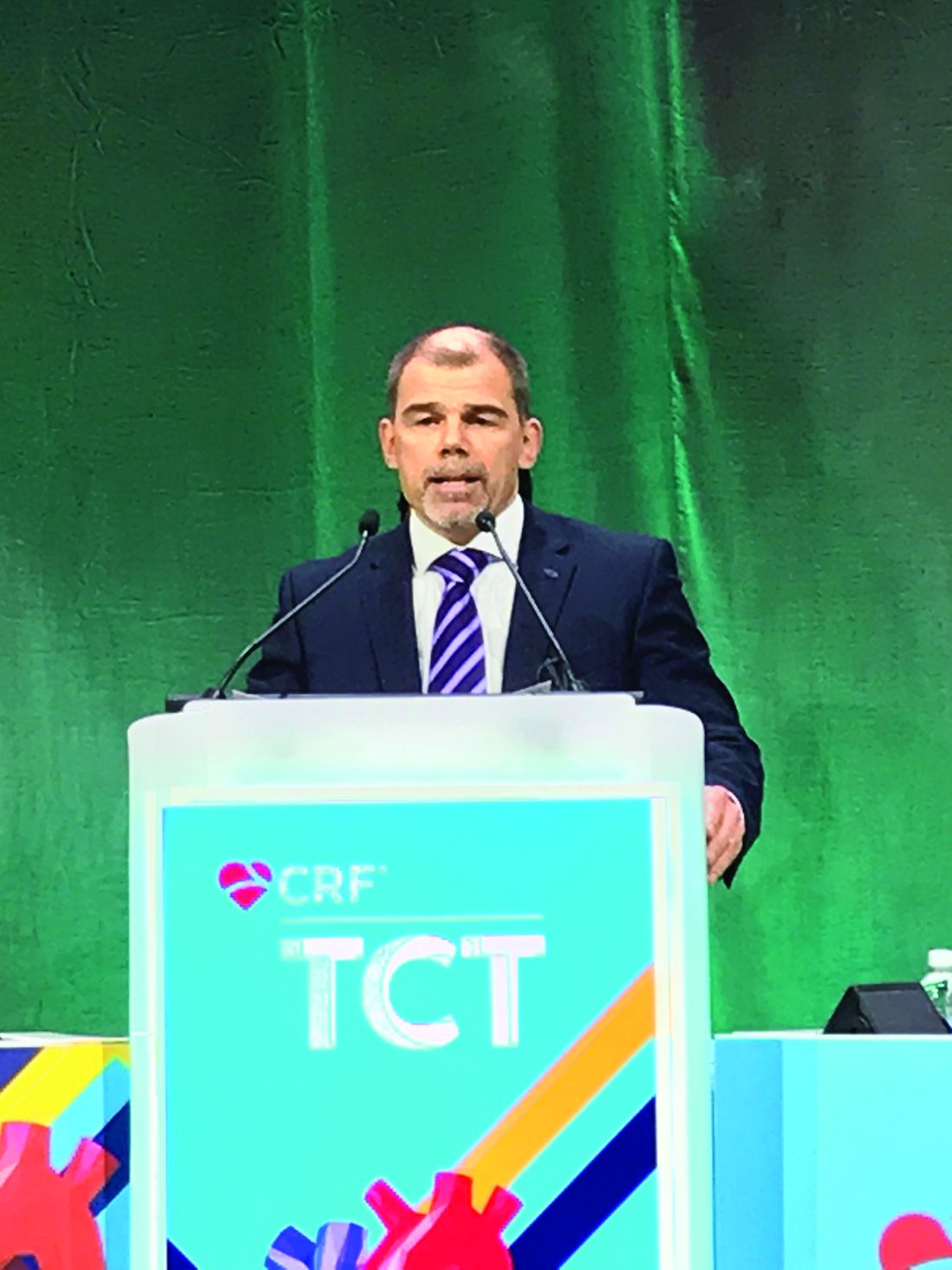
“We found that the post-PCI FFR had prognostic value both for the vessel and for the patient,” reported Zsolt Piroth, MD, PhD, deputy head, adult cardiology, György Gottsegen Institute of Cardiology, Budapest.
In this post hoc analysis, which was not a prespecified FAME 3 substudy, the goal was to look at the prognostic value of both post-PCI FFR and intravascular ultrasound, which were recommended in the study protocol. Several studies have addressed the value of these measures previously, according to Dr. Piroth, but he said the clinical value “has remained poorly defined” despite the currently available data.
The FAME 3 trial, published in the New England Journal of Medicine, was negative. It failed to confirm the study hypothesis that FFR-guided PCI is noninferior to CABG for the outcome of major adverse cardiac events (MACE) at 12 months.
However, this multinational trial has generated a large body of data with which to explore other issues relevant to revascularization. In this analysis, the goal was to evaluate whether post-PCI FFR predicted outcomes in complex multivessel revascularizations as it has been shown previously to do in single-vessel disease.
Presented at the Transcatheter Cardiovascular Therapeutics annual meeting, the focus of this analysis was on the 461 (61%) of patients in the 757-patient PCI arm of FAME 3 who underwent post-PCI FFR. The authors also looked at the predictive value of intravascular ultrasound, even though this was performed in just 11% of this group of trial participants.
As a continuous value, each 0.1-unit change in the post-PCI FFR was found to be prognostically significant for the outcome of TVF, defined as a composite of cardiac death, target vessel myocardial infarction, and target vessel revascularization (only postprocedural events were counted in this analysis). Specifically, for each 0.1-unit increase on a univariate analysis, the risk of TVF was reduced by about one-third (hazard ratio, 0.67; P = .0165).
On a patient level, a 0.1-unit increase in lowest post-PCI FFR of any assessed vessel was also associated with the same relative risk reduction (HR, 0.65; P = .0074) in the outcomes of cardiac death, target vessel MI, or target vessel revascularization, according to Dr. Piroth. On a receiver operating characteristic curve analysis, a value of 0.88 or below was predictive of TVF.
Although several other patient characteristics were also risk predictors of TVF on univariate analysis, only renal disease and the single lowest post-PCI FFR (as a continuous variable) emerged as predictors of TVF on multivariable analysis after adjustment for key clinical parameters, Dr. Piroth reported.
The reason why post-PCI FFR was not performed in almost 40% of patients randomized to PCI is unclear, but Dr. Piroth reported that the baseline characteristics of those who were or were not assessed with FFR after their procedure did not differ to any major degree.
Despite “a trend for improved outcomes in those who underwent post-PCI FFR,” Dr. Piroth, whose substudy was published in Circulation: Cardiovascular Interventions simultaneously with his TCT presentation, acknowledged that the reasons for a potential benefit cannot be derived from this post hoc analysis.
As for the prognostic value of IVUS, any conclusions are limited by the small proportion of patients who underwent this form of imaging. Overall, IVUS imaging was associated with longer procedures and more stents and “if anything, a signal for harm” in this analysis, but Dr. Piroth cautioned against any conclusions because of the small data pool.
The prognostic value of post-PCI FFR in complex multivessel disease is supported by these data, but the analysis was not designed to determine whether post-PCI FFR has relevance to intervention.
According to J. Dawn Abbott, MD, an FFR analysis conducted to identify lesions that are candidates for treatment should not be confused with FFR for physiologically guided PCI to optimize outcomes.
Noting that post-PCI FFR was encouraged in this study but not mandated and that these FFR values did not typically or necessarily lead to a change in management, take home messages about the value of post-PCI FFR in multivessel disease remain limited, said Dr. Abbott, director of interventional cardiology fellowship training, Brown University, Providence, R.I.
“There was a trend toward improved outcomes in patients who had this measurement done, but, unfortunately, we do not have data regarding whether these patients had further interventions performed,” Dr. Piroth acknowledged.
The post-PCI FFR values were made available to the treating physicians, but Dr. Piroth reiterated that it is unknown whether the physicians considered this information actionable. Moreover, “the vast majority had a nonsignificant post-PCI FFR” result, and “all of the patients had an angiographically successful PCI,” Dr. Piroth added.
Dr. Piroth has financial relationships with Abbott Vascular and Boston Scientific. Dr. Abbott reports financial relationships with Abbott Vascular, Boston Scientific, Medtronic, Microport, Philips, Penumbra, Recor, and Shockwave.
Risk by FFR is continuous variable
Risk by FFR is continuous variable
In a new analysis of the previously published FAME 3 trial, which compared fractional flow reserve–guided percutaneous coronary interventions to coronary artery bypass surgery (CABG) in patients with three-vessel disease, post-PCI FFR was shown to predict both target vessel failure (TVF) and risk of cardiac events.
“We found that the post-PCI FFR had prognostic value both for the vessel and for the patient,” reported Zsolt Piroth, MD, PhD, deputy head, adult cardiology, György Gottsegen Institute of Cardiology, Budapest.
In this post hoc analysis, which was not a prespecified FAME 3 substudy, the goal was to look at the prognostic value of both post-PCI FFR and intravascular ultrasound, which were recommended in the study protocol. Several studies have addressed the value of these measures previously, according to Dr. Piroth, but he said the clinical value “has remained poorly defined” despite the currently available data.
The FAME 3 trial, published in the New England Journal of Medicine, was negative. It failed to confirm the study hypothesis that FFR-guided PCI is noninferior to CABG for the outcome of major adverse cardiac events (MACE) at 12 months.
However, this multinational trial has generated a large body of data with which to explore other issues relevant to revascularization. In this analysis, the goal was to evaluate whether post-PCI FFR predicted outcomes in complex multivessel revascularizations as it has been shown previously to do in single-vessel disease.
Presented at the Transcatheter Cardiovascular Therapeutics annual meeting, the focus of this analysis was on the 461 (61%) of patients in the 757-patient PCI arm of FAME 3 who underwent post-PCI FFR. The authors also looked at the predictive value of intravascular ultrasound, even though this was performed in just 11% of this group of trial participants.
As a continuous value, each 0.1-unit change in the post-PCI FFR was found to be prognostically significant for the outcome of TVF, defined as a composite of cardiac death, target vessel myocardial infarction, and target vessel revascularization (only postprocedural events were counted in this analysis). Specifically, for each 0.1-unit increase on a univariate analysis, the risk of TVF was reduced by about one-third (hazard ratio, 0.67; P = .0165).
On a patient level, a 0.1-unit increase in lowest post-PCI FFR of any assessed vessel was also associated with the same relative risk reduction (HR, 0.65; P = .0074) in the outcomes of cardiac death, target vessel MI, or target vessel revascularization, according to Dr. Piroth. On a receiver operating characteristic curve analysis, a value of 0.88 or below was predictive of TVF.
Although several other patient characteristics were also risk predictors of TVF on univariate analysis, only renal disease and the single lowest post-PCI FFR (as a continuous variable) emerged as predictors of TVF on multivariable analysis after adjustment for key clinical parameters, Dr. Piroth reported.
The reason why post-PCI FFR was not performed in almost 40% of patients randomized to PCI is unclear, but Dr. Piroth reported that the baseline characteristics of those who were or were not assessed with FFR after their procedure did not differ to any major degree.
Despite “a trend for improved outcomes in those who underwent post-PCI FFR,” Dr. Piroth, whose substudy was published in Circulation: Cardiovascular Interventions simultaneously with his TCT presentation, acknowledged that the reasons for a potential benefit cannot be derived from this post hoc analysis.
As for the prognostic value of IVUS, any conclusions are limited by the small proportion of patients who underwent this form of imaging. Overall, IVUS imaging was associated with longer procedures and more stents and “if anything, a signal for harm” in this analysis, but Dr. Piroth cautioned against any conclusions because of the small data pool.
The prognostic value of post-PCI FFR in complex multivessel disease is supported by these data, but the analysis was not designed to determine whether post-PCI FFR has relevance to intervention.
According to J. Dawn Abbott, MD, an FFR analysis conducted to identify lesions that are candidates for treatment should not be confused with FFR for physiologically guided PCI to optimize outcomes.
Noting that post-PCI FFR was encouraged in this study but not mandated and that these FFR values did not typically or necessarily lead to a change in management, take home messages about the value of post-PCI FFR in multivessel disease remain limited, said Dr. Abbott, director of interventional cardiology fellowship training, Brown University, Providence, R.I.
“There was a trend toward improved outcomes in patients who had this measurement done, but, unfortunately, we do not have data regarding whether these patients had further interventions performed,” Dr. Piroth acknowledged.
The post-PCI FFR values were made available to the treating physicians, but Dr. Piroth reiterated that it is unknown whether the physicians considered this information actionable. Moreover, “the vast majority had a nonsignificant post-PCI FFR” result, and “all of the patients had an angiographically successful PCI,” Dr. Piroth added.
Dr. Piroth has financial relationships with Abbott Vascular and Boston Scientific. Dr. Abbott reports financial relationships with Abbott Vascular, Boston Scientific, Medtronic, Microport, Philips, Penumbra, Recor, and Shockwave.
FROM TCT 2022
Toward a new open-door model for psychiatric wards
If isolated, patients with mental disorders may end up having higher levels of social impairment. This has led several hospitals in Spain to set up open-door departments that are more accessible.
The purpose of the open-door model is to help remove the stigma from individuals who need to be admitted to a psychiatric ward because they have a mental disorder.
Traditional locked wards
According to the World Health Organization (WHO), in 2019, one in every eight people were living with a mental disorder. Having the least restrictive type of mental health care is one of the 10 basic principles listed in a 1996 reference document from the WHO.
Among people suffering from severe psychiatric disorders, there is a high probability of being involuntarily admitted to a psychiatry ward with locked doors (PWLD). Admission to a PWLD involves the application of a set of measures that restrict the individual’s freedom.
The main argument for keeping the doors locked is that it prevents suicides and self-harm behavior, as well as abscondment. But in recent years, efforts have been made to apply a model called open-door policy psychiatry wards (ODPWs).
Open wards model
Experiments were undertaken in various countries, including the United Kingdom, Australia, Switzerland, and Germany. Investigators found that the new forms of hospitalization led to a reduction in conflictive events; self-harm behavior; restrictive measures, such as seclusion, mechanical restraints, and chemical restraints; as well as forced medication. On the basis of these findings, ODPWs were launched.
According to Ignacio García Cabeza, MD, psychiatrist and coordinator of the department of psychiatry at Gregorio Marañón General University Hospital in Madrid, “The open wards model is founded on the idea of respecting the patient and their autonomy. In addition, it advocates a reduction in coercive measures.
“We wanted our department to be the same as the other departments in the hospital, with patients going in and out, receiving treatment, and being able to have family visits,” he explained. “A patient’s diagnosis should not factor into these things. People with schizophrenia, people with any type of mental disorder, should be able to enjoy this minimally restrictive environment.”
This model also implies fundamental changes in the interaction between health care professionals and patients. The implementation of new nursing care models, among which the Safewards model stands out, is a key element for the success of the project.
Based on a set of tools for preventing and managing conflict, the Safewards model seeks to modify the factors that regulate the relationship between staff and patients. Use of this model brought about a 15% reduction in the rate of conflictive events and a 23% reduction in the rate of coercive interventions, in comparison with a control group.
One of the major debates is about whether every patient should be able to choose this open-door system. For Dr. García Cabeza, the answer is yes, but with one caveat. “There’s a certain group of patients who perhaps need to be in locked wards, who perhaps require greater means of control – patients whose conditions put them at a high risk of suicide or of self-harm behavior or of absconding.”
He had no hesitation in saying that an open-door ward increases the patient’s self-esteem. It helps promote autonomy and a sense of control and of normalcy with respect to a community. “The idea is to get to the point where we’ve got an atmosphere, a climate, that serves to benefit the therapeutic actions that are going to continue to influence the patient’s future progress and their treatment.”
That’s why it’s important to bring about the kind of health care activities that can prevent the patient from experiencing some of the negative psychological effects, such as distrust and feeling removed from normalcy. “In traditional locked wards, the patient feels incapable of making decisions. They feel that they have very little to do with [and have] no say in the decisions made, and a lot of times, this leads to a situation where, after discharge, the patient ends up giving up on the treatments. If we can manage to break this perception held by the patient,” Dr. García Cabeza suggested, “it’s quite likely that we’ll manage to improve the course of their disorder in general.”
What the literature says
The effect of ODPW has been investigated through comparative studies with PWLD and research of the transition from PWLD to ODPW, both from a therapeutic and safety a point of view.
A 15-year observational study published in The Lancet Psychiatry found that, with respect to abscondment, suicide attempts, and suicide, there were no significant differences between hospitals with open-door policies and those without.
A subsequent study that was published in 2017 found that on open wards, any aggressive behavior and restraint or seclusion were less likely than on closed wards.
The Spanish situation
This system is already at work in some Spanish hospitals, among them Inca Comarcal Hospital (Palma de Mallorca), Elda General University Hospital (Alicante), Germans Trias i Pujol Hospital in Badalona, and Gregorio Marañón General University Hospital in Madrid.
“At Gregorio Marañón, we started the experiment just before the pandemic hit. We’re up and running now, but still with some limitations; the patient can go in and out, but not with the flexibility we’d like,” explained Dr. García Cabeza. “An open ward plays a clinical, patient-care role and a symbolic one as well. Locking the doors has a lot to do with the fear felt toward these patients. It’s a stigma that they’ve had to deal with and that they continue to have to deal with. In terms of the symbolic role, there’s also the fear that comes with giving these patients some rights.”
While the experiment at Gregorio Marañón’s psychiatric ward “is still very much in the early stages,” there have been no recorded incidents related to its open-door policy. Dr. García Cabeza is aware of the challenges of such a policy, “starting with assistance when conflictive events arise. Challenges faced by the staff – especially the nursing staff, as they’re the ones who are with the patients 24 hours day – and challenges faced by those in charge of providing care. In all of this, there are new things to learn and be aware of, new ways of understanding and looking at the patient-physician relationship. The fears are still there – they haven’t been done away with. But the way we conduct ourselves should be adjusted, matching how we act toward other patients. Although the differences have to be taken into account, we have to try to normalize, as much as possible, the environment where patients with mental disorders receive treatment.”
Dr. García Cabeza has no doubts. “The most sensible and reasonable decisions need to be made at these sites so as to allow the broadest applicability to cases. Anyone who needs psychiatric hospitalization and who is competent to consent to admission and who voluntarily agrees to be admitted – they can and must be placed in an open ward.”
The hope is that in the future, the number of open wards will increase and the number of locked wards – which have more stigma attached to them – will go down. The involvement of the staff and appropriate institutional support are essential to making this a reality.
This article was translated from Univadis Spain.
Complete endoscopic healing key when stopping anti-TNFs in IBD
The level of remission in patients with remitting inflammatory bowel disease (IBD) appears to play a major role in whether they will relapse after treatment when biologic therapies are discontinued, according to a new prospective study.
Patients with complete endoscopic healing have half the rate of relapse after withdrawal of anti-tumor necrosis factor alpha (anti-TNF) treatment than those with only partial healing, according to a study published online in Clinical Gastroenterology and Hepatology.
“Applying strict criteria for endoscopic healing and mesalamine treatment ... may lower the risk of relapse after withdrawal of anti-TNF treatment,” write Bas Oldenburg, MD, PhD, a professor at University Medical Center Utrecht, the Netherlands, and colleagues in their analysis of 81 patients.
De-escalation of anti-TNF treatment in IBD patients in remission has the potential to “reduce side effects, including risks of serious infections and malignancies, decrease health care expenditures, and meet patients’ preferences,” they note.
However, withdrawal of the drugs increases the risk of relapse by 30%-45% at 12 months. When patients relapse, reintroduction of anti-TNF therapy returns over 80% to remission.
Although no consensus exists on how to select patients for therapy de-escalation, evidence suggests that persistent inflammation affects outcomes and that the “depth” of endoscopic healing is a key indicator, the authors note.
Study details
To further the knowledge base, they conducted a prospective study of patients in remission to determine the relapse rate following de-escalation of anti-TNF therapy; evaluate relapse factors, including degree of endoscopic healing; and assess outcomes after reintroduction of anti-TNF therapy.
The study was limited to adult patients with IBD with at least 6 months of corticosteroid-free clinical remission, confirmed baseline clinical remission and endoscopic healing, no current hospitalization, and no pregnancy.
The patients underwent elective discontinuation of anti-TNF therapy between 2018 and 2020. The recommended protocol was to measure C-reactive protein (CRP) and fecal calprotectin at 3, 6, 12, and 24 months and to perform endoscopy at 12 months.
Patients also completed questionnaires at baseline and at 3, 6, 12, and 24 months. The authors selected the patient–Harvey-Bradshaw Index for patients with Crohn’s disease and the patient–Simple Clinical Colitis Activity Index for patients with ulcerative colitis and unclassified IBD, as well as the short IBD Quality of Life measure.
Of the 81 patients from 13 centers who took part, 51% had Crohn’s disease. The median duration of remission at baseline was 3.5 years, and the median disease duration was 9.1 years.
All patients had evidence of endoscopic healing, and 88% met the strict criteria for complete endoscopic healing. In 34%, trough levels of anti-TNF treatments were judged to be subtherapeutic.
After withdrawal of the drugs, 25.9% of patients continued on immunomodulators.
Over a median follow-up of 2 years, 49% of patients relapsed, which was confirmed via endoscopy, fecal calprotectin, or CRP in 83% of cases and inferred from treatment escalation for clinical flare in 17%. Rates of relapse were comparable between patients with Crohn’s disease and ulcerative colitis or unclassified IBD and between those discontinuing adalimumab and those stopping infliximab.
Better healing, better outcomes
However, analysis showed that partial endoscopic healing was independently associated with a higher risk of relapse, at an adjusted hazard ratio versus complete endoscopic healing of 3.28.
At 12 months, 70% of patients with partial endoscopic healing had relapsed versus 35% of those with complete endoscopic healing.
Treatment with the anti-inflammatory agent mesalamine (multiple brands) was independently associated with a reduced risk of relapse, at an adjusted hazard ratio of 0.08. No other potential predictors of relapse were identified.
Of the patients who relapsed, 75% restarted anti-TNF treatment, and the majority (87%) were restarted on the same agent at a median of 0.9 years since its withdrawal and a median of 24 days since the onset of relapse.
Clinical remission was achieved at 3 months in 73% of patients who restarted anti-TNF therapy, which was found to restore quality of life and well-being in relapsed patients, the authors report.
Reluctance remains
Stephen B. Hanauer, MD, professor of medicine (gastroenterology and hepatology) at Northwestern University Feinberg School of Medicine, Chicago, said the findings “reinforce the benefits of the maintenance versus the withdrawal of therapy” and “the deeper the remission” the more likely it is to be sustained.
The 35% relapse rate at 12 months, even in patients with compete endoscopic healing, indicates that treatment should be maintained, Dr. Hanauer said.
“What is also relevant, but was not evaluated, is the additional endpoint of histologic healing, which is likely to sustain remissions even longer,” he added.
Nevertheless, Dr. Hanauer said, the “observed relapse rate is important to discuss in shared decision-making with patients.”
The findings are interesting, but the study didn’t follow the patients for long enough to understand why 35% of those with complete endoscopic healing relapsed, Miguel Regueiro, MD, chair of the Cleveland Clinic’s Digestive Disease & Surgery Institute, told this news organization.
“Are there predictors, factors, or other treatments that could be used to reduce that 35% risk of relapse further?” he questioned.
Although the study didn’t clear up that question for Dr. Regueiro, he found it compelling that mesalamine continuation resulted in higher rates of sustained remission after anti-TNF withdrawal among patients with ulcerative colitis.
Dr. Regueiro said that he will not begin recommending withdrawal of advanced therapies, including anti-TNF drugs, in patients who have achieved a stable remission.
“We have not yet found the cure for IBD, and my concern is that patients may relapse with more severe disease than previously and that recurrence of inflammation could have potential risks for complications,” he said.
“Nonetheless, this study is intriguing and important, and at least prompts the discussion of withdrawing therapy in those who have achieved a deep endoscopic remission for a sustained period of time,” Dr. Regueiro added.
The study received support from the Dutch Health Insurance Innovation Fund.
Dr. Oldenburg declares relationships with AbbVie, Celltrion, Ferring, Takeda, Galapagos, Pfizer, Cablon, PBMS, Janssen, and MSD. Other authors also declare numerous relationships. The full list can be found with the original article. Dr. Hanauer declares relationships with AbbVie, Janssen, Pfizer, and Boehringer Ingelheim. Dr. Regueiro declares relationships with AbbVie, Janssen, UCB, Takeda, Pfizer, Miraca Labs, Amgen, Celgene, Seres, Allergan, Genentech, Gilead, Salix, Prometheus, Lilly, TARGET Pharma Solutions, ALFASIGMA, S.p.A., BMS, CME Outfitters, Imedex, GI Health Foundation, Cornerstones, Remedy, MJH Life Sciences, Medscape, MDEducation, WebMD, and HMPGlobal.
A version of this article first appeared on Medscape.com.
Mindfulness eases asthma burden
Adults with asthma who received mindfulness training showed significant improvement in symptoms compared to those who did not receive such training, based on data from 73 individuals.
Although previous research shows the contribution of psychological factors to poor asthma control and exacerbations, the ability of mindfulness-based stress reduction (MBSR) to improve asthma symptoms in particular has not been well studied, wrote Estelle T. Higgins, BA, of the University of Wisconsin, Madison, and colleagues.
they wrote. The researchers hypothesized that MBSR training would reduce the effect of psychological distress on asthma control and inflammation compared to asthma patients in a waitlist control group.
In a study published in Brain, Behavior, & Immunity – Health, the researchers randomized 38 adults with asthma to a program of MBSR and 24 to a waitlist. The participants ranged in age from 18 to 65 years, with a mean of 38.1 years, and 43 were female. All patients had an asthma diagnosis for at least 6 months; airway inflammation was based on measures of fraction of exhaled nitric oxide (FeNO) ≥ 30 ppb, 138 blood eosinophil count ≥ 150 cells/mcL, or percent sputum eosinophils ≥ 2% of total leukocytes. Individuals with ongoing medical conditions other than asthma were excluded.
The MBSR group had seven clinical data collection visits at approximately 1-month intervals. MBSR training sessions occurred within classes offered to the community over a period of 8 weekly sessions and one 6-hour retreat, and included breath-focused attention, body scan, and mindful awareness in seated positions, walking, and yoga. Participants completed questionnaires about mindfulness, distress, depression, and anxiety symptoms. These were assessed at baseline, post intervention, and at study completion. Chronic stress level was determined at baseline only.
The primary outcome was asthma control based on the Asthma Control Questionnaire 6-item version (ACQ6) Minimally Important Difference.
Overall, asthma control improved significantly among those randomized to MBSR compared to waitlisted controls (P = .01) and this difference persisted at 4 months after the intervention.
Nearly one-third (32%) of the MBSR participants met the criteria for clinically significant improvement in asthma symptoms, compared to 12% of those on the wait list.
In addition, MBSR-related improvement in asthma control was significantly associated with a reduced distress (P = .043), and was especially effective for individuals with the highest levels of depressive symptoms at baseline, the researchers noted. Individuals who received MBSR also showed significantly reduced levels of exhaled nitric oxide compared to waitlist controls (P < .05).
The study findings were limited by the lack of an active control group, the researchers noted. “Though a wait-list control group was employed to control for variation in outcome measures over time, it is possible that effects reported here were driven by factors that are not specific to training in mindfulness, such as social support or expectancy effects,” they wrote. However, the results support the value of mindfulness in reducing psychological stress, FeNO, and impairments related to asthma that were sustained over time, they said. The results also support the potential of MBSR to both augment and reduce the need for pharmacological treatment in asthma, and mindfulness may be an effective way to improve overall disease control by reducing the contribution of psychological factors to asthma morbidity, they concluded.
The study was supported by the National Center for Complementary and Integrative Health. Coauthor Richard J. Davidson, MD, is the founder and president, and serves on the board of directors for the nonprofit organization, Healthy Minds Innovations Inc. The researchers had no financial conflicts to disclose.
FROM BRAIN, BEHAVIOR, & IMMUNITY – HEALTH
How to make the best of your worst reviews
I have a love-hate relationship with patient reviews and satisfaction scores. I love good reviews and hate bad ones. Actually, I skim good reviews and then dwell for days on the negative ones, trying to rack my brain as to what I did wrong. Like everyone else, I have off days when I’m tired or distracted or just overwhelmed. Though I try to bring my A game to every patient visit, realistically, I know that I don’t always achieve this. But, for me, the difference in my best visits and good-enough visits is the difference between a 4-star and 5-star review. What’s up with those 1-star reviews?
Many, many years ago, when patient satisfaction scores were in their infancy, our clinic rewarded any physician who got a 100% satisfaction score. On the surface that makes perfect sense – of course, our patients should be satisfied 100% of the time, right? When I asked one of the winners of this competition how he did it (I never scored 100%), he told me, “I just do whatever the patient wants me to do.” Yikes, I thought at the time. That may be the recipe for an A+ for patient satisfaction but not for quality or outcomes.
Sometimes, I know that they are going to be unhappy, such as when I decline to refill their drug of abuse. Other times, I have to exercise my best medical judgment and hope that my explanation does not alienate the patient. After all, when I say “no” to a patient request, it is with their overall health and well-being in mind. But I would be lying if I said that I have matured to the point that I’m not bothered by a negative review or a patient choosing to take their care elsewhere.
Most of us seek and welcome feedback. Over time, I’ve learned to do this during the visit by asking “Am I giving you too much information?” or “What do you think of the plan?” or “What’s most important to you?” There are times when I conclude the visit and know that the patient is not satisfied but remain unable to ferret out where I let them down – even, on occasion, when I ask them directly. Ideally, any feedback we get from our patients, positive or negative, would be specific and actionable. It rarely is.
There is no doubt we have entered the era of consumer medicine. Everything from the physical appearance of our clinics to the response time to electronic messages is fair game in how patients judge us. As we all know, patients assume competence – they are not usually impressed by your training or quality outcomes because they already believe you are clinically competent (or arguably they’d never set foot in your office). Instead of judging us how we often judge ourselves, patients form opinions about us by how we enter the room, whether we sit or stand, how long they wait in the exam room before we come in, or whether they like the nurse with whom we work. So many subtle things – many of which are outside of our control.
I often struggle with staying on time. When I am invariably walking into the room late, I make a point of thanking patients for their patience. When I’m very late, I offer a more detailed, HIPAA-compliant explanation. What I wish my patients saw was that I am often accommodating a patient who arrives late for their appointment or who wants me to address every concern they’ve had for the past 5 years. While I aspire to not allow the patient’s perception of the visit to unduly influence how I handle the visit, it inevitably does. I do want to have patients who are happy with their experience.
One of my friends is enviously pragmatic in her view on patient experience. “I’m not their friend and they don’t have to like me.” She emphasizes the clinical care she is providing and does not allow patients who are upset with some aspect of the care to weigh heavy on her. It may be that specialists are more likely to enjoy the luxury of putting aside how patients feel about them personally. In primary care, the patient-physician relationship is so central to what we do that ignoring your “likability” has the potential to threaten your professional viability.
I conclude this blog much like I started it. My desire is to allow the negative reviews, particularly if they have nothing actionable in them, to roll off my back and to keep my focus on the clinical care that I am providing. In actuality, I care deeply about how my patients experience their visit with me and will likely continue to take my reviews to heart.
Dr. Frank is a family physician in Neenah, Wisc. She disclosed no relevant conflicts of interest.
A version of this article first appeared on Medscape.com.
Clinic and doc must pay millions after faulty hernia surgery
reports a story in the Bowling Green Daily News.
On May 31, 2013, Alice Duff went to Graves-Gilbert Clinic, a multispecialty group whose main campus is in Bowling Green, for hernia surgery. Her surgeon for the procedure at a nearby facility was Tage Haase, MD, a member of the clinic.
Originally, Mrs. Duff was expected to remain in the facility for 23 hours following the procedure. But her recovery didn’t proceed as expected, and that initial period was extended by roughly 3 days. During this time, Ms. Duff’s husband, Lloyd, allegedly requested multiple times that doctors order a CT scan for his wife.
Ten days after the procedure, and with Ms. Duff out of the hospital, the Duffs successfully urged their family physician to order a CT scan. It showed large amounts of free air in Alice’s abdomen, a condition that’s known as pneumoperitoneum.
On June 10, 2013, Mrs. Duff underwent a second surgery, during which doctors discovered that she had sustained a perforated bowel during the first procedure. As a result, her bowel contents had spilled into her abdomen, causing an infection that required an extended hospital stay and five additional surgeries. The infection also led to retinal damage that has left her legally blind.
In their claim against Graves-Gilbert and Dr. Haase, the Duffs argued that Dr. Haase and an assistant doctor who was not named in the suit had failed to meet the standard of care. Specifically, argued the Duffs, because bowel perforation is a known complication in hernia surgeries, Dr. Haase was negligent in not diagnosing and treating Ms. Duff’s problem earlier.
For their part, Graves-Gilbert and Dr. Haase maintained that there was no indication before the second surgery that Mrs. Duff was demonstrating symptoms that necessitated a follow-up procedure. Dr. Haase further argued that Mrs. Duff’s bowel perforation was probably caused by the sawblade-like effect of the suture material he had used to close her incision.
The jury didn’t see it this way, however. It awarded approximately $1.3 million to Mrs. Duff for past medical expenses, plus another $12 million for pain and suffering. Her husband, Lloyd, received an additional $8 million in damages.
The attorney representing the clinic and Dr. Haase has vowed to pursue “all available remedies to have the verdict vacated and the case set for a new trial.”
Case hinged on proper use of a ‘power morcellator’
A claim for punitive damages has been thrown out against a New Jersey doctor and the hospital he’s affiliated with, in a ruling that could help to clarify the standards for such damages in medical malpractice cases, according to a story first reported on NorthJersey.com.
In October 2014, Howard H. Jones, MD, director of minimally invasive gynecologic surgery at The Valley Hospital, in Ridgewood, N.J., treated a patient from nearby Nyack, N.Y., for uterine fibroid tumors. As part of that treatment at the hospital, Dr. Jones used a “laparoscopic power morcellator,” which during a myomectomy procedure cuts, or morcellates, fibroid tumors into pieces small enough to be removed through an incision that’s generally 2 cm or fewer.
While use of the device offers doctors an alternative to open surgery and its longer recovery times, it also risks spreading previously undiagnosed cancer cells throughout the abdomen, thereby shortening a patient’s life. Because of this risk, which has given rise to a number of malpractice cases around the country, the Food and Drug Administration issued a draft guidance in the spring of 2022 for the use of power morcellators.
One recommendation is that physicians employ a “compatible” containment system to catch morcellated tissue, including any with cancer cells. Another is that the device be used selectively, which is to say on patients who have a minimal cancer risk and who have been informed of the procedure’s possible side effects beforehand.
Within a month of the procedure, the patient was diagnosed with metastatic leiomyosarcoma and died in September 2015. Following her death, her sister, the executor of her estate, sued both The Valley Hospital and Dr. Jones. In her suit, the sister argued that the defendants knew, or should have known, the risks involved in using a power morcellator because of both an earlier (2014) FDA “safety communication” discouraging the use of the device and the death of another of Dr. Jones’ patients following a similar procedure the year before.
The suit further alleged that, even after the FDA had issued its safety caution, the hospital had used the device on 37 other patients, “without informing them of the [FDA] letter or obtaining their informed consent to use the device.”
In light of these alleged lapses, Mirian Rivera, the patient’s sister, sought both compensatory and punitive damages. Historically, punitive damages have been limited to the small number of med-mal cases where a doctor or hospital has been found to have acted with actual malice or “wanton and willful disregard.”
Both Valley and Dr. Jones strongly disagreed with Ms. Rivera’s claim, arguing that prior to the procedure Dr. Jones had in fact met with the patient several times and had conducted the proper cancer-detecting tests. Moreover, the defendants emphasized, the request for punitive damages in the absence of actual malice or other factors would almost certainly establish a dangerous legal precedent. Several industry groups – including the American Medical Association, the Medical Society of New Jersey, and the New Jersey Hospital Association – agreed and filed friend-of-court briefs in support of Valley Hospital and Dr. Jones.
But two lower courts refused to dismiss the plaintiff’s claims for punitive damages. That’s when attorneys for Valley and Dr. Jones appealed to the New Jersey Supreme Court. (Claims against the device manufacturer, a German company, had already been resolved.)
Ruling unanimously, the high court sided with the defendants: “As a matter of law, the evidence presented, even affording plaintiffs all favorable inferences, does not establish that defendants’ acts or omissions were motivated by actual malice or accompanied by wanton and willful disregard for the patient’s health and safety.”
The court also found that Valley had in fact reviewed hospital policy and drafted a patient-consent form after the release of the 2014 FDA safety communication on power morcellators. (The consent form had not been adopted before the surgery in question, however.)
The suit will now go back to Superior Court in Bergen County, New Jersey; unless a settlement is reached beforehand, the jury will weigh claims of negligence and compensatory damages.
At press time, no trial date had been set.
Will med-mal cases get tougher to defend in this state?
Late in August, the Pennsylvania Supreme Court reversed its own longstanding rule that required that medical malpractice cases be filed in the county where the alleged injury occurred, as an Associated Press story on NBCPhiladelphia.com, among other news sites, reports.
More than 2 decades ago, in response to what was then seen as a crisis in the med-mal system, the state legislature overwhelmingly passed MCARE (the Medical Care Availability and Reduction of Error Fund), which among other things restricted the venue of medical suits. The legislation was signed into law in March 2002 by then-Gov. Mark Schweiker.
The following year, the state’s high court adopted a similar venue rule.
Over the years, doctor and hospital groups have been big supporters of the rule, arguing that any attempt to shift cases back to allegedly more plaintiff-friendly courts in Philadelphia and other cities would likely retrigger a crisis of higher med-mal premiums, doctor flight, and worse health care.
But a 2020 report by Pennsylvania’s nonpartisan Legislative Budget and Finance Committee took issue with these conclusions. It said that, following a national trend, the cost of medical professional liability insurance had fallen in the state since 2007. The report concluded that nothing in the available data supports the “conclusion that changes in the availability, cost, and affordability of medical professional liability insurance are the result of changes in Pennsylvania law.”
A more recent report by the high court’s Civil Procedural Rules Committee reached a similar conclusion, noting that med-mal cases should be subject to the same rules as any other type of civil litigation. A majority of the high court agreed.
Predictably, this decision sits well with patient groups and officials representing trial attorneys in the Keystone State.
“Cases should be heard before 12 jurors that do not have a connection to a hospital or surgical center that is often times the largest employer in the county,” said Kila Baldwin, president of the Pennsylvania Association for Justice. “The new rule levels the playing field and will improve access to justice for all Pennsylvanians.”
Doctors, hospitals, and other health care providers, however, have predicted a “ruinous path” ahead.
A version of this article first appeared on Medscape.com.
Visual impairment more common in minority youth
Race, ethnicity, and socioeconomic status are closely associated with visual impairment in adolescents in the United States, researchers have found.
The study showed that adolescents who identified as Black, Mexican-American, of low income, or as non–U.S. citizens were two to three times more likely than White adolescents to report vision problems and to perform worse on objective tests of visual acuity.
Although disparities in visual impairment with respect to ethnic, racial, and socioeconomic status have been described among adults, little research has explored the adolescent population, according to the researchers, whose findings appear in JAMA Ophthalmology.
“The primary motivation behind trying to figure out exactly when these disparities emerge is that it gives us an opportunity to come up with ideas for interventions that can potentially address them before they manifest in a way that they are no longer treatable,” said Isdin Oke, MD, an instructor in ophthalmology at Harvard Medical School, Boston, who led the latest study.
Dr. Oke and his colleagues analyzed the records of 2,833 children and adolescents aged 12 through 18 years (mean, 15.5 years; 49% female) in the National Health and Nutrition Examination Survey. All the participants had completed a visual function questionnaire and had undergone an eye examination. The primary outcomes of the study were subjective (self-reported) poor vision and objective measures of visual function (visual acuity worse than 20/40 in the better-seeing eye).
Of the study participants, 14% were non-Hispanic Black, 11% were Mexican-American, 63% were non-Hispanic White, and 11% were of other race and ethnicity. Five percent of participants were not U.S. citizens, and 19% had a family income below the poverty threshold.
After accounting for potential confounders, self-reported poor vision was more common among Black (odds ratio, 2.85; 95% CI, 2.00-4.05; P < .001), Mexican-American (OR, 2.83; 95% CI, 1.70-4.73; P < .001), and low-income youth (OR, 2.44; 95% CI, 1.63-3.65; P < .001), the researchers report.
The study also found increased odds of visual acuity below 20/40 in the better-seeing eye among Black (OR, 2.13; 95% CI, 1.41-3.24; P = .001) and Mexican-American adolescents (OR, 2.13; 95% CI, 1.39-3.26; P = .001) and non–U.S. citizens (OR, 1.96; 95% CI, 1.10-3.49; P = .02).
Black and Mexican-American adolescents were almost three times more likely to suffer poor subjective visual function and twice as likely to have low objective visual acuity than non-Hispanic White youth, according to the researchers.
“I think it’s something that health care providers, and even the population as a whole, should be more aware of,” Dr. Oke said.
Opportunities for intervention
Dr. Oke said the findings likely reflect the underlying inequities in access to vision care experienced by these persons.
“There are a lot of opportunities for early intervention, whether it’s through vision screening or improving access to vision services for children and adolescents that can really make a big difference over the long term,” he told this news organization.
Michael F. Chiang, MD, director of the National Eye Institute at the National Institutes of Health, Bethesda, Md., agreed that more steps need to be taken to improve access to vision care for all Americans.
“There are good data showing that we don’t have a sufficient number of eye care providers and health-services clinical researchers who come from backgrounds that are currently underrepresented in medicine and science,” Dr. Chiang said. “Therefore, we need to find ways to inspire, recruit, and train a larger number of people to strengthen our vision workforce.”
Other ways to address these gaps in care, he added, include improving understanding of the social determinants of vision health as well as developing ways to improve access to eye care.
“That may include new models of eye care, like telehealth to improve outreach, and will likely need to include what we call implementation science, so clinicians can better adopt these measures,” he said.
Dr. Oke and Dr. Chiang reported no relevant financial relationships.
A version of this article first appeared on Medscape.com.
Urgent need for research into psychedelic therapy for older adults
new research shows.
“Geriatric psychiatrists and others caring for older adults are interested in how much is known about psychedelic use in older adults,” study investigator C. Bree Johnston, MD, MPH, University of Arizona, Tucson, told this news organization.
“A major concern is how safe psychedelic-assisted therapy is for patients with heart disease, hypertension, neurological disorders, and multimorbidity,” Dr. Johnston said.
The study is published online in the American Journal of Geriatric Psychiatry.
‘Groundswell’ of research
The past few years have brought a “groundswell” of interest and promising research into the potential therapeutic benefit of psychedelic-assisted therapy for a variety of conditions affecting adults, the researchers noted.
They include psilocybin-assisted therapy for the distress associated with a terminal diagnosis, depression, and addiction, and MDMA-assisted therapy for PTSD.
However, in most studies, psychedelic therapy has been tested in relatively young healthy adults, raising the question of how generalizable the study results are for the patients that most geropsychiatrists will be treating, the investigators noted.
They reviewed “the most important” research studies on psilocybin- and MDMA-assisted therapies published over the past 2 decades that are likely to be relevant for geriatric psychiatrists and other professionals caring for older adults.
The researchers point out that psychedelics and related compounds have shown efficacy for the treatment of a number of conditions that are common among older adults, including mood disorders, distress associated with a serious medical illness, PTSD, substance use problems, and prolonged grief.
Psychedelics also have properties that may provide for cognitive impairment and dementia and promote personal growth among healthy older adults.
Research has shown that psychedelics can be safely administered to healthy adults in controlled conditions.
However, both psilocybin and MDMA can increase blood pressure and heart rate, which could be a concern if used in older adults with cardiovascular disease, the investigators noted.
“Healthy older adults are likely to face similar risks when undergoing psychedelic-assisted therapy as healthy younger adults,” said Dr. Johnston.
“In carefully selected adults, those risks appear to be minor when psychedelics are administered in controlled conditions under the guidance of a skilled therapist,” she added.
Given the potential of psychedelic compounds to benefit older adults, the authors call for more research to establish the safety and efficacy among older adults, particularly those with multiple comorbidities.
Pressing knowledge gaps
The exclusion of older adults from clinical trials of novel treatments is “one of contemporary psychiatry’s more pressing problems – one that extends beyond psychedelics,” Ipsit V. Vahia, MD, associate chief of the division of geriatric psychiatry, McLean Hospital, Belmont, Mass., who wasn’t involved in the review, told this news organization.

“Currently, there is little evidence that clinicians can lean on while considering the use of psychedelics in older adults,” Dr. Vahia said.
This paper highlights “the most pressing gaps in the evidence that bear addressing in order to develop more substantial best practices around the use of these drugs,” he added.
For example, little is known about appropriate dosing, pharmacokinetics, and pharmacodynamics of psychedelics in older adults, Dr. Vahia said.
“Their risks, particularly cardiovascular risks, are barely studied, and almost nothing is known about how these drugs may impact those in their 80s or older, or those with serious medical comorbidities who use multiple medications,” Dr. Vahia said. “The majority of the existing literature has excluded older adults, and the extremely limited evidence that does exist has been collected in relatively healthy, and relatively young (aged below 75) persons.”
Dr. Vahia noted that, before psychedelics as a class can be considered viable treatment options for a broader group of older adults, “more research is needed, particularly to establish safety.”
This research had no specific funding. Dr. Johnston and Dr. Vahia have no relevant conflicts of interest.
A version of this article first appeared on Medscape.com.
new research shows.
“Geriatric psychiatrists and others caring for older adults are interested in how much is known about psychedelic use in older adults,” study investigator C. Bree Johnston, MD, MPH, University of Arizona, Tucson, told this news organization.
“A major concern is how safe psychedelic-assisted therapy is for patients with heart disease, hypertension, neurological disorders, and multimorbidity,” Dr. Johnston said.
The study is published online in the American Journal of Geriatric Psychiatry.
‘Groundswell’ of research
The past few years have brought a “groundswell” of interest and promising research into the potential therapeutic benefit of psychedelic-assisted therapy for a variety of conditions affecting adults, the researchers noted.
They include psilocybin-assisted therapy for the distress associated with a terminal diagnosis, depression, and addiction, and MDMA-assisted therapy for PTSD.
However, in most studies, psychedelic therapy has been tested in relatively young healthy adults, raising the question of how generalizable the study results are for the patients that most geropsychiatrists will be treating, the investigators noted.
They reviewed “the most important” research studies on psilocybin- and MDMA-assisted therapies published over the past 2 decades that are likely to be relevant for geriatric psychiatrists and other professionals caring for older adults.
The researchers point out that psychedelics and related compounds have shown efficacy for the treatment of a number of conditions that are common among older adults, including mood disorders, distress associated with a serious medical illness, PTSD, substance use problems, and prolonged grief.
Psychedelics also have properties that may provide for cognitive impairment and dementia and promote personal growth among healthy older adults.
Research has shown that psychedelics can be safely administered to healthy adults in controlled conditions.
However, both psilocybin and MDMA can increase blood pressure and heart rate, which could be a concern if used in older adults with cardiovascular disease, the investigators noted.
“Healthy older adults are likely to face similar risks when undergoing psychedelic-assisted therapy as healthy younger adults,” said Dr. Johnston.
“In carefully selected adults, those risks appear to be minor when psychedelics are administered in controlled conditions under the guidance of a skilled therapist,” she added.
Given the potential of psychedelic compounds to benefit older adults, the authors call for more research to establish the safety and efficacy among older adults, particularly those with multiple comorbidities.
Pressing knowledge gaps
The exclusion of older adults from clinical trials of novel treatments is “one of contemporary psychiatry’s more pressing problems – one that extends beyond psychedelics,” Ipsit V. Vahia, MD, associate chief of the division of geriatric psychiatry, McLean Hospital, Belmont, Mass., who wasn’t involved in the review, told this news organization.
“Currently, there is little evidence that clinicians can lean on while considering the use of psychedelics in older adults,” Dr. Vahia said.
This paper highlights “the most pressing gaps in the evidence that bear addressing in order to develop more substantial best practices around the use of these drugs,” he added.
For example, little is known about appropriate dosing, pharmacokinetics, and pharmacodynamics of psychedelics in older adults, Dr. Vahia said.
“Their risks, particularly cardiovascular risks, are barely studied, and almost nothing is known about how these drugs may impact those in their 80s or older, or those with serious medical comorbidities who use multiple medications,” Dr. Vahia said. “The majority of the existing literature has excluded older adults, and the extremely limited evidence that does exist has been collected in relatively healthy, and relatively young (aged below 75) persons.”
Dr. Vahia noted that, before psychedelics as a class can be considered viable treatment options for a broader group of older adults, “more research is needed, particularly to establish safety.”
This research had no specific funding. Dr. Johnston and Dr. Vahia have no relevant conflicts of interest.
A version of this article first appeared on Medscape.com.
FROM THE AMERICAN JOURNAL OF GERIATRIC PSYCHIATRY
Is corporate telepsychiatry the solution to access to care problems?
When Sue W’s mother died in 2018, she struggled terribly. She was already seeing a psychotherapist and was taking duloxetine, prescribed by her primary care physician. But her grief was profound, and her depression became paralyzing. She needed to see a psychiatrist, and there were many available in or near her hometown, a Connecticut suburb of New York City, but neither Sue, her therapist, nor her primary care doctor could find a psychiatrist who participated with her insurance. Finally, she was given the name of a psychiatrist in Manhattan who practiced online, and she made an appointment on the Skypiatrist (a telepsychiatry group founded in 2016) website.
“I hesitated about it at first,” Sue said. “The doctor was nice, and I liked the convenience. Appointments were 15 minutes long, although the first session was longer. He focused on the medications, which was okay because I already have a therapist. And it was really easy. I made appointments on their website and I saw the doctor through the same site, and I really liked that I could send him messages.” The psychiatrist was responsive when Sue had trouble coming off duloxetine, and he gave her instructions for a slower taper. The treatment was affordable and accessible, and she got better.

When you also consider that many private-practice psychiatrists do not participate with insurance panels, online companies that do accept insurance may add value, convenience, and access.
Cerebral, the largest online psychiatric service in the country, began seeing patients in January 2020, offering medications and psychotherapy. They participate with a number of commercial insurers, and this varies by state, but not with Medicaid or Medicare. Patients pay a monthly fee, and an initial 30-minute medication evaluation session is conducted, often with a nurse practitioner. They advertise wait times of less than 7 days.
Another company, Done, offers treatment specifically for ADHD. They don’t accept insurance for appointments; patients must submit their own claims for reimbursement. Their pricing structure involves a fee of $199 for the first month, then $79 a month thereafter, which does not include medications. Hims – another online company – targets men with a variety of health issues, including mental health problems.
Some of these internet companies have been in the news recently for concerns related to quality of care and prescribing practices. A The Wall Street Journal article of March 26, 2022, quoted clinicians who had previously worked for Cerebral and Done who left because they felt pressured to see patients quickly and to prescribe stimulants. Not all of the prescribers were unhappy, however. Yina Cruz-Harris, a nurse practitioner at Done who has a doctorate in nursing practice, said that she manages 2,300 patients with ADHD for Done. Virtually all are on stimulants. She renews each patient’s monthly prescription from her New Jersey home, based mostly on online forms filled out by the patients. She’s fast, doing two renewals per minute, and Done pays her almost $10 per patient, working out to around $20,000 in monthly earnings.
In May, the Department of Justice began looking into Cerebral’s practices around controlled substances and more recently, Cerebral has been in the news for complaints from patients that they have been unable to reach their prescribers when problems arise. Some pharmacy chains have refused to fill prescriptions for controlled medications from online telehealth providers, and some online providers, including Cerebral, are no longer prescribing controlled substances. A front-page The Wall Street Journal article on Aug. 19, 2022, told the story of a man with a history of addiction who was prescribed stimulants after a brief appointment with a prescriber at Done. Family and friends in his sober house believe that the stimulants triggered a relapse, and he died of an opioid overdose.
During the early days of the pandemic, nonemergency psychiatric care was shut down and we all became virtual psychiatrists. Many of us saw new patients and prescribed controlled medications to people we had never met in real life.
“John Brown,” MD, PhD, spoke with me on the condition that I don’t use his real name or the name of the practice he left. He was hired by a traditional group practice with a multidisciplinary staff and several offices in his state. Most of the clinicians worked part time and were contractual employees, and Dr. Brown was hired to develop a specialty service. He soon learned that the practice – which participates with a number of insurance plans – was not financially stable, and it was acquired by an investment firm with no medical experience.
“They wanted everyone to work 40-hour weeks and see 14 patients a day, including 3-4 new patients, and suddenly everyone was overextended and exhausted. Overnight, most of the therapists left, and they hired nurse practitioners to replace many of the psychiatrists. People weren’t getting good care.” While this was not a telepsychiatry startup, it was a corporate takeover of a traditional practice that was unable to remain financially solvent while participating with insurance panels.
Like Sue W, Elizabeth K struggled to get treatment for ADHD even before the pandemic.
“I work multiple part-time jobs, don’t own a car, and don’t have insurance. Before telehealth became available, it was difficult and discouraging for me to maintain consistent treatment. It took me months to get initial appointments with a doctor and I live in one of the largest cities in the country.” She was pleased with the care she received by Done.
“I was pleasantly surprised by the authenticity and thoroughness of my first telehealth provider,” Elizabeth noted. “She remembered and considered more about me, my medical history, and details of my personal life than nearly every psychiatric doctor I’ve ever seen. They informed me of the long-term effects of medications and the importance of routine cardiovascular check-ups. Also, they wouldn’t prescribe more than 5 mg of Adderall (even though I had been prescribed 30-90 mg a day for most of my life) until I completed a medical check-up with blood pressure and blood test results.”
Corporate telepsychiatry may fill an important void and provide care to many people who have been unable to access traditional treatment. Something, however, has to account for the fact that care is more affordable through startups than through traditional psychiatric practices. Startups have expensive technological and infrastructure costs and added layers of administration. This translates to either higher volumes with shorter appointments, less compensation for prescribers, or both. How this will affect the future of psychiatric care remains to be seen.
Dr. Miller, is a coauthor of “Committed: The Battle Over Involuntary Psychiatric Care” (Johns Hopkins University Press, 2016). She has a private practice and is assistant professor of psychiatry and behavioral sciences at Johns Hopkins University, Baltimore. She has disclosed no relevant financial relationships. A version of this article first appeared on Medscape.com.
When Sue W’s mother died in 2018, she struggled terribly. She was already seeing a psychotherapist and was taking duloxetine, prescribed by her primary care physician. But her grief was profound, and her depression became paralyzing. She needed to see a psychiatrist, and there were many available in or near her hometown, a Connecticut suburb of New York City, but neither Sue, her therapist, nor her primary care doctor could find a psychiatrist who participated with her insurance. Finally, she was given the name of a psychiatrist in Manhattan who practiced online, and she made an appointment on the Skypiatrist (a telepsychiatry group founded in 2016) website.
“I hesitated about it at first,” Sue said. “The doctor was nice, and I liked the convenience. Appointments were 15 minutes long, although the first session was longer. He focused on the medications, which was okay because I already have a therapist. And it was really easy. I made appointments on their website and I saw the doctor through the same site, and I really liked that I could send him messages.” The psychiatrist was responsive when Sue had trouble coming off duloxetine, and he gave her instructions for a slower taper. The treatment was affordable and accessible, and she got better.
When you also consider that many private-practice psychiatrists do not participate with insurance panels, online companies that do accept insurance may add value, convenience, and access.
Cerebral, the largest online psychiatric service in the country, began seeing patients in January 2020, offering medications and psychotherapy. They participate with a number of commercial insurers, and this varies by state, but not with Medicaid or Medicare. Patients pay a monthly fee, and an initial 30-minute medication evaluation session is conducted, often with a nurse practitioner. They advertise wait times of less than 7 days.
Another company, Done, offers treatment specifically for ADHD. They don’t accept insurance for appointments; patients must submit their own claims for reimbursement. Their pricing structure involves a fee of $199 for the first month, then $79 a month thereafter, which does not include medications. Hims – another online company – targets men with a variety of health issues, including mental health problems.
Some of these internet companies have been in the news recently for concerns related to quality of care and prescribing practices. A The Wall Street Journal article of March 26, 2022, quoted clinicians who had previously worked for Cerebral and Done who left because they felt pressured to see patients quickly and to prescribe stimulants. Not all of the prescribers were unhappy, however. Yina Cruz-Harris, a nurse practitioner at Done who has a doctorate in nursing practice, said that she manages 2,300 patients with ADHD for Done. Virtually all are on stimulants. She renews each patient’s monthly prescription from her New Jersey home, based mostly on online forms filled out by the patients. She’s fast, doing two renewals per minute, and Done pays her almost $10 per patient, working out to around $20,000 in monthly earnings.
In May, the Department of Justice began looking into Cerebral’s practices around controlled substances and more recently, Cerebral has been in the news for complaints from patients that they have been unable to reach their prescribers when problems arise. Some pharmacy chains have refused to fill prescriptions for controlled medications from online telehealth providers, and some online providers, including Cerebral, are no longer prescribing controlled substances. A front-page The Wall Street Journal article on Aug. 19, 2022, told the story of a man with a history of addiction who was prescribed stimulants after a brief appointment with a prescriber at Done. Family and friends in his sober house believe that the stimulants triggered a relapse, and he died of an opioid overdose.
During the early days of the pandemic, nonemergency psychiatric care was shut down and we all became virtual psychiatrists. Many of us saw new patients and prescribed controlled medications to people we had never met in real life.
“John Brown,” MD, PhD, spoke with me on the condition that I don’t use his real name or the name of the practice he left. He was hired by a traditional group practice with a multidisciplinary staff and several offices in his state. Most of the clinicians worked part time and were contractual employees, and Dr. Brown was hired to develop a specialty service. He soon learned that the practice – which participates with a number of insurance plans – was not financially stable, and it was acquired by an investment firm with no medical experience.
“They wanted everyone to work 40-hour weeks and see 14 patients a day, including 3-4 new patients, and suddenly everyone was overextended and exhausted. Overnight, most of the therapists left, and they hired nurse practitioners to replace many of the psychiatrists. People weren’t getting good care.” While this was not a telepsychiatry startup, it was a corporate takeover of a traditional practice that was unable to remain financially solvent while participating with insurance panels.
Like Sue W, Elizabeth K struggled to get treatment for ADHD even before the pandemic.
“I work multiple part-time jobs, don’t own a car, and don’t have insurance. Before telehealth became available, it was difficult and discouraging for me to maintain consistent treatment. It took me months to get initial appointments with a doctor and I live in one of the largest cities in the country.” She was pleased with the care she received by Done.
“I was pleasantly surprised by the authenticity and thoroughness of my first telehealth provider,” Elizabeth noted. “She remembered and considered more about me, my medical history, and details of my personal life than nearly every psychiatric doctor I’ve ever seen. They informed me of the long-term effects of medications and the importance of routine cardiovascular check-ups. Also, they wouldn’t prescribe more than 5 mg of Adderall (even though I had been prescribed 30-90 mg a day for most of my life) until I completed a medical check-up with blood pressure and blood test results.”
Corporate telepsychiatry may fill an important void and provide care to many people who have been unable to access traditional treatment. Something, however, has to account for the fact that care is more affordable through startups than through traditional psychiatric practices. Startups have expensive technological and infrastructure costs and added layers of administration. This translates to either higher volumes with shorter appointments, less compensation for prescribers, or both. How this will affect the future of psychiatric care remains to be seen.
Dr. Miller, is a coauthor of “Committed: The Battle Over Involuntary Psychiatric Care” (Johns Hopkins University Press, 2016). She has a private practice and is assistant professor of psychiatry and behavioral sciences at Johns Hopkins University, Baltimore. She has disclosed no relevant financial relationships. A version of this article first appeared on Medscape.com.
CRP levels could predict SSRI success
C-reactive protein (CRP) has been shown to predict antidepressant treatment outcomes in depressed patients, but previous studies have been small and under restricted conditions, and data from large, real-world studies are lacking, wrote Yuqian Pan of First Affiliated Hospital of Zhengzhou University, Henan, China, and colleagues.
In a study published in the Journal of Affective Disorders , the researchers identified depressed patients aged 12-60 years who had tested CRP levels. The participants were followed through outpatient visits or telephone interviews to collect information on medication use and assess efficacy based on the Clinical Global Impressions–Improvement scale.
CRP was separated into the low CRP group of 709 patients (CRP < 1 mg/L) and a high CRP group of 209 patients (CRP ≥ 1 mg/L). The primary outcome was efficacy defined as effective and ineffective for high and low CRP levels in patients using different medications: Selected serotonin reuptake inhibitors (SSRIs), serotonin-norepinephrine reuptake inhibitors, (SNRIs), melatonin receptor agonists (MTs), and norepinephrinergic and specific serotonergic antidepressants (NaSSAs).
The researchers compared efficacy in different groups according to CRP levels.
Overall, patients with low CRP showed significantly greater efficacy with SSRIs than did those with high CRP (hazard ratio [HR], 1.257, P = .047). SNRIs were more effective than SSRIs for treating patients with high CRP levels (HR, 1.652, P = .037).
A possible reason for the difference in efficacy is the correlation between CRP and body mass index; previous studies have shown that SSRIs may be less effective in obese individuals, the researchers said.
“Another possible explanation is that at high levels of inflammation, neurons, microglia, and macrophages respond to inflammatory challenges at the cellular level by activating metabolic pathways,” they said.
No significant changes in CRP levels were observed before and after starting medication use, which supports the stability of CRP as a biomarker under normal circumstances.
No difference in efficacy appeared between SSRIs and SNRIs in patients with low CRP, “which may indicate that SNRIs have stronger anti-inflammatory effects than SSRIs,” a finding consistent with previous studies, they said.
The study findings were limited by several factors including the small number of patients taking MT and NaSSA, the irregular time intervals for before and after SSRI treatment in 90 patients, the lack of classification by antidepressant type, and the potential for recall bias, the researchers noted.
However, the results suggest that CRP could predict the efficacy of SSRIs in depressed patients in a real-world setting, which may inform treatment decisions, they said.
The study received no outside funding. The researchers had no financial conflicts to disclose.
FROM THE JOURNAL OF AFFECTIVE DISORDERS