User login
Research Highlights From ESMO Breast Cancer
Among the topics the speakers addressed were breast cancer prevention, early breast cancer, advanced breast cancer, and supportive care.
In recent years, the way clinicians look at carcinogenesis in breast cancer has changed, and many new targets for potential early detection and prevention have emerged, said Suzette Delaloge, MD, of Gustave Roussy, Paris, France, in her presentation at the meeting.
Instant risk assessment at different time points could potentially intercept cancer among high-risk individuals, she said.
A study by Mikael Eriksson, PhD, and colleagues focused on external validation of the Profound AI tool to identify breast cancer risk in the general population. The researchers showed an area under the curve of 0.72 in their AI risk model, which has the potential to be clinically meaningful, although it must be prospectively validated, Dr. Delaloge said in her presentation.
She also reviewed two studies on the use of genes to further refine breast cancer risk among carriers. One of these, a prospective study presented in a session by Kelly-Anne Phillips, MD, of Peter MacCallum Cancer Center, Melbourne, Australia, used the CANRISK online risk assessment tool and validated increased breast cancer risk in BRCA1 and BRCA2 carriers, with AUCs of 0.79 and 0.78, respectively. The other study, which was by Maria Rezqallah Aron, MD, and colleagues examined polygenic scores as a way to refine breast cancer risk stratification among carriers of the ALM and PALB2 genes as well. These genes might be useful in identifying individuals who could benefit from early intervention, including surgery, Dr. Delaloge said.
Translational Research
“Preparing my talk, I felt like a kid in a candy store,” because of the amount of new translational research presented, including several studies of endocrine treatment–based approaches to therapy, said Marleen Kok, MD, of the Netherlands Cancer Institute, Amsterdam.
In her presentation, Dr. Kok highlighted findings from an analysis of patients in the monarchE study (a trial of high-risk patients) showing a consistent improvement in invasive disease-free survival for the subset of patients with germline BRCA1 and BRCA2 mutations who received abemaciclib plus endocrine therapy.
The value of tumor-infiltrating lymphocytes (TILs) on patients who are not receiving chemotherapy is important because of the focus on prognosis, and prospective trials are underway, she said.
A poster on the impact of chemotherapy and stromal tumor-infiltrating lymphocytes (sTILs) in stage I triple-negative breast cancer showed no association between chemotherapy and better outcomes regardless of sTILs in patients who did and did not receive chemotherapy, which has implications for potential treatment sparing in this population, Dr. Kok noted.
Artificial Intelligence (AI) was the subject of several posters at the meeting, and Dr. Kok identified a multisite European study of an automated HER2 scoring system as notable for its size and accuracy. In the study, the accuracy among pathologists was much higher with the assistance of AI, she said. Using AI for more complex analysis has shown success, she said.
Dr. Kok ended her talk with a poster that surveyed breast cancer patients about their understanding of their disease. The results showed that less than half (44%) of patients reported that their healthcare providers had given them enough information to learn about their breast cancer type, and less than one third could recall terminology about biomarkers; the study is important because it shows that clinicians need to do better in explaining these terms to patients, Dr. Kok said.
Early Breast Cancer
Right-sizing therapy, meaning identifying the right treatment for every patient, is a key element of new research in early breast cancer, said Erika Hamilton, MD, of the Sarah Cannon Research Institute, Nashville, Tenn.
She highlighted safety and treatment duration updates from the NATALEE study, which compared adjuvant ribociclib plus nonsteroidal aromatase inhibitor (NSAI) to NSAI alone for ER+/HER2- breast cancer. The current analysis presented at the meeting showed significant benefits with the addition of ribociclib and no evidence of new safety signals or adverse event exacerbations at 3 years, she said. Dose modifications had no significant impact on efficacy, she added.
The findings of no impact of dose reduction on efficacy in both the NATALEE and monarchE studies provide important information on whether dosage can be reduced in patients, which will increase the odds that patients will tolerate extended therapy with good outcomes and stay on their prescribed therapies, Dr. Hamilton emphasized.
The CARABELA study, a phase 2 trial of neoadjuvant letrozole plus abemaciclib vs adriamycin and cyclophosphamide (AC), showed clinically similar response rates but did not meet its endpoint for residual cancer burden (RCB) scores. These data add to results from other studies and show that it is too soon to universally replace neoadjuvant chemotherapy as first-line treatment for highly proliferative ER+ breast cancer, Dr. Hamilton said in her presentation.
Advanced Breast Cancer
Take-home messages about advanced breast cancer include growing evidence for the potential benefits of antibody drug conjugates (ADCs), said Eva Ciruelos, MD, of University Hospital, Madrid, Spain. The TROPION-BREAST01 study, a phase 3 randomized trial, showed significant and clinically meaningful improvement in progression-free survival in patients with previously treated, inoperable, or metastatic HR+/HER2- breast cancer who received datopotamab deruxtecan (Dato-DXd) compared with those who received chemotherapy.
Data from an additional safety analysis were presented at the meeting; although Dato-DXd, a trophoblast cell-surface antigen 2 (TROP2)–directed antibody-drug conjugate, was well-tolerated, it is important to remain aware of toxicities, notably oral mucositis, which occurred in 55.6% of the patients in the study across all grades, and ocular surface toxicity, which occurred in 40% of patients across all grades, Dr. Ciruelos emphasized.
Key research in the area of advanced triple-negative breast cancer included data from the IMPASSION 132 study. This study is “specifically centered on early relapsers,” a population often excluded from other trials, Dr. Ciruelos said. In this study, patients with advanced triple-negative breast cancer were randomized to chemotherapy with or without atezolizumab, and the study showed no benefits with atezolizumab for overall survival, progression-free survival, or overall response rate, she said. “This is something to work with, because this is a very refractory population,” Dr. Ciruelos noted.
New immunotherapy combinations are needed to improve survival in advanced breast cancer patients, Dr. Ciruelos said. At the meeting, researchers presented interim data from a subset of patients in the MORPHEUS-pan breast cancer trial, a phase 1B/2 study involving multiple treatment combinations in locally advanced/metastatic breast cancer patients.
The interim analysis included 18-week data from triple-negative breast cancer patients and compared outcomes for patients randomized to atezolizumab with or without sacituzumab govitecan (SG).
The study was small, with only 31 patients in the combination arm and 11 controls, but the results were promising, with an overall response rate of 76.7% in the combination arm vs 66.7% in the control arm, Dr. Ciruelos said.
Supportive Care
Key supportive care takeaways included data on pregnancy in young breast cancer survivors and the safety of vaginal estrogen therapy in breast cancer patients with genitourinary symptoms, said Anne May, MD, of the University Medical Center Utrecht, Utrecht, Netherlands.
A study previously published in JAMA including nearly 5000 BRCA carriers who were diagnosed with invasive breast cancer at age 40 years or younger showed no association between pregnancy after breast cancer and adverse maternal or fetal outcomes, and pregnancy had no significant impact on overall survival. The authors presented new data on the safety of assisted reproductive techniques (ART) based on the 543 pregnancies in the original study, at the meeting. Of these, 436 conceived naturally, and 107 used ART. After a median of 9.1 years, ART had no effect on disease-free survival compared to natural conception (hazard ratio [HR], 0.64). Based on these findings, fertility preservation should be offered to all women who receive a breast cancer diagnosis and are interested in future fertility, Dr. May said.
Conceiving after breast cancer treatment and follow-up should not be contraindicated for young BRCA carriers, she added.No trial data are available for the effects of vaginal estrogen therapy (VET) on disease-free survival in breast cancer survivors with genitourinary symptoms caused by declining estrogen levels, Dr. May said. However, researchers in France and Switzerland conducted an emulation of a hypothetical target trial using data from the French National social security system for more than 130,000 individuals. Although VET therapy had no impact on disease-free survival in most breast cancer survivors overall, it did have a negative impact in a subset of patients with HR-positive and HR-negative tumors who were treated with aromatase inhibitors. The study was hypothetical, but important because the results suggest that clinicians can safely propose VTE to patients who report genitourinary symptoms after treatment for early-stage breast cancer with tamoxifen, but VTE should be avoided in patients treated with aromatase inhibitors, Dr. May said.
Dr. Delaloge disclosed research support to her institution from AstraZeneca, MSD, Bristol Myers Squibb, Sanofi, Taiho, Novartis, European Commission, INCa, Banque des Territoires, and Fondation Philanthropia. She also disclosed honoraria to her institution from AstraZeneca, Gilead, Novartis, Elsan, Besins, Sanofi, Exact Sciences, and Lilly, as well as travel support from Novartis.
Dr. Kok disclosed research funding from AstraZeneca, Bristol Myers Squibb, Daichi, and Roche, and advisory board membership/speaker’s fees from Alderaan Biotechnology, BIONTECH, Domain Therapeutics, AstraZeneca, Daichi, Bristol Myers Squibb, Gilead, Medscape, MSD, and Roche.
Dr. Hamilton disclosed a consulting advisory role (to her institution) for Accutar Biotechology, AstraZeneca, Daiichi Sankyo, Ellipses Pharma, Entos, Forsum Pharma, Gilead Sciences, Greenwich LifeSciences, Jazz Pharmaceuticals, Lilly, Medical Pharma Services, Mersana, Novartis, Olema Pharmaceuticals, Orum Therapeutics, Roche/Genentech, Stemline Therapeutics, ands others. She also disclosed contracted research/grant support to her institution only from Abbvie, Acerta Pharma, Accutar Biotechnology , ADC Therapeutics, AKESOBIO Australia , Amgen, Aravive, ArQule, Artios, Arvinas, AstraZeneca, AtlasMedx, BeiGene, Black Diamond and others.
Dr. Ciruelos disclosed serving as an external advisor for Roche, MSD, Gilead, AstraZeneca, Daichii Sankyo, Reveal Genomics, Pfizer, Novartis, and Lilly, as well as serving as a speaker for Roche, MSD, Gilead, AstraZeneca, Daichii Sankyo, Reveal Genomics, Pfizer, Novartis, Lilly, and Pierre Fabre. She also disclosed travel grants from Roche, Pfizer, and AstraZeneca, and research grants from Seagen and Roche.
Dr. May had no financial conflicts to disclose.
FROM ESMO BREAST CANCER 2024
Does More Systemic Treatment for Advanced Cancer Improve Survival?
This conclusion of a new study published online May 16 in JAMA Oncology may help reassure oncologists that giving systemic anticancer therapy (SACT) at the most advanced stages of cancer will not improve the patient’s life, the authors wrote. It also may encourage them to instead focus more on honest communication with patients about their choices, Maureen E. Canavan, PhD, at the Cancer and Outcomes, Public Policy and Effectiveness Research (COPPER) Center at the Yale School of Medicine in New Haven, Connecticut, and colleagues, wrote in their paper.
How Was the Study Conducted?
Researchers used Flatiron Health, a nationwide electronic health records database of academic and community practices throughout the United State. They identified 78,446 adults with advanced or metastatic stages of one of six common cancers (breast, colorectal, urothelial, non–small cell lung cancer [NSCLC], pancreatic and renal cell carcinoma) who were treated at healthcare practices from 2015 to 2019. They then stratified practices into quintiles based on how often the practices treated patients with any systemic therapy, including chemotherapy and immunotherapy, in their last 14 days of life. They compared whether patients in practices with greater use of systemic treatment at very advanced stages had longer overall survival.
What Were the Main Findings?
“We saw that there were absolutely no survival differences between the practices that used more systemic therapy for very advanced cancer than the practices that use less,” said senior author Kerin Adelson, MD, chief quality and value officer at MD Anderson Cancer Center in Houston, Texas. In some cancers, those in the lowest quintile (those with the lowest rates of systemic end-of-life care) lived fewer years compared with those in the highest quintiles. In other cancers, those in the lowest quintiles lived more years than those in the highest quintiles.
“What’s important is that none of those differences, after you control for other factors, was statistically significant,” Dr. Adelson said. “That was the same in every cancer type we looked at.”
An example is seen in advanced urothelial cancer. Those in the first quintile (lowest rates of systemic care at end of life) had an SACT rate range of 4.0-9.1. The SACT rate range in the highest quintile was 19.8-42.6. But the median overall survival (OS) rate for those in the lowest quintile was 12.7 months, not statistically different from the median OS in the highest quintile (11 months.)
How Does This Study Add to the Literature?
The American Society of Clinical Oncology (ASCO) and the National Quality Forum (NQF) developed a cancer quality metric to reduce SACT at the end of life. The NQF 0210 is a ratio of patients who get systemic treatment within 14 days of death over all patients who die of cancer. The quality metric has been widely adopted and used in value-based care reporting.
But the metric has been criticized because it focuses only on people who died and not people who lived longer because they benefited from the systemic therapy, the authors wrote.
Dr. Canavan’s team focused on all patients treated in the practice, not just those who died, Dr. Adelson said. This may put that criticism to rest, Dr. Adelson said.
“I personally believed the ASCO and NQF metric was appropriate and the criticisms were off base,” said Otis Brawley, MD, associate director of community outreach and engagement at the Sidney Kimmel Comprehensive Cancer Center at Johns Hopkins University School of Medicine in Baltimore. “Canavan’s study is evidence suggesting the metrics were appropriate.”
This study included not just chemotherapy, as some other studies have, but targeted therapies and immunotherapies as well. Dr. Adelson said some think that the newer drugs might change the prognosis at end of life. But this study shows “even those drugs are not helping patients to survive with very advanced cancer,” she said.
Could This Change Practice?
The authors noted that end-of life SACT has been linked with more acute care use, delays in conversations about care goals, late enrollment in hospice, higher costs, and potentially shorter and poorer quality life.
Dr. Adelson said she’s hoping that the knowledge that there’s no survival benefit for use of SACT for patients with advanced solid tumors who are nearing the end of life will lead instead to more conversations about prognosis with patients and transitions to palliative care.
“Palliative care has actually been shown to improve quality of life and, in some studies, even survival,” she said.
“I doubt it will change practice, but it should,” Dr. Brawley said. “The study suggests that doctors and patients have too much hope for chemotherapy as patients’ disease progresses. In the US especially, there is a tendency to believe we have better therapies than we truly do and we have difficulty accepting that the patient is dying. Many patients get third- and fourth-line chemotherapy that is highly likely to increase suffering without realistic hope of prolonging life and especially no hope of prolonging life with good quality.”
Dr. Adelson disclosed ties with AbbVie, Quantum Health, Gilead, ParetoHealth, and Carrum Health. Various coauthors disclosed ties with Roche, AbbVie, Johnson & Johnson, Genentech, the National Comprehensive Cancer Network, and AstraZeneca. The study was funded by Flatiron Health, an independent member of the Roche group. Dr. Brawley reports no relevant financial disclosures.
FROM JAMA ONCOLOGY
Merkel Cell: Immunotherapy Not Used for Many Patients With Metastatic Disease
PHOENIX — Immunotherapy has revolutionized outcomes for patients are better at high-volume centers.
The study has important implications, said study author Shayan Cheraghlou, MD, an incoming fellow in Mohs surgery at New York University, New York City. “We can see that in a real-world setting, these agents have an impact on survival,” he said. “We also found high-volume centers were significantly more likely to use the agents than low-volume centers.” He presented the findings at the annual meeting of the American College of Mohs Surgery.
MCC is a neuroendocrine skin cancer with a high rate of mortality, and even though it remains relatively rare, its incidence has been rising rapidly since the late 1990s and continues to increase. There were no approved treatments available until 2017, when the US Food and Drug Administration (FDA) approved the immunotherapy drug avelumab (Bavencio) to treat advanced MCC. Two years later, pembrolizumab (Keytruda) also received regulatory approval for MCC, and these two agents have revolutionized outcomes.
“In clinical trial settings, these agents led to significant and durable responses, and they are now the recommended treatments in guidelines for metastatic Merkel cell carcinoma,” said Dr. Cheraghlou. “However, we don’t have data as to how they are being used in the real-world setting and if survival outcomes are similar.”
Real World vs Clinical Trials
Real-world outcomes can differ from clinical trial data, and the adoption of novel therapeutics can be gradual. The goal of this study was to see if clinical trial data matched what was being observed in actual clinical use and if the agents were being used uniformly in centers across the United States.
The authors used data from the National Cancer Database that included patients diagnosed with cancer from 2004 to 2019 and identified 1017 adult cases of metastatic MCC. They then looked at the association of a variety of patient characteristics, tumors, and system factors with the likelihood of receiving systemic treatment for their disease.
“Our first finding was maybe the least surprising,” he said. “Patients who received these therapeutic agents had significantly improved survival compared to those who have not.”
Those who received immunotherapy had a 35% decrease in the risk for death per year compared with those who did not. The 1-, 3-, and 5-year survival rates were 47.2%, 21.8%, and 16.5%, respectively, for patients who did not receive immunotherapy compared with 62.7%, 34.4%, and 23.6%, respectively, for those who were treated with these agents.
Dr. Cheraghlou noted that they started to get some “surprising” findings when they looked at utilization data. “While it has been increasing over time, it is not as high as it should be,” he emphasized.
From 2017 to 2019, 54.2% of patients with metastatic MCC received immunotherapy. The data also showed an increase in use from 45.1% in 2017 to 63.0% in 2019. “This is an effective treatment for aggressive malignancy, so we have to ask why more patients aren’t getting them,” said Dr. Cheraghlou.
Their findings did suggest one possible reason, and that was that high-volume centers were significantly more likely to use the agents than low-volume centers. Centers that were in the top percentile for MCC case volume were three times as likely to use immunotherapy for MCC compared with other institutions. “So, if you have metastatic Merkel cell carcinoma and go to a low volume center, you may be less likely to get potential lifesaving treatment,” he noted.
Implications Going Forward
Dr. Cheraghlou concluded his presentation by pointing out that this study has important implications. The data showed that in a real-world setting, these agents have an impact on survival, but all eligible patients do not have access. “In other countries, there are established referral patterns for all patients with aggressive rare malignancies and really all cancers,” he added. “But in the US, cancer care is more decentralized. Studies like this and others show that high-volume centers have much better outcomes for aggressive rare malignancies, and we should be looking at why this is the case and mitigating these disparities and outcomes.”
Commenting on the study results, Jeffrey M. Farma, MD, co-director of the Melanoma and Skin Cancer Program and professor of surgical oncology at Fox Chase Cancer Center, Philadelphia, referred to the two immunotherapies that have been approved for MCC since 2017, which have demonstrated a survival benefit and improved outcomes in patients with metastatic MCC.
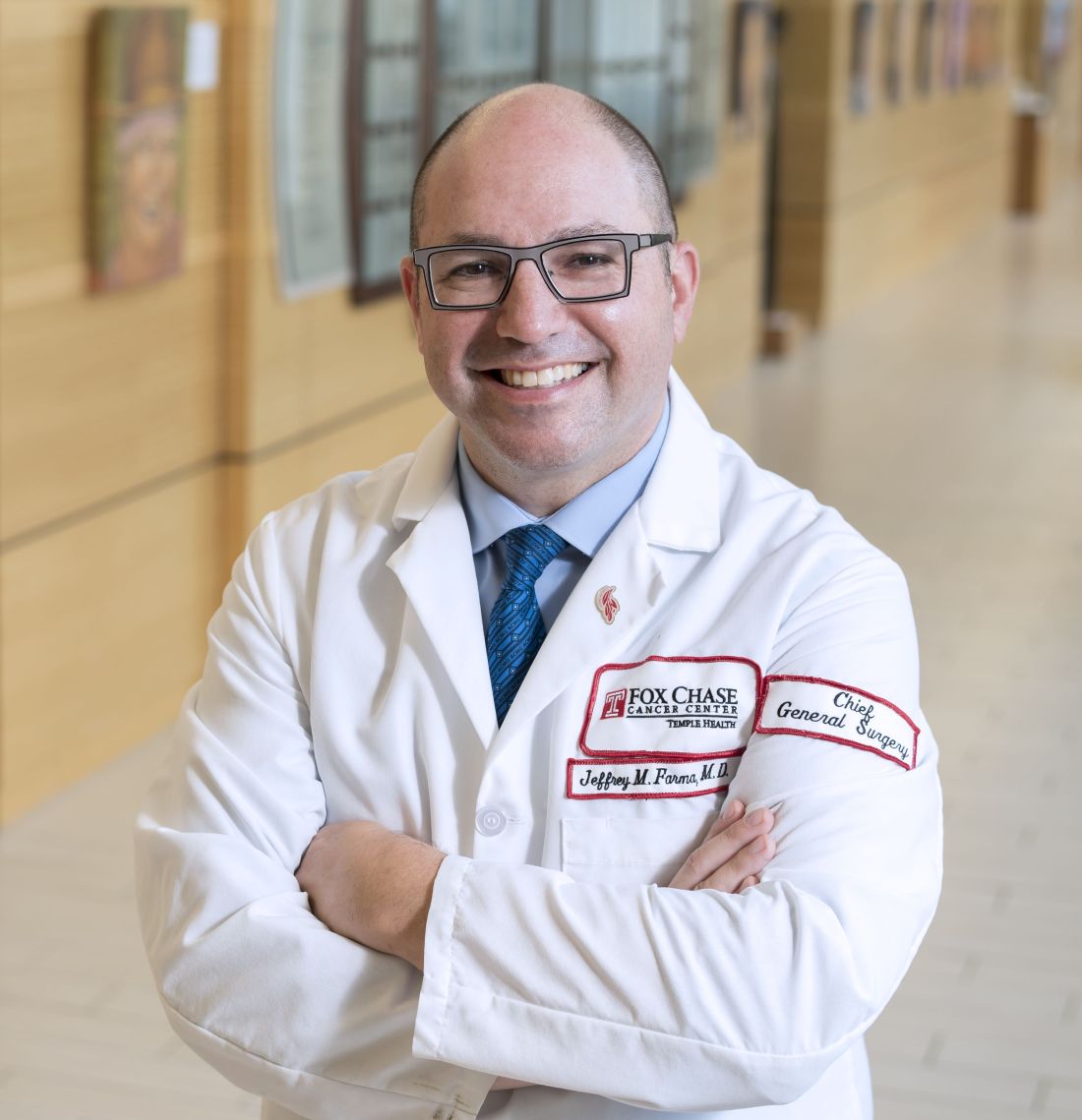
“In their study, immunotherapy was associated with improved outcomes,” said Dr. Farma. “This study highlights the continued lag of implementation of guidelines when new therapies are approved, and that for rare cancers like Merkel cell carcinoma, being treated at high-volume centers and the regionalization of care can lead to improved outcomes for patients.”
Dr. Cheraghlou and Dr. Farma had no disclosures.
A version of this article appeared on Medscape.com.
PHOENIX — Immunotherapy has revolutionized outcomes for patients are better at high-volume centers.
The study has important implications, said study author Shayan Cheraghlou, MD, an incoming fellow in Mohs surgery at New York University, New York City. “We can see that in a real-world setting, these agents have an impact on survival,” he said. “We also found high-volume centers were significantly more likely to use the agents than low-volume centers.” He presented the findings at the annual meeting of the American College of Mohs Surgery.
MCC is a neuroendocrine skin cancer with a high rate of mortality, and even though it remains relatively rare, its incidence has been rising rapidly since the late 1990s and continues to increase. There were no approved treatments available until 2017, when the US Food and Drug Administration (FDA) approved the immunotherapy drug avelumab (Bavencio) to treat advanced MCC. Two years later, pembrolizumab (Keytruda) also received regulatory approval for MCC, and these two agents have revolutionized outcomes.
“In clinical trial settings, these agents led to significant and durable responses, and they are now the recommended treatments in guidelines for metastatic Merkel cell carcinoma,” said Dr. Cheraghlou. “However, we don’t have data as to how they are being used in the real-world setting and if survival outcomes are similar.”
Real World vs Clinical Trials
Real-world outcomes can differ from clinical trial data, and the adoption of novel therapeutics can be gradual. The goal of this study was to see if clinical trial data matched what was being observed in actual clinical use and if the agents were being used uniformly in centers across the United States.
The authors used data from the National Cancer Database that included patients diagnosed with cancer from 2004 to 2019 and identified 1017 adult cases of metastatic MCC. They then looked at the association of a variety of patient characteristics, tumors, and system factors with the likelihood of receiving systemic treatment for their disease.
“Our first finding was maybe the least surprising,” he said. “Patients who received these therapeutic agents had significantly improved survival compared to those who have not.”
Those who received immunotherapy had a 35% decrease in the risk for death per year compared with those who did not. The 1-, 3-, and 5-year survival rates were 47.2%, 21.8%, and 16.5%, respectively, for patients who did not receive immunotherapy compared with 62.7%, 34.4%, and 23.6%, respectively, for those who were treated with these agents.
Dr. Cheraghlou noted that they started to get some “surprising” findings when they looked at utilization data. “While it has been increasing over time, it is not as high as it should be,” he emphasized.
From 2017 to 2019, 54.2% of patients with metastatic MCC received immunotherapy. The data also showed an increase in use from 45.1% in 2017 to 63.0% in 2019. “This is an effective treatment for aggressive malignancy, so we have to ask why more patients aren’t getting them,” said Dr. Cheraghlou.
Their findings did suggest one possible reason, and that was that high-volume centers were significantly more likely to use the agents than low-volume centers. Centers that were in the top percentile for MCC case volume were three times as likely to use immunotherapy for MCC compared with other institutions. “So, if you have metastatic Merkel cell carcinoma and go to a low volume center, you may be less likely to get potential lifesaving treatment,” he noted.
Implications Going Forward
Dr. Cheraghlou concluded his presentation by pointing out that this study has important implications. The data showed that in a real-world setting, these agents have an impact on survival, but all eligible patients do not have access. “In other countries, there are established referral patterns for all patients with aggressive rare malignancies and really all cancers,” he added. “But in the US, cancer care is more decentralized. Studies like this and others show that high-volume centers have much better outcomes for aggressive rare malignancies, and we should be looking at why this is the case and mitigating these disparities and outcomes.”
Commenting on the study results, Jeffrey M. Farma, MD, co-director of the Melanoma and Skin Cancer Program and professor of surgical oncology at Fox Chase Cancer Center, Philadelphia, referred to the two immunotherapies that have been approved for MCC since 2017, which have demonstrated a survival benefit and improved outcomes in patients with metastatic MCC.
“In their study, immunotherapy was associated with improved outcomes,” said Dr. Farma. “This study highlights the continued lag of implementation of guidelines when new therapies are approved, and that for rare cancers like Merkel cell carcinoma, being treated at high-volume centers and the regionalization of care can lead to improved outcomes for patients.”
Dr. Cheraghlou and Dr. Farma had no disclosures.
A version of this article appeared on Medscape.com.
FROM ACMS 2024
Low-FODMAP, Low-Carb Diets May Beat Medical Treatment for IBS
According to a new study, evidence was found that these dietary interventions were more efficacious at 4 weeks, suggesting their potential as first-line treatments.
“IBS is a disorder that may have different underlying causes, and it can manifest in different ways among patients. It is also likely that the most effective treatment option can differ in patients,” said lead author Sanna Nybacka, RD, PhD, a postdoctoral researcher in molecular and clinical medicine at the University of Gothenburg’s Sahlgrenska Academy, Gothenburg, Sweden.
“Up to 80% of patients with IBS report that their symptoms are exacerbated by dietary factors, and dietary modifications are considered a promising avenue for alleviating IBS symptoms,” she said. “However, as not all patients respond to dietary modifications, we need studies comparing the effectiveness of dietary vs pharmacological treatments in IBS to better understand which patients are more likely to benefit from which treatment.”
The study was published online in The Lancet Gastroenterology and Hepatology.
Treatment Comparison
Dr. Nybacka and colleagues conducted a single-blind randomized controlled trial at a specialized outpatient clinic at Sahlgrenska University Hospital in Gothenburg, Sweden, between January 2017 and September 2021. They included adults with moderate to severe IBS, which was defined as ≥ 175 points on the IBS Severity Scoring System (IBS-SSS), and who had no other serious diseases or food allergies.
The participants were assigned 1:1:1 to receive a low-FODMAP diet plus traditional dietary advice (50% carbohydrates, 33% fat, 17% protein), a fiber-optimized diet with low carbohydrates and high protein and fat (10% carbohydrates, 67% fat, 23% protein), or optimized medical treatment based on predominant IBS symptoms. Participants were masked to the names of the diets, but the pharmacological treatment was open-label.
After 4 weeks, participants were unmasked and encouraged to continue their diets.
During 6 months of follow-up, those in the low-FODMAP group were instructed on how to reintroduce FODMAPs, and those in the pharmacological treatment group were offered personalized diet counseling and to continue their medication.
Among 1104 participants assessed for eligibility, 304 were randomly assigned. However, 10 participants did not receive their intervention after randomization, so only 294 participants were included in the modified intention-to-treat population: 96 in the low-FODMAP group, 97 in the low-carbohydrate group, and 101 in the optimized medical treatment group. Overall, 82% were women, and the mean age was 38 years.
Following the 4-week intervention, 73 of 96 participants (76%) in the low-FODMAP group, 69 of 97 participants (71%) in the low-carbohydrate group, and 59 of 101 participants (58%) in the optimized medical treatment group had a reduction of ≥ 50 points in the IBS-SSS compared with baseline.
A stricter score reduction of ≥ 100 points was observed in 61% of the low-FODMAP group, 58% of the low-carbohydrate group, and 39% of the optimized medical treatment group.
In both the low-FODMAP group and the low-carbohydrate group, 95% of participants completed the 4-week intervention compared with 90% among the pharmacological group. Two people in each group said adverse events prompted their discontinuation, and five in the medical treatment group stopped prematurely due to side effects. No serious adverse events or treatment-related deaths occurred.
“We were surprised by the effectiveness of the fiber-optimized low-carbohydrate diet, which demonstrated comparable efficacy to the combined low-FODMAP and traditional IBS diet,” Dr. Nybacka said. “While previous knowledge suggested that high-fat intake could worsen symptoms in some individuals, the synergy with low-carbohydrate intake appeared to render the diet more tolerable for these patients.”
The authors noted that since all three treatment options showed significant and clinically meaningful efficacy, patient preference, ease of implementation, compliance, cost-effectiveness, and long-term effects, including those on nutritional status and gut microbiota, should be considered in personalized plans.
Future Practice Considerations
Dr. Nybacka and colleagues recommended additional trials before implementing the low-carbohydrate diet in clinical practice. “Worse blood lipid levels among some participants in the low-carbohydrate group point to an area for caution,” she said.
The research team also plans to evaluate changes in microbiota composition and metabolomic profiles among participants to further understand factors associated with positive treatment outcomes.
“Approximately two thirds of patients with IBS report that certain foods trigger symptoms of IBS, which is why many patients are interested in exploring dietary interventions for their symptoms,” said Brian Lacy, MD, professor of medicine and program director of the GI fellowship program at the Mayo Clinic in Jacksonville, Florida. “One of the most commonly employed diets for the treatment of IBS is the low-FODMAP diet.”
Dr. Lacy, who wasn’t involved with this study, co-authored the 2021 American College of Gastroenterology clinical guideline for the management of IBS.
He and his colleagues recommended a limited trial of a low-FODMAP diet to improve symptoms, as well as targeted use of medications for IBS subtypes with constipation or diarrhea and gut-directed psychotherapy for overall IBS symptoms.
“However, there are problems with the low-FODMAP diet, as it can be difficult to institute, it can be fairly restrictive, and long-term use has the potential to lead to micronutrient deficiencies,” he said. “Importantly, large studies comparing dietary interventions directly to medical therapies are absent, which led to the study by Nybacka and colleagues.”
Dr. Lacy noted several limitations, including the single-center focus, short-term intervention, and variety of therapies used among the medical arm of the study. In addition, some therapies available in the United States aren’t available in Europe, so the varying approaches to medical management in the former may lead to different results. At the same time, he said, the study is important and will be widely discussed among patients and clinicians.
“I think it will likely stand the test of time,” Dr. Lacy said. “An easy-to-use diet with common sense advice that improves symptoms will likely eventually translate into first-line therapy for IBS patients.”
The study was funded by grants from the Healthcare Board Region Västra Götaland, Swedish Research Council, Swedish Research Council for Health, Working Life and Welfare, and AFA Insurance; the ALF agreement between the Swedish government and county councils; Wilhelm and Martina Lundgren Science Foundation; Skandia; Dietary Science Foundation; and Nanna Swartz Foundation. Several authors declared grants, consulting fees, and advisory board roles with various pharmaceutical companies. Dr. Lacy reported no relevant disclosures.
A version of this article appeared on Medscape.com.
FROM THE LANCET GASTROENTEROLOGY AND HEPATOLOGY
Use of Radiotherapy for Nonmelanoma Skin Cancer Increasing, Study Finds
PHOENIX — More specifically, the persistent growth in the use of superficial radiotherapy (SRT) devices and electronic brachytherapy (eBT) to treat nonmelanoma skin cancer (NMSC) has exceeded that of traditional procedures among dermatologists using these modalities, according to Christian Gronbeck, MD, a resident in dermatology at the University of Connecticut Health Center, Farmington.
“These services increased substantially over the study period,” Dr. Gronbeck said at the annual meeting of the American College of Mohs Surgery, where he presented the results of the study. “Our findings suggest that those using eBT/SRT were frequently general dermatologists and non-fellowship–trained Mohs surgeons who have less formalized surgical training.”
He also noted that billing for these services also rose substantially, which is being driven by growing utilization and an increased SRT payment rate.
Surgical approaches are standard for most NMSC cases, but some patients are not good surgical candidates because of medical comorbidities and/or other factors, and radiotherapy is emerging as a potential treatment option for those patients. Traditionally, radiotherapy was administered by radiation oncologists, but with the growing availability of SRT devices and the introduction of eBT, dermatologists are now treating patients with these modalities.
“It is a potential treatment option for nonmelanoma skin cancer and keloids, and these lower energy devices can be used in the outpatient setting,” said Dr. Gronbeck. “Treatment typically involves a series of fractions over a period of several weeks. There has been recent growth in the use of radiotherapy despite this being a secondary option in skin cancer, primarily when surgery is contraindicated.”
Steady Expansion of Use
Dr. Gronbeck and colleagues sought to gain a better understanding of the use of SRT and eBT for NMSC among dermatologists, as well as trends in cost. Data were obtained from the 2016-2021 Medicare Public Use Files to evaluate the trend in the volume of Medicare Part B claims for eBT (CPT 0394T) and SRT (CPT 77401) by dermatologists, and they also looked at related billable services for radiotherapy.
Of 12,050 dermatologists, 293 (2.4%) were identified as utilizing eBT or SRT in 2021, representing a 75.4% increase from 2016. The usage of both eBT and SRT increased by 59.6% and 148.4%, respectively, from 2016 to 2021.
There were notable geographic differences in the utilization of radiotherapy. “Florida, California, Texas, and Arizona had the highest utilization,” Dr. Gronbeck said, although during the study period, utilization increased in other states, including North Carolina and Alabama.
When looking at geographic regions as a whole, the highest number of dermatologists using radiotherapy were located in the South (n = 143, 50.9%), followed by the West (n = 69, 23.6%). Utilization was more common in metro areas than in nonmetro/rural areas (86% vs 14%).
Differences were also noted among dermatologists. Those who performed eBT/SRT than those who did not were significantly more likely to have had 15 or more years of independent practice (70.1% vs 48.6%), be in a small private dermatology practice (62.7% vs 47.5%), and be non–fellowship-trained Mohs surgeons (33.5% vs 10.2%). Dermatologists utilizing radiotherapy were also more likely to treat Medicare beneficiaries who were older, with a mean age over 75 years (39.3% vs 31.1%) and a mean hierarchical condition category (HCC) score, above the national average (55.2% vs 44.6%).
Dr. Gronbeck and colleagues also looked at cost. The number of direct payments for eBT/SRT payments increased throughout the study period, from 3,678,224 in 2016 to 11,680,925 in 2021, nearly a 218% increase. The change in payments for services related to eBT/SRT, such as radiotherapy simulation, radiotherapy dosing, and ultrasound guidance, increased by 621.4% during this same timeframe.
Radiotherapy in dermatology has primarily been assessed through retrospective studies. “Our findings suggest that eBT and SRT are more frequently utilized by dermatologists managing older and sicker patients, but further studies are needed to identify whether these interventions are truly addressing poor surgical candidates,” Dr. Gronbeck said.
The Centers for Medicare & Medicaid Services (CMS) has recently proposed changes in Medicare coverage in seven states for Image-Guided Superficial Radiation Therapy (image-guided SRT or IGSRT) for the treatment of NMSC. The proposed local coverage determination, or LCD, if finalized in its current form, would affect residents in North Carolina, South Carolina, Virginia, West Virginia, Alabama, Georgia, and Tennessee.
“These changes would mean more restrictive coverage,” said Dr. Gronbeck, and further support the need for “improved clinical data and development of guidelines to support evidence-based utilization.”
Surgical Management Standard, but SRT Has a Role
Asked to comment on the findings, Seemal R. Desai, MD, president of the American Academy of Dermatology (AAD), who was not involved with the study, reiterated that according to this abstract, efficacy has mainly been assessed through retrospective studies, and results are likely inferior to Mohs surgery, require multiple treatment visits, and are associated with significant costs. More study is needed for the use of radiation therapy in dermatology, he told this news organization.

“The Academy supports continued research and studies for therapies that can help improve patient outcomes and offer treatment options, as well as further studies on long-term outcomes for treatments like superficial radiation therapy,” he said.
“Well-designed studies can certainly be helpful to better assess efficacy and outcomes,” Dr. Desai continued. “This is why the Academy supports the idea of scientific studies that continue to expand the body of literature and data, which can help dermatologists tailor therapeutic options for their patients.”
As for general dermatologists using radiation therapy, he pointed out that SRT was developed within the dermatology specialty with dermatologists being the experts in delivering SRT for patients with NMSCs when indicated. “SRT has been used for over 100 years to treat skin cancer,” said Dr. Desai, of the department of dermatology, UT Southwestern Medical Center, Dallas. “While certain radiation devices have historically been used by dermatologists, dermatologists engaged in providing superficial radiation therapy must have adequate education and training to administer this therapy safely and effectively.”
The AAD Association (AADA) has a position statement that supports the use of SRT as an option for the treatment of basal cell carcinoma and squamous cell carcinoma in certain circumstances. “This could be when surgical intervention is contraindicated or refused and after the benefits and risk of treatment alternatives have been discussed with the patient,” he said. “Based on current evidence, surgical management remains the most effective treatment for NMSC.”
Dr. Desai added that the AADA is also concerned that if the Proposed LCD is finalized by CMS, it “could restrict dermatologists from performing SRT and impede patient access to SRT as a potential treatment when indicated.”
The study was independently supported. Dr. Gronbeck and Dr. Desai reported no relevant financial relationships.
A version of this article appeared on Medscape.com.
PHOENIX — More specifically, the persistent growth in the use of superficial radiotherapy (SRT) devices and electronic brachytherapy (eBT) to treat nonmelanoma skin cancer (NMSC) has exceeded that of traditional procedures among dermatologists using these modalities, according to Christian Gronbeck, MD, a resident in dermatology at the University of Connecticut Health Center, Farmington.
“These services increased substantially over the study period,” Dr. Gronbeck said at the annual meeting of the American College of Mohs Surgery, where he presented the results of the study. “Our findings suggest that those using eBT/SRT were frequently general dermatologists and non-fellowship–trained Mohs surgeons who have less formalized surgical training.”
He also noted that billing for these services also rose substantially, which is being driven by growing utilization and an increased SRT payment rate.
Surgical approaches are standard for most NMSC cases, but some patients are not good surgical candidates because of medical comorbidities and/or other factors, and radiotherapy is emerging as a potential treatment option for those patients. Traditionally, radiotherapy was administered by radiation oncologists, but with the growing availability of SRT devices and the introduction of eBT, dermatologists are now treating patients with these modalities.
“It is a potential treatment option for nonmelanoma skin cancer and keloids, and these lower energy devices can be used in the outpatient setting,” said Dr. Gronbeck. “Treatment typically involves a series of fractions over a period of several weeks. There has been recent growth in the use of radiotherapy despite this being a secondary option in skin cancer, primarily when surgery is contraindicated.”
Steady Expansion of Use
Dr. Gronbeck and colleagues sought to gain a better understanding of the use of SRT and eBT for NMSC among dermatologists, as well as trends in cost. Data were obtained from the 2016-2021 Medicare Public Use Files to evaluate the trend in the volume of Medicare Part B claims for eBT (CPT 0394T) and SRT (CPT 77401) by dermatologists, and they also looked at related billable services for radiotherapy.
Of 12,050 dermatologists, 293 (2.4%) were identified as utilizing eBT or SRT in 2021, representing a 75.4% increase from 2016. The usage of both eBT and SRT increased by 59.6% and 148.4%, respectively, from 2016 to 2021.
There were notable geographic differences in the utilization of radiotherapy. “Florida, California, Texas, and Arizona had the highest utilization,” Dr. Gronbeck said, although during the study period, utilization increased in other states, including North Carolina and Alabama.
When looking at geographic regions as a whole, the highest number of dermatologists using radiotherapy were located in the South (n = 143, 50.9%), followed by the West (n = 69, 23.6%). Utilization was more common in metro areas than in nonmetro/rural areas (86% vs 14%).
Differences were also noted among dermatologists. Those who performed eBT/SRT than those who did not were significantly more likely to have had 15 or more years of independent practice (70.1% vs 48.6%), be in a small private dermatology practice (62.7% vs 47.5%), and be non–fellowship-trained Mohs surgeons (33.5% vs 10.2%). Dermatologists utilizing radiotherapy were also more likely to treat Medicare beneficiaries who were older, with a mean age over 75 years (39.3% vs 31.1%) and a mean hierarchical condition category (HCC) score, above the national average (55.2% vs 44.6%).
Dr. Gronbeck and colleagues also looked at cost. The number of direct payments for eBT/SRT payments increased throughout the study period, from 3,678,224 in 2016 to 11,680,925 in 2021, nearly a 218% increase. The change in payments for services related to eBT/SRT, such as radiotherapy simulation, radiotherapy dosing, and ultrasound guidance, increased by 621.4% during this same timeframe.
Radiotherapy in dermatology has primarily been assessed through retrospective studies. “Our findings suggest that eBT and SRT are more frequently utilized by dermatologists managing older and sicker patients, but further studies are needed to identify whether these interventions are truly addressing poor surgical candidates,” Dr. Gronbeck said.
The Centers for Medicare & Medicaid Services (CMS) has recently proposed changes in Medicare coverage in seven states for Image-Guided Superficial Radiation Therapy (image-guided SRT or IGSRT) for the treatment of NMSC. The proposed local coverage determination, or LCD, if finalized in its current form, would affect residents in North Carolina, South Carolina, Virginia, West Virginia, Alabama, Georgia, and Tennessee.
“These changes would mean more restrictive coverage,” said Dr. Gronbeck, and further support the need for “improved clinical data and development of guidelines to support evidence-based utilization.”
Surgical Management Standard, but SRT Has a Role
Asked to comment on the findings, Seemal R. Desai, MD, president of the American Academy of Dermatology (AAD), who was not involved with the study, reiterated that according to this abstract, efficacy has mainly been assessed through retrospective studies, and results are likely inferior to Mohs surgery, require multiple treatment visits, and are associated with significant costs. More study is needed for the use of radiation therapy in dermatology, he told this news organization.
“The Academy supports continued research and studies for therapies that can help improve patient outcomes and offer treatment options, as well as further studies on long-term outcomes for treatments like superficial radiation therapy,” he said.
“Well-designed studies can certainly be helpful to better assess efficacy and outcomes,” Dr. Desai continued. “This is why the Academy supports the idea of scientific studies that continue to expand the body of literature and data, which can help dermatologists tailor therapeutic options for their patients.”
As for general dermatologists using radiation therapy, he pointed out that SRT was developed within the dermatology specialty with dermatologists being the experts in delivering SRT for patients with NMSCs when indicated. “SRT has been used for over 100 years to treat skin cancer,” said Dr. Desai, of the department of dermatology, UT Southwestern Medical Center, Dallas. “While certain radiation devices have historically been used by dermatologists, dermatologists engaged in providing superficial radiation therapy must have adequate education and training to administer this therapy safely and effectively.”
The AAD Association (AADA) has a position statement that supports the use of SRT as an option for the treatment of basal cell carcinoma and squamous cell carcinoma in certain circumstances. “This could be when surgical intervention is contraindicated or refused and after the benefits and risk of treatment alternatives have been discussed with the patient,” he said. “Based on current evidence, surgical management remains the most effective treatment for NMSC.”
Dr. Desai added that the AADA is also concerned that if the Proposed LCD is finalized by CMS, it “could restrict dermatologists from performing SRT and impede patient access to SRT as a potential treatment when indicated.”
The study was independently supported. Dr. Gronbeck and Dr. Desai reported no relevant financial relationships.
A version of this article appeared on Medscape.com.
FROM ACMS 2024
Post–Mohs Surgery Opioid Prescribing More Common in Some Patient Groups
PHOENIX — The study also found that patients who do receive opioids postoperatively are at an increased risk for chronic opioid use and complications.
This report represents the largest analysis to date of opioid prescribing after dermatologic surgery, said lead author Kyle C. Lauck, MD, a dermatology resident at Baylor University Medical Center, Dallas, Texas. “Females, African Americans, and Latino patients may be at a higher risk of opioid prescription after dermatologic surgery. Surgeons should be aware of these populations and the risks they face when determining candidacy for postsurgical opioid analgesia.”
He presented the results at the annual meeting of the American College of Mohs Surgery.
The opioid epidemic is a concern across all areas of medicine, and the majority of opioid prescriptions in dermatology are given following surgery. Dr. Lauck noted that even though guidelines delegate opioids as second line for pain control, the existing data on opioid prescribing in dermatologic surgery is mixed. For example, some reports have shown that up to 58% of patients receive opioids postoperatively. “No consensus exists when we should routinely give opioids to these patients,” he said.
Even though most surgeons prescribe short courses of opioids, even brief regimens are associated with increased risks for overuse and substance abuse. Population-level data are limited concerning opioid prescriptions in dermatologic surgery, and in particular, there is an absence of data on the risk for long-term complications associated with use.
Certain Populations at Risk
To evaluate opioid prescription rates in dermatologic surgery, focusing on disparities between demographic populations, as well as the risk for long-term complications of postoperative opioid prescriptions, Dr. Lauck and colleagues conducted a retrospective study that included 914,721 dermatologic surgery patients, with billing codes for Mohs micrographic surgery. Patient data were obtained from TriNetX, a federated health research network.
The mean age of patients in this cohort was 54 years, and 124,494 (13.6%) were prescribed postsurgical oral opioids. The most common was oxycodone, prescribed to 43% of patients. Dr. Lauck noted that, according to their data, certain groups appeared more likely to receive a prescription for opioids following surgery. These included Black or African American patients (23.75% vs 12.86% for White patients), females (13.73% vs 13.16% for males), and Latino or Hispanic patients (17.02% vs 13.61% non-Latino/Hispanic patients).
Patients with a history of prior oral opioid prescription, prior opioid abuse or dependence, and any type of substance abuse had a significant increase in absolute risk of being prescribed postsurgical opioids (P < .0001).
The type of surgery also was associated with prescribed postop opioids. For a malignant excision, 18.29% of patients were prescribed postop opioids compared with 14.9% for a benign excision. About a third of patients (34.9%) undergoing a graft repair received opioids.
There was an elevated rate of postop opioid prescribing that was specific to the site of surgery, with the highest rates observed with eyelids, scalp and neck, trunk, and genital sites. The highest overall rates of opioid prescriptions were for patients who underwent excisions in the genital area (54.5%).
Long-Term Consequences
The authors also looked at the longer-term consequences of postop opioid use. “Nearly one in three patients who were prescribed opioids needed subsequent prescriptions down the line,” said Dr. Lauck.
From 3 months to 5 years after surgery, patients who received postsurgical opioids were at significantly higher risk for not only subsequent oral opioid prescription but also opiate abuse, any substance abuse, overdose by opioid narcotics, constipation, and chronic pain. “An opioid prescription may confer further risks of longitudinal complications of chronic opioid use,” he concluded.
Commenting on the study, Jesse M. Lewin, MD, chief of Mohs micrographic and dermatologic surgery at Icahn School of Medicine at Mount Sinai, New York City, noted an important finding of this study was the long-term sequelae of patients who did receive postop opioids.
“This is striking given that postsurgical opiate prescriptions are for short durations and limited number of pills,” he told this news organization. “This study highlights the potential danger of even short course of opiates and should serve as a reminder to dermatologic surgeons to be judicious about opiate prescribing.”
Dr. Lauck and Dr. Lewin had no disclosures.
A version of this article appeared on Medscape.com.
FROM ACMS 2024
Mirikizumab Shows Promise for Moderate to Severe Crohn’s Disease
WASHINGTON — , according to results of the phase 3 randomized, double blind, treat-through VIVID-1 study.
Bruce E. Sands, MD, AGAF, chief of gastroenterology at the Icahn School of Medicine at Mount Sinai in New York, reported the findings in a poster (Abstract Su1801) at the annual Digestive Disease Week® (DDW).
The FDA approved mirikizumab (Omvoh, Eli Lilly) to treat moderate to severe ulcerative colitis in October 2023.
Dr. Sands and a team of US and international collaborators studied 1065 adults with Crohn’s disease or fistulizing Crohn’s disease for 3 months or more, with a mean duration of more than 7 years. At baseline, participants had a Simple Endoscopic Score for Crohn’s Disease (SES-CD) of 7 or more and reported an inadequate response, lost response, or intolerance to other therapy.
A total of 579 people were randomly assigned to mirikizumab and another 199 to placebo. Another 287 patients received ustekinumab; though they were not included in this current analysis, the findings were presented separately at DDW 2024.
Mean age of study participants was 30 years, and men comprised 57%-59% of the groups. Nearly half (49%) of each group previously failed biologic therapy.
A primary composite endpoint was clinical response at 12 weeks according to patient reported outcome and endoscopic response at 52 weeks measured with the SES-CD. A second primary endpoint was clinical response at 12 weeks by patient reported outcome combined with clinical remission on Crohn’s Disease Activity Index (CDAI) at 52 weeks.
Researchers also tracked 12 major secondary endpoints for mirikizumab vs placebo, including clinical response, endoscopic response, and clinical remission at week 12 and week 52.
Efficacy Findings
A higher percentage of participants in the mirikizumab group achieved 12-week secondary endpoints compared with placebo. In the treatment group, 32.5% reached endoscopic response vs 12.6% in the placebo group, a statistically significant difference (P < .000001). In addition, 17.6% achieved endoscopic remission in the treatment group vs 7.0% in the placebo group at 12 weeks (P < .000213).
The “treat-through” results at 52 weeks revealed that a higher proportion of the group taking mirikizumab met the co-primary endpoints compared with placebo. A total of 48.4% in the mirikizumab group vs 9.0% in the placebo group achieved endoscopic response (P < .000001). Similarly, a higher proportion met clinical remission on the CDAI, 54.1% in the treatment group vs 19.6% in the placebo group (P < .000001).
Overall, 38% of mirikizumab-treated patients vs 9% of the placebo group reached a composite endpoint of patient reported clinical response at week 12 and endoscopic response by SES-CD at week 52 (P < .000001).
Dr. Sands and colleagues also combined clinical response reported by patients at 12 weeks with CDAI findings for clinical remission at week 52. A total of 45.4% in the treatment group met the combined endpoint compared with 19.6% of the placebo group (P < .000001).
In an additional analysis, the researchers looked at this composite endpoint in patients in both groups who had failed or not failed a prior biologic for a total of 43.4% vs 12.4%, and 47.3% vs 26.5%, respectively.
“Mirikizumab demonstrated statistically significant and clinically meaningful improvements” in the study co-primary endpoints and secondary endpoints compared with placebo, the researchers concluded.
Safety Findings
Safety outcomes during the 52-week study were “consistent with the known safety profile” of mirikizumab, the researchers noted.
Treatment-emergent adverse events occurred in 78.6% of mirikizumab participants vs 73.0% of the placebo group. The most common were COVID-19, anemia, and arthralgia. Serious adverse events were reported in 10.3% of the mirikizumab group vs 17.1% of the placebo group. There were seven opportunistic infections in the treatment group, including herpes zoster and Candida, compared with none in the placebo group.
One person in the placebo cohort died of a pulmonary embolism; there were no deaths in the mirikizumab group.
People randomly assigned to placebo without a response at 12 weeks were switched over to mirikizumab. However, the findings from this group between 12 and 52 weeks were excluded from the 1-year data presented at DDW 2024, including one death from worsening Crohn’s disease during that time.
Mirikizumab looked particularly robust in this study, and it may turn out to be a critically important option for our patients, said Jordan Axelrad, MD, MPH, co-director of the Inflammatory Bowel Disease Center at NYU Langone Health in New York City. Dr. Axelrad was not involved in this study.
Of importance, effect sizes were similar for “bio-naive and previously biologic-exposed patients,” he added.
These data “really underscore that therapies targeting IL-23 may be clinically useful for Crohn’s disease patients with prior biologic failure, representing a significant departure from our previous experience with other biologic classes,” Dr. Axelrad said.
The study was funded by Eli Lilly and Company. Dr. Sands is a consultant and receives grant funding from Lilly. Dr. Axelrad had no relevant disclosures.
A version of this article appeared on Medscape.com.
FROM DDW 2024
Liposomal Irinotecan for Pancreatic Cancer: Is It Worth It?
In February, the US Food and Drug Administration (FDA) approved irinotecan liposome (Onivyde) as part of a new regimen for first-line metastatic pancreatic adenocarcinoma called NALIRIFOX.
The main difference between NALIRIFOX and a standard go-to regimen for the indication, modified FOLFIRINOX, is that liposomal irinotecan — irinotecan encased in a lipid nanoparticle — is used instead of free irinotecan.
Trial data suggested a better overall response rate, a slight progression-free survival advantage, and potentially fewer adverse events with the liposomal formulation.
The substitution, however, raises the cost of treatment substantially. According to one estimate, a single cycle of FOLFIRINOX costs about $500 at a body surface area of 2 m2, while the equivalent single cycle of NALIRIFOX costs $7800 — over 15-fold more expensive.
While some oncologists have called the NALIRIFOX regimen a potential new standard first-line treatment for metastatic pancreatic adenocarcinoma, others have expressed serious doubts about whether the potential benefits are worth the extra cost.
“I can’t really see a single scenario where I would recommend NALIRIFOX over FOLFIRINOX” Ignacio Garrido-Laguna, MD, PhD, a gastrointestinal oncologist and pancreatic cancer researcher at the University of Utah, Salt Lake City, told this news organization. “Most of us in the academic setting have the same take on this.”
No Head-to-Head Comparison
Instead, the 770-patient NAPOLI 3 trial compared NALIRIFOX — which also includes oxaliplatin, fluorouracil, and leucovorin — with a two-drug regimen, nab-paclitaxel and gemcitabine. In the trial, overall survival and other outcomes were moderately better with NALIRIFOX.
Oncologists have said that the true value of the trial is that it conclusively demonstrates that a four-drug regimen is superior to a two-drug regimen for patients who can tolerate the more intensive therapy.
Eileen M. O’Reilly, MD, the senior investigator on NAPOLI 3, made this point when she presented the phase 3 results at the 2023 ASCO annual meeting.
The trial “answers the question of four drugs versus two” for first-line metastatic pancreatic cancer but “does not address the question of NALIRIFOX versus FOLFIRINOX,” said Dr. O’Reilly, a pancreatic and hepatobiliary oncologist and researcher at Memorial Sloan Kettering Cancer Center in New York City.
Comparing them directly in the study “probably wouldn’t have been in the interest of the sponsor,” said Dr. O’Reilly.
With no head-to-head comparison, oncologists have been comparing NAPOLI 3 results with those from PRODIGE 4, the 2011 trial that won FOLFIRINOX its place as a first-line regimen.
When comparing the trials, median overall survival was exactly the same for the two regimens — 11.1 months. FOLFIRINOX was associated with a slightly higher 1-year survival rate — 48.4% with FOLFIRINOX vs 45.6% with NALIRIFOX.
However, Dr. O’Reilly and her colleagues also highlighted comparisons between the two trials that favored NAPOLI 3.
NAPOLI 3 had no age limit, while PRODIGE subjects were no older than 75 years. Median progression-free survival was 1 month longer among patients receiving NALIRIFOX — 7.4 months vs 6.4 months in PRODIGE — and overall response rates were higher as well — 41.8% in NAPOLI 3 vs 31.6%. Patients receiving NALIRIFOX also had lower rates of grade 3/4 neutropenia (23.8% vs 45.7%, respectively) and peripheral sensory neuropathy (3.5% vs 9.0%, respectively).
The authors explained that the lower rate of neuropathy could be because NALIRIFOX uses a lower dose of oxaliplatin (FOLFIRINOX), at 60 mg/m2 instead of 85 mg/m2.
Is It Worth It?
During a presentation of the phase 3 findings last year, study author Zev A. Wainberg, MD, of the University of California, Los Angeles, said the NALIRIFOX regimen can be considered the new reference regimen for first-line treatment of metastatic pancreatic adenocarcinoma.
The study discussant, Laura Goff, MD, MSCI, of Vanderbilt-Ingram Cancer Center, Nashville, Tennessee, agreed that the results support the NALIRIFOX regimen as “the new standard for fit patients.”
However, other oncologists remain skeptical about the benefits of the new regimen over FOLFIRINOX for patients with metastatic pancreatic adenocarcinoma.
In a recent editorial, Dr. Garrido-Laguna and University of Utah gastrointestinal oncologist Christopher Nevala-Plagemann, MD, compared the evidence for both regimens.
The experts pointed out that overall response rates were assessed by investigators in NAPOLI 3 and not by an independent review committee, as in PRODIGE 4, and might have been overestimated.
Although the lack of an age limit was touted as a benefit of NAPOLI 3, Dr. Garrido-Laguna and Dr. Nevala-Plagemann doubt whether enough patients over 75 years old participated to draw any meaningful conclusions about using NALIRIFOX in older, frailer patients. If anything, patients in PRODIGE 4 might have been less fit because, among other things, the trial allowed patients with serum albumins < 3 g/dL.
On the adverse event front, the authors highlighted the higher incidences of grade 3 or worse diarrhea with NALIRIFOX (20% vs 12.7%) and questioned if there truly is less neutropenia with NALIRIFOX because high-risk patients in NAPOLI 3 were treated with granulocyte colony-stimulating factor to prevent it. The pair also questioned whether the differences in neuropathy rates between the two trials were big enough to be clinically meaningful.
Insights from a recent meta-analysis may further clarify some of the lingering questions about the efficacy of NALIRIFOX vs FOLFIRINOX.
In the analysis, the team found no meaningful difference in overall and progression-free survival between the two regimens. Differences in rates of peripheral neuropathy and diarrhea were not statistically significant, but NALIRIFOX did carry a statistically significant advantage in lower rates of febrile neutropenia, thrombocytopenia, and vomiting.
The team concluded that “NALIRIFOX and FOLFIRINOX may provide equal efficacy as first-line treatment of metastatic pancreatic cancer, but with different toxicity profiles,” and called for careful patient selection when choosing between the two regimens as well as consideration of financial toxicity.
Dr. Garrido-Laguna had a different take. With the current data, NALIRIFOX does not seem to “add anything substantially different to what we already” have with FOLFIRINOX, he told this news organization. Given that, “we can’t really justify NALIRIFOX over FOLFIRINOX without more of a head-to-head comparison.”
The higher cost of NALIRIFOX, in particular, remains a major drawback.
“We think it would be an economic disservice to our healthcare systems if we used NALIRIFOX instead of FOLFIRINOX for these patients on the basis of [NAPOLI 3] data,” Bishal Gyawali, MD, PhD, and Christopher Booth, MD, gastrointestinal oncologists at Queen’s University in Kingston, Ontario, Canada, said in a recent essay.
Dr. Garrido-Laguna and Dr. Nevala-Plagemann reiterated this concern.
Overall, “NALIRIFOX does not seem to raise the bar but rather exposes patients and healthcare systems to financial toxicities,” Dr. Garrido-Laguna and Dr. Nevala-Plagemann wrote in their review.
NAPOLI 3 was funded by Ipsen and PRODIGE 4 was funded by the government of France. No funding source was reported for the meta-analysis. NAPOLI 3 investigators included Ipsen employees. Dr. O’Reilly disclosed grants or contracts from Ipsen and many other companies. Dr. Garrido-Laguna reported institutional research funding from Bristol Myers Squibb, Novartis, Pfizer, and other companies, but not Ipsen. Dr. Nevala-Plagemann is an advisor for Seagen and reported institutional research funding from Theriva. Dr. Gyawali is a consultant for Vivio Health; Dr. Booth had no disclosures. Two meta-analysis authors reported grants or personal fees from Ipsen as well as ties to other companies.
A version of this article appeared on Medscape.com.
Study Highlights Atopic Dermatitis Features, Treatments Among Older Patients
.
The researchers reviewed charts of patients aged 60 years and older who were seen at either a private or county dermatology clinic in Houston between 2009 and 2020 and had been diagnosed with AD by a dermatologist. The findings of their cross-sectional study further supports that AD in this age group “presents as a unique phenotype compared to AD in younger ages, which may inform dermatologists’ diagnosis of AD in these patients” they wrote.
The 791 patients in the study had an average age of 69.3 years, were predominantly women (60.1%), and were racially diverse, with almost 40% being non-Hispanic White individuals. Others were non-Hispanic Black individuals (21.8%), Hispanics (20.4%), and non-Hispanic Asian/Pacific Islanders (11.7%).
Use of topicals, mainly topical corticosteroids (92.2%), was the most frequent treatment prescribed. Oral corticosteroids and antihistamines were “frequent systemic treatments” in this population, prescribed to 10.4% and 12.1%, respectively, “likely due to management prior to a diagnosis of AD by a dermatologist,” wrote first author Hannah Y. Wang, Baylor College of Medicine, Houston, and her coauthors, including Soo Jung Kim, MD, PhD, of the department of dermatology at Baylor.
Other treatments included dupilumab in 5.4%, systemic immunosuppressants (including methotrexate, cyclosporine, and mycophenolate) in 5.4%, and UVB-phototherapy in 2.7%.
Approximately 40% of the patients had a history of allergic rhinitis, while 20% had a history of asthma. Lichenification was noted in 14.5% of patients and nummular lesions in almost 13%. Other rash characteristics — ichthyosis and hyperpigmented patches — were less frequent, seen in 9.7% and 9.1%, respectively.
AD in this older population was most commonly documented on the extensors (49.9%) and the trunk (46%) and less commonly on the hands (19.8%) and feet (9%) — a distribution that is similar to past reports, the authors wrote.
Asked to comment on the findings, Adam Friedman, MD, professor and chair of dermatology at George Washington University, Washington, DC, told this news organization that the data relating to clinical morphology are consistent with past reports and with his own experiences. Lichenification is a “tell-tale sign of chronic disease” and may indicate undertreatment, and the frequency of nummular plaques is unsurprising because “nummular dermatitis as an independent eczema tends to occur more so in the elderly.”

More important, he said, was the finding regarding the use of oral corticosteroid and antihistamine, “both of which are advocated against in the management of AD.”
More research is “needed to elucidate the unique features of elderly AD in pathophysiology and optimal treatments,” the authors wrote, noting that age-related factors potentially affecting AD in this population include reduced skin barrier function, immune dysregulation, and environmental exposures.
The study, Dr. Friedman said, “shines a spotlight on this demographic — they exist, they suffer, and they are at times being managed with less-than-optimal options.” Clinical trials of “the welcome additions to our historically limited armament often lack a substantial elderly study population,” he said, and Medicare makes it “painful to get these game-changing drugs for this large patient population.”
The study authors and Dr. Friedman, who was not involved with the study, reported no conflicts of interest.
A version of this article appeared on Medscape.com.
.
The researchers reviewed charts of patients aged 60 years and older who were seen at either a private or county dermatology clinic in Houston between 2009 and 2020 and had been diagnosed with AD by a dermatologist. The findings of their cross-sectional study further supports that AD in this age group “presents as a unique phenotype compared to AD in younger ages, which may inform dermatologists’ diagnosis of AD in these patients” they wrote.
The 791 patients in the study had an average age of 69.3 years, were predominantly women (60.1%), and were racially diverse, with almost 40% being non-Hispanic White individuals. Others were non-Hispanic Black individuals (21.8%), Hispanics (20.4%), and non-Hispanic Asian/Pacific Islanders (11.7%).
Use of topicals, mainly topical corticosteroids (92.2%), was the most frequent treatment prescribed. Oral corticosteroids and antihistamines were “frequent systemic treatments” in this population, prescribed to 10.4% and 12.1%, respectively, “likely due to management prior to a diagnosis of AD by a dermatologist,” wrote first author Hannah Y. Wang, Baylor College of Medicine, Houston, and her coauthors, including Soo Jung Kim, MD, PhD, of the department of dermatology at Baylor.
Other treatments included dupilumab in 5.4%, systemic immunosuppressants (including methotrexate, cyclosporine, and mycophenolate) in 5.4%, and UVB-phototherapy in 2.7%.
Approximately 40% of the patients had a history of allergic rhinitis, while 20% had a history of asthma. Lichenification was noted in 14.5% of patients and nummular lesions in almost 13%. Other rash characteristics — ichthyosis and hyperpigmented patches — were less frequent, seen in 9.7% and 9.1%, respectively.
AD in this older population was most commonly documented on the extensors (49.9%) and the trunk (46%) and less commonly on the hands (19.8%) and feet (9%) — a distribution that is similar to past reports, the authors wrote.
Asked to comment on the findings, Adam Friedman, MD, professor and chair of dermatology at George Washington University, Washington, DC, told this news organization that the data relating to clinical morphology are consistent with past reports and with his own experiences. Lichenification is a “tell-tale sign of chronic disease” and may indicate undertreatment, and the frequency of nummular plaques is unsurprising because “nummular dermatitis as an independent eczema tends to occur more so in the elderly.”
More important, he said, was the finding regarding the use of oral corticosteroid and antihistamine, “both of which are advocated against in the management of AD.”
More research is “needed to elucidate the unique features of elderly AD in pathophysiology and optimal treatments,” the authors wrote, noting that age-related factors potentially affecting AD in this population include reduced skin barrier function, immune dysregulation, and environmental exposures.
The study, Dr. Friedman said, “shines a spotlight on this demographic — they exist, they suffer, and they are at times being managed with less-than-optimal options.” Clinical trials of “the welcome additions to our historically limited armament often lack a substantial elderly study population,” he said, and Medicare makes it “painful to get these game-changing drugs for this large patient population.”
The study authors and Dr. Friedman, who was not involved with the study, reported no conflicts of interest.
A version of this article appeared on Medscape.com.
FROM JAAD INTERNATIONAL
Subcutaneous Antifibrinolytic Reduces Bleeding After Mohs Surgery
“Though Mohs micrographic surgery is associated with low bleeding complication rates, around 1% of patients in the literature report postoperative bleeding,” corresponding author Abigail H. Waldman, MD, director of the Mohs and Dermatologic Surgery Center, at Brigham and Women’s Hospital, Boston, and colleagues wrote in the study, which was published online in the Journal of the American Academy of Dermatology. “Intravenous tranexamic acid has been used across surgical specialties to reduce perioperative blood loss. Prior studies have shown topical TXA, an antifibrinolytic agent, following MMS may be effective in reducing postoperative bleeding complications, but there are no large cohort studies on injectable TXA utilization in all patients undergoing MMS.”
To improve the understanding of this intervention, the researchers examined the impact of off-label, locally injected TXA on postoperative bleeding outcomes following MMS conducted at Brigham and Women’s Hospital. They evaluated two cohorts: 1843 patients who underwent MMS from January 1, 2019, to December 31, 2019 (the pre-TXA cohort), and 2101 patients who underwent MMS from July 1, 2022, to June 30, 2023 (the TXA cohort), and extracted data, including patient and tumor characteristics, MMS procedure details, antithrombotic medication use, systemic conditions that predispose to bleeding, encounters reporting postoperative bleeding, and interventions required for postoperative bleeding, from electronic medical records. Patients reconstructed by a non-MMS surgeon were excluded from the analysis.
Overall, 2509 cases among 1843 patients and 2818 cases among 2101 were included in the pre-TXA and TXA cohorts, respectively. The researchers found that local subcutaneous injection of TXA reduced the risk for postoperative phone calls or visits for bleeding by 25% (RR [risk ratio], 0.75; 0.57-0.99) and risk for bleeding necessitating a medical visit by 51% (RR, 0.49; 0.32-0.77).
The use of preoperative TXA in several subgroups of patients also was also associated with a reduction in visits for bleeding, including those using alcohol (52% reduction; RR, 0.47; 0.26-0.85), cigarettes (57% reduction; RR, 0.43; 0.23-0.82), oral anticoagulants (61% reduction; RR, 0.39; 0.20-0.77), or antiplatelets (60% reduction; RR, 0.40; 0.20-0.79). The use of TXA was also associated with reduced visits for bleeding in tumors of the head and neck (RR, 0.45; 0.26-0.77) and tumors with a preoperative diameter > 2 cm (RR, 0.37; 0.15-0.90).
Impact of Surgical Repair Type
In other findings, the type of surgical repair was a potential confounder, the authors reported. Grafts and flaps were associated with an increased risk for bleeding across both cohorts (RR, 2.36 [1.5-3.6] and 1.7 [1.1-2.6], respectively) and together comprised 15% of all procedures in the pre-TXA cohort compared with 11.1% in TXA cohort. Two patients in the TXA cohort (0.11%) developed deep vein thrombosis (DVT) 10- and 20-days postoperation, a rate that the authors said is comparable to that of the general population. The two patients had risk factors for hypercoagulability, including advanced cancer and recurrent DVT.
“Overall, local injection of TXA was an effective method for reducing the risk of clinically significant bleeding following MMS,” the researchers concluded. “Perioperative TXA may help to limit the risk of bleeding overall, as well as in populations predisposed to bleeding.” Adverse events with TXA use were rare “and delayed beyond the activity of TXA, indicating a low likelihood of being due to TXA,” they wrote.
“Dermatologists performing MMS may consider incorporating local TXA injection into their regular practice,” they noted, adding that “legal counsel on adverse effects in the setting of off-label pharmaceutical usage may be advised.”
In an interview, Patricia M. Richey, MD, director of Mohs surgery at Boston Medical Center, who was asked to comment on the study, said that postoperative bleeding is one of the most commonly encountered Mohs surgery complications. “Because of increased clinic visits and phone calls, it can also often result in decreased patient satisfaction,” she said.
“This study is particularly notable in that we see that local subcutaneous TXA injection decreased visits for bleeding even in those using oral anticoagulants, antiplatelets, alcohol, and cigarettes. Dermatologic surgery has a very low complication rate, even in patients on anticoagulant and antiplatelet medications, but this study shows that TXA is a fantastic option for Mohs surgeons and patients.”
Neither the study authors nor Dr. Richey reported having financial disclosures.
A version of this article first appeared on Medscape.com.
FROM JOURNAL OF THE AMERICAN ACADEMY OF DERMATOLOGY