User login
Most pregnant women want guidance on prenatal whole-genome sequencing
according to results from a survey published in Obstetrics & Gynecology.

Nearly half said they would want clear guidance from clinicians before undergoing the noninvasive procedure.
“Prenatal whole-genome sequencing offers significantly more fetal information than women can currently receive, and it is not surprising that, when faced with a tremendous range of information, many women want recommendations from their clinicians,” Haley K. Sullivan from the National Institutes of Health Clinical Center and National Human Genome Research Institute and colleagues wrote. “Our data suggest that most women prefer a directive interaction with their clinician when deciding what types of genetic information to receive from prenatal whole-genome sequencing.”
Research coordinators from the Inova Translational Medicine Institute offered 805 pregnant women a survey on their preferences for prenatal whole-genome sequencing between June and August 2017; of these, 553 women answered (69% response rate). The women responded to questions about what type of information they would like to receive if they were to undergo prenatal whole-genome sequencing and what role a clinician would preferably play in the decision-making process. The researchers divided the survey into sections based on actionability, severity, prevalence, and age of onset.
According to the survey results, 90% of respondents wanted information on serious treatable childhood-onset diseases from prenatal whole-genome sequencing results, while 40% said they did not want to receive results based on nonmedical traits such as eye color, height, or athletic ability.
With regard to clinician role, 45% of women said they wanted all options presented with clear recommendations from a clinician on which tests to order, 26% wanted all options presented but with a joint decision-making process, 13% wanted all options presented but independent decision making, and 11% wanted the clear recommendation from clinicians alone.
The respondents said the most common reason for wanting to undergo prenatal whole-genome sequencing was to prepare “financially, medically, or psychologically” for a child with special needs, the researchers said.
“This represents a departure from the current state of genetic counseling, where nondirectiveness is a central tenet, and is contrary to the 45% of ob.gyns. who said in a previous survey that they should not be at all directive when counseling patients on prenatal whole-genome sequencing,” the authors wrote. “Given this clear patient desire for guidance, there is a vital opportunity for the American College of Obstetricians and Gynecologists to provide leadership and recommendations as prenatal whole-genome sequencing is adopted into clinical practice.”
Limitations in the study include asking the respondents to make hypothetical decisions, using examples to describe genetic conditions that might have skewed decision making; asking women to pick only one reason for wanting the sequencing information from a list of predetermined options, when many reasons may be important to them; social desirability bias in the responses, if women are reluctant to pick a choice they perceive as less socially acceptable; and a potential systematic difference between women who were and were not enrolled as survey participants. The respondents also were from the Northern Virginia area, which may not be generalizable to a national population of patients, the researchers said.
This study was supported by the Intramural Research Program of the National Human Genome Research Institute and the Clinical Center Department of Bioethics, National Institutes of Health. The authors reported no relevant conflicts of interest.
SOURCE: Sullivan HK et al. Obstet Gynecol. 2019 Mar. doi: 10.1097/AOG.0000000000003121.
according to results from a survey published in Obstetrics & Gynecology.
Nearly half said they would want clear guidance from clinicians before undergoing the noninvasive procedure.
“Prenatal whole-genome sequencing offers significantly more fetal information than women can currently receive, and it is not surprising that, when faced with a tremendous range of information, many women want recommendations from their clinicians,” Haley K. Sullivan from the National Institutes of Health Clinical Center and National Human Genome Research Institute and colleagues wrote. “Our data suggest that most women prefer a directive interaction with their clinician when deciding what types of genetic information to receive from prenatal whole-genome sequencing.”
Research coordinators from the Inova Translational Medicine Institute offered 805 pregnant women a survey on their preferences for prenatal whole-genome sequencing between June and August 2017; of these, 553 women answered (69% response rate). The women responded to questions about what type of information they would like to receive if they were to undergo prenatal whole-genome sequencing and what role a clinician would preferably play in the decision-making process. The researchers divided the survey into sections based on actionability, severity, prevalence, and age of onset.
According to the survey results, 90% of respondents wanted information on serious treatable childhood-onset diseases from prenatal whole-genome sequencing results, while 40% said they did not want to receive results based on nonmedical traits such as eye color, height, or athletic ability.
With regard to clinician role, 45% of women said they wanted all options presented with clear recommendations from a clinician on which tests to order, 26% wanted all options presented but with a joint decision-making process, 13% wanted all options presented but independent decision making, and 11% wanted the clear recommendation from clinicians alone.
The respondents said the most common reason for wanting to undergo prenatal whole-genome sequencing was to prepare “financially, medically, or psychologically” for a child with special needs, the researchers said.
“This represents a departure from the current state of genetic counseling, where nondirectiveness is a central tenet, and is contrary to the 45% of ob.gyns. who said in a previous survey that they should not be at all directive when counseling patients on prenatal whole-genome sequencing,” the authors wrote. “Given this clear patient desire for guidance, there is a vital opportunity for the American College of Obstetricians and Gynecologists to provide leadership and recommendations as prenatal whole-genome sequencing is adopted into clinical practice.”
Limitations in the study include asking the respondents to make hypothetical decisions, using examples to describe genetic conditions that might have skewed decision making; asking women to pick only one reason for wanting the sequencing information from a list of predetermined options, when many reasons may be important to them; social desirability bias in the responses, if women are reluctant to pick a choice they perceive as less socially acceptable; and a potential systematic difference between women who were and were not enrolled as survey participants. The respondents also were from the Northern Virginia area, which may not be generalizable to a national population of patients, the researchers said.
This study was supported by the Intramural Research Program of the National Human Genome Research Institute and the Clinical Center Department of Bioethics, National Institutes of Health. The authors reported no relevant conflicts of interest.
SOURCE: Sullivan HK et al. Obstet Gynecol. 2019 Mar. doi: 10.1097/AOG.0000000000003121.
FROM OBSTETRICS & GYNECOLOGY
Key clinical point: A majority of pregnant women surveyed said they wanted information on childhood-onset genetic diseases, with almost half wanting clear clinical recommendations before deciding to undergo noninvasive prenatal whole-genome sequencing.
Major finding: Of the respondents, 90% said they wanted information on serious treatable childhood-onset conditions.
Study details: A survey of 553 pregnant women coordinated by the Inova Translational Medicine Institute.
Disclosures: This study was supported by the Intramural Research Program of the National Human Genome Research Institute and the Clinical Center Department of Bioethics, National Institutes of Health. The authors reported no relevant conflicts of interest.
Source: Sullivan HK et al. Obstet Gynecol. 2019 Mar. doi: 10.1097/AOG.0000000000003121.
Hearing drills into patient impact of ACA legal challenge
If Republican state attorneys general prevail in their legal bid to overturn the Affordable Care Act, patients with preexisting conditions will lose their coverage protections, and an additional 24 million Americans could become uninsured, according to testimony presented at a Feb. 6 hearing of the House Energy and Commerce Health Subcommittee.
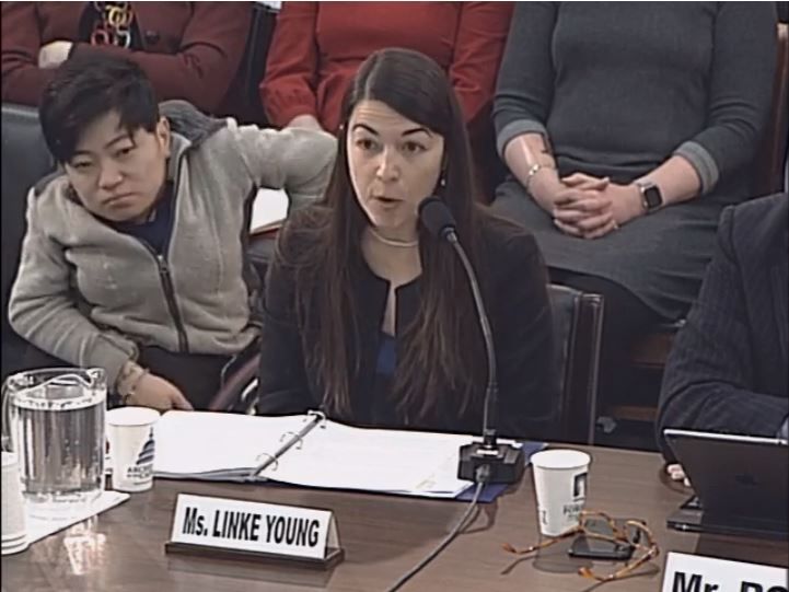
A ruling for the plaintiffs in Texas v. United States also would mean the ACA’s consumer protections for employer-based coverage would be eliminated, affecting more than 150 million Americans, said Christen Linke Young, an attorney and fellow for the liberal-leaning USC-Brookings Schaeffer Initiative on Health Policy.
“The ACA’s changes to Medicare would be undone, reinstating copays on preventive services and reopening the prescription drug ‘donut hole,’ ” Ms. Young testified at the hearing. “It would also create major confusion in Medicare payment, as the ACA policies that are today fully integrated into the Medicare payment rules would suddenly lack a legislative basis.”
Ms. Linke Young was one of five witnesses who testified before lawmakers about the implications of Texas v. United States, an ongoing legal case that centers on whether a part of the health care law should be severed and if so, whether the entire law should then fall.
A group of Republican state attorneys general sued over the law in 2018, arguing that, because budget legislation in 2017 zeroed out the penalties associated with the ACA’s individual mandate, the mandate is invalid. The attorneys general also argue that, if the mandate is severed, the entire ACA should be struck down.
In response to the suit, the Trump administration agreed that the mandate is unconstitutional and should be parsed. Attorneys for the administration wrote that, if the mandate is found unconstitutional, the court should also consider finding two other provisions – the guaranteed issue and community rating requirements – of the ACA invalid. Guaranteed issue refers to insurers in the individual market offering coverage to all citizens, regardless of preexisting conditions, while community rating refers to charging equal premiums to every patient, no matter their past health status. The remainder of the ACA can stand without the three linked provisions, according to the Trump administration, which refused to defend the case.
A coalition of 17 Democratic attorneys general have stepped in to defend the case.
In December, a district court declared the entire ACA to be invalid, a decision immediately appealed to the 5th Circuit by the Democratic attorneys general. In January, the circuit court froze the case in light of the federal government’s partial shutdown. The case remains on hold.

At the hearing, Thomas P. Miller, a resident fellow at the conservative-leaning American Enterprise Institute, testified that a 5th Circuit decision would not likely come before summer 2019, followed by a potential U.S. Supreme Court reading in 2020 – if the case gets that far.
“The probability of a Supreme Court ruling that would overturn the entire ACA remains very low, despite last December’s decision at the federal district court level reaching exactly that legal conclusion,” Mr. Miller testified. “Given the murkiness of divining legislative intent in harder cases like the ACA, challenges to the individual mandate, past and present, it’s better to conclude that, although several different severability settings are hypothetically conceivable, it remains all but certain that an ultimate Supreme Court ruling in this case will, at a minimum, follow its previous inclinations revealed in the 2012 and 2015 ACA challenges and try to save as much of the law as possible.”
If the individual mandate is ultimately severed from the ACA, the decision would have little impact on beneficiaries or function of the health care law, according to Simon Lazarus, a lawyer and writer on constitutional and legal issues.
“Such a result will have virtually no impact on the operation of the ACA, nor on the millions of Americans – in reality, substantially all Americans – who depend on the ACA and its guarantees for people with preexisting conditions and myriad other protections that now are ‘baked into’ the national health care system,” Mr. Lazarus testified at the hearing. “To declare invalid the law’s shared responsibility payment provision, when that provision has no financial penalty behind it, will, by itself, have little if any depressive effect on the number of enrollees in health insurance plans.”
However, Mr. Lazarus noted that the guaranteed issue and community rating provisions of the law are “critical protections” for people with preexisting conditions.
Avik Roy, president for the Foundation for Research on Equal Opportunity, a nonpartisan organization that supports universal health care, testified that the Trump administration’s position about the mandate being tied to the guaranteed issue and community rating provisions is being mischaracterized as implying the administration opposes protecting people with preexisting conditions. President Trump has repeatedly expressed that any reforms or replacements of the ACA cover those with preexisting conditions, he said.
Mr. Roy recommended that Congress pass a bill reiterating the guaranteed issue and community rating requirements in the individual market to ensure protection for patients with preexisting conditions in light of Texas v. United States.
“I understand that a motion to produce such legislation was proposed by House Republicans during floor debate at the beginning of this Congress, [a bill] that would guarantee that no American could be denied coverage, or be charged higher premiums or cost sharing, as a result of a previous or current illness – and that the motion was defeated by the majority,” Mr. Roy testified. To me, this is a shame, as such legislation would ensure that Americans with preexisting conditions would be protected whatever the courts decide. I hope that Congress will reconsider its position.”
A number of subcommittee members pledged their support for protecting people with preexisting conditions and encouraged discussion of further legislation proposals.
“Let me speak on behalf of Republicans; we fully support protecting Americans with preexisting conditions,” said Rep. Greg Walden (R-Ore.), ranking member of the full committee. “We’ve said this repeatedly, we’ve acted accordingly, and we mean it completely. We could and should inject certainly into the system by passing legislation to protect those with preexisting conditions.”

Rep. Michael Burgess, MD, (R-Texas) questioned why the subcommittee was having a hearing on Texas v. United States, rather than focusing on making specific health policy improvements.
“It’s unfortunate we’re having a hearing that doesn’t move toward the development of any policies that will actually improve health care for Americans,” Rep. Burgess said during the hearing. “To that effect, there are numerous options that you could bring before us that could moot [Texas v. United States], but the subcommittee apparently has chosen not to do so.”
Committee Chairman Frank Pallone Jr. (D-N.J.), who called for the hearing, took offense at Rep. Burgess’ statements, expressing the importance of the hearing and the case at large.

“I saw no effort at all in the time that you were the chairman [of the health subcommittee] to try to work toward solutions in improving the ACA,” Rep. Pallone said. “What I saw were constant efforts to join with President Trump to sabotage it. ... and the reason that this hearing is so important is because the ultimate sabotage would be to have the courts rule that the ACA was unconstitutional, which is totally bogus.”
The Subcommittee on Health will hold another hearing on Feb. 13 to discuss ACA legislation and protections for patients with preexisting conditions.
If Republican state attorneys general prevail in their legal bid to overturn the Affordable Care Act, patients with preexisting conditions will lose their coverage protections, and an additional 24 million Americans could become uninsured, according to testimony presented at a Feb. 6 hearing of the House Energy and Commerce Health Subcommittee.
A ruling for the plaintiffs in Texas v. United States also would mean the ACA’s consumer protections for employer-based coverage would be eliminated, affecting more than 150 million Americans, said Christen Linke Young, an attorney and fellow for the liberal-leaning USC-Brookings Schaeffer Initiative on Health Policy.
“The ACA’s changes to Medicare would be undone, reinstating copays on preventive services and reopening the prescription drug ‘donut hole,’ ” Ms. Young testified at the hearing. “It would also create major confusion in Medicare payment, as the ACA policies that are today fully integrated into the Medicare payment rules would suddenly lack a legislative basis.”
Ms. Linke Young was one of five witnesses who testified before lawmakers about the implications of Texas v. United States, an ongoing legal case that centers on whether a part of the health care law should be severed and if so, whether the entire law should then fall.
A group of Republican state attorneys general sued over the law in 2018, arguing that, because budget legislation in 2017 zeroed out the penalties associated with the ACA’s individual mandate, the mandate is invalid. The attorneys general also argue that, if the mandate is severed, the entire ACA should be struck down.
In response to the suit, the Trump administration agreed that the mandate is unconstitutional and should be parsed. Attorneys for the administration wrote that, if the mandate is found unconstitutional, the court should also consider finding two other provisions – the guaranteed issue and community rating requirements – of the ACA invalid. Guaranteed issue refers to insurers in the individual market offering coverage to all citizens, regardless of preexisting conditions, while community rating refers to charging equal premiums to every patient, no matter their past health status. The remainder of the ACA can stand without the three linked provisions, according to the Trump administration, which refused to defend the case.
A coalition of 17 Democratic attorneys general have stepped in to defend the case.
In December, a district court declared the entire ACA to be invalid, a decision immediately appealed to the 5th Circuit by the Democratic attorneys general. In January, the circuit court froze the case in light of the federal government’s partial shutdown. The case remains on hold.
At the hearing, Thomas P. Miller, a resident fellow at the conservative-leaning American Enterprise Institute, testified that a 5th Circuit decision would not likely come before summer 2019, followed by a potential U.S. Supreme Court reading in 2020 – if the case gets that far.
“The probability of a Supreme Court ruling that would overturn the entire ACA remains very low, despite last December’s decision at the federal district court level reaching exactly that legal conclusion,” Mr. Miller testified. “Given the murkiness of divining legislative intent in harder cases like the ACA, challenges to the individual mandate, past and present, it’s better to conclude that, although several different severability settings are hypothetically conceivable, it remains all but certain that an ultimate Supreme Court ruling in this case will, at a minimum, follow its previous inclinations revealed in the 2012 and 2015 ACA challenges and try to save as much of the law as possible.”
If the individual mandate is ultimately severed from the ACA, the decision would have little impact on beneficiaries or function of the health care law, according to Simon Lazarus, a lawyer and writer on constitutional and legal issues.
“Such a result will have virtually no impact on the operation of the ACA, nor on the millions of Americans – in reality, substantially all Americans – who depend on the ACA and its guarantees for people with preexisting conditions and myriad other protections that now are ‘baked into’ the national health care system,” Mr. Lazarus testified at the hearing. “To declare invalid the law’s shared responsibility payment provision, when that provision has no financial penalty behind it, will, by itself, have little if any depressive effect on the number of enrollees in health insurance plans.”
However, Mr. Lazarus noted that the guaranteed issue and community rating provisions of the law are “critical protections” for people with preexisting conditions.
Avik Roy, president for the Foundation for Research on Equal Opportunity, a nonpartisan organization that supports universal health care, testified that the Trump administration’s position about the mandate being tied to the guaranteed issue and community rating provisions is being mischaracterized as implying the administration opposes protecting people with preexisting conditions. President Trump has repeatedly expressed that any reforms or replacements of the ACA cover those with preexisting conditions, he said.
Mr. Roy recommended that Congress pass a bill reiterating the guaranteed issue and community rating requirements in the individual market to ensure protection for patients with preexisting conditions in light of Texas v. United States.
“I understand that a motion to produce such legislation was proposed by House Republicans during floor debate at the beginning of this Congress, [a bill] that would guarantee that no American could be denied coverage, or be charged higher premiums or cost sharing, as a result of a previous or current illness – and that the motion was defeated by the majority,” Mr. Roy testified. To me, this is a shame, as such legislation would ensure that Americans with preexisting conditions would be protected whatever the courts decide. I hope that Congress will reconsider its position.”
A number of subcommittee members pledged their support for protecting people with preexisting conditions and encouraged discussion of further legislation proposals.
“Let me speak on behalf of Republicans; we fully support protecting Americans with preexisting conditions,” said Rep. Greg Walden (R-Ore.), ranking member of the full committee. “We’ve said this repeatedly, we’ve acted accordingly, and we mean it completely. We could and should inject certainly into the system by passing legislation to protect those with preexisting conditions.”
Rep. Michael Burgess, MD, (R-Texas) questioned why the subcommittee was having a hearing on Texas v. United States, rather than focusing on making specific health policy improvements.
“It’s unfortunate we’re having a hearing that doesn’t move toward the development of any policies that will actually improve health care for Americans,” Rep. Burgess said during the hearing. “To that effect, there are numerous options that you could bring before us that could moot [Texas v. United States], but the subcommittee apparently has chosen not to do so.”
Committee Chairman Frank Pallone Jr. (D-N.J.), who called for the hearing, took offense at Rep. Burgess’ statements, expressing the importance of the hearing and the case at large.
“I saw no effort at all in the time that you were the chairman [of the health subcommittee] to try to work toward solutions in improving the ACA,” Rep. Pallone said. “What I saw were constant efforts to join with President Trump to sabotage it. ... and the reason that this hearing is so important is because the ultimate sabotage would be to have the courts rule that the ACA was unconstitutional, which is totally bogus.”
The Subcommittee on Health will hold another hearing on Feb. 13 to discuss ACA legislation and protections for patients with preexisting conditions.
REPORTING FROM A HEALTH SUBCOMMITTEE HEARING
FDA: 246 new reports on breast implant-associated lymphoma
The Food and Drug Administration has identified 457 unique cases of breast implant–associated anaplastic large cell lymphoma (BIA-ALCL) and 9 related deaths since 2010, and received 246 new medical device reports (MDRs) regarding BIA-ALCL between September 2017 and September 2018, according to an update from the agency’s Center for Devices and Radiological Health.

That brings the total number of reports to 660; however, that number reflects duplicative cases, Binita Ashar, MD, a general surgeon and the director of the division of surgical devices at the center, said in a statement.
“These types of increases in the MDRs are to be expected and may include past cases that were not previously reported to the FDA,” Dr. Ashar said, addressing the high number of new reports. “The increased number of MDRs contributes to our evolving understanding of BIA-ALCL and represents a more thorough and comprehensive analysis.”
BIA-ALCL is a type of non-Hodgkin lymphoma and a known risk from breast implants that was first communicated by the FDA in 2011. Regular updates have been provided with respect to related medical device reports, cases, deaths, and known risks.
“We hope that this information prompts providers and patients to have important, informed conversations about breast implants and the risk of BIA-ALCL. At the same time, we remain committed to working in partnership with all stakeholders to continue to study, understand, and provide updates about this important public health issue,” Dr. Ashar said.
To that end, the center also issued a Letter to Health Care Providers to “encourage those who regularly treat patients, including primary care physicians and gynecologists, to learn about BIA-ALCL in patients with breast implants.”
Patients and providers are encouraged to file MDRs with the FDA via MedWatch, the FDA Safety Information and Adverse Event Reporting program, she said.
The Food and Drug Administration has identified 457 unique cases of breast implant–associated anaplastic large cell lymphoma (BIA-ALCL) and 9 related deaths since 2010, and received 246 new medical device reports (MDRs) regarding BIA-ALCL between September 2017 and September 2018, according to an update from the agency’s Center for Devices and Radiological Health.
That brings the total number of reports to 660; however, that number reflects duplicative cases, Binita Ashar, MD, a general surgeon and the director of the division of surgical devices at the center, said in a statement.
“These types of increases in the MDRs are to be expected and may include past cases that were not previously reported to the FDA,” Dr. Ashar said, addressing the high number of new reports. “The increased number of MDRs contributes to our evolving understanding of BIA-ALCL and represents a more thorough and comprehensive analysis.”
BIA-ALCL is a type of non-Hodgkin lymphoma and a known risk from breast implants that was first communicated by the FDA in 2011. Regular updates have been provided with respect to related medical device reports, cases, deaths, and known risks.
“We hope that this information prompts providers and patients to have important, informed conversations about breast implants and the risk of BIA-ALCL. At the same time, we remain committed to working in partnership with all stakeholders to continue to study, understand, and provide updates about this important public health issue,” Dr. Ashar said.
To that end, the center also issued a Letter to Health Care Providers to “encourage those who regularly treat patients, including primary care physicians and gynecologists, to learn about BIA-ALCL in patients with breast implants.”
Patients and providers are encouraged to file MDRs with the FDA via MedWatch, the FDA Safety Information and Adverse Event Reporting program, she said.
Measles outbreak sends vaccine demand soaring, even among the hesitant
Demand for measles vaccine has surged in the Washington county in which the highly contagious virus is linked to more than 50 confirmed illnesses this year – including among people who had previously shunned the shots.
Orders for two types of measles vaccines in Clark County were up nearly 500% in January, compared with the same month last year, jumping from 530 doses to 3,150, according to state health department figures.
Area health clinics are scrambling to keep up with sudden demand, mostly among parents of children who had not been inoculated.
“During an outbreak is when you see an influx of patients who would otherwise be vaccine hesitant,” said Virginia Ramos, infection control nurse with Sea Mar Community Health Center, which runs six sites that offer vaccines in Clark County.
“We’re just happy that we’re prepared and that there is vaccine available.”
The Vancouver Clinic, which operates medical offices and urgent care centers in the area, reported that shots administered jumped from 263 in January 2018 to 1,444 last month, a nearly 450% increase.
That’s a huge rise in a county in which vaccination rates lag – only 76.5% of kindergartners had all the required immunizations for the 2017-2018 school year. Health officials have long worried about the potential for an outbreak in the region.
Statewide in Washington, orders for measles vaccine jumped about 30% in January, compared with the same month last year, climbing from 12,140 doses to 15,780 doses, figures showed. The vaccines include MMR, which protects against measles, mumps and rubella, and MMR-V, which also protects against the varicella-zoster virus, which causes chickenpox. The vaccine takes effect within 72 hours, health officials said.
The orders represent only state-supplied vaccines requested through the federal Vaccines for Children program, which provides free immunizations to children who otherwise couldn’t afford them.
But it’s a snapshot of the scare an outbreak can cause, said Alan Melnick, MD, the health officer and public health director for Clark County overseeing the response.

“I would rather it not take an outbreak for this to happen,” he said.
Since Jan. 1, 2019, 50 cases of measles have been confirmed in Clark County, with 11 more cases suspected, officials said. The Pacific Northwest outbreak includes one confirmed case in King County, where Seattle is located, and four in Multnomah County, which includes Portland, Ore.
On Feb. 6, officials sent letters to families of 5,000 children in Multnomah County telling them they’ll be excluded from school if they don’t have up-to-date immunizations or valid exemptions by Feb. 20.
Most of the infections have occurred in children, under age 18 years, who were unvaccinated. The outbreak includes 43 cases among those who were not immunized, 6 cases in which immunization has not been verified, and 1 case in which the person had received only a single dose of vaccine.
The Centers for Disease Control and Prevention recommends two doses of measles vaccine, one given at between 12 and 15 months of age and one between ages 4 and 6. Health officials say the shots are safe and effective, providing about 93% protection with one dose and 97% with two doses.
The Northwest cases are among three ongoing measles outbreaks in the United States that sickened 79 people in January, according to the CDC. Last year, 372 measles cases were confirmed nationwide, the most since an outbreak in 2014 sickened 667 people.
Washington and Oregon are among 17 states that allow nonmedical exemptions from vaccination requirements for school entry, according to the National Conference of State Legislatures.
Washington state Rep. Paul Harris (R-Vancouver) has introduced a measure that would remove personal belief exemptions for the MMR vaccine.
Research has confirmed that vaccines don’t cause autism, a common reason cited by parents who reject vaccinations. Others object to the timing and combinations of the vaccines and to being forced to inoculate their children.
Kaiser Health News is a nonprofit national health policy news service. It is an editorially independent program of the Henry J. Kaiser Family Foundation that is not affiliated with Kaiser Permanente.
Demand for measles vaccine has surged in the Washington county in which the highly contagious virus is linked to more than 50 confirmed illnesses this year – including among people who had previously shunned the shots.
Orders for two types of measles vaccines in Clark County were up nearly 500% in January, compared with the same month last year, jumping from 530 doses to 3,150, according to state health department figures.
Area health clinics are scrambling to keep up with sudden demand, mostly among parents of children who had not been inoculated.
“During an outbreak is when you see an influx of patients who would otherwise be vaccine hesitant,” said Virginia Ramos, infection control nurse with Sea Mar Community Health Center, which runs six sites that offer vaccines in Clark County.
“We’re just happy that we’re prepared and that there is vaccine available.”
The Vancouver Clinic, which operates medical offices and urgent care centers in the area, reported that shots administered jumped from 263 in January 2018 to 1,444 last month, a nearly 450% increase.
That’s a huge rise in a county in which vaccination rates lag – only 76.5% of kindergartners had all the required immunizations for the 2017-2018 school year. Health officials have long worried about the potential for an outbreak in the region.
Statewide in Washington, orders for measles vaccine jumped about 30% in January, compared with the same month last year, climbing from 12,140 doses to 15,780 doses, figures showed. The vaccines include MMR, which protects against measles, mumps and rubella, and MMR-V, which also protects against the varicella-zoster virus, which causes chickenpox. The vaccine takes effect within 72 hours, health officials said.
The orders represent only state-supplied vaccines requested through the federal Vaccines for Children program, which provides free immunizations to children who otherwise couldn’t afford them.
But it’s a snapshot of the scare an outbreak can cause, said Alan Melnick, MD, the health officer and public health director for Clark County overseeing the response.
“I would rather it not take an outbreak for this to happen,” he said.
Since Jan. 1, 2019, 50 cases of measles have been confirmed in Clark County, with 11 more cases suspected, officials said. The Pacific Northwest outbreak includes one confirmed case in King County, where Seattle is located, and four in Multnomah County, which includes Portland, Ore.
On Feb. 6, officials sent letters to families of 5,000 children in Multnomah County telling them they’ll be excluded from school if they don’t have up-to-date immunizations or valid exemptions by Feb. 20.
Most of the infections have occurred in children, under age 18 years, who were unvaccinated. The outbreak includes 43 cases among those who were not immunized, 6 cases in which immunization has not been verified, and 1 case in which the person had received only a single dose of vaccine.
The Centers for Disease Control and Prevention recommends two doses of measles vaccine, one given at between 12 and 15 months of age and one between ages 4 and 6. Health officials say the shots are safe and effective, providing about 93% protection with one dose and 97% with two doses.
The Northwest cases are among three ongoing measles outbreaks in the United States that sickened 79 people in January, according to the CDC. Last year, 372 measles cases were confirmed nationwide, the most since an outbreak in 2014 sickened 667 people.
Washington and Oregon are among 17 states that allow nonmedical exemptions from vaccination requirements for school entry, according to the National Conference of State Legislatures.
Washington state Rep. Paul Harris (R-Vancouver) has introduced a measure that would remove personal belief exemptions for the MMR vaccine.
Research has confirmed that vaccines don’t cause autism, a common reason cited by parents who reject vaccinations. Others object to the timing and combinations of the vaccines and to being forced to inoculate their children.
Kaiser Health News is a nonprofit national health policy news service. It is an editorially independent program of the Henry J. Kaiser Family Foundation that is not affiliated with Kaiser Permanente.
Nonsurgical OSA treatment ineffective in children with Down syndrome
CORONADO, CALIF. – Resolution of who were treated nonsurgically with either medication, observation, or supplemental oxygen was low, results from a small cohort study showed.

“This suggests that we should consider early treatment options, including multimodal approaches, for children with mild OSA and Down syndrome,” one of the study authors, Javier J.M. Howard, MPH, said at the Triological Society’s Combined Sections Meeting. “Prospective studies with longer follow-up are needed to better understand treatment outcomes in children with Down syndrome and mild OSA.”
An estimated 1%-6% of otherwise healthy children have obstructive sleep apnea, but the prevalence in children with Down syndrome is estimated to be between 30% and 70%, said Mr. Howard, a medical student at the University of Cincinnati. Additionally, those with Down syndrome tend to have more severe phenotypes, including significant hypoxemia and hypoventilation, compared with children without Down syndrome. “Nasal steroids, oral antileukotrienes, and supplemental oxygen have shown efficacy in the treatment of mild OSA in otherwise healthy children,” he said. “Observation is also employed in children with mild OSA, as a proportion of them will resolve spontaneously. The efficacy of these approaches in children with Down syndrome is unknown.”
In a study led by senior author Stacey L. Ishman, MD, MPH, researchers set out to examine the efficacy of single-medication therapy with either montelukast or intransal steroids versus observation versus oxygen on polysomnographic (PSG) outcomes in children with Down syndrome. They conducted a retrospective chart review of 24 children diagnosed with Down syndrome and mild OSA. The children were surgically naive and were treated between 2012 and 2017 with either supplemental oxygen, a single medication, or were observed. They had a follow-up PSG 3-12 months after initiation of treatment. The primary outcome was obstructive apnea hypopnea index (AHI), while secondary outcomes were oxygen saturation nadir, percent of total sleep time in rapid eye movement, and percentage of total sleep time with end-tidal carbon dioxide of greater than 50 mm Hg.
Of the 24 children, 58% were female, 67% were white, 13 were treated with observation, one was treated with oxygen, and 10 were treated with medication. Their baseline obstructive AHIs were 2.9, 3.5, and 3.3 events per hour, respectively. The follow-up PSGs revealed no statistically significant changes in obstructive AHI, oxygen saturation nadir, percentage of total sleep time in rapid eye movement, or percentage of total sleep time with end-tidal carbon dioxide greater than 50 mm Hg for any treatment group. OSA resolved in one patient in the observation group and in two patients in the medication group. At the same time, OSA worsened in two patients each in the medication and observation groups. Resolution of OSA was observed in 20% of patients in the medication group, compared with 7.1% of those in the observation or oxygen group (P = .82).
Mr. Howard acknowledged certain limitations of the study, including the potential for selection bias, its retrospective design, and its small sample size. “Resolution of mild OSA was low for all of our treatment groups after 3-12 months of treatment,” he said. “Resolution with medication was lower in our study, compared to published studies in otherwise healthy children.”
The researchers reported having no financial disclosures. The meeting was jointly sponsored by the Triological Society and the American College of Surgeons.
SOURCE: Howard J et al .Triological CSM, Abstracts.
CORONADO, CALIF. – Resolution of who were treated nonsurgically with either medication, observation, or supplemental oxygen was low, results from a small cohort study showed.
“This suggests that we should consider early treatment options, including multimodal approaches, for children with mild OSA and Down syndrome,” one of the study authors, Javier J.M. Howard, MPH, said at the Triological Society’s Combined Sections Meeting. “Prospective studies with longer follow-up are needed to better understand treatment outcomes in children with Down syndrome and mild OSA.”
An estimated 1%-6% of otherwise healthy children have obstructive sleep apnea, but the prevalence in children with Down syndrome is estimated to be between 30% and 70%, said Mr. Howard, a medical student at the University of Cincinnati. Additionally, those with Down syndrome tend to have more severe phenotypes, including significant hypoxemia and hypoventilation, compared with children without Down syndrome. “Nasal steroids, oral antileukotrienes, and supplemental oxygen have shown efficacy in the treatment of mild OSA in otherwise healthy children,” he said. “Observation is also employed in children with mild OSA, as a proportion of them will resolve spontaneously. The efficacy of these approaches in children with Down syndrome is unknown.”
In a study led by senior author Stacey L. Ishman, MD, MPH, researchers set out to examine the efficacy of single-medication therapy with either montelukast or intransal steroids versus observation versus oxygen on polysomnographic (PSG) outcomes in children with Down syndrome. They conducted a retrospective chart review of 24 children diagnosed with Down syndrome and mild OSA. The children were surgically naive and were treated between 2012 and 2017 with either supplemental oxygen, a single medication, or were observed. They had a follow-up PSG 3-12 months after initiation of treatment. The primary outcome was obstructive apnea hypopnea index (AHI), while secondary outcomes were oxygen saturation nadir, percent of total sleep time in rapid eye movement, and percentage of total sleep time with end-tidal carbon dioxide of greater than 50 mm Hg.
Of the 24 children, 58% were female, 67% were white, 13 were treated with observation, one was treated with oxygen, and 10 were treated with medication. Their baseline obstructive AHIs were 2.9, 3.5, and 3.3 events per hour, respectively. The follow-up PSGs revealed no statistically significant changes in obstructive AHI, oxygen saturation nadir, percentage of total sleep time in rapid eye movement, or percentage of total sleep time with end-tidal carbon dioxide greater than 50 mm Hg for any treatment group. OSA resolved in one patient in the observation group and in two patients in the medication group. At the same time, OSA worsened in two patients each in the medication and observation groups. Resolution of OSA was observed in 20% of patients in the medication group, compared with 7.1% of those in the observation or oxygen group (P = .82).
Mr. Howard acknowledged certain limitations of the study, including the potential for selection bias, its retrospective design, and its small sample size. “Resolution of mild OSA was low for all of our treatment groups after 3-12 months of treatment,” he said. “Resolution with medication was lower in our study, compared to published studies in otherwise healthy children.”
The researchers reported having no financial disclosures. The meeting was jointly sponsored by the Triological Society and the American College of Surgeons.
SOURCE: Howard J et al .Triological CSM, Abstracts.
REPORTING FROM TRIOLOGICAL CSM
Key clinical point: Resolution of mild OSA was low for all treatment groups after 3-12 months of treatment.
Major finding: Resolution of OSA was observed in 20% of patients in the medication group, compared with 7.1% of those in the observation or oxygen group (P = .82).
Study details: A retrospective chart review of 24 children diagnosed with Down syndrome and mild OSA.
Disclosures: The researchers reported having no financial disclosures.
Source: Howard J et al. Triological CSM, Abstracts.
New findings raise questions about the role of ANAs in SLE
Antinuclear antibodies (ANAs) have long been considered an important marker in rheumatologic conditions, particularly for the diagnosis and classification of patients with systemic lupus erythematosus, but recent findings are raising new questions about their role.

“We’ve measured ANAs for a long time – it’s a very important test in rheumatology,” David S. Pisetsky, MD, PhD, explained in an interview.
However, even though this test has been around for decades, “some interesting things have developed around it that have made a lot of people, including me, take a second look,” said Dr. Pisetsky, professor of medicine and immunology at Duke University, Durham, N.C.
He elaborated on those recent findings, which relate to the findings of ANA negativity in patients with an established diagnosis of systemic lupus erythematosus (SLE) and to variability among ANA test kit findings, during a presentation at the Winter Rheumatology Symposium sponsored by the American College of Rheumatology.
“Screening of patients during clinical trials for new treatments of SLE suggest that a significant number of people with lupus – 20%-30%, in fact – are ANA negative despite disease activity at the time the test is done,” he said.
For example, unpublished (but recently submitted) data from a phase 2 trial looking at the efficacy and safety of an interleukin-6 monoclonal antibody for the treatment of SLE showed that 23.8% of baseline samples from 183 SLE patients with positive historical ANA and clinically active lupus prior to randomization were ANA negative.
A particular concern with respect to such findings is that ANA positivity is typically a criterion for entry into clinical trials of therapies for lupus and prescription of medications approved for active lupus, Dr. Pisetsky said.
“On the other hand, about 20% of otherwise healthy people – especially women – can be ANA positive, so it’s always been problematic as a screening test due to these false positives, but these new findings suggest that in lupus a real concern is false negatives,” he said. “It’s quite a surprise.”
The findings raise questions about whether ANA negativity in SLE reflects the natural history of the disease, an effect of treatments, or a problem with the assays.
It appears an important problem relates to test kit variability, he said.
“There are lots of different ANA test kits. Their performance characteristics are very different. The performance of ANA tests is much more variable than people realize,” he said, citing data from an analysis that he and his colleagues conducted using 103 samples from a cohort of patients with established SLE.
In that 2017 study, an ANA enzyme-linked immunosorbent assay showed an ANA-negativity rate of 11.7% with zero indeterminate tests, whereas three different test kits showed ANA-negativity rates of 22.3% (with 8.7% of samples reported as indeterminate), 9.7% (with another 9.7% indeterminate), and 4.9% (with another 1.9% indeterminate), respectively. Multiplex testing showed a 13.6% ANA-negativity rate and an indeterminate rate of 7.8% (Ann Rheum Dis. 2018;77:911-3).
Only one sample tested negative for ANA on all three test kits, and disagreement about ANA negativity occurred in one-third of the samples, he said.
Anti–double-stranded DNA assays
Recent findings also raise questions about the use of assays that specifically assess for anti–double-stranded DNA (anti-dsDNA) antibodies, which are highly associated with SLE and have been used as a biomarker for the disease, Dr. Pisetsky said.
For example, a comparison of two anti-dsDNA assays showed discordant results with respect to negativity for anti-dsDNA antibodies in 64 of 181 samples from SLE patients. One assay showed a 70.7% rate of anti-dsDNA negativity and the other showed a 37.6% rate.
The concern regarding test variability relates to the issue of ANA positivity and eligibility for study enrollment and certain treatments; test variability can affect the diagnosis of patients with SLE because ANA positivity is an important finding in routine clinical care, and for anti-dsDNA, test variability can affect assessment of disease activity, he explained.
Tests may differ in a number of ways, such as in their specificity, sensitivity, avidity, and range of epitopes detected. Unfortunately, not enough is known at this point to make specific recommendations regarding best test kits, and while there are alternative technologies that could be useful for ANA testing, none has been validated for particular use in the assessment of trial eligibility, Dr. Pisetsky said.
Nonetheless, awareness of the test variability is important, especially when it comes to assessing patients for trial eligibility and prescribing medications, he added. “For practical, real-world utilization, people need to know about this.”
Dr. Pisetsky reported receiving ANA-related research support from Pfizer, conducting collaborative research with Bio-Rad and EuroImmun, and serving as an adviser to ImmunArray.
Antinuclear antibodies (ANAs) have long been considered an important marker in rheumatologic conditions, particularly for the diagnosis and classification of patients with systemic lupus erythematosus, but recent findings are raising new questions about their role.
“We’ve measured ANAs for a long time – it’s a very important test in rheumatology,” David S. Pisetsky, MD, PhD, explained in an interview.
However, even though this test has been around for decades, “some interesting things have developed around it that have made a lot of people, including me, take a second look,” said Dr. Pisetsky, professor of medicine and immunology at Duke University, Durham, N.C.
He elaborated on those recent findings, which relate to the findings of ANA negativity in patients with an established diagnosis of systemic lupus erythematosus (SLE) and to variability among ANA test kit findings, during a presentation at the Winter Rheumatology Symposium sponsored by the American College of Rheumatology.
“Screening of patients during clinical trials for new treatments of SLE suggest that a significant number of people with lupus – 20%-30%, in fact – are ANA negative despite disease activity at the time the test is done,” he said.
For example, unpublished (but recently submitted) data from a phase 2 trial looking at the efficacy and safety of an interleukin-6 monoclonal antibody for the treatment of SLE showed that 23.8% of baseline samples from 183 SLE patients with positive historical ANA and clinically active lupus prior to randomization were ANA negative.
A particular concern with respect to such findings is that ANA positivity is typically a criterion for entry into clinical trials of therapies for lupus and prescription of medications approved for active lupus, Dr. Pisetsky said.
“On the other hand, about 20% of otherwise healthy people – especially women – can be ANA positive, so it’s always been problematic as a screening test due to these false positives, but these new findings suggest that in lupus a real concern is false negatives,” he said. “It’s quite a surprise.”
The findings raise questions about whether ANA negativity in SLE reflects the natural history of the disease, an effect of treatments, or a problem with the assays.
It appears an important problem relates to test kit variability, he said.
“There are lots of different ANA test kits. Their performance characteristics are very different. The performance of ANA tests is much more variable than people realize,” he said, citing data from an analysis that he and his colleagues conducted using 103 samples from a cohort of patients with established SLE.
In that 2017 study, an ANA enzyme-linked immunosorbent assay showed an ANA-negativity rate of 11.7% with zero indeterminate tests, whereas three different test kits showed ANA-negativity rates of 22.3% (with 8.7% of samples reported as indeterminate), 9.7% (with another 9.7% indeterminate), and 4.9% (with another 1.9% indeterminate), respectively. Multiplex testing showed a 13.6% ANA-negativity rate and an indeterminate rate of 7.8% (Ann Rheum Dis. 2018;77:911-3).
Only one sample tested negative for ANA on all three test kits, and disagreement about ANA negativity occurred in one-third of the samples, he said.
Anti–double-stranded DNA assays
Recent findings also raise questions about the use of assays that specifically assess for anti–double-stranded DNA (anti-dsDNA) antibodies, which are highly associated with SLE and have been used as a biomarker for the disease, Dr. Pisetsky said.
For example, a comparison of two anti-dsDNA assays showed discordant results with respect to negativity for anti-dsDNA antibodies in 64 of 181 samples from SLE patients. One assay showed a 70.7% rate of anti-dsDNA negativity and the other showed a 37.6% rate.
The concern regarding test variability relates to the issue of ANA positivity and eligibility for study enrollment and certain treatments; test variability can affect the diagnosis of patients with SLE because ANA positivity is an important finding in routine clinical care, and for anti-dsDNA, test variability can affect assessment of disease activity, he explained.
Tests may differ in a number of ways, such as in their specificity, sensitivity, avidity, and range of epitopes detected. Unfortunately, not enough is known at this point to make specific recommendations regarding best test kits, and while there are alternative technologies that could be useful for ANA testing, none has been validated for particular use in the assessment of trial eligibility, Dr. Pisetsky said.
Nonetheless, awareness of the test variability is important, especially when it comes to assessing patients for trial eligibility and prescribing medications, he added. “For practical, real-world utilization, people need to know about this.”
Dr. Pisetsky reported receiving ANA-related research support from Pfizer, conducting collaborative research with Bio-Rad and EuroImmun, and serving as an adviser to ImmunArray.
EXPERT ANALYSIS FROM THE WINTER RHEUMATOLOGY SYMPOSIUM
Obstetric hospitalists can screen for postpartum depression
Postpartum depression (PPD) is the most common complication of pregnancy, and onset can occur at any time from pregnancy until up to 1 year post partum.1,2 The immediate postpartum period is a time during which care is shared among multiple providers for both mother and child, and the transition from inpatient to outpatient postpartum care can impede communication between those caring for the patient in each setting. In 2018, the American College of Obstetricians and Gynecologists published a committee opinion emphasizing the importance of the “fourth trimester” and calling for health care providers to assist women in navigating the transition from pre- to postpartum care.3 An important consideration of perinatal care is mental health care for the mother, including screening and care for postpartum depression; however, the optimal role for the obstetric hospitalist in providing such services has been unclear.

Estimates of the prevalence of PPD in new mothers in the United States varied by state from 8% to 20% in 2012, with an overall average of 12%.2 Left untreated, PPD may result in significant negative outcomes for women, their children, and families. The depressive symptoms of PPD may persist for months or years afterward,4 with one study finding elevated depressive symptoms in women up to 11 years post partum.5 Suicide is also a leading cause of pregnancy-related mortality associated with depressive symptoms.6-9 In addition, maternal postpartum depression symptoms have been associated with impaired mother-infant bonding at 6 months of age10 and decreased cognitive and fine motor development of children at 18 months.11
Importance of screening
Evidence from the literature shows that, without proper screening, approximately 50% of cases of PPD go undiagnosed, and that increasing the number of women being screened by perinatal providers is an important first step to improving outcomes.12-18 Current recommendations for the timing and frequency of screening for PPD vary among the published guidelines. ACOG recommends screening at least once during the perinatal period for depression and anxiety using a standardized, validated tool; an update of the ACOG committee opinion in 2018 also states: “It is recommended that all obstetrician-gynecologists and other obstetric care providers complete a full assessment of mood and emotional well-being (including screening for PPD and anxiety with a validated instrument) during the comprehensive postpartum visit for each patient.”19 The American Medical Association adopted new policies in 2017 promoting the implementation of a routine protocol for depression screening of perinatal women.20 The American Academy of Pediatrics recommends more frequent screening, with assessments at the 1-, 2-, 4-, and 6-month visits.21 Finally, the U.S. Preventive Services Task Force recommends screening for depression in the general population including pregnant and postpartum women.22
Multiple standardized, validated screening instruments are available for detecting possible symptoms of PPD, including the most widely used tools: the Edinburgh Postnatal Depression Scale (EPDS)19,23 and the Patient Health Questionnaire (PHQ-9).24 Two recent studies have shown that screening women for symptoms of PPD with a validated tool may reduce the duration or severity of depressive symptoms,25,26 further reinforcing the need to ensure that women experiencing symptoms of PPD are identified and treated early.
The inpatient hospitalization for labor, delivery, and birth of a child has not traditionally been viewed as an opportunity for PPD screening. While private practitioners and obstetric medical group practices typically have inquired about and documented the individual patient’s mental health history and risk factors for PPD, the obstetric hospitalist is most commonly meeting a patient in labor or in a postpartum encounter for the first time. As obstetric practices grow ever more consolidated, and as obstetric hospitalist care is implemented for a variety of reasons including, but not limited to, preventing burnout among private practitioners, serving as a safety net for all inpatient obstetric services, and increasing standardization in obstetric triage and obstetric emergency departments,
Barriers remain
Despite the need for early detection of PPD, screening practices remain inconsistent. A literature review of health care provider practices showed only one in four physicians reported using screening tools; obstetrician-gynecologists were most likely (36%) to use screening tools, followed by family practitioners (31%), with pediatricians the least likely (7%).27 This low rate is at least partially the result of perceived barriers to screening among health care providers, which contributes to underdiagnosis. A survey of more than 200 physicians who were members of ACOG showed that the top three barriers restricting screening practices were time constraints, inadequate training, and a lack of knowledge of the diagnostic criteria.28
Since 2017, Dignity Health has instituted routine screening of all inpatient postpartum patients at its 29 birth centers in Arizona, California, and Nevada. In this program, of which I am a physician participant, more than 30,000 women have been screened with the EPDS. In addition to providing screening, Dignity Health staff (physicians, certified nurse midwives, nurse practitioners, registered nurses, social workers, mental health therapists, lactation consultants, health educators, and others) have received in-person Perinatal Mental Health training. In this way, the entire care team coordinates inpatient screening and referral to outpatient care providers – thus bridging the gap in postpartum mental health care. For those patients who screen positive while an inpatient, a psychiatric telemedicine appointment is provided and, if necessary, short-course medications can be prescribed until the patient has outpatient follow-up and continuity of care. While we as obstetric hospitalists and community obstetrician-gynecologists recognize that inpatient postpartum screening may be limited in its sensitivity for capturing all women who will go on to develop PPD, there is definitely a benefit to having a discussion about PPD and maternal mental health early and often throughout the postpartum period. For many women suffering in silence, a 6-week postpartum outpatient visit is too late, especially given that approximately one-third of women are lost to postpartum follow-up.29,30

Addressing barriers
A growing number of states have enacted policies to address the challenge of peripartum behavioral health needs, and several states – Illinois, Massachusetts, New Jersey, and West Virginia – now mandate routine PPD screening by health care providers.31 However, few of these laws or policies contain specific guidance, such as the optimal timing for screening, instead leaving the details to providers.32 The proper identification and management of PPD cannot be achieved by state-level policy mandates alone, but must include clinician buy-in and participation.
Obstetricians play an essential role in the identification and treatment of PPD. Among nonpsychiatric specialists, obstetrician-gynecologists are the most likely providers to see and screen during the perinatal period.33 In addition, women prefer to receive help for PPD from either their obstetric practitioners or a mental-health specialists located at the obstetric clinic, and are more likely to receive mental-health services if they are provided at the same location as that of the obstetric provider.34,35 According to ACOG’s new guidance on the fourth trimester, obstetricians are encouraged to take responsibility for women’s care immediately after birth, and this care would include contact with all mothers within the first 3 weeks post partum, at follow-up visits as needed, and for a comprehensive postpartum visit at 12 weeks.3
Our specialty has and will continue to evolve, and obstetric hospitalists will play an ever more essential role in the care of women during their inpatient obstetric admission. Whether we are a patient’s primary inpatient obstetric provider or a practice extender for single or multigroup practice, we are in a unique role to screen, begin treatment for, and offer anticipatory guidance for maternal mental health and postpartum depression disorders. Obstetric hospitalists can be a bridge between inpatient and outpatient follow-up and catalysts for implementing universal inpatient PPD screening. Our role presents an opportunity to start the discussion early and often in the fourth trimester and to make a significant difference in addressing this critical unmet need in postnatal care.
Dr. van Dis is the medical director of the Ob Hospitalist Group in Burbank, Calif. She disclosed she received editorial assistance from Erik MacLaren, PhD, of Boston Strategic Partners Inc., with funding support from Sage Therapeutics Inc. E-mail [email protected].
References
1. Centers for Disease Control and Prevention. Postpartum Depression. 2017.
2. Morb Mortal Wkly Rep. 2017;66(6):153-8.
3. Obstet Gynecol. 2018;131(5):e140-e150.
4. Harv Rev Psychiatry. 2014;22(1):1-22.
5. JAMA Psychiatry. 2018;75(3):247-53.
6. J Womens Health (Larchmt). 2016;25(12):1219-24.
7. J Psychiatr Res. 2017;84:284-91.
8. Br J Psychiatry. 2003;183:279-81.
9. Obstet Gynecol Surv. 2005;60(3):183-90.
10. Arch Womens Ment Health. 2016;19(1):87-94.
11. Soc Psychiatry Psychiatr Epidemiol. 2013;48(8):1335-45.
12. J Reprod Med. 1999;44(4):351-8.
13. J Behav Health Serv Res. 2004;31(2):117-33.
14. J Clin Psychiatry. 2016;77(9):1189-200.
15. Am J Obstet Gynecol. 2000;182(5):1080-2.
16. J Fam Pract. 2001;50(2):117-22.
17. Obstet Gynecol. 1999;93(5 Pt 1):653-7.
18. J Womens Health (Larchmt). 2010;19(3):477-90.
19. Obstet Gynecol. 2018;132:e208-12.
20. “Physicians back programs to address maternal mortality, depression,” AMA, Nov. 15, 2017
21. Pediatrics. 2019 Jan 1;143(1):e20183260.
22. JAMA. 2016;315(4):380-7.
23. Br J Psychiatry. 1987;150:782-6.
24. Ann Fam Med. 2009;7(1):63-70.
25. Obstet Gynecol. 2016;127(5):917-25.
26. Pediatrics. 2017 Oct;140(4). pii: e20170110.
27. Womens Health Issues. 2015;25(6):703-10.
28. J Psychosom Obstet Gynaecol. 2011;32(1):27-34.
29. Matern Child Health J. 2016;20(Suppl 1):22-7.
30. National Committee for Quality Assurance. Prenatal and Postpartum Care (PPC). 2018.
31. Psychiatr Serv. 2015;66(3):324-8.
32. Postpartum Support International. Legislation. 2018.
33. American Academy of Pediatrics, American College of Obstetricians and Gynecologists, eds. Guidelines for Perinatal Care. 7th ed. (Elk Grove Village, IL: Washington, DC: American Academy of Pediatrics; American College of Obstetricians and Gynecologists; Oct 2012.)
34. Birth. 2009;36(1):60-9.
35. Gen Hosp Psychiatry. 2009;31(2):155-62.
Postpartum depression (PPD) is the most common complication of pregnancy, and onset can occur at any time from pregnancy until up to 1 year post partum.1,2 The immediate postpartum period is a time during which care is shared among multiple providers for both mother and child, and the transition from inpatient to outpatient postpartum care can impede communication between those caring for the patient in each setting. In 2018, the American College of Obstetricians and Gynecologists published a committee opinion emphasizing the importance of the “fourth trimester” and calling for health care providers to assist women in navigating the transition from pre- to postpartum care.3 An important consideration of perinatal care is mental health care for the mother, including screening and care for postpartum depression; however, the optimal role for the obstetric hospitalist in providing such services has been unclear.
Estimates of the prevalence of PPD in new mothers in the United States varied by state from 8% to 20% in 2012, with an overall average of 12%.2 Left untreated, PPD may result in significant negative outcomes for women, their children, and families. The depressive symptoms of PPD may persist for months or years afterward,4 with one study finding elevated depressive symptoms in women up to 11 years post partum.5 Suicide is also a leading cause of pregnancy-related mortality associated with depressive symptoms.6-9 In addition, maternal postpartum depression symptoms have been associated with impaired mother-infant bonding at 6 months of age10 and decreased cognitive and fine motor development of children at 18 months.11
Importance of screening
Evidence from the literature shows that, without proper screening, approximately 50% of cases of PPD go undiagnosed, and that increasing the number of women being screened by perinatal providers is an important first step to improving outcomes.12-18 Current recommendations for the timing and frequency of screening for PPD vary among the published guidelines. ACOG recommends screening at least once during the perinatal period for depression and anxiety using a standardized, validated tool; an update of the ACOG committee opinion in 2018 also states: “It is recommended that all obstetrician-gynecologists and other obstetric care providers complete a full assessment of mood and emotional well-being (including screening for PPD and anxiety with a validated instrument) during the comprehensive postpartum visit for each patient.”19 The American Medical Association adopted new policies in 2017 promoting the implementation of a routine protocol for depression screening of perinatal women.20 The American Academy of Pediatrics recommends more frequent screening, with assessments at the 1-, 2-, 4-, and 6-month visits.21 Finally, the U.S. Preventive Services Task Force recommends screening for depression in the general population including pregnant and postpartum women.22
Multiple standardized, validated screening instruments are available for detecting possible symptoms of PPD, including the most widely used tools: the Edinburgh Postnatal Depression Scale (EPDS)19,23 and the Patient Health Questionnaire (PHQ-9).24 Two recent studies have shown that screening women for symptoms of PPD with a validated tool may reduce the duration or severity of depressive symptoms,25,26 further reinforcing the need to ensure that women experiencing symptoms of PPD are identified and treated early.
The inpatient hospitalization for labor, delivery, and birth of a child has not traditionally been viewed as an opportunity for PPD screening. While private practitioners and obstetric medical group practices typically have inquired about and documented the individual patient’s mental health history and risk factors for PPD, the obstetric hospitalist is most commonly meeting a patient in labor or in a postpartum encounter for the first time. As obstetric practices grow ever more consolidated, and as obstetric hospitalist care is implemented for a variety of reasons including, but not limited to, preventing burnout among private practitioners, serving as a safety net for all inpatient obstetric services, and increasing standardization in obstetric triage and obstetric emergency departments,
Barriers remain
Despite the need for early detection of PPD, screening practices remain inconsistent. A literature review of health care provider practices showed only one in four physicians reported using screening tools; obstetrician-gynecologists were most likely (36%) to use screening tools, followed by family practitioners (31%), with pediatricians the least likely (7%).27 This low rate is at least partially the result of perceived barriers to screening among health care providers, which contributes to underdiagnosis. A survey of more than 200 physicians who were members of ACOG showed that the top three barriers restricting screening practices were time constraints, inadequate training, and a lack of knowledge of the diagnostic criteria.28
Since 2017, Dignity Health has instituted routine screening of all inpatient postpartum patients at its 29 birth centers in Arizona, California, and Nevada. In this program, of which I am a physician participant, more than 30,000 women have been screened with the EPDS. In addition to providing screening, Dignity Health staff (physicians, certified nurse midwives, nurse practitioners, registered nurses, social workers, mental health therapists, lactation consultants, health educators, and others) have received in-person Perinatal Mental Health training. In this way, the entire care team coordinates inpatient screening and referral to outpatient care providers – thus bridging the gap in postpartum mental health care. For those patients who screen positive while an inpatient, a psychiatric telemedicine appointment is provided and, if necessary, short-course medications can be prescribed until the patient has outpatient follow-up and continuity of care. While we as obstetric hospitalists and community obstetrician-gynecologists recognize that inpatient postpartum screening may be limited in its sensitivity for capturing all women who will go on to develop PPD, there is definitely a benefit to having a discussion about PPD and maternal mental health early and often throughout the postpartum period. For many women suffering in silence, a 6-week postpartum outpatient visit is too late, especially given that approximately one-third of women are lost to postpartum follow-up.29,30
Addressing barriers
A growing number of states have enacted policies to address the challenge of peripartum behavioral health needs, and several states – Illinois, Massachusetts, New Jersey, and West Virginia – now mandate routine PPD screening by health care providers.31 However, few of these laws or policies contain specific guidance, such as the optimal timing for screening, instead leaving the details to providers.32 The proper identification and management of PPD cannot be achieved by state-level policy mandates alone, but must include clinician buy-in and participation.
Obstetricians play an essential role in the identification and treatment of PPD. Among nonpsychiatric specialists, obstetrician-gynecologists are the most likely providers to see and screen during the perinatal period.33 In addition, women prefer to receive help for PPD from either their obstetric practitioners or a mental-health specialists located at the obstetric clinic, and are more likely to receive mental-health services if they are provided at the same location as that of the obstetric provider.34,35 According to ACOG’s new guidance on the fourth trimester, obstetricians are encouraged to take responsibility for women’s care immediately after birth, and this care would include contact with all mothers within the first 3 weeks post partum, at follow-up visits as needed, and for a comprehensive postpartum visit at 12 weeks.3
Our specialty has and will continue to evolve, and obstetric hospitalists will play an ever more essential role in the care of women during their inpatient obstetric admission. Whether we are a patient’s primary inpatient obstetric provider or a practice extender for single or multigroup practice, we are in a unique role to screen, begin treatment for, and offer anticipatory guidance for maternal mental health and postpartum depression disorders. Obstetric hospitalists can be a bridge between inpatient and outpatient follow-up and catalysts for implementing universal inpatient PPD screening. Our role presents an opportunity to start the discussion early and often in the fourth trimester and to make a significant difference in addressing this critical unmet need in postnatal care.
Dr. van Dis is the medical director of the Ob Hospitalist Group in Burbank, Calif. She disclosed she received editorial assistance from Erik MacLaren, PhD, of Boston Strategic Partners Inc., with funding support from Sage Therapeutics Inc. E-mail [email protected].
References
1. Centers for Disease Control and Prevention. Postpartum Depression. 2017.
2. Morb Mortal Wkly Rep. 2017;66(6):153-8.
3. Obstet Gynecol. 2018;131(5):e140-e150.
4. Harv Rev Psychiatry. 2014;22(1):1-22.
5. JAMA Psychiatry. 2018;75(3):247-53.
6. J Womens Health (Larchmt). 2016;25(12):1219-24.
7. J Psychiatr Res. 2017;84:284-91.
8. Br J Psychiatry. 2003;183:279-81.
9. Obstet Gynecol Surv. 2005;60(3):183-90.
10. Arch Womens Ment Health. 2016;19(1):87-94.
11. Soc Psychiatry Psychiatr Epidemiol. 2013;48(8):1335-45.
12. J Reprod Med. 1999;44(4):351-8.
13. J Behav Health Serv Res. 2004;31(2):117-33.
14. J Clin Psychiatry. 2016;77(9):1189-200.
15. Am J Obstet Gynecol. 2000;182(5):1080-2.
16. J Fam Pract. 2001;50(2):117-22.
17. Obstet Gynecol. 1999;93(5 Pt 1):653-7.
18. J Womens Health (Larchmt). 2010;19(3):477-90.
19. Obstet Gynecol. 2018;132:e208-12.
20. “Physicians back programs to address maternal mortality, depression,” AMA, Nov. 15, 2017
21. Pediatrics. 2019 Jan 1;143(1):e20183260.
22. JAMA. 2016;315(4):380-7.
23. Br J Psychiatry. 1987;150:782-6.
24. Ann Fam Med. 2009;7(1):63-70.
25. Obstet Gynecol. 2016;127(5):917-25.
26. Pediatrics. 2017 Oct;140(4). pii: e20170110.
27. Womens Health Issues. 2015;25(6):703-10.
28. J Psychosom Obstet Gynaecol. 2011;32(1):27-34.
29. Matern Child Health J. 2016;20(Suppl 1):22-7.
30. National Committee for Quality Assurance. Prenatal and Postpartum Care (PPC). 2018.
31. Psychiatr Serv. 2015;66(3):324-8.
32. Postpartum Support International. Legislation. 2018.
33. American Academy of Pediatrics, American College of Obstetricians and Gynecologists, eds. Guidelines for Perinatal Care. 7th ed. (Elk Grove Village, IL: Washington, DC: American Academy of Pediatrics; American College of Obstetricians and Gynecologists; Oct 2012.)
34. Birth. 2009;36(1):60-9.
35. Gen Hosp Psychiatry. 2009;31(2):155-62.

Asthma, obesity, and the risk for severe sleep apnea in children
CORONADO, CALIF. –

“We have a good idea that obesity and asthma independently increase the risk of OSA, but a lot of the time in the pediatric population, these risk factors are found comorbid,” Ajay Narayanan at the Triological Society’s Combined Sections Meeting. “For this study we asked, how does the presence of asthma change the likelihood of having severe OSA in a cohort of obese patients? Knowing that both asthma and obesity independently increase the risk for OSA, we hypothesized that when they were comorbid, asthma would have a synergistic effect with obesity, causing severe OSA.”
Mr. Narayanan, a third-year student at the University of Texas Southwestern Medical Center, Dallas, and his colleagues performed a retrospective chart review of 367 children aged 9-17 years referred for a full-night polysomnography (PSG) for suspicion of having OSA. Demographic variables recorded included race, body mass index, rhinitis, gastroesophageal reflux disease, and tonsillar hypertrophy. Sleep variables recorded included apnea hypopnea index (AHI), sleep efficiency, rapid eye movement, and the peripheral capillary oxygen saturation (SpO2) nadir. The primary outcome was severe OSA defined as an AHI of 10 or greater on the PSG. They used logistic modeling to determine the association between asthma, obesity, and severe OSA.
The mean age of the study population was 14 years, 56% were male, and 43% were Hispanic. Of the 367 patients, 77 were neither obese nor asthmatic, 93 were nonobese but were asthmatic, 102 were obese but were nonasthmatic, and 95 were both obese and asthmatic. PSG results confirmed that obesity was associated with more signs of sleep apnea. For example, the nonobese, nonasthmatic group had a mean AHI of 11 events per hour, while the obese, nonasthmatic group had a mean AHI of 19 events per hour. “We observed a similar trend amongst our asthmatic population,” Mr. Narayanan said. “We observed an increase in the mean AHI amongst our asthmatic kids when we added obesity to the picture. Surprisingly, we found that asthma was associated with having fewer signs of sleep apnea.” Specifically, while the nonobese, nonasthmatic group had a mean AHI of 11 events per hour, those in the nonobese, asthmatic group had a mean of 5.6 events per hour (P = .005). “The finding was similar amongst our obese kids,” he said. “We saw a decrease in the mean AHI of our obese kids when we added asthma to the picture.”
On logistic regression analysis using obesity and asthma as independent variables, the researchers found that obesity increased the risk of severe OSA by 2.4-fold, but asthma decreased the odds of having severe OSA by about half (0.55). On multiple logistic regression controlling for commonly associated factors such as tonsillar hypertrophy, black race, and Hispanic ethnicity, obesity increased the risk of severe OSA by 2.2-fold, while asthma decreased the odds of having severe OSA by about half (0.51).
“In trying to explain this finding, we can turn to how these diseases are treated,” Mr. Narayanan said. “I say this because of the proven association between preexisting asthma and new onset OSA. Some of the reasons for this association include the tendency for airway collapsibility and systemwide inflammation seen in asthma, which then might contribute to the development of OSA. If we treat asthma symptoms early on, it might prevent the progression to sleep apnea down the line.”
Considering how prevalent comorbid asthma and OSA is, he continued, “we need to confirm that it is in fact well-controlled asthma that is associated with lowering the risk of severe OSA. Once we do this, we can ask the question: Can we use asthma pharmacotherapy to treat OSA? Some studies have shown that inhaled corticosteroids and montelukast (Singulair) may be effective treatment options for kids with OSA, but there’s definitely room for more research in this field, [such as determining] which patients would most benefit from this pharmacotherapy.” The researchers reported having no financial disclosures.
SOURCE: Narayanan A et al. Triological CSM, Abstracts.
CORONADO, CALIF. –
“We have a good idea that obesity and asthma independently increase the risk of OSA, but a lot of the time in the pediatric population, these risk factors are found comorbid,” Ajay Narayanan at the Triological Society’s Combined Sections Meeting. “For this study we asked, how does the presence of asthma change the likelihood of having severe OSA in a cohort of obese patients? Knowing that both asthma and obesity independently increase the risk for OSA, we hypothesized that when they were comorbid, asthma would have a synergistic effect with obesity, causing severe OSA.”
Mr. Narayanan, a third-year student at the University of Texas Southwestern Medical Center, Dallas, and his colleagues performed a retrospective chart review of 367 children aged 9-17 years referred for a full-night polysomnography (PSG) for suspicion of having OSA. Demographic variables recorded included race, body mass index, rhinitis, gastroesophageal reflux disease, and tonsillar hypertrophy. Sleep variables recorded included apnea hypopnea index (AHI), sleep efficiency, rapid eye movement, and the peripheral capillary oxygen saturation (SpO2) nadir. The primary outcome was severe OSA defined as an AHI of 10 or greater on the PSG. They used logistic modeling to determine the association between asthma, obesity, and severe OSA.
The mean age of the study population was 14 years, 56% were male, and 43% were Hispanic. Of the 367 patients, 77 were neither obese nor asthmatic, 93 were nonobese but were asthmatic, 102 were obese but were nonasthmatic, and 95 were both obese and asthmatic. PSG results confirmed that obesity was associated with more signs of sleep apnea. For example, the nonobese, nonasthmatic group had a mean AHI of 11 events per hour, while the obese, nonasthmatic group had a mean AHI of 19 events per hour. “We observed a similar trend amongst our asthmatic population,” Mr. Narayanan said. “We observed an increase in the mean AHI amongst our asthmatic kids when we added obesity to the picture. Surprisingly, we found that asthma was associated with having fewer signs of sleep apnea.” Specifically, while the nonobese, nonasthmatic group had a mean AHI of 11 events per hour, those in the nonobese, asthmatic group had a mean of 5.6 events per hour (P = .005). “The finding was similar amongst our obese kids,” he said. “We saw a decrease in the mean AHI of our obese kids when we added asthma to the picture.”
On logistic regression analysis using obesity and asthma as independent variables, the researchers found that obesity increased the risk of severe OSA by 2.4-fold, but asthma decreased the odds of having severe OSA by about half (0.55). On multiple logistic regression controlling for commonly associated factors such as tonsillar hypertrophy, black race, and Hispanic ethnicity, obesity increased the risk of severe OSA by 2.2-fold, while asthma decreased the odds of having severe OSA by about half (0.51).
“In trying to explain this finding, we can turn to how these diseases are treated,” Mr. Narayanan said. “I say this because of the proven association between preexisting asthma and new onset OSA. Some of the reasons for this association include the tendency for airway collapsibility and systemwide inflammation seen in asthma, which then might contribute to the development of OSA. If we treat asthma symptoms early on, it might prevent the progression to sleep apnea down the line.”
Considering how prevalent comorbid asthma and OSA is, he continued, “we need to confirm that it is in fact well-controlled asthma that is associated with lowering the risk of severe OSA. Once we do this, we can ask the question: Can we use asthma pharmacotherapy to treat OSA? Some studies have shown that inhaled corticosteroids and montelukast (Singulair) may be effective treatment options for kids with OSA, but there’s definitely room for more research in this field, [such as determining] which patients would most benefit from this pharmacotherapy.” The researchers reported having no financial disclosures.
SOURCE: Narayanan A et al. Triological CSM, Abstracts.
REPORTING FROM THE TRIOLOGICAL CSM
Key clinical point: In children, having asthma could decrease the risk of having severe obstructive sleep apnea, regardless of their obesity status.
Major finding: On multiple logistic regression, obesity increased the risk of severe OSA by 2.2-fold, while asthma decreased the odds of having severe OSA by about half.
Study details: A retrospective review of 367 children referred for a full-night polysomnography for suspicion of having OSA.
Disclosures: The researchers reported having no financial disclosures.
Source: Narayanan A et al. Triological CSM, Abstracts.
ADHD: When and how do we choose to start medications?
- A 9-year-old boy has poor impulse control, throws things in class, and cannot sit still. Teachers ask: Is this ADHD and should we start a medication?
- A 9-year-old girl is an inattentive daydreamer with poor class performance and trouble turning in homework. Her parents and teachers ask: Is this ADHD and should we start a medication?
- A 17-year-old boy who is a high achiever is taking the upcoming SATs and does poorly on timed tests because of poor focus and is now wondering: Do I have ADHD and would a medication help me perform better?
- A 17-year-old boy had poor grades for much of his early school years, but his parents always thought he was just a “lazy kid” although he insists he is trying his best. His parents now ask: Is this ADHD and has it been all along?
The above cases may sound familiar to you. They are an oversimplification of the patients who may come to you with two questions: Do I or someone I care about have ADHD and should they have medication for it? What may matter even more is how they are doing with that inattentiveness and how much it impacts their lives.
Sigmund Freud was known to think about goals for treatment as “liebe und arbeit” translated into “to love and to work.” As in, can someone live, love, and work or are their psychiatric symptoms impairing those functionalities? For a child, to live, work, and play (well with others) is most apt here. It is often more helpful to think in terms of childhood daily life when choosing to begin a medication or not. With inattention, a child can range from having a parent hoping for performance enhancement to having a severe impairment in their day-to-day functioning in a classroom. In the above case examples, each child or adolescent has varying impairments in performance – one is a high academic performer with very few issues outside of testing and another is a young child who can’t even sit still in a classroom to learn. Who should be prescribed a stimulant? Any or all of the above? It’s not as easy an answer as you may suspect, and there may not be one “right” answer either.
We know that stimulants can help a great deal of patients. They have the highest effect size for ADHD in that about 80% of children can benefit from stimulant treatment for ADHD. Specifically, “a high response rate of 70%-85% has been noted with methylphenidate and amphetamine formulations. The response rate is lower for atomoxetine [60%-65%] and guanfacine [30%-40%]” (Venkat B, Hechtman L. Considerations in selecting pharmacological treatments for attention deficit hyperactivity disorder. Clinical Pharmacist. 2016 Feb 11). In thinking about when to prescribe, we want to balance offering nonpharmacologic means to address symptoms of inattention (like mindfulness, exercise, and school supports such as individualized learning plans where applicable). We also do not want to withhold helpful treatments such as stimulants or other nonstimulant medications or trend toward overprescribing potentially habit-forming and imperfect medications.
It is important to make that distinction between impairment and the desire for medications to “enhance” life and optimize performance rather than treating symptoms of a disorder. It is most helpful to gain skills to navigate those challenges simultaneously to ultimately help our patients live, learn, love, and play to the best of their abilities.
Where to begin
When I was in training, I had difficulty teasing out the various ADHD stimulant formations. There were and are so many Ritalin preparations! Mostly there is a variation in shorter-acting to longer-acting effects. If the diagnosis is highly suspected and uncomplicated ADHD, I usually choose to start with Concerta 18 mg daily (a long-acting methylphenidate) for children aged over 6 years. Many times I don’t see the need to titrate that upwards much further toward the maximum clinically used dose of 54 mg daily (despite guidelines saying otherwise up to 72 mg daily, which I have found unnecessary usually and poorly tolerated). Concerta has an immediate effect (20%) and then slowly peaks until 12 p.m. (80%) and then is out of system by about 3 p.m. (for a total of 7 hours duration of action). There also are shorter-acting preparations (Ritalin, Methylin) which are “on/off” in 4 hours and use of these is more consistent with an antiquated way of prescribing, often up to twice daily and three times daily dosing schedules with the risk of the harder to tolerate “drop-off” effects with stimulants. And, if there is not an effect, I often reconsider the diagnosis and any co-occurring anxiety disorder, stressful life events, or depression or other illness with the knowledge that these medications so often are effective.
Anxiety + ADHD
If there is prominent anxiety, anxiety disorder, or tics, I often consider Strattera 10-20 mg daily up to around 40 mg. I tend to dose this lower than as written for tolerability and in a “dose low and go slow” approach with kids, which often results in better experiences with the medication. This medication also is recommended to be dosed by weight; this should be taken into account as well. Atomoxetine is a selective serotonin and norepinephrine reuptake inhibitor which is likely similar to Cymbalta (duloxetine). It may have a lower effect size of around less than 60% but this also is around the reported effect sizes for selective serotonin reuptake inhibitors (SSRIs) for depression. If a patient has both ADHD and an anxiety disorder, I often consider an SSRI alternatively first to manage attention issues associated with anxiety and then would add on a stimulant if attention issues persist once anxiety is better treated.
Second/third line ADHD treatments
As a second-line approach to long-acting Ritalin and if there is not a response to it, I would consider extended-release Adderall preparations such as Vyvanse, which is an amphetamine preparation supposedly less abusable than Adderall (one can’t snort it), but I also caution that it releases dopamine, peaks faster, and does not reduce to zero stimulant in 24 hours because of a variable half-life.

In this way, I always have imagined that these amphetamines may be more theoretically concerning than Ritalin/methylphenidate because they increase dopamine dumping into the synapse (which is a different and extra mechanism than just reuptake). For a third line, I may consider guanfacine depending on weight daily, which is an Food and Drug Administration–approved, nonstimulant alpha-2 agonist, which also acts longer than clonidine and may be better for hyperactivity symptoms. I may begin with doses as low as 0.25-0.5 mg in the evening for concerns with sedation or groggy aftereffects in the morning.
Throughout all treatment with medication, I emphasize the importance of assertively managing ADHD symptoms which may be in the form of “behavioral treatment,” like cognitive behavioral therapy, organizational coaching available at some educational centers, or even finding ways to train one’s focus with athletics or practices such as yoga and mindfulness. In addition to this combined approach to treatment, stimulants are not perfect medications. All stimulants have a “drop-off effect” and were made to work during a school day lasting from 8 a.m. to 3 p.m. Some patients and families complain about the drop-off effect and may want to “dose” around a medication more frequently, in the late afternoon and in the evening, which can lead to poor appetite at dinner and insomnia.
My answers to the cases above would be that all the patients could have ADHD, but they also may have anxiety or stress-related disorders, depression, worries about performance, or poor skills to manage inattention. They may not yet have received school supports, coaching, or found ways to manage these symptoms either. Because stimulants can improve and enhance performance but also have their own drawbacks and risks not covered here, it’s important to consider each case as a whole with thoughtfulness about a child’s unique ability to “live and work” in this world.
Dr. Pawlowski is an adult, adolescent, and child psychiatrist at the University of Vermont Medical Center and an assistant professor of psychiatry at UVM, both in Burlington. She reported no relevant financial disclosures. Email her at [email protected].
- A 9-year-old boy has poor impulse control, throws things in class, and cannot sit still. Teachers ask: Is this ADHD and should we start a medication?
- A 9-year-old girl is an inattentive daydreamer with poor class performance and trouble turning in homework. Her parents and teachers ask: Is this ADHD and should we start a medication?
- A 17-year-old boy who is a high achiever is taking the upcoming SATs and does poorly on timed tests because of poor focus and is now wondering: Do I have ADHD and would a medication help me perform better?
- A 17-year-old boy had poor grades for much of his early school years, but his parents always thought he was just a “lazy kid” although he insists he is trying his best. His parents now ask: Is this ADHD and has it been all along?
The above cases may sound familiar to you. They are an oversimplification of the patients who may come to you with two questions: Do I or someone I care about have ADHD and should they have medication for it? What may matter even more is how they are doing with that inattentiveness and how much it impacts their lives.
Sigmund Freud was known to think about goals for treatment as “liebe und arbeit” translated into “to love and to work.” As in, can someone live, love, and work or are their psychiatric symptoms impairing those functionalities? For a child, to live, work, and play (well with others) is most apt here. It is often more helpful to think in terms of childhood daily life when choosing to begin a medication or not. With inattention, a child can range from having a parent hoping for performance enhancement to having a severe impairment in their day-to-day functioning in a classroom. In the above case examples, each child or adolescent has varying impairments in performance – one is a high academic performer with very few issues outside of testing and another is a young child who can’t even sit still in a classroom to learn. Who should be prescribed a stimulant? Any or all of the above? It’s not as easy an answer as you may suspect, and there may not be one “right” answer either.
We know that stimulants can help a great deal of patients. They have the highest effect size for ADHD in that about 80% of children can benefit from stimulant treatment for ADHD. Specifically, “a high response rate of 70%-85% has been noted with methylphenidate and amphetamine formulations. The response rate is lower for atomoxetine [60%-65%] and guanfacine [30%-40%]” (Venkat B, Hechtman L. Considerations in selecting pharmacological treatments for attention deficit hyperactivity disorder. Clinical Pharmacist. 2016 Feb 11). In thinking about when to prescribe, we want to balance offering nonpharmacologic means to address symptoms of inattention (like mindfulness, exercise, and school supports such as individualized learning plans where applicable). We also do not want to withhold helpful treatments such as stimulants or other nonstimulant medications or trend toward overprescribing potentially habit-forming and imperfect medications.
It is important to make that distinction between impairment and the desire for medications to “enhance” life and optimize performance rather than treating symptoms of a disorder. It is most helpful to gain skills to navigate those challenges simultaneously to ultimately help our patients live, learn, love, and play to the best of their abilities.
Where to begin
When I was in training, I had difficulty teasing out the various ADHD stimulant formations. There were and are so many Ritalin preparations! Mostly there is a variation in shorter-acting to longer-acting effects. If the diagnosis is highly suspected and uncomplicated ADHD, I usually choose to start with Concerta 18 mg daily (a long-acting methylphenidate) for children aged over 6 years. Many times I don’t see the need to titrate that upwards much further toward the maximum clinically used dose of 54 mg daily (despite guidelines saying otherwise up to 72 mg daily, which I have found unnecessary usually and poorly tolerated). Concerta has an immediate effect (20%) and then slowly peaks until 12 p.m. (80%) and then is out of system by about 3 p.m. (for a total of 7 hours duration of action). There also are shorter-acting preparations (Ritalin, Methylin) which are “on/off” in 4 hours and use of these is more consistent with an antiquated way of prescribing, often up to twice daily and three times daily dosing schedules with the risk of the harder to tolerate “drop-off” effects with stimulants. And, if there is not an effect, I often reconsider the diagnosis and any co-occurring anxiety disorder, stressful life events, or depression or other illness with the knowledge that these medications so often are effective.
Anxiety + ADHD
If there is prominent anxiety, anxiety disorder, or tics, I often consider Strattera 10-20 mg daily up to around 40 mg. I tend to dose this lower than as written for tolerability and in a “dose low and go slow” approach with kids, which often results in better experiences with the medication. This medication also is recommended to be dosed by weight; this should be taken into account as well. Atomoxetine is a selective serotonin and norepinephrine reuptake inhibitor which is likely similar to Cymbalta (duloxetine). It may have a lower effect size of around less than 60% but this also is around the reported effect sizes for selective serotonin reuptake inhibitors (SSRIs) for depression. If a patient has both ADHD and an anxiety disorder, I often consider an SSRI alternatively first to manage attention issues associated with anxiety and then would add on a stimulant if attention issues persist once anxiety is better treated.
Second/third line ADHD treatments
As a second-line approach to long-acting Ritalin and if there is not a response to it, I would consider extended-release Adderall preparations such as Vyvanse, which is an amphetamine preparation supposedly less abusable than Adderall (one can’t snort it), but I also caution that it releases dopamine, peaks faster, and does not reduce to zero stimulant in 24 hours because of a variable half-life.
In this way, I always have imagined that these amphetamines may be more theoretically concerning than Ritalin/methylphenidate because they increase dopamine dumping into the synapse (which is a different and extra mechanism than just reuptake). For a third line, I may consider guanfacine depending on weight daily, which is an Food and Drug Administration–approved, nonstimulant alpha-2 agonist, which also acts longer than clonidine and may be better for hyperactivity symptoms. I may begin with doses as low as 0.25-0.5 mg in the evening for concerns with sedation or groggy aftereffects in the morning.
Throughout all treatment with medication, I emphasize the importance of assertively managing ADHD symptoms which may be in the form of “behavioral treatment,” like cognitive behavioral therapy, organizational coaching available at some educational centers, or even finding ways to train one’s focus with athletics or practices such as yoga and mindfulness. In addition to this combined approach to treatment, stimulants are not perfect medications. All stimulants have a “drop-off effect” and were made to work during a school day lasting from 8 a.m. to 3 p.m. Some patients and families complain about the drop-off effect and may want to “dose” around a medication more frequently, in the late afternoon and in the evening, which can lead to poor appetite at dinner and insomnia.
My answers to the cases above would be that all the patients could have ADHD, but they also may have anxiety or stress-related disorders, depression, worries about performance, or poor skills to manage inattention. They may not yet have received school supports, coaching, or found ways to manage these symptoms either. Because stimulants can improve and enhance performance but also have their own drawbacks and risks not covered here, it’s important to consider each case as a whole with thoughtfulness about a child’s unique ability to “live and work” in this world.
Dr. Pawlowski is an adult, adolescent, and child psychiatrist at the University of Vermont Medical Center and an assistant professor of psychiatry at UVM, both in Burlington. She reported no relevant financial disclosures. Email her at [email protected].
‘Simple’ way to cut PAD risk, misguided ED visits for atrial fib, and more
This week in MDedge Cardiocast: Elevated CAC in highly active men doesn’t raise risk of death, Life’s Simple 7 scores can be used to modify PAD risk, medical guidance often leads atrial fibrillation patients to needlessly seek emergency department care, and thinking of pregnancy as a stress test can help predict women’s future cardiovascular risk.
Amazon AlexaApple Podcasts
Google Podcasts
TuneIn