User login
Magnetic LES augmentation for Barrett’s regression debated
SEATTLE –
The study caught the attention of audience members – and raised a few eyebrows – at the 2018 World Congress of Endoscopic Surgery, where its results were presented, because the regression rate with the current standard operation for medically refractory gastroesophageal reflux – Nissen fundoplication – is only about 40%.

Lead investigator Evan Alicuben, MD, a general surgery resident at the university, cautioned that “longer-term follow-up is required to make a meaningful comparison with results following fundoplication.”
Fundoplication has been studied for decades, whereas the new study is likely the very first to look at the rates of Barrett’s regression after magnetic augmentation, and the 70% regression rate was based on postop endoscopies a median of 1.2 years after the procedure, not after the 5, 10, or even more years typically seen in fundoplication studies.
Magnetic sphincter augmentation (LINX Reflux Management System) was approved by the Food and Drug Administration in 2012 for reflux that persists despite maximum drug therapy. Patients have a band of magnetic titanium beads surgically placed around their LES; the band opens to let food pass, but tightens again to bolster the LES and prevent reflux.
The approach is gaining popularity. “We now know that it’s effective at controlling reflux symptoms, taking patients off proton pump inhibitors, and curing esophagitis,” at least in the short term. “One of the issues with [fundoplication] is that it may not last forever; the wrap comes undone or it slips. This device may give longer lasting” protection, Dr. Alicuben said.
“The main criticism is that it’s relatively new; people are still questioning it. The optimist in me wants to say that this is the answer we’ve been looking for; the pessimist [says] we need to wait to see what longer-term data show,” he said.
Barrett’s esophagus was confirmed by endoscopy in all 67 subjects before the magnets were placed, and each had at least one postop endoscopy.
At baseline, 29 had ultrashort-segment disease, which means there was no visible Barrett’s, but did have columnar epithelium with goblet cells on pathology. Thirty patients had short-segment disease, with up to 3 cm of visible involvement confirmed by pathology, while eight had long-segment disease, with involvement extending 3 cm or more.
Of the 67 patients, 48 had no evidence of Barrett’s after the procedure, for an overall regression rate of 71.6%. The regression rate was 82.8% in the ultrashort group (24/29); 73.3% in the short segment group (22/30); and 25% in the long segment group (2/8). Long-segment disease is notorious for persisting despite treatment; both patients had 3-cm lesions.
Among the 34 patients with two or more postop endoscopies, the regression rate was 73.5% (25).
There’s a lot of debate about whether ultrashort-segment disease is truly Barrett’s and whether it carries the same risk of malignant transformation, as one surgeon in the audience noted pointedly, worrying that including ultrashort patients oversold the results.
Dr. Alicuben countered that the regression rate remained strong even when ultrashort patients were excluded: 63% (24/38). “This is every bit as good if not better than the results of fundoplication,” another surgeon in the audience said.
The subjects were aged about 60 years, on average, with more men than women. Most had hiatal hernias, often measuring 3 cm or more. The mean body mass index was 27.3 kg/m2, but BMI ranged as high as 44.3.
Mean operative time was 66 minutes, and there were no major complications. None of the patients progressed to dysplasia or carcinoma. Median DeMeester scores fell from 35.3 to 9.2 after the operation in the 47 patients who had postop pH testing.
Surgeons have worried about esophageal erosion with the LINX system. A recent paper by Dr. Alicuben and his colleagues found 29 cases among almost 10,000 patients, which makes for an erosion rate of 0.3% at a median of about 2 years (J Gastrointest Surg. 2018 Apr 17. doi: 10.1007/s11605-018-3775-0).
About 500 LINX systems have been placed at the University of Southern California. Procedures in the study were performed between 2012 and late 2017.
Dr. Alicuben had no disclosures. Two investigators, including senior author John Lipham, MD, are paid consultants for Torax Medical, the maker of the LINX system, and Johnson & Johnson, which owns Torax through a subsidiary. There was no company funding for the review.
The World Congress of Endoscopic Surgery is hosted by the Society of American Gastrointestinal and Endoscopic Surgeons and the Canadian Association of General Surgeons
SOURCE: Alicuben E et al. WCE 2018, Abstract S095.
The enthusiasm and reservation surrounding the presented results supporting relatively high rates of regression of Barrett’s esophagus in patients undergoing LES magnetic sphincter augmentation are both well founded. Barrett’s regression, which has been observed to occur spontaneously as well as following antireflux interventions, is always a rich topic for debate. The reported rate of regression in this study being higher than that of complete fundoplication (Nissen fundoplication) is perplexing. The premise for the development of magnetic sphincter augmentation at a focal site was based on the theory that complete fundoplication is supraphysiologic, resulting in desired resolution of regurgitation with unwanted sequelae of dysphagia and bloat in a substantial number of patients. The development of magnetic sphincter augmentation was inspired by the concept that it would provide very reproducible control of regurgitation nearing that of complete fundoplication in a permanent fashion and do so at a focal point (< 1 cm) at the level of the lower esophageal sphincter, minimizing dysphagia and bloat. Since the design is one to replicate appropriate physiology without over treating the targeted reflux disease, theoretically any regression of Barrett’s esophagus should likewise approach but not exceed that of complete fundoplication.
I anticipate further studies will add to this rich debate. Any reservations about the results of this study should not overshadow the inherent advantages of magnetic sphincter augmentation. Its implantation is fairly straightforward to teach to surgeons who have a practice focused on antireflux surgery and due to the limited dissection/tissue mobilization required, most patients can return home a few hours after surgery and immediately resume a diet of solid foods.

Dr. Alicuban and colleagues discuss the small but real concern of erosion, however, another point of inherent concern is the binary function of the device. It is either implanted or not, there is no ability beyond endoscopic dilation to treat relative outflow obstruction and no means to convert the device to a “partial wrap.”
As we forge ahead with increasingly creative ways to address reflux disease the lessons we learn will contribute to the development of better medical, endoscopic and minimally invasive surgical technologies and the robust, civil debate seen here is something we can all enthusiastically anticipate.
Kevin M. Reavis, MD, FACS, is with the Division of Gastrointestinal and Minimally Invasive Surgery The Oregon Clinic; associate professor, Oregon Health & Science University, Portland, and President, Oregon Medical Society.
The enthusiasm and reservation surrounding the presented results supporting relatively high rates of regression of Barrett’s esophagus in patients undergoing LES magnetic sphincter augmentation are both well founded. Barrett’s regression, which has been observed to occur spontaneously as well as following antireflux interventions, is always a rich topic for debate. The reported rate of regression in this study being higher than that of complete fundoplication (Nissen fundoplication) is perplexing. The premise for the development of magnetic sphincter augmentation at a focal site was based on the theory that complete fundoplication is supraphysiologic, resulting in desired resolution of regurgitation with unwanted sequelae of dysphagia and bloat in a substantial number of patients. The development of magnetic sphincter augmentation was inspired by the concept that it would provide very reproducible control of regurgitation nearing that of complete fundoplication in a permanent fashion and do so at a focal point (< 1 cm) at the level of the lower esophageal sphincter, minimizing dysphagia and bloat. Since the design is one to replicate appropriate physiology without over treating the targeted reflux disease, theoretically any regression of Barrett’s esophagus should likewise approach but not exceed that of complete fundoplication.
I anticipate further studies will add to this rich debate. Any reservations about the results of this study should not overshadow the inherent advantages of magnetic sphincter augmentation. Its implantation is fairly straightforward to teach to surgeons who have a practice focused on antireflux surgery and due to the limited dissection/tissue mobilization required, most patients can return home a few hours after surgery and immediately resume a diet of solid foods.
Dr. Alicuban and colleagues discuss the small but real concern of erosion, however, another point of inherent concern is the binary function of the device. It is either implanted or not, there is no ability beyond endoscopic dilation to treat relative outflow obstruction and no means to convert the device to a “partial wrap.”
As we forge ahead with increasingly creative ways to address reflux disease the lessons we learn will contribute to the development of better medical, endoscopic and minimally invasive surgical technologies and the robust, civil debate seen here is something we can all enthusiastically anticipate.
Kevin M. Reavis, MD, FACS, is with the Division of Gastrointestinal and Minimally Invasive Surgery The Oregon Clinic; associate professor, Oregon Health & Science University, Portland, and President, Oregon Medical Society.
The enthusiasm and reservation surrounding the presented results supporting relatively high rates of regression of Barrett’s esophagus in patients undergoing LES magnetic sphincter augmentation are both well founded. Barrett’s regression, which has been observed to occur spontaneously as well as following antireflux interventions, is always a rich topic for debate. The reported rate of regression in this study being higher than that of complete fundoplication (Nissen fundoplication) is perplexing. The premise for the development of magnetic sphincter augmentation at a focal site was based on the theory that complete fundoplication is supraphysiologic, resulting in desired resolution of regurgitation with unwanted sequelae of dysphagia and bloat in a substantial number of patients. The development of magnetic sphincter augmentation was inspired by the concept that it would provide very reproducible control of regurgitation nearing that of complete fundoplication in a permanent fashion and do so at a focal point (< 1 cm) at the level of the lower esophageal sphincter, minimizing dysphagia and bloat. Since the design is one to replicate appropriate physiology without over treating the targeted reflux disease, theoretically any regression of Barrett’s esophagus should likewise approach but not exceed that of complete fundoplication.
I anticipate further studies will add to this rich debate. Any reservations about the results of this study should not overshadow the inherent advantages of magnetic sphincter augmentation. Its implantation is fairly straightforward to teach to surgeons who have a practice focused on antireflux surgery and due to the limited dissection/tissue mobilization required, most patients can return home a few hours after surgery and immediately resume a diet of solid foods.
Dr. Alicuban and colleagues discuss the small but real concern of erosion, however, another point of inherent concern is the binary function of the device. It is either implanted or not, there is no ability beyond endoscopic dilation to treat relative outflow obstruction and no means to convert the device to a “partial wrap.”
As we forge ahead with increasingly creative ways to address reflux disease the lessons we learn will contribute to the development of better medical, endoscopic and minimally invasive surgical technologies and the robust, civil debate seen here is something we can all enthusiastically anticipate.
Kevin M. Reavis, MD, FACS, is with the Division of Gastrointestinal and Minimally Invasive Surgery The Oregon Clinic; associate professor, Oregon Health & Science University, Portland, and President, Oregon Medical Society.
SEATTLE –
The study caught the attention of audience members – and raised a few eyebrows – at the 2018 World Congress of Endoscopic Surgery, where its results were presented, because the regression rate with the current standard operation for medically refractory gastroesophageal reflux – Nissen fundoplication – is only about 40%.
Lead investigator Evan Alicuben, MD, a general surgery resident at the university, cautioned that “longer-term follow-up is required to make a meaningful comparison with results following fundoplication.”
Fundoplication has been studied for decades, whereas the new study is likely the very first to look at the rates of Barrett’s regression after magnetic augmentation, and the 70% regression rate was based on postop endoscopies a median of 1.2 years after the procedure, not after the 5, 10, or even more years typically seen in fundoplication studies.
Magnetic sphincter augmentation (LINX Reflux Management System) was approved by the Food and Drug Administration in 2012 for reflux that persists despite maximum drug therapy. Patients have a band of magnetic titanium beads surgically placed around their LES; the band opens to let food pass, but tightens again to bolster the LES and prevent reflux.
The approach is gaining popularity. “We now know that it’s effective at controlling reflux symptoms, taking patients off proton pump inhibitors, and curing esophagitis,” at least in the short term. “One of the issues with [fundoplication] is that it may not last forever; the wrap comes undone or it slips. This device may give longer lasting” protection, Dr. Alicuben said.
“The main criticism is that it’s relatively new; people are still questioning it. The optimist in me wants to say that this is the answer we’ve been looking for; the pessimist [says] we need to wait to see what longer-term data show,” he said.
Barrett’s esophagus was confirmed by endoscopy in all 67 subjects before the magnets were placed, and each had at least one postop endoscopy.
At baseline, 29 had ultrashort-segment disease, which means there was no visible Barrett’s, but did have columnar epithelium with goblet cells on pathology. Thirty patients had short-segment disease, with up to 3 cm of visible involvement confirmed by pathology, while eight had long-segment disease, with involvement extending 3 cm or more.
Of the 67 patients, 48 had no evidence of Barrett’s after the procedure, for an overall regression rate of 71.6%. The regression rate was 82.8% in the ultrashort group (24/29); 73.3% in the short segment group (22/30); and 25% in the long segment group (2/8). Long-segment disease is notorious for persisting despite treatment; both patients had 3-cm lesions.
Among the 34 patients with two or more postop endoscopies, the regression rate was 73.5% (25).
There’s a lot of debate about whether ultrashort-segment disease is truly Barrett’s and whether it carries the same risk of malignant transformation, as one surgeon in the audience noted pointedly, worrying that including ultrashort patients oversold the results.
Dr. Alicuben countered that the regression rate remained strong even when ultrashort patients were excluded: 63% (24/38). “This is every bit as good if not better than the results of fundoplication,” another surgeon in the audience said.
The subjects were aged about 60 years, on average, with more men than women. Most had hiatal hernias, often measuring 3 cm or more. The mean body mass index was 27.3 kg/m2, but BMI ranged as high as 44.3.
Mean operative time was 66 minutes, and there were no major complications. None of the patients progressed to dysplasia or carcinoma. Median DeMeester scores fell from 35.3 to 9.2 after the operation in the 47 patients who had postop pH testing.
Surgeons have worried about esophageal erosion with the LINX system. A recent paper by Dr. Alicuben and his colleagues found 29 cases among almost 10,000 patients, which makes for an erosion rate of 0.3% at a median of about 2 years (J Gastrointest Surg. 2018 Apr 17. doi: 10.1007/s11605-018-3775-0).
About 500 LINX systems have been placed at the University of Southern California. Procedures in the study were performed between 2012 and late 2017.
Dr. Alicuben had no disclosures. Two investigators, including senior author John Lipham, MD, are paid consultants for Torax Medical, the maker of the LINX system, and Johnson & Johnson, which owns Torax through a subsidiary. There was no company funding for the review.
The World Congress of Endoscopic Surgery is hosted by the Society of American Gastrointestinal and Endoscopic Surgeons and the Canadian Association of General Surgeons
SOURCE: Alicuben E et al. WCE 2018, Abstract S095.
SEATTLE –
The study caught the attention of audience members – and raised a few eyebrows – at the 2018 World Congress of Endoscopic Surgery, where its results were presented, because the regression rate with the current standard operation for medically refractory gastroesophageal reflux – Nissen fundoplication – is only about 40%.
Lead investigator Evan Alicuben, MD, a general surgery resident at the university, cautioned that “longer-term follow-up is required to make a meaningful comparison with results following fundoplication.”
Fundoplication has been studied for decades, whereas the new study is likely the very first to look at the rates of Barrett’s regression after magnetic augmentation, and the 70% regression rate was based on postop endoscopies a median of 1.2 years after the procedure, not after the 5, 10, or even more years typically seen in fundoplication studies.
Magnetic sphincter augmentation (LINX Reflux Management System) was approved by the Food and Drug Administration in 2012 for reflux that persists despite maximum drug therapy. Patients have a band of magnetic titanium beads surgically placed around their LES; the band opens to let food pass, but tightens again to bolster the LES and prevent reflux.
The approach is gaining popularity. “We now know that it’s effective at controlling reflux symptoms, taking patients off proton pump inhibitors, and curing esophagitis,” at least in the short term. “One of the issues with [fundoplication] is that it may not last forever; the wrap comes undone or it slips. This device may give longer lasting” protection, Dr. Alicuben said.
“The main criticism is that it’s relatively new; people are still questioning it. The optimist in me wants to say that this is the answer we’ve been looking for; the pessimist [says] we need to wait to see what longer-term data show,” he said.
Barrett’s esophagus was confirmed by endoscopy in all 67 subjects before the magnets were placed, and each had at least one postop endoscopy.
At baseline, 29 had ultrashort-segment disease, which means there was no visible Barrett’s, but did have columnar epithelium with goblet cells on pathology. Thirty patients had short-segment disease, with up to 3 cm of visible involvement confirmed by pathology, while eight had long-segment disease, with involvement extending 3 cm or more.
Of the 67 patients, 48 had no evidence of Barrett’s after the procedure, for an overall regression rate of 71.6%. The regression rate was 82.8% in the ultrashort group (24/29); 73.3% in the short segment group (22/30); and 25% in the long segment group (2/8). Long-segment disease is notorious for persisting despite treatment; both patients had 3-cm lesions.
Among the 34 patients with two or more postop endoscopies, the regression rate was 73.5% (25).
There’s a lot of debate about whether ultrashort-segment disease is truly Barrett’s and whether it carries the same risk of malignant transformation, as one surgeon in the audience noted pointedly, worrying that including ultrashort patients oversold the results.
Dr. Alicuben countered that the regression rate remained strong even when ultrashort patients were excluded: 63% (24/38). “This is every bit as good if not better than the results of fundoplication,” another surgeon in the audience said.
The subjects were aged about 60 years, on average, with more men than women. Most had hiatal hernias, often measuring 3 cm or more. The mean body mass index was 27.3 kg/m2, but BMI ranged as high as 44.3.
Mean operative time was 66 minutes, and there were no major complications. None of the patients progressed to dysplasia or carcinoma. Median DeMeester scores fell from 35.3 to 9.2 after the operation in the 47 patients who had postop pH testing.
Surgeons have worried about esophageal erosion with the LINX system. A recent paper by Dr. Alicuben and his colleagues found 29 cases among almost 10,000 patients, which makes for an erosion rate of 0.3% at a median of about 2 years (J Gastrointest Surg. 2018 Apr 17. doi: 10.1007/s11605-018-3775-0).
About 500 LINX systems have been placed at the University of Southern California. Procedures in the study were performed between 2012 and late 2017.
Dr. Alicuben had no disclosures. Two investigators, including senior author John Lipham, MD, are paid consultants for Torax Medical, the maker of the LINX system, and Johnson & Johnson, which owns Torax through a subsidiary. There was no company funding for the review.
The World Congress of Endoscopic Surgery is hosted by the Society of American Gastrointestinal and Endoscopic Surgeons and the Canadian Association of General Surgeons
SOURCE: Alicuben E et al. WCE 2018, Abstract S095.
REPORTING FROM WCE 2018
Key clinical point: Magnetic lower esophageal sphincter augmentation might offer an easier and more effective fix for gastroesophageal reflux than the current standard, Nissen fundoplication.
Major finding: The overall regression rate of Barrett’s esophagus topped 70%.
Study details: Review of 67 patients
Disclosures: There was no industry funding, and the presenter had no disclosures. Two authors are consultants for Torax Medical, the company that makes the device.
Source: Alicuben E et al. WCE 2018, Abstract S095

Monoclonal antibody reduced alpha-synuclein levels in Parkinson’s patients
Adults with mild to moderate Parkinson’s disease showed reductions in free serum alpha-synuclein levels without notable side effects after intravenous treatment with a monoclonal antibody known as PRX002.
“Pathologically, PD [Parkinson’s disease] is typically associated with an accumulation of aggregated alpha-synuclein protein in the central nervous system and the peripheral nervous system,” making alpha-synuclein a target for treatment in preclinical studies, wrote Joseph Jankovic, MD, of Baylor College of Medicine, Houston, and his colleagues.
“Notably, rapid and robust reductions in free serum alpha-synuclein levels were achieved without seriously affecting safety,” the researchers said. Overall, reductions in free serum alpha-synuclein occurred quickly and were similar throughout the study period, and treatment with PRX002 was safe, well tolerated, and effective at doses up to 60 mg/kg.
The most relevant adverse events were mild to moderate infusion-related reactions in four patients in the highest-dose group; two of these patients discontinued the study. No anti-PRX002 antibodies were seen, and no serious adverse events or deaths occurred during the study period.
Statistically significant reductions from baseline were noted at 1 and 4 hours after the first and third infusion in all dose groups, compared with placebo, and these reductions lasted longer after the higher doses.
Over the longer term, statistically significant reductions after the third infusion were noted at day 64 for the 1.0-mg/kg through 60-mg/kg dose groups, day 71 for the 1.0-mg/kg through 60-mg/kg dose groups, and at day 85 for the 3-mg/kg through 60-mg/kg dose groups.
The study findings were limited by several factors, including the relatively small sample size, short period of exposure to the treatment, homogeneous population, and lack of imaging to monitor brain pathology, the researchers noted. However, the results support the safety of PRX002 and the progression of the follow-up phase 2 study known as PASADENA.
The study was funded by Prothena Biosciences and F. Hoffmann-LaRoche. Lead author Dr. Jankovich disclosed relationships with both of those companies and has received funding from the Parkinson’s Foundation. Many of the other authors are employees of Prothena Biosciences or F. Hoffmann-LaRoche or a subsidiary.
SOURCE: Jankovic J et al. JAMA Neurol. 2018 June 18. doi: 10.1001/jamaneurol.2018.1487.
The study results met the endpoints for safety and tolerance; however, “the question remains: To what extent does this process reflect the role of alpha-synuclein in the causal mechanisms of Parkinson disease?” wrote Fredric P. Manfredsson, PhD; Malú G. Tansey, PhD; and Todd E. Golde, PhD, in an accompanying editorial (JAMA Neurol. 2018 June 18. doi: 10.1001/jamaneurol.2018.0346).
The trio noted that the potential of alpha-synuclein for cell-to-cell transmission and disease propagation and progression remains unknown and the research behind the passive immunization technique remains limited and controversial at the preclinical level. In addition, they emphasized the need to consider the potential for neurotoxicity with the removal of soluble alpha-synuclein from neurons.
“Thus, the potential negative consequences following sustained treatment with PRX002 must also be heavily scrutinized before it can be said to be safe for long-term use in elderly individuals,” they wrote.
The study also lacked data on whether the antibody directly engaged its target in the CNS, they said.
“Although the PRX002 trial met its primary goals and is now poised to move forward into efficacy trials, it is clear that progress within the synuclein basic science field needs to follow suit,” they concluded.
Dr. Manfredsson is affiliated with Michigan State University in Grand Rapids. Dr. Tansey is affiliated with Emory University in Atlanta. Dr. Golde is affiliated with the University of Florida, Gainesville. Dr. Tansey disclosed relationships with INmune Bio, Above and Beyond, Hygieia Sciences, UCB, and the Michael J. Fox Foundation for Parkinson’s Research.
The study results met the endpoints for safety and tolerance; however, “the question remains: To what extent does this process reflect the role of alpha-synuclein in the causal mechanisms of Parkinson disease?” wrote Fredric P. Manfredsson, PhD; Malú G. Tansey, PhD; and Todd E. Golde, PhD, in an accompanying editorial (JAMA Neurol. 2018 June 18. doi: 10.1001/jamaneurol.2018.0346).
The trio noted that the potential of alpha-synuclein for cell-to-cell transmission and disease propagation and progression remains unknown and the research behind the passive immunization technique remains limited and controversial at the preclinical level. In addition, they emphasized the need to consider the potential for neurotoxicity with the removal of soluble alpha-synuclein from neurons.
“Thus, the potential negative consequences following sustained treatment with PRX002 must also be heavily scrutinized before it can be said to be safe for long-term use in elderly individuals,” they wrote.
The study also lacked data on whether the antibody directly engaged its target in the CNS, they said.
“Although the PRX002 trial met its primary goals and is now poised to move forward into efficacy trials, it is clear that progress within the synuclein basic science field needs to follow suit,” they concluded.
Dr. Manfredsson is affiliated with Michigan State University in Grand Rapids. Dr. Tansey is affiliated with Emory University in Atlanta. Dr. Golde is affiliated with the University of Florida, Gainesville. Dr. Tansey disclosed relationships with INmune Bio, Above and Beyond, Hygieia Sciences, UCB, and the Michael J. Fox Foundation for Parkinson’s Research.
The study results met the endpoints for safety and tolerance; however, “the question remains: To what extent does this process reflect the role of alpha-synuclein in the causal mechanisms of Parkinson disease?” wrote Fredric P. Manfredsson, PhD; Malú G. Tansey, PhD; and Todd E. Golde, PhD, in an accompanying editorial (JAMA Neurol. 2018 June 18. doi: 10.1001/jamaneurol.2018.0346).
The trio noted that the potential of alpha-synuclein for cell-to-cell transmission and disease propagation and progression remains unknown and the research behind the passive immunization technique remains limited and controversial at the preclinical level. In addition, they emphasized the need to consider the potential for neurotoxicity with the removal of soluble alpha-synuclein from neurons.
“Thus, the potential negative consequences following sustained treatment with PRX002 must also be heavily scrutinized before it can be said to be safe for long-term use in elderly individuals,” they wrote.
The study also lacked data on whether the antibody directly engaged its target in the CNS, they said.
“Although the PRX002 trial met its primary goals and is now poised to move forward into efficacy trials, it is clear that progress within the synuclein basic science field needs to follow suit,” they concluded.
Dr. Manfredsson is affiliated with Michigan State University in Grand Rapids. Dr. Tansey is affiliated with Emory University in Atlanta. Dr. Golde is affiliated with the University of Florida, Gainesville. Dr. Tansey disclosed relationships with INmune Bio, Above and Beyond, Hygieia Sciences, UCB, and the Michael J. Fox Foundation for Parkinson’s Research.
Adults with mild to moderate Parkinson’s disease showed reductions in free serum alpha-synuclein levels without notable side effects after intravenous treatment with a monoclonal antibody known as PRX002.
“Pathologically, PD [Parkinson’s disease] is typically associated with an accumulation of aggregated alpha-synuclein protein in the central nervous system and the peripheral nervous system,” making alpha-synuclein a target for treatment in preclinical studies, wrote Joseph Jankovic, MD, of Baylor College of Medicine, Houston, and his colleagues.
“Notably, rapid and robust reductions in free serum alpha-synuclein levels were achieved without seriously affecting safety,” the researchers said. Overall, reductions in free serum alpha-synuclein occurred quickly and were similar throughout the study period, and treatment with PRX002 was safe, well tolerated, and effective at doses up to 60 mg/kg.
The most relevant adverse events were mild to moderate infusion-related reactions in four patients in the highest-dose group; two of these patients discontinued the study. No anti-PRX002 antibodies were seen, and no serious adverse events or deaths occurred during the study period.
Statistically significant reductions from baseline were noted at 1 and 4 hours after the first and third infusion in all dose groups, compared with placebo, and these reductions lasted longer after the higher doses.
Over the longer term, statistically significant reductions after the third infusion were noted at day 64 for the 1.0-mg/kg through 60-mg/kg dose groups, day 71 for the 1.0-mg/kg through 60-mg/kg dose groups, and at day 85 for the 3-mg/kg through 60-mg/kg dose groups.
The study findings were limited by several factors, including the relatively small sample size, short period of exposure to the treatment, homogeneous population, and lack of imaging to monitor brain pathology, the researchers noted. However, the results support the safety of PRX002 and the progression of the follow-up phase 2 study known as PASADENA.
The study was funded by Prothena Biosciences and F. Hoffmann-LaRoche. Lead author Dr. Jankovich disclosed relationships with both of those companies and has received funding from the Parkinson’s Foundation. Many of the other authors are employees of Prothena Biosciences or F. Hoffmann-LaRoche or a subsidiary.
SOURCE: Jankovic J et al. JAMA Neurol. 2018 June 18. doi: 10.1001/jamaneurol.2018.1487.
Adults with mild to moderate Parkinson’s disease showed reductions in free serum alpha-synuclein levels without notable side effects after intravenous treatment with a monoclonal antibody known as PRX002.
“Pathologically, PD [Parkinson’s disease] is typically associated with an accumulation of aggregated alpha-synuclein protein in the central nervous system and the peripheral nervous system,” making alpha-synuclein a target for treatment in preclinical studies, wrote Joseph Jankovic, MD, of Baylor College of Medicine, Houston, and his colleagues.
“Notably, rapid and robust reductions in free serum alpha-synuclein levels were achieved without seriously affecting safety,” the researchers said. Overall, reductions in free serum alpha-synuclein occurred quickly and were similar throughout the study period, and treatment with PRX002 was safe, well tolerated, and effective at doses up to 60 mg/kg.
The most relevant adverse events were mild to moderate infusion-related reactions in four patients in the highest-dose group; two of these patients discontinued the study. No anti-PRX002 antibodies were seen, and no serious adverse events or deaths occurred during the study period.
Statistically significant reductions from baseline were noted at 1 and 4 hours after the first and third infusion in all dose groups, compared with placebo, and these reductions lasted longer after the higher doses.
Over the longer term, statistically significant reductions after the third infusion were noted at day 64 for the 1.0-mg/kg through 60-mg/kg dose groups, day 71 for the 1.0-mg/kg through 60-mg/kg dose groups, and at day 85 for the 3-mg/kg through 60-mg/kg dose groups.
The study findings were limited by several factors, including the relatively small sample size, short period of exposure to the treatment, homogeneous population, and lack of imaging to monitor brain pathology, the researchers noted. However, the results support the safety of PRX002 and the progression of the follow-up phase 2 study known as PASADENA.
The study was funded by Prothena Biosciences and F. Hoffmann-LaRoche. Lead author Dr. Jankovich disclosed relationships with both of those companies and has received funding from the Parkinson’s Foundation. Many of the other authors are employees of Prothena Biosciences or F. Hoffmann-LaRoche or a subsidiary.
SOURCE: Jankovic J et al. JAMA Neurol. 2018 June 18. doi: 10.1001/jamaneurol.2018.1487.
FROM JAMA NEUROLOGY
Key clinical point: Treatment with a monoclonal antibody for alpha-synuclein known as PRX002 was safe and effective in a preliminary study of Parkinson’s disease patients.
Major finding: Significant reductions in free serum alpha-synuclein levels persisted at 85 days after the third infusion in the 3-mg/kg through 60-mg/kg dose groups.
Study details: The data come from a randomized, phase 1b trial of 80 adults aged 40-80 years with Parkinson’s disease.
Disclosures: The study was funded by Prothena Biosciences and F. Hoffmann-LaRoche. Lead author Dr. Jankovich disclosed relationships with both of those companies and has received funding from the Parkinson’s Foundation. Many of the other authors are employees of Prothena Biosciences or F. Hoffmann-LaRoche or a subsidiary.
Source: Jankovic J et al. JAMA Neurol. 2018 June 18. doi: 10.1001/jamaneurol.2018.1487.
TNF inhibitor linked to one-third drop in total mortality
AMSTERDAM – Patients treated with a tumor necrosis factor inhibitor for any indication had their mortality rate cut by about one third, compared with the general population, in a combined analysis of safety findings from 78 trials that involved nearly 30,000 patients.
This first indication that treatment with a tumor necrosis factor inhibitor (TNFi) significantly cut overall mortality only became apparent because of the very large number of patients and patient-years of treatment analyzed, and is likely a real effect – not an artifact – that’s probably linked in part to the anti-inflammatory effect from treatment and its favorable impact on cardiovascular disease events, Gerd R. Burmester, MD, said at the European Congress of Rheumatology.
The cut in overall mortality might also partially result from a “healthy cohort effect,” in which patients enrolled in trials pay more attention to their diet and other aspects of a healthy lifestyle, compared with the general population. But Dr. Burmester cited the recent results from the CANTOS trial that showed treatment with the anti-inflammatory drug canakinumab (Ilaris) was linked with a significant 12% relative reduction in cardiovascular death, myocardial infarction, and stroke (New Engl J Med. 2017 Sept 21;377[12]:1119-31).
“It may be that the anticytokine effect of TNFi works the same way as canakinumab,” Dr. Burmester said in an interview.
The results also confirmed previous reports, based on trial data from fewer numbers of TNFi-treated patients, of low rates of serious infections and malignancies, said Dr. Burmester, professor and director of the department of rheumatology and clinical immunology at Charité Medical University in Berlin.
The data he presented came from both randomized trials and open-label studies of adalimumab (Humira) conducted in several countries worldwide through the end of 2016. The various studies enrolled a total of 29,987 patients treated with adalimumab for 56,951 patient-years who had any of 11 different diseases, including rheumatologic, gastrointestinal, and dermatologic diseases. The most common condition treated in the studies was rheumatoid arthritis (in 33 of the 78 studies), followed by psoriasis (13 studies), and Crohn’s disease (11 studies).
The studies included 9,363 patients treated for at least 2 years, and 4,003 patients treated for at least 5 years. The median duration of adalimumab exposure was 0.7 years and the maximum exposure was just over 12 years.
The overall rate of serious infections in treated patients was 3.7 per 100 patient-years. The most common serious infections were pneumonia, at a rate of 0.6 per 100 patient-years, followed by cellulitis, at a rate of 0.2 per 100 patient-years. Active tuberculosis infections also occurred at a rate of 0.2 per 100 patient-years. Malignancies occurred at a rate of 0.6 per 100 patient-years. These rates were similar to those reported by Dr. Burmester and his associates in 2013 using data from a small pool of patients – 23,458 – enrolled in 71 studies of adalimumab (Ann Rheum Dis. 2013 Apr;72[4]:517-24).
In the current study, Dr. Burmester and his coauthors analyzed the observed mortality rate of the adalimumab-treated patients against the mortality rates for the general populations in the various countries in which the studies were run, based on World Health Organization statistics for the period 1997-2006, and adjusted so that the age and sex of the comparison general populations matched the age and sex of the treated patients. This analysis showed an overall, statistically significant mortality reduction in patients receiving adalimumab of 35%, which was consistent in both the subgroups of men and women.
The observed mortality reduction linked with TNFi treatment is likely a class effect, Dr. Burmester said, although similar analyses have not been conducted using data from patients treated with other TNFis. So far, he has been unsuccessful in getting similar, large-scale trial data from manufacturers of other TNFis that he has approached, but Dr. Burmester said he hopes to eventually receive these data so that he can perform an even larger analysis.
The study was sponsored by AbbVie, the company that markets adalimumab (Humira). Dr. Burmester has been a consultant to and speaker on behalf of AbbVie, as well as for Bristol Myers Squibb, Merk, Pfizer, Roche, and UCB.
SOURCE: Burmester GR et al. Ann Rheum Dis. 2018;77(Suppl 2):165. Abstract OP0233.
AMSTERDAM – Patients treated with a tumor necrosis factor inhibitor for any indication had their mortality rate cut by about one third, compared with the general population, in a combined analysis of safety findings from 78 trials that involved nearly 30,000 patients.
This first indication that treatment with a tumor necrosis factor inhibitor (TNFi) significantly cut overall mortality only became apparent because of the very large number of patients and patient-years of treatment analyzed, and is likely a real effect – not an artifact – that’s probably linked in part to the anti-inflammatory effect from treatment and its favorable impact on cardiovascular disease events, Gerd R. Burmester, MD, said at the European Congress of Rheumatology.
The cut in overall mortality might also partially result from a “healthy cohort effect,” in which patients enrolled in trials pay more attention to their diet and other aspects of a healthy lifestyle, compared with the general population. But Dr. Burmester cited the recent results from the CANTOS trial that showed treatment with the anti-inflammatory drug canakinumab (Ilaris) was linked with a significant 12% relative reduction in cardiovascular death, myocardial infarction, and stroke (New Engl J Med. 2017 Sept 21;377[12]:1119-31).
“It may be that the anticytokine effect of TNFi works the same way as canakinumab,” Dr. Burmester said in an interview.
The results also confirmed previous reports, based on trial data from fewer numbers of TNFi-treated patients, of low rates of serious infections and malignancies, said Dr. Burmester, professor and director of the department of rheumatology and clinical immunology at Charité Medical University in Berlin.
The data he presented came from both randomized trials and open-label studies of adalimumab (Humira) conducted in several countries worldwide through the end of 2016. The various studies enrolled a total of 29,987 patients treated with adalimumab for 56,951 patient-years who had any of 11 different diseases, including rheumatologic, gastrointestinal, and dermatologic diseases. The most common condition treated in the studies was rheumatoid arthritis (in 33 of the 78 studies), followed by psoriasis (13 studies), and Crohn’s disease (11 studies).
The studies included 9,363 patients treated for at least 2 years, and 4,003 patients treated for at least 5 years. The median duration of adalimumab exposure was 0.7 years and the maximum exposure was just over 12 years.
The overall rate of serious infections in treated patients was 3.7 per 100 patient-years. The most common serious infections were pneumonia, at a rate of 0.6 per 100 patient-years, followed by cellulitis, at a rate of 0.2 per 100 patient-years. Active tuberculosis infections also occurred at a rate of 0.2 per 100 patient-years. Malignancies occurred at a rate of 0.6 per 100 patient-years. These rates were similar to those reported by Dr. Burmester and his associates in 2013 using data from a small pool of patients – 23,458 – enrolled in 71 studies of adalimumab (Ann Rheum Dis. 2013 Apr;72[4]:517-24).
In the current study, Dr. Burmester and his coauthors analyzed the observed mortality rate of the adalimumab-treated patients against the mortality rates for the general populations in the various countries in which the studies were run, based on World Health Organization statistics for the period 1997-2006, and adjusted so that the age and sex of the comparison general populations matched the age and sex of the treated patients. This analysis showed an overall, statistically significant mortality reduction in patients receiving adalimumab of 35%, which was consistent in both the subgroups of men and women.
The observed mortality reduction linked with TNFi treatment is likely a class effect, Dr. Burmester said, although similar analyses have not been conducted using data from patients treated with other TNFis. So far, he has been unsuccessful in getting similar, large-scale trial data from manufacturers of other TNFis that he has approached, but Dr. Burmester said he hopes to eventually receive these data so that he can perform an even larger analysis.
The study was sponsored by AbbVie, the company that markets adalimumab (Humira). Dr. Burmester has been a consultant to and speaker on behalf of AbbVie, as well as for Bristol Myers Squibb, Merk, Pfizer, Roche, and UCB.
SOURCE: Burmester GR et al. Ann Rheum Dis. 2018;77(Suppl 2):165. Abstract OP0233.
AMSTERDAM – Patients treated with a tumor necrosis factor inhibitor for any indication had their mortality rate cut by about one third, compared with the general population, in a combined analysis of safety findings from 78 trials that involved nearly 30,000 patients.
This first indication that treatment with a tumor necrosis factor inhibitor (TNFi) significantly cut overall mortality only became apparent because of the very large number of patients and patient-years of treatment analyzed, and is likely a real effect – not an artifact – that’s probably linked in part to the anti-inflammatory effect from treatment and its favorable impact on cardiovascular disease events, Gerd R. Burmester, MD, said at the European Congress of Rheumatology.
The cut in overall mortality might also partially result from a “healthy cohort effect,” in which patients enrolled in trials pay more attention to their diet and other aspects of a healthy lifestyle, compared with the general population. But Dr. Burmester cited the recent results from the CANTOS trial that showed treatment with the anti-inflammatory drug canakinumab (Ilaris) was linked with a significant 12% relative reduction in cardiovascular death, myocardial infarction, and stroke (New Engl J Med. 2017 Sept 21;377[12]:1119-31).
“It may be that the anticytokine effect of TNFi works the same way as canakinumab,” Dr. Burmester said in an interview.
The results also confirmed previous reports, based on trial data from fewer numbers of TNFi-treated patients, of low rates of serious infections and malignancies, said Dr. Burmester, professor and director of the department of rheumatology and clinical immunology at Charité Medical University in Berlin.
The data he presented came from both randomized trials and open-label studies of adalimumab (Humira) conducted in several countries worldwide through the end of 2016. The various studies enrolled a total of 29,987 patients treated with adalimumab for 56,951 patient-years who had any of 11 different diseases, including rheumatologic, gastrointestinal, and dermatologic diseases. The most common condition treated in the studies was rheumatoid arthritis (in 33 of the 78 studies), followed by psoriasis (13 studies), and Crohn’s disease (11 studies).
The studies included 9,363 patients treated for at least 2 years, and 4,003 patients treated for at least 5 years. The median duration of adalimumab exposure was 0.7 years and the maximum exposure was just over 12 years.
The overall rate of serious infections in treated patients was 3.7 per 100 patient-years. The most common serious infections were pneumonia, at a rate of 0.6 per 100 patient-years, followed by cellulitis, at a rate of 0.2 per 100 patient-years. Active tuberculosis infections also occurred at a rate of 0.2 per 100 patient-years. Malignancies occurred at a rate of 0.6 per 100 patient-years. These rates were similar to those reported by Dr. Burmester and his associates in 2013 using data from a small pool of patients – 23,458 – enrolled in 71 studies of adalimumab (Ann Rheum Dis. 2013 Apr;72[4]:517-24).
In the current study, Dr. Burmester and his coauthors analyzed the observed mortality rate of the adalimumab-treated patients against the mortality rates for the general populations in the various countries in which the studies were run, based on World Health Organization statistics for the period 1997-2006, and adjusted so that the age and sex of the comparison general populations matched the age and sex of the treated patients. This analysis showed an overall, statistically significant mortality reduction in patients receiving adalimumab of 35%, which was consistent in both the subgroups of men and women.
The observed mortality reduction linked with TNFi treatment is likely a class effect, Dr. Burmester said, although similar analyses have not been conducted using data from patients treated with other TNFis. So far, he has been unsuccessful in getting similar, large-scale trial data from manufacturers of other TNFis that he has approached, but Dr. Burmester said he hopes to eventually receive these data so that he can perform an even larger analysis.
The study was sponsored by AbbVie, the company that markets adalimumab (Humira). Dr. Burmester has been a consultant to and speaker on behalf of AbbVie, as well as for Bristol Myers Squibb, Merk, Pfizer, Roche, and UCB.
SOURCE: Burmester GR et al. Ann Rheum Dis. 2018;77(Suppl 2):165. Abstract OP0233.
REPORTING FROM THE EULAR 2018 CONGRESS
Key clinical point: Major finding: Patients on a TNF inhibitor had 35% fewer deaths, compared with the age- and sex-matched general population.
Study details: Post-hoc analysis of data from 29,987 patients treated with adalimumab in 78 studies.
Disclosures: The study was sponsored by AbbVie, the company that markets adalimumab (Humira). Dr. Burmester has been a consultant to and speaker on behalf of AbbVie, as well as for Bristol Myers Squibb, Merk, Pfizer, Roche, and UCB.
Source: Burmester GR et al. Ann Rheum Dis. 2018;77(Suppl 2):165. Abstract OP0233.
Watch for substance use risks among never-deployed reservists
SAN DIEGO – Reserve soldiers who have never been deployed are just as likely to have poor outcomes related to mental health and substance use as their peers who have been deployed, results from a novel survey suggest.
“We tend to focus on screening and intervention efforts for soldiers who have been deployed and who have experienced combat, but we should also focus our intervention and screening efforts on soldiers who have never been deployed,” lead study author Rachel A. Hoopsick, MPH, said in an interview at the annual meeting of the College on Problems of Drug Dependence. “Both are at risk for substance use and mental illness.”
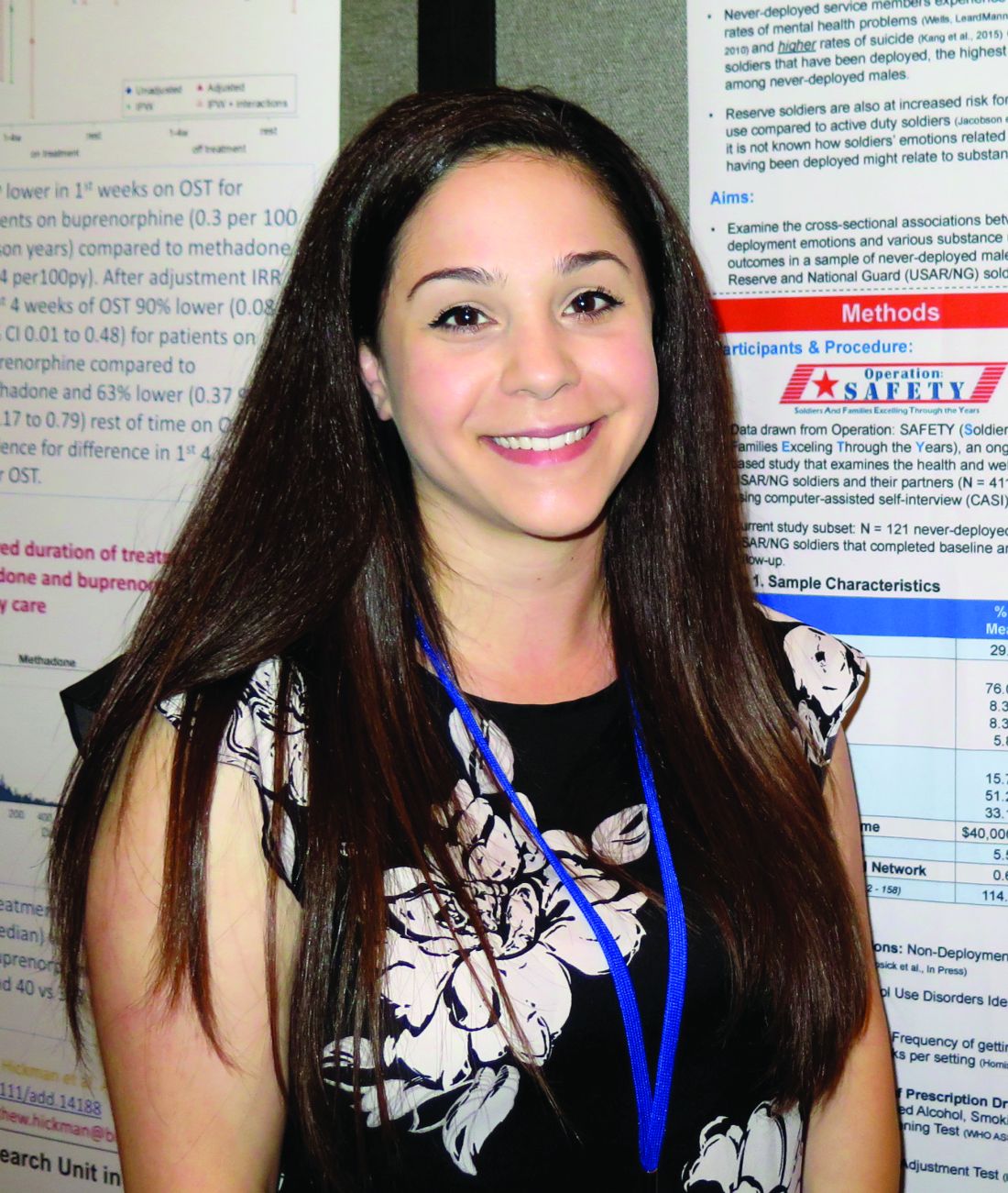
Measures included the Non-Deployment Emotions Questionnaire, the Alcohol Use Disorders Identification Test, the National Institute on Drug Abuse Modified Alcohol, Smoking and Substance Involvement Screening Test 2.0, and the Marital Adjustment Test. The researchers used separate models to examine the relationship between nondeployment emotions and alcohol problems, frequent heavy drinking, current nonmedical use of prescription drugs, and current illicit drug use, and controlled for years of military service, number of military friends in the social network, and marital satisfaction.
Ms. Hoopsick reported results from 121 never-deployed, male soldiers who completed the survey at baseline and at a 1-year follow-up. Their mean age was 30 years, 76% were non-Hispanic white, and 84% had at least some college education. In addition, the soldiers had served a mean of 5.5 years, had a mean of 0.6 military friends in the social network, and had a mean marital satisfaction score of 114 out of 158 possible points.
On responses to the Non-Deployment Emotions Questionnaire, 65% of nondeployed soldiers felt guilt, 56% felt decreased value within his unit, 50% felt decreased camaraderie within his unit, and 50% felt decreased connectedness within his unit for having never been deployed. “We did anticipate that some of these soldiers were going to have poor outcomes related to their nondeployment emotions, but it was surprising to see how many of the soldiers did express having negative emotions related to having never been deployed,” Ms. Hoopsick said. After the researchers controlled for years of military service, the number of military friends in the soldier’s social network, and marital satisfaction, more negative nondeployment emotions were associated with a greater likelihood of alcohol problems (adjusted risk ratio, 1.06), frequent heavy drinking (ARR, 1.03), and current nonmedical use of prescription drugs (ARR, 1.21).
She acknowledged certain limitations of the study, including its cross-sectional nature. she said.
The National Institute on Drug Abuse provided funding to Ms. Hoopsick’s mentor and coauthor, Gregory G. Homish, PhD, and the study received additional funding from the National Center for Advancing Translational Sciences and the Health Resources and Services Administration.
SAN DIEGO – Reserve soldiers who have never been deployed are just as likely to have poor outcomes related to mental health and substance use as their peers who have been deployed, results from a novel survey suggest.
“We tend to focus on screening and intervention efforts for soldiers who have been deployed and who have experienced combat, but we should also focus our intervention and screening efforts on soldiers who have never been deployed,” lead study author Rachel A. Hoopsick, MPH, said in an interview at the annual meeting of the College on Problems of Drug Dependence. “Both are at risk for substance use and mental illness.”
Measures included the Non-Deployment Emotions Questionnaire, the Alcohol Use Disorders Identification Test, the National Institute on Drug Abuse Modified Alcohol, Smoking and Substance Involvement Screening Test 2.0, and the Marital Adjustment Test. The researchers used separate models to examine the relationship between nondeployment emotions and alcohol problems, frequent heavy drinking, current nonmedical use of prescription drugs, and current illicit drug use, and controlled for years of military service, number of military friends in the social network, and marital satisfaction.
Ms. Hoopsick reported results from 121 never-deployed, male soldiers who completed the survey at baseline and at a 1-year follow-up. Their mean age was 30 years, 76% were non-Hispanic white, and 84% had at least some college education. In addition, the soldiers had served a mean of 5.5 years, had a mean of 0.6 military friends in the social network, and had a mean marital satisfaction score of 114 out of 158 possible points.
On responses to the Non-Deployment Emotions Questionnaire, 65% of nondeployed soldiers felt guilt, 56% felt decreased value within his unit, 50% felt decreased camaraderie within his unit, and 50% felt decreased connectedness within his unit for having never been deployed. “We did anticipate that some of these soldiers were going to have poor outcomes related to their nondeployment emotions, but it was surprising to see how many of the soldiers did express having negative emotions related to having never been deployed,” Ms. Hoopsick said. After the researchers controlled for years of military service, the number of military friends in the soldier’s social network, and marital satisfaction, more negative nondeployment emotions were associated with a greater likelihood of alcohol problems (adjusted risk ratio, 1.06), frequent heavy drinking (ARR, 1.03), and current nonmedical use of prescription drugs (ARR, 1.21).
She acknowledged certain limitations of the study, including its cross-sectional nature. she said.
The National Institute on Drug Abuse provided funding to Ms. Hoopsick’s mentor and coauthor, Gregory G. Homish, PhD, and the study received additional funding from the National Center for Advancing Translational Sciences and the Health Resources and Services Administration.
SAN DIEGO – Reserve soldiers who have never been deployed are just as likely to have poor outcomes related to mental health and substance use as their peers who have been deployed, results from a novel survey suggest.
“We tend to focus on screening and intervention efforts for soldiers who have been deployed and who have experienced combat, but we should also focus our intervention and screening efforts on soldiers who have never been deployed,” lead study author Rachel A. Hoopsick, MPH, said in an interview at the annual meeting of the College on Problems of Drug Dependence. “Both are at risk for substance use and mental illness.”
Measures included the Non-Deployment Emotions Questionnaire, the Alcohol Use Disorders Identification Test, the National Institute on Drug Abuse Modified Alcohol, Smoking and Substance Involvement Screening Test 2.0, and the Marital Adjustment Test. The researchers used separate models to examine the relationship between nondeployment emotions and alcohol problems, frequent heavy drinking, current nonmedical use of prescription drugs, and current illicit drug use, and controlled for years of military service, number of military friends in the social network, and marital satisfaction.
Ms. Hoopsick reported results from 121 never-deployed, male soldiers who completed the survey at baseline and at a 1-year follow-up. Their mean age was 30 years, 76% were non-Hispanic white, and 84% had at least some college education. In addition, the soldiers had served a mean of 5.5 years, had a mean of 0.6 military friends in the social network, and had a mean marital satisfaction score of 114 out of 158 possible points.
On responses to the Non-Deployment Emotions Questionnaire, 65% of nondeployed soldiers felt guilt, 56% felt decreased value within his unit, 50% felt decreased camaraderie within his unit, and 50% felt decreased connectedness within his unit for having never been deployed. “We did anticipate that some of these soldiers were going to have poor outcomes related to their nondeployment emotions, but it was surprising to see how many of the soldiers did express having negative emotions related to having never been deployed,” Ms. Hoopsick said. After the researchers controlled for years of military service, the number of military friends in the soldier’s social network, and marital satisfaction, more negative nondeployment emotions were associated with a greater likelihood of alcohol problems (adjusted risk ratio, 1.06), frequent heavy drinking (ARR, 1.03), and current nonmedical use of prescription drugs (ARR, 1.21).
She acknowledged certain limitations of the study, including its cross-sectional nature. she said.
The National Institute on Drug Abuse provided funding to Ms. Hoopsick’s mentor and coauthor, Gregory G. Homish, PhD, and the study received additional funding from the National Center for Advancing Translational Sciences and the Health Resources and Services Administration.
REPORTING FROM CPDD 2018
Key clinical point: All military personnel, regardless of deployment status, could be at risk for substance use.
Major finding:. More negative nondeployment emotions were associated with a greater likelihood of alcohol problems (adjusted risk ratio, 1.06), frequent heavy drinking (ARR, 1.03), and current nonmedical use of prescription drugs (ARR, 1.21).
Study details: Responses from 121 never-deployed, male U.S. Army Reserve/National Guard soldiers who completed surveys at baseline and at a 1-year follow-up.
Disclosures: The National Institute on Drug Abuse provided funding to Ms. Hoopsick’s mentor and coauthor, Gregory G. Homish, PhD, and the study received additional funding from the National Center for Advancing Translational Sciences and the Health Resources and Services Administration.
Launching the Moderate to Severe Asthma Center of Excellence
The American College of Chest Physicians (CHEST) announces a new partnership with Medscape focused on supporting physicians in addressing the challenges of diagnosing and treating moderate to severe asthma. The Moderate to Severe Asthma Center of Excellence will provide news, expert commentary, and insights on challenging cases to physicians specializing in chest medicine, allergy, primary care, pediatrics, and emergency medicine.

Medscape is a leading source of clinical news, health information, and point-of-care tools for physicians and health-care professionals. This new Center of Excellence available on Medscape.com will explore the diagnostic, therapeutic, and prevention strategies associated with moderate to severe asthma, including the latest research and breakthroughs. Topics will include challenges in classifying and diagnosing disease; risks, benefits, and barriers to treatment; and impact on patients’ quality of life.
Don’t miss Dr. Aaron Holley’s video on “Diagnosing Severe Asthma: ‘Not as Easy as It Sounds’ ”
Visit the Moderate to Severe Asthma Center of Excellence at https://www.medscape.com/resource/moderate-severe-asthma
The American College of Chest Physicians (CHEST) announces a new partnership with Medscape focused on supporting physicians in addressing the challenges of diagnosing and treating moderate to severe asthma. The Moderate to Severe Asthma Center of Excellence will provide news, expert commentary, and insights on challenging cases to physicians specializing in chest medicine, allergy, primary care, pediatrics, and emergency medicine.
Medscape is a leading source of clinical news, health information, and point-of-care tools for physicians and health-care professionals. This new Center of Excellence available on Medscape.com will explore the diagnostic, therapeutic, and prevention strategies associated with moderate to severe asthma, including the latest research and breakthroughs. Topics will include challenges in classifying and diagnosing disease; risks, benefits, and barriers to treatment; and impact on patients’ quality of life.
Don’t miss Dr. Aaron Holley’s video on “Diagnosing Severe Asthma: ‘Not as Easy as It Sounds’ ”
Visit the Moderate to Severe Asthma Center of Excellence at https://www.medscape.com/resource/moderate-severe-asthma
The American College of Chest Physicians (CHEST) announces a new partnership with Medscape focused on supporting physicians in addressing the challenges of diagnosing and treating moderate to severe asthma. The Moderate to Severe Asthma Center of Excellence will provide news, expert commentary, and insights on challenging cases to physicians specializing in chest medicine, allergy, primary care, pediatrics, and emergency medicine.
Medscape is a leading source of clinical news, health information, and point-of-care tools for physicians and health-care professionals. This new Center of Excellence available on Medscape.com will explore the diagnostic, therapeutic, and prevention strategies associated with moderate to severe asthma, including the latest research and breakthroughs. Topics will include challenges in classifying and diagnosing disease; risks, benefits, and barriers to treatment; and impact on patients’ quality of life.
Don’t miss Dr. Aaron Holley’s video on “Diagnosing Severe Asthma: ‘Not as Easy as It Sounds’ ”
Visit the Moderate to Severe Asthma Center of Excellence at https://www.medscape.com/resource/moderate-severe-asthma
Galectin-3: A new post-MI prognostic biomarker?
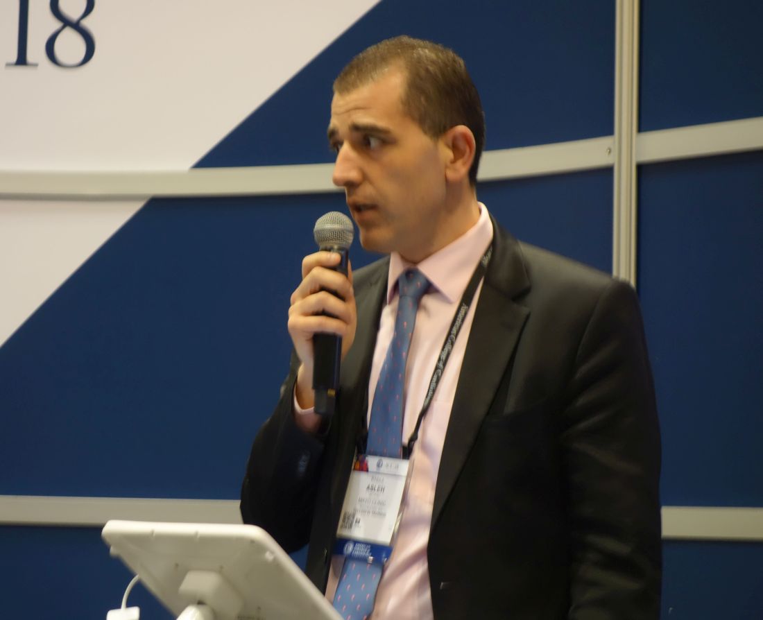
ORLANDO – An elevated circulating galactin-3 level after an acute MI is a potent long-term predictor of both heart failure and mortality, independent of known prognostic markers, Rabea Asleh, MD, PhD, reported at the annual meeting of the American College of Cardiology.
“These findings suggest that galectin-3 measurement may have a role in the risk stratification of patients presenting with MI,” according to Dr. Asleh, an Israeli cardiologist doing a fellowship in advanced heart failure and transplant cardiology at the Mayo Clinic in Rochester, Minn.
“The changing clinical presentation of MI necessitates evolution in our approach to risk stratification,” he explained. “Over the last 2 decades we’ve observed a change in the epidemiology of MI, with more patients developing non-ST-elevation MI compared to STEMI. They present at an older age and develop heart failure with preserved ejection fraction more than heart failure with reduced ejection fraction.”
He presented a prospective population-based community cohort study of 1,401 Olmsted County, Minn., residents who had a validated MI during 2002-2012. Their mean age was 67 years, 61% were men, and 79% presented with non-STEMI. During a mean follow-up of 5.3 years, 389 of the participants developed heart failure and 512 patients died.
Galectin-3 was measured a median of 2 days post MI. The median level was 18.4 ng/mL. Patients were divided into tertiles based upon their galactin-3 measurement: Tertile 1 required a post-MI galectin-3 level below 15.2 ng/mL; tertile 2 had a level of 15.2-22.6 ng/mL; and the top tertile was for individuals with a galectin-3 above 22.6 ng/mL.
Of note, patients with a higher galectin-3 level were older, had a higher prevalence of diabetes, hypertension, hyperlipidemia, anterior MI, a higher Killip class, a higher Charlson comorbidity score, and a lower peak troponin T level. They also had a lower estimated glomerular filtration rate; indeed, the median eGFR in the top tertile for galactin-3 was 48 mL/min per 1.73 m2, compared with 68 mL/min in the lowest galectin-3 tertile. Women accounted for 27% of patients in tertile 1, 41% in tertile 2, and fully half of those in tertile 3.
In an unadjusted analysis, the risk of mortality during follow-up was sixfold greater for patients in galectin-3 tertile 3 than in tertile 1; the risk of heart failure was increased 5.5-fold.
More meaningfully, in a Cox multivariate analysis extensively adjusted for age, gender, comorbidities, malignancy, standard cardiovascular risk factors, MI characteristics, eGFR, Killip class, cardiac troponin T, and other potential confounders, patients in galectin-3 tertile 2 had a 1.6-fold increased risk of death and a 1.62-fold increased likelihood of heart failure during follow-up, compared with subjects in tertile 1, Dr. Asleh noted.
Patients in tertile 3 had a 2.4-fold increased risk of death and were at 2.1 times greater risk of heart failure than those in tertile 1. The degree of risk for heart failure associated with elevated galactin-3 was virtually identical for heart failure with preserved as compared with reduced ejection fraction, he added.
Session cochair L. Kristin Newby, MD, of Duke University, Durham, N.C., noted that the Mayo study did not adjust for brain natriuretic peptide (BNP) or N-terminal pro hormone BNP (NT-proBNP), both of which are known to be strong predictors of both heart failure and mortality after acute MI. Doesn’t their absence weaken the strength of galactin-3’s prognostic power as demonstrated in the study? she asked.
Dr. Asleh replied that those biomarkers weren’t collected in this study, which began in 2002.
“What I can tell you is, other studies show there is only a weak correlation between galactin-3 and NT-proBNP post MI. Some studies have even shown an inverse correlation,” he said. “The pathophysiological explanation is that galactin-3 is more implicated in fibrosis before the stage of development of left ventricular loading and stretching of the myocardium. So galactin-3 may be implicated in LV fibrosis leading to heart failure before the NT-proBNP comes into play.”
Also, he cited a study by other investigators conducted in patients with a left ventricular assist device for advanced heart failure. Upon device-induced left ventricular unloading the patients’ NT-proBNP levels dropped significantly while their galactin-3 remained high and unchanged. This suggests the two biomarkers are implicated in different disease pathways.
Both animal and human studies indicate galactin-3 is involved specifically in fibrosis, as opposed to, say, C-reactive protein, a well established marker of systemic inflammation, the cardiologist added.
Dr. Asleh reported having no financial conflicts of interest regarding his study, which was supported by the National Institutes of Health.
ORLANDO – An elevated circulating galactin-3 level after an acute MI is a potent long-term predictor of both heart failure and mortality, independent of known prognostic markers, Rabea Asleh, MD, PhD, reported at the annual meeting of the American College of Cardiology.
“These findings suggest that galectin-3 measurement may have a role in the risk stratification of patients presenting with MI,” according to Dr. Asleh, an Israeli cardiologist doing a fellowship in advanced heart failure and transplant cardiology at the Mayo Clinic in Rochester, Minn.
“The changing clinical presentation of MI necessitates evolution in our approach to risk stratification,” he explained. “Over the last 2 decades we’ve observed a change in the epidemiology of MI, with more patients developing non-ST-elevation MI compared to STEMI. They present at an older age and develop heart failure with preserved ejection fraction more than heart failure with reduced ejection fraction.”
He presented a prospective population-based community cohort study of 1,401 Olmsted County, Minn., residents who had a validated MI during 2002-2012. Their mean age was 67 years, 61% were men, and 79% presented with non-STEMI. During a mean follow-up of 5.3 years, 389 of the participants developed heart failure and 512 patients died.
Galectin-3 was measured a median of 2 days post MI. The median level was 18.4 ng/mL. Patients were divided into tertiles based upon their galactin-3 measurement: Tertile 1 required a post-MI galectin-3 level below 15.2 ng/mL; tertile 2 had a level of 15.2-22.6 ng/mL; and the top tertile was for individuals with a galectin-3 above 22.6 ng/mL.
Of note, patients with a higher galectin-3 level were older, had a higher prevalence of diabetes, hypertension, hyperlipidemia, anterior MI, a higher Killip class, a higher Charlson comorbidity score, and a lower peak troponin T level. They also had a lower estimated glomerular filtration rate; indeed, the median eGFR in the top tertile for galactin-3 was 48 mL/min per 1.73 m2, compared with 68 mL/min in the lowest galectin-3 tertile. Women accounted for 27% of patients in tertile 1, 41% in tertile 2, and fully half of those in tertile 3.
In an unadjusted analysis, the risk of mortality during follow-up was sixfold greater for patients in galectin-3 tertile 3 than in tertile 1; the risk of heart failure was increased 5.5-fold.
More meaningfully, in a Cox multivariate analysis extensively adjusted for age, gender, comorbidities, malignancy, standard cardiovascular risk factors, MI characteristics, eGFR, Killip class, cardiac troponin T, and other potential confounders, patients in galectin-3 tertile 2 had a 1.6-fold increased risk of death and a 1.62-fold increased likelihood of heart failure during follow-up, compared with subjects in tertile 1, Dr. Asleh noted.
Patients in tertile 3 had a 2.4-fold increased risk of death and were at 2.1 times greater risk of heart failure than those in tertile 1. The degree of risk for heart failure associated with elevated galactin-3 was virtually identical for heart failure with preserved as compared with reduced ejection fraction, he added.
Session cochair L. Kristin Newby, MD, of Duke University, Durham, N.C., noted that the Mayo study did not adjust for brain natriuretic peptide (BNP) or N-terminal pro hormone BNP (NT-proBNP), both of which are known to be strong predictors of both heart failure and mortality after acute MI. Doesn’t their absence weaken the strength of galactin-3’s prognostic power as demonstrated in the study? she asked.
Dr. Asleh replied that those biomarkers weren’t collected in this study, which began in 2002.
“What I can tell you is, other studies show there is only a weak correlation between galactin-3 and NT-proBNP post MI. Some studies have even shown an inverse correlation,” he said. “The pathophysiological explanation is that galactin-3 is more implicated in fibrosis before the stage of development of left ventricular loading and stretching of the myocardium. So galactin-3 may be implicated in LV fibrosis leading to heart failure before the NT-proBNP comes into play.”
Also, he cited a study by other investigators conducted in patients with a left ventricular assist device for advanced heart failure. Upon device-induced left ventricular unloading the patients’ NT-proBNP levels dropped significantly while their galactin-3 remained high and unchanged. This suggests the two biomarkers are implicated in different disease pathways.
Both animal and human studies indicate galactin-3 is involved specifically in fibrosis, as opposed to, say, C-reactive protein, a well established marker of systemic inflammation, the cardiologist added.
Dr. Asleh reported having no financial conflicts of interest regarding his study, which was supported by the National Institutes of Health.
ORLANDO – An elevated circulating galactin-3 level after an acute MI is a potent long-term predictor of both heart failure and mortality, independent of known prognostic markers, Rabea Asleh, MD, PhD, reported at the annual meeting of the American College of Cardiology.
“These findings suggest that galectin-3 measurement may have a role in the risk stratification of patients presenting with MI,” according to Dr. Asleh, an Israeli cardiologist doing a fellowship in advanced heart failure and transplant cardiology at the Mayo Clinic in Rochester, Minn.
“The changing clinical presentation of MI necessitates evolution in our approach to risk stratification,” he explained. “Over the last 2 decades we’ve observed a change in the epidemiology of MI, with more patients developing non-ST-elevation MI compared to STEMI. They present at an older age and develop heart failure with preserved ejection fraction more than heart failure with reduced ejection fraction.”
He presented a prospective population-based community cohort study of 1,401 Olmsted County, Minn., residents who had a validated MI during 2002-2012. Their mean age was 67 years, 61% were men, and 79% presented with non-STEMI. During a mean follow-up of 5.3 years, 389 of the participants developed heart failure and 512 patients died.
Galectin-3 was measured a median of 2 days post MI. The median level was 18.4 ng/mL. Patients were divided into tertiles based upon their galactin-3 measurement: Tertile 1 required a post-MI galectin-3 level below 15.2 ng/mL; tertile 2 had a level of 15.2-22.6 ng/mL; and the top tertile was for individuals with a galectin-3 above 22.6 ng/mL.
Of note, patients with a higher galectin-3 level were older, had a higher prevalence of diabetes, hypertension, hyperlipidemia, anterior MI, a higher Killip class, a higher Charlson comorbidity score, and a lower peak troponin T level. They also had a lower estimated glomerular filtration rate; indeed, the median eGFR in the top tertile for galactin-3 was 48 mL/min per 1.73 m2, compared with 68 mL/min in the lowest galectin-3 tertile. Women accounted for 27% of patients in tertile 1, 41% in tertile 2, and fully half of those in tertile 3.
In an unadjusted analysis, the risk of mortality during follow-up was sixfold greater for patients in galectin-3 tertile 3 than in tertile 1; the risk of heart failure was increased 5.5-fold.
More meaningfully, in a Cox multivariate analysis extensively adjusted for age, gender, comorbidities, malignancy, standard cardiovascular risk factors, MI characteristics, eGFR, Killip class, cardiac troponin T, and other potential confounders, patients in galectin-3 tertile 2 had a 1.6-fold increased risk of death and a 1.62-fold increased likelihood of heart failure during follow-up, compared with subjects in tertile 1, Dr. Asleh noted.
Patients in tertile 3 had a 2.4-fold increased risk of death and were at 2.1 times greater risk of heart failure than those in tertile 1. The degree of risk for heart failure associated with elevated galactin-3 was virtually identical for heart failure with preserved as compared with reduced ejection fraction, he added.
Session cochair L. Kristin Newby, MD, of Duke University, Durham, N.C., noted that the Mayo study did not adjust for brain natriuretic peptide (BNP) or N-terminal pro hormone BNP (NT-proBNP), both of which are known to be strong predictors of both heart failure and mortality after acute MI. Doesn’t their absence weaken the strength of galactin-3’s prognostic power as demonstrated in the study? she asked.
Dr. Asleh replied that those biomarkers weren’t collected in this study, which began in 2002.
“What I can tell you is, other studies show there is only a weak correlation between galactin-3 and NT-proBNP post MI. Some studies have even shown an inverse correlation,” he said. “The pathophysiological explanation is that galactin-3 is more implicated in fibrosis before the stage of development of left ventricular loading and stretching of the myocardium. So galactin-3 may be implicated in LV fibrosis leading to heart failure before the NT-proBNP comes into play.”
Also, he cited a study by other investigators conducted in patients with a left ventricular assist device for advanced heart failure. Upon device-induced left ventricular unloading the patients’ NT-proBNP levels dropped significantly while their galactin-3 remained high and unchanged. This suggests the two biomarkers are implicated in different disease pathways.
Both animal and human studies indicate galactin-3 is involved specifically in fibrosis, as opposed to, say, C-reactive protein, a well established marker of systemic inflammation, the cardiologist added.
Dr. Asleh reported having no financial conflicts of interest regarding his study, which was supported by the National Institutes of Health.
REPORTING FROM ACC 18
Key clinical point: Galectin-3 level post-MI is a potent long-term predictor of both heart failure and mortality independent of known prognostic markers.
Major finding: Post-MI patients in the top tertile of circulating galectin-3 were at an adjusted 2.4-fold increased mortality risk and a 2.05-fold greater risk of developing heart failure compared with those in the lowest tertile.
Study details: This prospective population-based cohort study included 1,401 MI patients followed for a mean of 5.3 years.
Disclosures: The National Institutes of Health supported the study. The presenter reported having no financial conflicts of interest.
Continuous Cryotherapy vs Ice Following Total Shoulder Arthroplasty: A Randomized Control Trial
ABSTRACT
Postoperative pain management is an important component of total shoulder arthroplasty (TSA). Continuous cryotherapy (CC) has been proposed as a means of improving postoperative pain control. However, CC represents an increased cost not typically covered by insurance. The purpose of this study is to compare CC to plain ice (ICE) following TSA. The hypothesis was that CC would lead to lower pain scores and decreased narcotic usage during the first 2 weeks postoperatively.
A randomized controlled trial was performed to compare CC to ICE. Forty patients were randomized to receive either CC or ICE following TSA. The rehabilitation and pain control protocols were otherwise standardized. Visual analog scales (VAS) for pain, satisfaction with cold therapy, and quality of sleep were recorded preoperatively and postoperatively at 24 hours, 3 days, 7 days, and 14 days following surgery. Narcotic usage in morphine equivalents was also recorded.
No significant differences in preoperative pain (5.9 vs 6.8; P = .121), or postoperative pain at 24 hours (4.2 vs 4.3; P = .989), 3 days (4.8 vs 4.7; P = .944), 7 days (2.9 vs 3.3; P = .593) or 14 days (2.5 vs 2.7; P = .742) were observed between the CC and ICE groups. Similarly, no differences in quality of sleep, satisfaction with the cold therapy, or narcotic usage at any time interval were observed between the 2 groups.
No differences in pain control, quality of sleep, patient satisfaction, or narcotic usage were detected between CC and ICE following TSA. CC may offer convenience as an advantage, but the increased cost associated with this type of treatment may not be justified.
The number of total shoulder arthroplasties (TSAs) performed annually is increasing dramatically.1 At the same time, there has been a push toward decreased length of hospital stay and earlier mobilization following joint replacement surgery. Central to these goals is adequate pain control. Multimodal pain pathways exist, and one of the safest and cheapest methods of pain control is cold therapy, which can be accomplished with continuous cryotherapy (CC) or plain ice (ICE).
Continue to: The mechanism of cryotherapy...
The mechanism of cryotherapy for controlling pain is poorly understood. Cryotherapy reduces leukocyte migration and slows down nerve signal transmission, which reduces inflammation, thereby producing a short-term analgesic effect. Stalman and colleagues2 reported on a randomized control study that evaluated the effects of postoperative cooling after knee arthroscopy. Measurements of metabolic and inflammatory markers in the synovial membrane were used to assess whether cryotherapy provides a temperature-sensitive release of prostaglandin E2. Cryotherapy lowered the temperature in the postoperative knee, and synovial prostaglandin concentrations were correlated with temperature. Because prostaglandin is a marker of inflammation and pain, the conclusion was that postoperative cooling appeared to have an anti-inflammatory effect.
The knee literature contains multiple studies that have examined the benefits of cryotherapy after both arthroscopic and arthroplasty procedures. The clinical benefits on pain have been equivocal with some studies showing improvements using cryotherapy3,4 and others showing no difference in the treatment group.5,6
Few studies have examined cryotherapy for the shoulder. Speer and colleagues7 demonstrated that postoperative use of CC was effective in reducing recovery time after shoulder surgery. However; they did not provide an ICE comparative group and did not focus specifically on TSA. In another study, Kraeutler and colleagues8 examined only arthroscopic shoulder surgery cases in a randomized prospective trial and found no significant different between CC and ICE. They concluded that there did not appear to be a significant benefit in using CC over ICE for arthroscopic shoulder procedures.
The purpose of this study is to prospectively evaluate CC and ICE following TSA. The hypothesis was that CC leads to improved pain control, less narcotic consumption, and improved quality of sleep compared to ICE in the immediate postoperative period following TSA.
MATERIALS AND METHODS
This was a prospective randomized control study of patients undergoing TSA receiving either CC or ICE postoperatively. Institutional Review Board approval was obtained before commencement of the study. Inclusion criteria included patients aged 30 to 90 years old undergoing a primary or revision shoulder arthroplasty procedure between June 2015 and January 2016. Exclusion criteria included hemiarthroplasty procedures.
Continue to: Three patients refused...
Three patients refused to participate in the study. Enrollment was performed until 40 patients were enrolled in the study (20 patients in each group). Randomization was performed with a random number generator, and patients were assigned to a treatment group following consent to participate. Complete follow-up was available for all patients. There were 13 (65%) male patients in the CC group. The average age of the CC group at the time of surgery was 68.7 years (range). There were 11 male patients in the ICE group. The average age of the ICE group at the time of surgery was 73.2 years (range). The dominant extremity was involved in 9 (45%) patients in the CC group and in 11 patients (55%) in the ICE group. Surgical case specifics are summarized in Table 1.
Table 1. Summary of Surgical Cases
| CC group (n = 20) | ICE group (n = 20) |
Primary TSA | 7 (35%) | 9 (45%) |
Primary RSA | 12 (60%) | 9 (45%) |
Revision arthroplasty | 1 (5%) | 2 (10%) |
Abbreviations: CC, continuous cryotherapy; ICE, plain ice; RSA, reverse shoulder arthroplasty; TSA, total shoulder arthroplasty.
All surgeries were performed by Dr. Denard. All patients received a single-shot interscalene nerve block prior to the procedure. A deltopectoral approach was utilized, and the subscapularis was managed with the peel technique.9 All patients were admitted to the hospital following surgery. Standard postoperative pain control consisted of as-needed intravenous morphine (1-2 mg every 2 hours, as needed) or an oral narcotic (hydrocodone/acetaminophen 5/325mg, 1-2 every 4 hours, as needed) which was also provided at discharge. However, total narcotic usage was recorded in morphine equivalents to account for substitutions. No non-steroidal anti-inflammatory drugs were allowed until 3 months postoperatively.
The CC group received treatment from a commercially available cryotherapy unit (Polar Care; Breg). All patients received instructions by a medical professional on how to use the unit. The unit was applied immediately postoperatively and set at a temperature of 45°F to 55°F. Patients were instructed to use the unit continuously during postoperative days 0 to 3. This cryotherapy was administered by a nurse while in the hospital but was left to the responsibility of the patient upon discharge. Patients were instructed to use the unit as needed for pain control during the day and continuously while asleep from days 4 to14.
The ICE group used standard ice packs postoperatively. The patients were instructed to apply an ice pack for 20 min every 2 hours while awake during days 0 to 3. This therapy was administered by a nurse while in the hospital but left to the responsibility of the patient upon discharge. Patients were instructed to use ice packs as needed for pain control during the day at a maximum of 20 minutes per hour on postoperative days 4 to 14. Compliance by both groups was monitored using a patient survey after hospital discharge. The number of hours that patients used either the CC or ICE per 24-hour period was recorded at 24 hours, 3 days, 7 days, and 14 days. The nursing staff recorded the number of hours of use of either cold modality for each patient prior to hospital discharge. The average length of stay as an inpatient was 1.2 days for the CC group and 1.3 days for the ICE group.
Visual analog scales (VAS) for pain, satisfaction with the cold therapy, and quality of sleep were recorded preoperatively and postoperatively at 24 hours, 3 days, 7 days, and 14 days following surgery.
Continue to: The Wilcoxon rank-sum test...
STATISTICAL METHOD
The Wilcoxon rank-sum test was used to assess whether scores changed significantly from the preoperative period to the different postoperative time intervals, as well as to assess the values for pain, quality of sleep, and patient satisfaction. P-values <.05 were considered significant.
RESULTS
No differences were observed in the baseline characteristics between the 2 groups. Both groups showed improvements in pain, quality of sleep, and satisfaction with the cold therapy from the preoperative period to the final follow-up.
The VAS pain scores were not different between the CC and ICE groups preoperatively (5.9 vs 6.8; P = .121) or postoperatively at 24 hours (4.2 vs 4.3; P = .989), 3 days (4.8 vs 4.7; P = .944), 7 days (2.9 vs 3.3; P = .593), or 14 days (2.5 vs 2.7; P = .742). Both cohorts demonstrated improved overall pain throughout the study period. These findings are summarized in Table 2.
Table 2. Summary of VAS Pain Scores With Cold Therapy
| CC group (mean ± SD) | ICE group (mean ± SD) | P value | 95% CI |
Preoperative | 5.9 ± 4.1 | 6.8 ± 5.3 | .121 | 3.3-8.3 |
24 hours | 4.2 ± 3.0 | 4.3 ± 3.1 | .989 | 2.9-5.7 |
3 days | 4.8 ± 2.7 | 4.7 ± 3.2 | .944 | 3.2-6.3 |
7 days | 2.9 ± 1.8 | 3.3 ± 2.5 | .593 | 2.1-4.4 |
14 days | 2.5 ± 2.1 | 2.7 ± 1.8 | .742 | 1.5-3.6 |
Abbreviations: CC, continuous cryotherapy; CI, confidence interval; ICE, plain ice; VAS, visual analog scales.
The number of morphine equivalents of pain medication was not different between the CC and ICE groups postoperatively at 24 hours (43 vs 38 mg; P = .579), 3 days (149 vs 116 mg; P = .201), 7 days (308 vs 228 mg; P = .181), or 14 days (431 vs 348 mg; P = .213). Both groups showed increased narcotic consumption from 24 hours postoperatively until the 2-week follow-up. Narcotic consumption is summarized in Table 3.
Table 3. Summary of Narcotic Consumption in Morphine Equivalents
| CC group (mean ± SD) | ICE group (mean ± SD) | P value | 95% CI |
24 hours | 43.0 ± 36.7 | 38.0 ± 42.9 | .579 | 17.9-60.1 |
3 days | 149.0 ± 106.5 | 116.3 ± 108.9 | .201 | 63.4-198.7 |
7 days | 308.1 ± 234.0 | 228 ± 258.3 | .181 | 107.1-348.9 |
14 days | 430.8 ± 384.2 | 347.5 ± 493.4 | .213 | 116.6-610.6 |
Abbreviations: CC, continuous cryotherapy; CI, confidence interval; ICE, plain ice.
VAS for quality of sleep improved in both groups from 24 hours postoperatively until the final follow-up. However, no significant differences in sleep quality were observed between the CC and ICE groups postoperatively at 24 hours (5.1 vs 4.3; P = .382), 3 days (5.1 vs 5.3; P = .601), 7 days (6.0 vs 6.7; P = .319), or 14 days (6.5 vs 7.1; P = .348). The VAS scores for sleep quality are reported in Table 4.
Table 4. Summary of VAS Sleep Quality With Cold Therapya
| CC group (mean ± SD) | ICE group (mean ± SD) | P value | 95% CI |
24 hours | 5.1 ± 2.8 | 4.3 ± 2.4 | .382 | 3.2-6.4 |
3 days | 5.1 ± 1.9 | 5.3 ± 2.3 | .601 | 4.2-6.5 |
7 days | 6.0 ± 2.3 | 6.7 ± 2.1 | .319 | 4.9-7.7 |
14 days | 6.5 ± 2.3 | 7.1 ± 2.5 | .348 | 5.3-8.4 |
a0-10 rating with 10 being the highest possible score.
Abbreviations: CC, continuous cryotherapy; CI, confidence interval; ICE, plain ice; VAS, visual analog scales.
Continue to: Finally, VAS patient satisfaction...
Finally, VAS patient satisfaction scores were not different between the CC and ICE groups postoperatively at 24 hours (7.3 vs 6.1; P = .315), 3 days (6.1 vs 6.6; P = .698), 7 days (6.6 vs 6.9; P = .670), or 14 days (7.1 vs 6.3; P = .288).
While compliance within each group utilizing the randomly assigned cold modality was similar, the usage by the CC group was consistently higher at all time points recorded. No complications or reoperations were observed in either group.
DISCUSSION
The optimal method for managing postoperative pain from an arthroplasty procedure is controversial. This prospective randomized study attempted to confirm the hypothesis that CC infers better pain control, improves quality of sleep, and decreases narcotic usage compared to ICE in the first 2 weeks after a TSA procedure. The results of this study refuted our hypothesis, demonstrating no significant difference in pain control, satisfaction, narcotic usage, or sleep quality between the CC and ICE cohorts at all time points studied.
Studies on knees and lower extremities demonstrate equivocal results for the role CC plays in providing improved postoperative pain control. Thienpont10 evaluated CC in a randomized control trial comparing plain ice packs postoperatively in patients who underwent TKA. The author found no significant difference in VAS for pain or narcotic consumption in morphine equivalents. Thienpont10 recommended that CC not be used for outpatient knee arthroplasty as it is an additional cost that does not improve pain significantly. Healy and colleagues5 reported similar results that CC did not demonstrate a difference in narcotic requirement or pain control compared to plain ice packs, as well as no difference in local postoperative swelling or wound drainage. However, a recently published randomized trial by Su and colleagues11 comparing a cryopneumatic device and ICE with static compression in patients who underwent TKA demonstrated significantly lower narcotic consumption and increased ambulation distances in the treatment group. The treatment group consumed approximately 170 mg morphine equivalents less than the control group between discharge and the 2-week postoperative visit. In addition, a significant difference was observed in the satisfaction scores in the treatment group.11 Similarly, a meta-analysis by Raynor and colleagues12 on randomized clinical trials comparing cryotherapy to a placebo group after anterior cruciate ligament reconstruction showed that cryotherapy is associated with significantly lower postoperative pain (P = .02), but demonstrated no difference in postoperative drainage (P = .23) or range of motion (P = .25).
Although multiple studies have been published regarding the efficacy of cryotherapy after knee surgery, very few studies have compared CC to conventional ICE after shoulder surgery. A prospective randomized trial was performed by Singh and colleagues13 to compare CC vs no ICE in open and arthroscopic shoulder surgery patients. Both the open and arthroscopic groups receiving CC demonstrated significant reductions in pain frequency and more restful sleep at the 7-day, 14-day, and 21-day intervals compared to the control group. However, they did not compare the commercial unit to ICE. In contrast, a study by Kraeutler and colleagues8 randomized 46 patients to receive either CC or ICE in the setting of arthroscopic shoulder surgery. Although no significant difference was observed in morphine equivalent dosage between the 2 groups, the CC group used more pain medication on every postoperative day during the first week after surgery. They found no difference between the 2 groups with regards to narcotic consumption or pain scores. The results of this study mirror those by Kraeutler and colleagues,8 demonstrating no difference in pain scores, sleep quality, or narcotic consumption.
Continue to: With rising costs in the US...
With rising costs in the US healthcare system, a great deal of interest has developed in the application of value-based principles to healthcare. Value can be defined as a gain in benefits over the costs expended.14 The average cost for a commercial CC unit used in this study was $260. A pack of ICE is a nominal cost. Based on the results of this study, the cost of the commercial CC device may not be justified when compared to the cost of an ice pack.
The major strengths of this study are the randomized design and multiple data points during the early postoperative period. However, there are several limitations. First, we did not objectively measure compliance of either therapy and relied only on a patient survey. Usage of the commercial CC unit in hours decreased over half between days 3 and 14. This occurred despite training on the application and specific instructions. We believe this reflects “real-world” usage, but it is possible that compliance affected our results. Second, all patients in this study had a single-shot interscalene block. While this is standard at our institution, it is possible that either CC or ICE would have a more significant effect in the absence of an interscalene block. Finally, we did not evaluate final outcomes in this study and therefore cannot determine if the final outcome was different between the 2 groups. Our goal was simply to evaluate the first 2 weeks following surgery, as this is the most painful period following TSA.
CONCLUSION
There was no difference between CC and ICE in terms of pain control, quality of sleep, patient satisfaction, or narcotic consumption following TSA. CC may offer convenience advantages, but the increased cost associated with this type of unit may not be justified.
1. Kim SH, Wise BL, Zhang Y, Szabo RM. Increasing incidence of shoulder arthroplasty in the United States. J Bone Joint Surg Am. 2011;93(24):2249-2254. doi:10.2106/jbjs.j.01994.
2. Stalman A, Berglund L, Dungnerc E, Arner P, Fellander-Tsai L. Temperature sensitive release of prostaglandin E2 and diminished energy requirements in synovial tissue with postoperative cryotherapy: a prospective randomized study after knee arthroscopy. J Bone Joint Surg Am. 2011;93(21):1961-1968. doi:10.2016/jbjs.j.01790.
3. Levy AS, Marmar E. The role of cold compression dressings in the postoperative treatment of total knee arthroplasty. Clin Orthop Relat Res. 1993;297:174-178. doi:10.1097/00003086-199312000-00029.
4. Webb JM, Williams D, Ivory JP, Day S, Williamson DM. The use of cold compression dressings after total knee replacement: a randomized controlled trial. Orthopaedics 1998;21(1):59-61.
5. Healy WL, Seidman J, Pfeifer BA, Brown DG. Cold compressive dressing after total knee arthroplasty. Clin Orthop Relat Res. 1994;299:143-146. doi:10.1097/00003086-199402000-00019.
6. Whitelaw GP, DeMuth KA, Demos HA, Schepsis A, Jacques E. The use of Cryo/Cuff versus ice and elastic wrap in the postoperative care of knee arthroscopy patients. Am J Knee Surg. 1995;8(1):28-30.
7. Speer KP, Warren RF, Horowitz L. The efficacy of cryotherapy in the postoperative shoulder. J Shoulder Elbow Surg. 1996;5(1):62-68. doi:10.16/s1058-2746(96)80032-2.
8. Kraeutler MJ, Reynolds KA, Long C, McCarthy EC. Compressive cryotherapy versus ice- a prospective, randomized study on postoperative pain in patients undergoing arthroscopic rotator cuff repair or subacromial decompression. J Shoulder Elbow Surg. 2015;24(6):854-859. doi:10.1016/j.jse.2015.02.004.
9. DeFranco MJ, Higgins LD, Warner JP. Subscapularis management in open shoulder surgery. J Am Acad Orthop Surg. 2010;18(12):707-717. doi:10.5435/00124635-201012000-00001.
10. Thienpont E. Does advanced cryotherapy reduce pain and narcotic consumption after knee arthroplasty. Clin Orthop Relat Res. 2014;472(11):3417-3423. doi:10.1007/s11999-014-3810-8.
11. Su EP, Perna M, Boettner F, Mayman DJ, et al. A prospective, multicenter, randomized trial to evaluate the efficacy of a cryopneumatic device on total knee arthroplasty recovery. J Bone Joint Surg Br. 2012;94(11 Suppl A):153-156. doi:10.1302/0301-620x.94B11.30832.
12. Raynor MC, Pietrobon R, Guller U, Higgins LD. Cryotherapy after ACL reconstruction- a meta analysis. J Knee Surg. 2005;18(2):123-129. doi:10.1055/s-0030-1248169.
13. Singh H, Osbahr DC, Holovacs TF, Cawley PW, Speer KP. The efficacy of continuous cryotherapy on the postoperative shoulder: a prospective randomized investigation. J Shoulder Elbow Surg. 2001;10(6):522-525. doi:10.1067/mse.2001.118415.
14. Black EM, Higgins LD, Warner JP. Value based shoulder surgery: outcomes driven, cost-conscious care. J Shoulder Elbow Surg. 2013;22(7):1-10. doi:10.1016/j.se.2013.02.008.
ABSTRACT
Postoperative pain management is an important component of total shoulder arthroplasty (TSA). Continuous cryotherapy (CC) has been proposed as a means of improving postoperative pain control. However, CC represents an increased cost not typically covered by insurance. The purpose of this study is to compare CC to plain ice (ICE) following TSA. The hypothesis was that CC would lead to lower pain scores and decreased narcotic usage during the first 2 weeks postoperatively.
A randomized controlled trial was performed to compare CC to ICE. Forty patients were randomized to receive either CC or ICE following TSA. The rehabilitation and pain control protocols were otherwise standardized. Visual analog scales (VAS) for pain, satisfaction with cold therapy, and quality of sleep were recorded preoperatively and postoperatively at 24 hours, 3 days, 7 days, and 14 days following surgery. Narcotic usage in morphine equivalents was also recorded.
No significant differences in preoperative pain (5.9 vs 6.8; P = .121), or postoperative pain at 24 hours (4.2 vs 4.3; P = .989), 3 days (4.8 vs 4.7; P = .944), 7 days (2.9 vs 3.3; P = .593) or 14 days (2.5 vs 2.7; P = .742) were observed between the CC and ICE groups. Similarly, no differences in quality of sleep, satisfaction with the cold therapy, or narcotic usage at any time interval were observed between the 2 groups.
No differences in pain control, quality of sleep, patient satisfaction, or narcotic usage were detected between CC and ICE following TSA. CC may offer convenience as an advantage, but the increased cost associated with this type of treatment may not be justified.
The number of total shoulder arthroplasties (TSAs) performed annually is increasing dramatically.1 At the same time, there has been a push toward decreased length of hospital stay and earlier mobilization following joint replacement surgery. Central to these goals is adequate pain control. Multimodal pain pathways exist, and one of the safest and cheapest methods of pain control is cold therapy, which can be accomplished with continuous cryotherapy (CC) or plain ice (ICE).
Continue to: The mechanism of cryotherapy...
The mechanism of cryotherapy for controlling pain is poorly understood. Cryotherapy reduces leukocyte migration and slows down nerve signal transmission, which reduces inflammation, thereby producing a short-term analgesic effect. Stalman and colleagues2 reported on a randomized control study that evaluated the effects of postoperative cooling after knee arthroscopy. Measurements of metabolic and inflammatory markers in the synovial membrane were used to assess whether cryotherapy provides a temperature-sensitive release of prostaglandin E2. Cryotherapy lowered the temperature in the postoperative knee, and synovial prostaglandin concentrations were correlated with temperature. Because prostaglandin is a marker of inflammation and pain, the conclusion was that postoperative cooling appeared to have an anti-inflammatory effect.
The knee literature contains multiple studies that have examined the benefits of cryotherapy after both arthroscopic and arthroplasty procedures. The clinical benefits on pain have been equivocal with some studies showing improvements using cryotherapy3,4 and others showing no difference in the treatment group.5,6
Few studies have examined cryotherapy for the shoulder. Speer and colleagues7 demonstrated that postoperative use of CC was effective in reducing recovery time after shoulder surgery. However; they did not provide an ICE comparative group and did not focus specifically on TSA. In another study, Kraeutler and colleagues8 examined only arthroscopic shoulder surgery cases in a randomized prospective trial and found no significant different between CC and ICE. They concluded that there did not appear to be a significant benefit in using CC over ICE for arthroscopic shoulder procedures.
The purpose of this study is to prospectively evaluate CC and ICE following TSA. The hypothesis was that CC leads to improved pain control, less narcotic consumption, and improved quality of sleep compared to ICE in the immediate postoperative period following TSA.
MATERIALS AND METHODS
This was a prospective randomized control study of patients undergoing TSA receiving either CC or ICE postoperatively. Institutional Review Board approval was obtained before commencement of the study. Inclusion criteria included patients aged 30 to 90 years old undergoing a primary or revision shoulder arthroplasty procedure between June 2015 and January 2016. Exclusion criteria included hemiarthroplasty procedures.
Continue to: Three patients refused...
Three patients refused to participate in the study. Enrollment was performed until 40 patients were enrolled in the study (20 patients in each group). Randomization was performed with a random number generator, and patients were assigned to a treatment group following consent to participate. Complete follow-up was available for all patients. There were 13 (65%) male patients in the CC group. The average age of the CC group at the time of surgery was 68.7 years (range). There were 11 male patients in the ICE group. The average age of the ICE group at the time of surgery was 73.2 years (range). The dominant extremity was involved in 9 (45%) patients in the CC group and in 11 patients (55%) in the ICE group. Surgical case specifics are summarized in Table 1.
Table 1. Summary of Surgical Cases
| CC group (n = 20) | ICE group (n = 20) |
Primary TSA | 7 (35%) | 9 (45%) |
Primary RSA | 12 (60%) | 9 (45%) |
Revision arthroplasty | 1 (5%) | 2 (10%) |
Abbreviations: CC, continuous cryotherapy; ICE, plain ice; RSA, reverse shoulder arthroplasty; TSA, total shoulder arthroplasty.
All surgeries were performed by Dr. Denard. All patients received a single-shot interscalene nerve block prior to the procedure. A deltopectoral approach was utilized, and the subscapularis was managed with the peel technique.9 All patients were admitted to the hospital following surgery. Standard postoperative pain control consisted of as-needed intravenous morphine (1-2 mg every 2 hours, as needed) or an oral narcotic (hydrocodone/acetaminophen 5/325mg, 1-2 every 4 hours, as needed) which was also provided at discharge. However, total narcotic usage was recorded in morphine equivalents to account for substitutions. No non-steroidal anti-inflammatory drugs were allowed until 3 months postoperatively.
The CC group received treatment from a commercially available cryotherapy unit (Polar Care; Breg). All patients received instructions by a medical professional on how to use the unit. The unit was applied immediately postoperatively and set at a temperature of 45°F to 55°F. Patients were instructed to use the unit continuously during postoperative days 0 to 3. This cryotherapy was administered by a nurse while in the hospital but was left to the responsibility of the patient upon discharge. Patients were instructed to use the unit as needed for pain control during the day and continuously while asleep from days 4 to14.
The ICE group used standard ice packs postoperatively. The patients were instructed to apply an ice pack for 20 min every 2 hours while awake during days 0 to 3. This therapy was administered by a nurse while in the hospital but left to the responsibility of the patient upon discharge. Patients were instructed to use ice packs as needed for pain control during the day at a maximum of 20 minutes per hour on postoperative days 4 to 14. Compliance by both groups was monitored using a patient survey after hospital discharge. The number of hours that patients used either the CC or ICE per 24-hour period was recorded at 24 hours, 3 days, 7 days, and 14 days. The nursing staff recorded the number of hours of use of either cold modality for each patient prior to hospital discharge. The average length of stay as an inpatient was 1.2 days for the CC group and 1.3 days for the ICE group.
Visual analog scales (VAS) for pain, satisfaction with the cold therapy, and quality of sleep were recorded preoperatively and postoperatively at 24 hours, 3 days, 7 days, and 14 days following surgery.
Continue to: The Wilcoxon rank-sum test...
STATISTICAL METHOD
The Wilcoxon rank-sum test was used to assess whether scores changed significantly from the preoperative period to the different postoperative time intervals, as well as to assess the values for pain, quality of sleep, and patient satisfaction. P-values <.05 were considered significant.
RESULTS
No differences were observed in the baseline characteristics between the 2 groups. Both groups showed improvements in pain, quality of sleep, and satisfaction with the cold therapy from the preoperative period to the final follow-up.
The VAS pain scores were not different between the CC and ICE groups preoperatively (5.9 vs 6.8; P = .121) or postoperatively at 24 hours (4.2 vs 4.3; P = .989), 3 days (4.8 vs 4.7; P = .944), 7 days (2.9 vs 3.3; P = .593), or 14 days (2.5 vs 2.7; P = .742). Both cohorts demonstrated improved overall pain throughout the study period. These findings are summarized in Table 2.
Table 2. Summary of VAS Pain Scores With Cold Therapy
| CC group (mean ± SD) | ICE group (mean ± SD) | P value | 95% CI |
Preoperative | 5.9 ± 4.1 | 6.8 ± 5.3 | .121 | 3.3-8.3 |
24 hours | 4.2 ± 3.0 | 4.3 ± 3.1 | .989 | 2.9-5.7 |
3 days | 4.8 ± 2.7 | 4.7 ± 3.2 | .944 | 3.2-6.3 |
7 days | 2.9 ± 1.8 | 3.3 ± 2.5 | .593 | 2.1-4.4 |
14 days | 2.5 ± 2.1 | 2.7 ± 1.8 | .742 | 1.5-3.6 |
Abbreviations: CC, continuous cryotherapy; CI, confidence interval; ICE, plain ice; VAS, visual analog scales.
The number of morphine equivalents of pain medication was not different between the CC and ICE groups postoperatively at 24 hours (43 vs 38 mg; P = .579), 3 days (149 vs 116 mg; P = .201), 7 days (308 vs 228 mg; P = .181), or 14 days (431 vs 348 mg; P = .213). Both groups showed increased narcotic consumption from 24 hours postoperatively until the 2-week follow-up. Narcotic consumption is summarized in Table 3.
Table 3. Summary of Narcotic Consumption in Morphine Equivalents
| CC group (mean ± SD) | ICE group (mean ± SD) | P value | 95% CI |
24 hours | 43.0 ± 36.7 | 38.0 ± 42.9 | .579 | 17.9-60.1 |
3 days | 149.0 ± 106.5 | 116.3 ± 108.9 | .201 | 63.4-198.7 |
7 days | 308.1 ± 234.0 | 228 ± 258.3 | .181 | 107.1-348.9 |
14 days | 430.8 ± 384.2 | 347.5 ± 493.4 | .213 | 116.6-610.6 |
Abbreviations: CC, continuous cryotherapy; CI, confidence interval; ICE, plain ice.
VAS for quality of sleep improved in both groups from 24 hours postoperatively until the final follow-up. However, no significant differences in sleep quality were observed between the CC and ICE groups postoperatively at 24 hours (5.1 vs 4.3; P = .382), 3 days (5.1 vs 5.3; P = .601), 7 days (6.0 vs 6.7; P = .319), or 14 days (6.5 vs 7.1; P = .348). The VAS scores for sleep quality are reported in Table 4.
Table 4. Summary of VAS Sleep Quality With Cold Therapya
| CC group (mean ± SD) | ICE group (mean ± SD) | P value | 95% CI |
24 hours | 5.1 ± 2.8 | 4.3 ± 2.4 | .382 | 3.2-6.4 |
3 days | 5.1 ± 1.9 | 5.3 ± 2.3 | .601 | 4.2-6.5 |
7 days | 6.0 ± 2.3 | 6.7 ± 2.1 | .319 | 4.9-7.7 |
14 days | 6.5 ± 2.3 | 7.1 ± 2.5 | .348 | 5.3-8.4 |
a0-10 rating with 10 being the highest possible score.
Abbreviations: CC, continuous cryotherapy; CI, confidence interval; ICE, plain ice; VAS, visual analog scales.
Continue to: Finally, VAS patient satisfaction...
Finally, VAS patient satisfaction scores were not different between the CC and ICE groups postoperatively at 24 hours (7.3 vs 6.1; P = .315), 3 days (6.1 vs 6.6; P = .698), 7 days (6.6 vs 6.9; P = .670), or 14 days (7.1 vs 6.3; P = .288).
While compliance within each group utilizing the randomly assigned cold modality was similar, the usage by the CC group was consistently higher at all time points recorded. No complications or reoperations were observed in either group.
DISCUSSION
The optimal method for managing postoperative pain from an arthroplasty procedure is controversial. This prospective randomized study attempted to confirm the hypothesis that CC infers better pain control, improves quality of sleep, and decreases narcotic usage compared to ICE in the first 2 weeks after a TSA procedure. The results of this study refuted our hypothesis, demonstrating no significant difference in pain control, satisfaction, narcotic usage, or sleep quality between the CC and ICE cohorts at all time points studied.
Studies on knees and lower extremities demonstrate equivocal results for the role CC plays in providing improved postoperative pain control. Thienpont10 evaluated CC in a randomized control trial comparing plain ice packs postoperatively in patients who underwent TKA. The author found no significant difference in VAS for pain or narcotic consumption in morphine equivalents. Thienpont10 recommended that CC not be used for outpatient knee arthroplasty as it is an additional cost that does not improve pain significantly. Healy and colleagues5 reported similar results that CC did not demonstrate a difference in narcotic requirement or pain control compared to plain ice packs, as well as no difference in local postoperative swelling or wound drainage. However, a recently published randomized trial by Su and colleagues11 comparing a cryopneumatic device and ICE with static compression in patients who underwent TKA demonstrated significantly lower narcotic consumption and increased ambulation distances in the treatment group. The treatment group consumed approximately 170 mg morphine equivalents less than the control group between discharge and the 2-week postoperative visit. In addition, a significant difference was observed in the satisfaction scores in the treatment group.11 Similarly, a meta-analysis by Raynor and colleagues12 on randomized clinical trials comparing cryotherapy to a placebo group after anterior cruciate ligament reconstruction showed that cryotherapy is associated with significantly lower postoperative pain (P = .02), but demonstrated no difference in postoperative drainage (P = .23) or range of motion (P = .25).
Although multiple studies have been published regarding the efficacy of cryotherapy after knee surgery, very few studies have compared CC to conventional ICE after shoulder surgery. A prospective randomized trial was performed by Singh and colleagues13 to compare CC vs no ICE in open and arthroscopic shoulder surgery patients. Both the open and arthroscopic groups receiving CC demonstrated significant reductions in pain frequency and more restful sleep at the 7-day, 14-day, and 21-day intervals compared to the control group. However, they did not compare the commercial unit to ICE. In contrast, a study by Kraeutler and colleagues8 randomized 46 patients to receive either CC or ICE in the setting of arthroscopic shoulder surgery. Although no significant difference was observed in morphine equivalent dosage between the 2 groups, the CC group used more pain medication on every postoperative day during the first week after surgery. They found no difference between the 2 groups with regards to narcotic consumption or pain scores. The results of this study mirror those by Kraeutler and colleagues,8 demonstrating no difference in pain scores, sleep quality, or narcotic consumption.
Continue to: With rising costs in the US...
With rising costs in the US healthcare system, a great deal of interest has developed in the application of value-based principles to healthcare. Value can be defined as a gain in benefits over the costs expended.14 The average cost for a commercial CC unit used in this study was $260. A pack of ICE is a nominal cost. Based on the results of this study, the cost of the commercial CC device may not be justified when compared to the cost of an ice pack.
The major strengths of this study are the randomized design and multiple data points during the early postoperative period. However, there are several limitations. First, we did not objectively measure compliance of either therapy and relied only on a patient survey. Usage of the commercial CC unit in hours decreased over half between days 3 and 14. This occurred despite training on the application and specific instructions. We believe this reflects “real-world” usage, but it is possible that compliance affected our results. Second, all patients in this study had a single-shot interscalene block. While this is standard at our institution, it is possible that either CC or ICE would have a more significant effect in the absence of an interscalene block. Finally, we did not evaluate final outcomes in this study and therefore cannot determine if the final outcome was different between the 2 groups. Our goal was simply to evaluate the first 2 weeks following surgery, as this is the most painful period following TSA.
CONCLUSION
There was no difference between CC and ICE in terms of pain control, quality of sleep, patient satisfaction, or narcotic consumption following TSA. CC may offer convenience advantages, but the increased cost associated with this type of unit may not be justified.
ABSTRACT
Postoperative pain management is an important component of total shoulder arthroplasty (TSA). Continuous cryotherapy (CC) has been proposed as a means of improving postoperative pain control. However, CC represents an increased cost not typically covered by insurance. The purpose of this study is to compare CC to plain ice (ICE) following TSA. The hypothesis was that CC would lead to lower pain scores and decreased narcotic usage during the first 2 weeks postoperatively.
A randomized controlled trial was performed to compare CC to ICE. Forty patients were randomized to receive either CC or ICE following TSA. The rehabilitation and pain control protocols were otherwise standardized. Visual analog scales (VAS) for pain, satisfaction with cold therapy, and quality of sleep were recorded preoperatively and postoperatively at 24 hours, 3 days, 7 days, and 14 days following surgery. Narcotic usage in morphine equivalents was also recorded.
No significant differences in preoperative pain (5.9 vs 6.8; P = .121), or postoperative pain at 24 hours (4.2 vs 4.3; P = .989), 3 days (4.8 vs 4.7; P = .944), 7 days (2.9 vs 3.3; P = .593) or 14 days (2.5 vs 2.7; P = .742) were observed between the CC and ICE groups. Similarly, no differences in quality of sleep, satisfaction with the cold therapy, or narcotic usage at any time interval were observed between the 2 groups.
No differences in pain control, quality of sleep, patient satisfaction, or narcotic usage were detected between CC and ICE following TSA. CC may offer convenience as an advantage, but the increased cost associated with this type of treatment may not be justified.
The number of total shoulder arthroplasties (TSAs) performed annually is increasing dramatically.1 At the same time, there has been a push toward decreased length of hospital stay and earlier mobilization following joint replacement surgery. Central to these goals is adequate pain control. Multimodal pain pathways exist, and one of the safest and cheapest methods of pain control is cold therapy, which can be accomplished with continuous cryotherapy (CC) or plain ice (ICE).
Continue to: The mechanism of cryotherapy...
The mechanism of cryotherapy for controlling pain is poorly understood. Cryotherapy reduces leukocyte migration and slows down nerve signal transmission, which reduces inflammation, thereby producing a short-term analgesic effect. Stalman and colleagues2 reported on a randomized control study that evaluated the effects of postoperative cooling after knee arthroscopy. Measurements of metabolic and inflammatory markers in the synovial membrane were used to assess whether cryotherapy provides a temperature-sensitive release of prostaglandin E2. Cryotherapy lowered the temperature in the postoperative knee, and synovial prostaglandin concentrations were correlated with temperature. Because prostaglandin is a marker of inflammation and pain, the conclusion was that postoperative cooling appeared to have an anti-inflammatory effect.
The knee literature contains multiple studies that have examined the benefits of cryotherapy after both arthroscopic and arthroplasty procedures. The clinical benefits on pain have been equivocal with some studies showing improvements using cryotherapy3,4 and others showing no difference in the treatment group.5,6
Few studies have examined cryotherapy for the shoulder. Speer and colleagues7 demonstrated that postoperative use of CC was effective in reducing recovery time after shoulder surgery. However; they did not provide an ICE comparative group and did not focus specifically on TSA. In another study, Kraeutler and colleagues8 examined only arthroscopic shoulder surgery cases in a randomized prospective trial and found no significant different between CC and ICE. They concluded that there did not appear to be a significant benefit in using CC over ICE for arthroscopic shoulder procedures.
The purpose of this study is to prospectively evaluate CC and ICE following TSA. The hypothesis was that CC leads to improved pain control, less narcotic consumption, and improved quality of sleep compared to ICE in the immediate postoperative period following TSA.
MATERIALS AND METHODS
This was a prospective randomized control study of patients undergoing TSA receiving either CC or ICE postoperatively. Institutional Review Board approval was obtained before commencement of the study. Inclusion criteria included patients aged 30 to 90 years old undergoing a primary or revision shoulder arthroplasty procedure between June 2015 and January 2016. Exclusion criteria included hemiarthroplasty procedures.
Continue to: Three patients refused...
Three patients refused to participate in the study. Enrollment was performed until 40 patients were enrolled in the study (20 patients in each group). Randomization was performed with a random number generator, and patients were assigned to a treatment group following consent to participate. Complete follow-up was available for all patients. There were 13 (65%) male patients in the CC group. The average age of the CC group at the time of surgery was 68.7 years (range). There were 11 male patients in the ICE group. The average age of the ICE group at the time of surgery was 73.2 years (range). The dominant extremity was involved in 9 (45%) patients in the CC group and in 11 patients (55%) in the ICE group. Surgical case specifics are summarized in Table 1.
Table 1. Summary of Surgical Cases
| CC group (n = 20) | ICE group (n = 20) |
Primary TSA | 7 (35%) | 9 (45%) |
Primary RSA | 12 (60%) | 9 (45%) |
Revision arthroplasty | 1 (5%) | 2 (10%) |
Abbreviations: CC, continuous cryotherapy; ICE, plain ice; RSA, reverse shoulder arthroplasty; TSA, total shoulder arthroplasty.
All surgeries were performed by Dr. Denard. All patients received a single-shot interscalene nerve block prior to the procedure. A deltopectoral approach was utilized, and the subscapularis was managed with the peel technique.9 All patients were admitted to the hospital following surgery. Standard postoperative pain control consisted of as-needed intravenous morphine (1-2 mg every 2 hours, as needed) or an oral narcotic (hydrocodone/acetaminophen 5/325mg, 1-2 every 4 hours, as needed) which was also provided at discharge. However, total narcotic usage was recorded in morphine equivalents to account for substitutions. No non-steroidal anti-inflammatory drugs were allowed until 3 months postoperatively.
The CC group received treatment from a commercially available cryotherapy unit (Polar Care; Breg). All patients received instructions by a medical professional on how to use the unit. The unit was applied immediately postoperatively and set at a temperature of 45°F to 55°F. Patients were instructed to use the unit continuously during postoperative days 0 to 3. This cryotherapy was administered by a nurse while in the hospital but was left to the responsibility of the patient upon discharge. Patients were instructed to use the unit as needed for pain control during the day and continuously while asleep from days 4 to14.
The ICE group used standard ice packs postoperatively. The patients were instructed to apply an ice pack for 20 min every 2 hours while awake during days 0 to 3. This therapy was administered by a nurse while in the hospital but left to the responsibility of the patient upon discharge. Patients were instructed to use ice packs as needed for pain control during the day at a maximum of 20 minutes per hour on postoperative days 4 to 14. Compliance by both groups was monitored using a patient survey after hospital discharge. The number of hours that patients used either the CC or ICE per 24-hour period was recorded at 24 hours, 3 days, 7 days, and 14 days. The nursing staff recorded the number of hours of use of either cold modality for each patient prior to hospital discharge. The average length of stay as an inpatient was 1.2 days for the CC group and 1.3 days for the ICE group.
Visual analog scales (VAS) for pain, satisfaction with the cold therapy, and quality of sleep were recorded preoperatively and postoperatively at 24 hours, 3 days, 7 days, and 14 days following surgery.
Continue to: The Wilcoxon rank-sum test...
STATISTICAL METHOD
The Wilcoxon rank-sum test was used to assess whether scores changed significantly from the preoperative period to the different postoperative time intervals, as well as to assess the values for pain, quality of sleep, and patient satisfaction. P-values <.05 were considered significant.
RESULTS
No differences were observed in the baseline characteristics between the 2 groups. Both groups showed improvements in pain, quality of sleep, and satisfaction with the cold therapy from the preoperative period to the final follow-up.
The VAS pain scores were not different between the CC and ICE groups preoperatively (5.9 vs 6.8; P = .121) or postoperatively at 24 hours (4.2 vs 4.3; P = .989), 3 days (4.8 vs 4.7; P = .944), 7 days (2.9 vs 3.3; P = .593), or 14 days (2.5 vs 2.7; P = .742). Both cohorts demonstrated improved overall pain throughout the study period. These findings are summarized in Table 2.
Table 2. Summary of VAS Pain Scores With Cold Therapy
| CC group (mean ± SD) | ICE group (mean ± SD) | P value | 95% CI |
Preoperative | 5.9 ± 4.1 | 6.8 ± 5.3 | .121 | 3.3-8.3 |
24 hours | 4.2 ± 3.0 | 4.3 ± 3.1 | .989 | 2.9-5.7 |
3 days | 4.8 ± 2.7 | 4.7 ± 3.2 | .944 | 3.2-6.3 |
7 days | 2.9 ± 1.8 | 3.3 ± 2.5 | .593 | 2.1-4.4 |
14 days | 2.5 ± 2.1 | 2.7 ± 1.8 | .742 | 1.5-3.6 |
Abbreviations: CC, continuous cryotherapy; CI, confidence interval; ICE, plain ice; VAS, visual analog scales.
The number of morphine equivalents of pain medication was not different between the CC and ICE groups postoperatively at 24 hours (43 vs 38 mg; P = .579), 3 days (149 vs 116 mg; P = .201), 7 days (308 vs 228 mg; P = .181), or 14 days (431 vs 348 mg; P = .213). Both groups showed increased narcotic consumption from 24 hours postoperatively until the 2-week follow-up. Narcotic consumption is summarized in Table 3.
Table 3. Summary of Narcotic Consumption in Morphine Equivalents
| CC group (mean ± SD) | ICE group (mean ± SD) | P value | 95% CI |
24 hours | 43.0 ± 36.7 | 38.0 ± 42.9 | .579 | 17.9-60.1 |
3 days | 149.0 ± 106.5 | 116.3 ± 108.9 | .201 | 63.4-198.7 |
7 days | 308.1 ± 234.0 | 228 ± 258.3 | .181 | 107.1-348.9 |
14 days | 430.8 ± 384.2 | 347.5 ± 493.4 | .213 | 116.6-610.6 |
Abbreviations: CC, continuous cryotherapy; CI, confidence interval; ICE, plain ice.
VAS for quality of sleep improved in both groups from 24 hours postoperatively until the final follow-up. However, no significant differences in sleep quality were observed between the CC and ICE groups postoperatively at 24 hours (5.1 vs 4.3; P = .382), 3 days (5.1 vs 5.3; P = .601), 7 days (6.0 vs 6.7; P = .319), or 14 days (6.5 vs 7.1; P = .348). The VAS scores for sleep quality are reported in Table 4.
Table 4. Summary of VAS Sleep Quality With Cold Therapya
| CC group (mean ± SD) | ICE group (mean ± SD) | P value | 95% CI |
24 hours | 5.1 ± 2.8 | 4.3 ± 2.4 | .382 | 3.2-6.4 |
3 days | 5.1 ± 1.9 | 5.3 ± 2.3 | .601 | 4.2-6.5 |
7 days | 6.0 ± 2.3 | 6.7 ± 2.1 | .319 | 4.9-7.7 |
14 days | 6.5 ± 2.3 | 7.1 ± 2.5 | .348 | 5.3-8.4 |
a0-10 rating with 10 being the highest possible score.
Abbreviations: CC, continuous cryotherapy; CI, confidence interval; ICE, plain ice; VAS, visual analog scales.
Continue to: Finally, VAS patient satisfaction...
Finally, VAS patient satisfaction scores were not different between the CC and ICE groups postoperatively at 24 hours (7.3 vs 6.1; P = .315), 3 days (6.1 vs 6.6; P = .698), 7 days (6.6 vs 6.9; P = .670), or 14 days (7.1 vs 6.3; P = .288).
While compliance within each group utilizing the randomly assigned cold modality was similar, the usage by the CC group was consistently higher at all time points recorded. No complications or reoperations were observed in either group.
DISCUSSION
The optimal method for managing postoperative pain from an arthroplasty procedure is controversial. This prospective randomized study attempted to confirm the hypothesis that CC infers better pain control, improves quality of sleep, and decreases narcotic usage compared to ICE in the first 2 weeks after a TSA procedure. The results of this study refuted our hypothesis, demonstrating no significant difference in pain control, satisfaction, narcotic usage, or sleep quality between the CC and ICE cohorts at all time points studied.
Studies on knees and lower extremities demonstrate equivocal results for the role CC plays in providing improved postoperative pain control. Thienpont10 evaluated CC in a randomized control trial comparing plain ice packs postoperatively in patients who underwent TKA. The author found no significant difference in VAS for pain or narcotic consumption in morphine equivalents. Thienpont10 recommended that CC not be used for outpatient knee arthroplasty as it is an additional cost that does not improve pain significantly. Healy and colleagues5 reported similar results that CC did not demonstrate a difference in narcotic requirement or pain control compared to plain ice packs, as well as no difference in local postoperative swelling or wound drainage. However, a recently published randomized trial by Su and colleagues11 comparing a cryopneumatic device and ICE with static compression in patients who underwent TKA demonstrated significantly lower narcotic consumption and increased ambulation distances in the treatment group. The treatment group consumed approximately 170 mg morphine equivalents less than the control group between discharge and the 2-week postoperative visit. In addition, a significant difference was observed in the satisfaction scores in the treatment group.11 Similarly, a meta-analysis by Raynor and colleagues12 on randomized clinical trials comparing cryotherapy to a placebo group after anterior cruciate ligament reconstruction showed that cryotherapy is associated with significantly lower postoperative pain (P = .02), but demonstrated no difference in postoperative drainage (P = .23) or range of motion (P = .25).
Although multiple studies have been published regarding the efficacy of cryotherapy after knee surgery, very few studies have compared CC to conventional ICE after shoulder surgery. A prospective randomized trial was performed by Singh and colleagues13 to compare CC vs no ICE in open and arthroscopic shoulder surgery patients. Both the open and arthroscopic groups receiving CC demonstrated significant reductions in pain frequency and more restful sleep at the 7-day, 14-day, and 21-day intervals compared to the control group. However, they did not compare the commercial unit to ICE. In contrast, a study by Kraeutler and colleagues8 randomized 46 patients to receive either CC or ICE in the setting of arthroscopic shoulder surgery. Although no significant difference was observed in morphine equivalent dosage between the 2 groups, the CC group used more pain medication on every postoperative day during the first week after surgery. They found no difference between the 2 groups with regards to narcotic consumption or pain scores. The results of this study mirror those by Kraeutler and colleagues,8 demonstrating no difference in pain scores, sleep quality, or narcotic consumption.
Continue to: With rising costs in the US...
With rising costs in the US healthcare system, a great deal of interest has developed in the application of value-based principles to healthcare. Value can be defined as a gain in benefits over the costs expended.14 The average cost for a commercial CC unit used in this study was $260. A pack of ICE is a nominal cost. Based on the results of this study, the cost of the commercial CC device may not be justified when compared to the cost of an ice pack.
The major strengths of this study are the randomized design and multiple data points during the early postoperative period. However, there are several limitations. First, we did not objectively measure compliance of either therapy and relied only on a patient survey. Usage of the commercial CC unit in hours decreased over half between days 3 and 14. This occurred despite training on the application and specific instructions. We believe this reflects “real-world” usage, but it is possible that compliance affected our results. Second, all patients in this study had a single-shot interscalene block. While this is standard at our institution, it is possible that either CC or ICE would have a more significant effect in the absence of an interscalene block. Finally, we did not evaluate final outcomes in this study and therefore cannot determine if the final outcome was different between the 2 groups. Our goal was simply to evaluate the first 2 weeks following surgery, as this is the most painful period following TSA.
CONCLUSION
There was no difference between CC and ICE in terms of pain control, quality of sleep, patient satisfaction, or narcotic consumption following TSA. CC may offer convenience advantages, but the increased cost associated with this type of unit may not be justified.
1. Kim SH, Wise BL, Zhang Y, Szabo RM. Increasing incidence of shoulder arthroplasty in the United States. J Bone Joint Surg Am. 2011;93(24):2249-2254. doi:10.2106/jbjs.j.01994.
2. Stalman A, Berglund L, Dungnerc E, Arner P, Fellander-Tsai L. Temperature sensitive release of prostaglandin E2 and diminished energy requirements in synovial tissue with postoperative cryotherapy: a prospective randomized study after knee arthroscopy. J Bone Joint Surg Am. 2011;93(21):1961-1968. doi:10.2016/jbjs.j.01790.
3. Levy AS, Marmar E. The role of cold compression dressings in the postoperative treatment of total knee arthroplasty. Clin Orthop Relat Res. 1993;297:174-178. doi:10.1097/00003086-199312000-00029.
4. Webb JM, Williams D, Ivory JP, Day S, Williamson DM. The use of cold compression dressings after total knee replacement: a randomized controlled trial. Orthopaedics 1998;21(1):59-61.
5. Healy WL, Seidman J, Pfeifer BA, Brown DG. Cold compressive dressing after total knee arthroplasty. Clin Orthop Relat Res. 1994;299:143-146. doi:10.1097/00003086-199402000-00019.
6. Whitelaw GP, DeMuth KA, Demos HA, Schepsis A, Jacques E. The use of Cryo/Cuff versus ice and elastic wrap in the postoperative care of knee arthroscopy patients. Am J Knee Surg. 1995;8(1):28-30.
7. Speer KP, Warren RF, Horowitz L. The efficacy of cryotherapy in the postoperative shoulder. J Shoulder Elbow Surg. 1996;5(1):62-68. doi:10.16/s1058-2746(96)80032-2.
8. Kraeutler MJ, Reynolds KA, Long C, McCarthy EC. Compressive cryotherapy versus ice- a prospective, randomized study on postoperative pain in patients undergoing arthroscopic rotator cuff repair or subacromial decompression. J Shoulder Elbow Surg. 2015;24(6):854-859. doi:10.1016/j.jse.2015.02.004.
9. DeFranco MJ, Higgins LD, Warner JP. Subscapularis management in open shoulder surgery. J Am Acad Orthop Surg. 2010;18(12):707-717. doi:10.5435/00124635-201012000-00001.
10. Thienpont E. Does advanced cryotherapy reduce pain and narcotic consumption after knee arthroplasty. Clin Orthop Relat Res. 2014;472(11):3417-3423. doi:10.1007/s11999-014-3810-8.
11. Su EP, Perna M, Boettner F, Mayman DJ, et al. A prospective, multicenter, randomized trial to evaluate the efficacy of a cryopneumatic device on total knee arthroplasty recovery. J Bone Joint Surg Br. 2012;94(11 Suppl A):153-156. doi:10.1302/0301-620x.94B11.30832.
12. Raynor MC, Pietrobon R, Guller U, Higgins LD. Cryotherapy after ACL reconstruction- a meta analysis. J Knee Surg. 2005;18(2):123-129. doi:10.1055/s-0030-1248169.
13. Singh H, Osbahr DC, Holovacs TF, Cawley PW, Speer KP. The efficacy of continuous cryotherapy on the postoperative shoulder: a prospective randomized investigation. J Shoulder Elbow Surg. 2001;10(6):522-525. doi:10.1067/mse.2001.118415.
14. Black EM, Higgins LD, Warner JP. Value based shoulder surgery: outcomes driven, cost-conscious care. J Shoulder Elbow Surg. 2013;22(7):1-10. doi:10.1016/j.se.2013.02.008.
1. Kim SH, Wise BL, Zhang Y, Szabo RM. Increasing incidence of shoulder arthroplasty in the United States. J Bone Joint Surg Am. 2011;93(24):2249-2254. doi:10.2106/jbjs.j.01994.
2. Stalman A, Berglund L, Dungnerc E, Arner P, Fellander-Tsai L. Temperature sensitive release of prostaglandin E2 and diminished energy requirements in synovial tissue with postoperative cryotherapy: a prospective randomized study after knee arthroscopy. J Bone Joint Surg Am. 2011;93(21):1961-1968. doi:10.2016/jbjs.j.01790.
3. Levy AS, Marmar E. The role of cold compression dressings in the postoperative treatment of total knee arthroplasty. Clin Orthop Relat Res. 1993;297:174-178. doi:10.1097/00003086-199312000-00029.
4. Webb JM, Williams D, Ivory JP, Day S, Williamson DM. The use of cold compression dressings after total knee replacement: a randomized controlled trial. Orthopaedics 1998;21(1):59-61.
5. Healy WL, Seidman J, Pfeifer BA, Brown DG. Cold compressive dressing after total knee arthroplasty. Clin Orthop Relat Res. 1994;299:143-146. doi:10.1097/00003086-199402000-00019.
6. Whitelaw GP, DeMuth KA, Demos HA, Schepsis A, Jacques E. The use of Cryo/Cuff versus ice and elastic wrap in the postoperative care of knee arthroscopy patients. Am J Knee Surg. 1995;8(1):28-30.
7. Speer KP, Warren RF, Horowitz L. The efficacy of cryotherapy in the postoperative shoulder. J Shoulder Elbow Surg. 1996;5(1):62-68. doi:10.16/s1058-2746(96)80032-2.
8. Kraeutler MJ, Reynolds KA, Long C, McCarthy EC. Compressive cryotherapy versus ice- a prospective, randomized study on postoperative pain in patients undergoing arthroscopic rotator cuff repair or subacromial decompression. J Shoulder Elbow Surg. 2015;24(6):854-859. doi:10.1016/j.jse.2015.02.004.
9. DeFranco MJ, Higgins LD, Warner JP. Subscapularis management in open shoulder surgery. J Am Acad Orthop Surg. 2010;18(12):707-717. doi:10.5435/00124635-201012000-00001.
10. Thienpont E. Does advanced cryotherapy reduce pain and narcotic consumption after knee arthroplasty. Clin Orthop Relat Res. 2014;472(11):3417-3423. doi:10.1007/s11999-014-3810-8.
11. Su EP, Perna M, Boettner F, Mayman DJ, et al. A prospective, multicenter, randomized trial to evaluate the efficacy of a cryopneumatic device on total knee arthroplasty recovery. J Bone Joint Surg Br. 2012;94(11 Suppl A):153-156. doi:10.1302/0301-620x.94B11.30832.
12. Raynor MC, Pietrobon R, Guller U, Higgins LD. Cryotherapy after ACL reconstruction- a meta analysis. J Knee Surg. 2005;18(2):123-129. doi:10.1055/s-0030-1248169.
13. Singh H, Osbahr DC, Holovacs TF, Cawley PW, Speer KP. The efficacy of continuous cryotherapy on the postoperative shoulder: a prospective randomized investigation. J Shoulder Elbow Surg. 2001;10(6):522-525. doi:10.1067/mse.2001.118415.
14. Black EM, Higgins LD, Warner JP. Value based shoulder surgery: outcomes driven, cost-conscious care. J Shoulder Elbow Surg. 2013;22(7):1-10. doi:10.1016/j.se.2013.02.008.
TAKE-HOME POINTS
- CC has been proposed as a means of improving postoperative pain control.
- CC represents a cost typically not covered by insurances.
- No difference was noted between the 2 groups in quality of sleep, satisfaction with the cold therapy, or narcotic usage at any time interval.
- While CC may offer convenience advantages, the increased cost associated with this type of unit may not be justified.
- The mechanism for CC for pain control is poorly understood.

Ketogenic Diet Found Effective and Well Tolerated in Children With RSE
A ketogenic diet appears to be effective for children with refractory status epilepticus (RSE) suggests a small trial that included 14 patients.
- A study conducted by the Status Epilepticus Research Group from January 2011 to December 2016 found that 71% of patients with refractory status epilepticus who received a ketogenic diet experienced seizure resolution, verified by EEG findings, within 7 days of starting the regimen.
- 79% of the children with RSE were weaned off enteral infusions of the diet within 14 days.
- Possible adverse effects from the ketogenic diet occurred in 3 of 14 patients, including gastrointestinal paresis and elevated triglyceride levels.
- The regimen produced ketosis within a median of 2 days after it was initiated.
- By 3 months, 4 patients were still seizure free and 3 had fewer seizures.
pediatric Status Epilepticus Research Group (pSERG). Efficacy and safety of ketogenic diet for treatment of pediatric convulsive refractory status epilepticus. Epilepsy Res. 2018;144:1-6.
A ketogenic diet appears to be effective for children with refractory status epilepticus (RSE) suggests a small trial that included 14 patients.
- A study conducted by the Status Epilepticus Research Group from January 2011 to December 2016 found that 71% of patients with refractory status epilepticus who received a ketogenic diet experienced seizure resolution, verified by EEG findings, within 7 days of starting the regimen.
- 79% of the children with RSE were weaned off enteral infusions of the diet within 14 days.
- Possible adverse effects from the ketogenic diet occurred in 3 of 14 patients, including gastrointestinal paresis and elevated triglyceride levels.
- The regimen produced ketosis within a median of 2 days after it was initiated.
- By 3 months, 4 patients were still seizure free and 3 had fewer seizures.
pediatric Status Epilepticus Research Group (pSERG). Efficacy and safety of ketogenic diet for treatment of pediatric convulsive refractory status epilepticus. Epilepsy Res. 2018;144:1-6.
A ketogenic diet appears to be effective for children with refractory status epilepticus (RSE) suggests a small trial that included 14 patients.
- A study conducted by the Status Epilepticus Research Group from January 2011 to December 2016 found that 71% of patients with refractory status epilepticus who received a ketogenic diet experienced seizure resolution, verified by EEG findings, within 7 days of starting the regimen.
- 79% of the children with RSE were weaned off enteral infusions of the diet within 14 days.
- Possible adverse effects from the ketogenic diet occurred in 3 of 14 patients, including gastrointestinal paresis and elevated triglyceride levels.
- The regimen produced ketosis within a median of 2 days after it was initiated.
- By 3 months, 4 patients were still seizure free and 3 had fewer seizures.
pediatric Status Epilepticus Research Group (pSERG). Efficacy and safety of ketogenic diet for treatment of pediatric convulsive refractory status epilepticus. Epilepsy Res. 2018;144:1-6.
Understanding Focal Cortical Dysplasia-Induced Epilepsy
The epilepsy associated with focal cortical dysplasia remains a major challenge, but early recognition of the disorder will allow clinicians to consider the possibility of resective surgery, which has been shown to eliminate seizures in some patients.
- A recent review of the medical literature found that most children with focal cortical dysplasia have intractable focal epilepsy.
- The epilepsy observed in patients with focal cortical dysplasia is related to activation of the mTOR pathway and altered receptor neurotransmission.
- The literature review discusses the epidemiology, natural history, and mechanisms that precipitate seizures in children with focal cortical dysplasia.
- Between 25% and 29% of children in a surgical series had focal cortical dysplasia.
Challenges in managing epilepsy associated with focal cortical dysplasia in children. Epilepsy Res. 2018;145:1-17.
The epilepsy associated with focal cortical dysplasia remains a major challenge, but early recognition of the disorder will allow clinicians to consider the possibility of resective surgery, which has been shown to eliminate seizures in some patients.
- A recent review of the medical literature found that most children with focal cortical dysplasia have intractable focal epilepsy.
- The epilepsy observed in patients with focal cortical dysplasia is related to activation of the mTOR pathway and altered receptor neurotransmission.
- The literature review discusses the epidemiology, natural history, and mechanisms that precipitate seizures in children with focal cortical dysplasia.
- Between 25% and 29% of children in a surgical series had focal cortical dysplasia.
Challenges in managing epilepsy associated with focal cortical dysplasia in children. Epilepsy Res. 2018;145:1-17.
The epilepsy associated with focal cortical dysplasia remains a major challenge, but early recognition of the disorder will allow clinicians to consider the possibility of resective surgery, which has been shown to eliminate seizures in some patients.
- A recent review of the medical literature found that most children with focal cortical dysplasia have intractable focal epilepsy.
- The epilepsy observed in patients with focal cortical dysplasia is related to activation of the mTOR pathway and altered receptor neurotransmission.
- The literature review discusses the epidemiology, natural history, and mechanisms that precipitate seizures in children with focal cortical dysplasia.
- Between 25% and 29% of children in a surgical series had focal cortical dysplasia.
Challenges in managing epilepsy associated with focal cortical dysplasia in children. Epilepsy Res. 2018;145:1-17.
Medication Patterns Changing for Pregnant Women with Epilepsy
Drug therapy for pregnant women with epilepsy has changed markedly in recent years according to analysis of data from the Maternal Outcomes and Neurodevelopmental Effects of Antiepileptic Drugs (MONEAD) study.
- MONEAD, an NIH-funded, observational, multicenter study that looked at pregnancy outcomes in mothers and their children, included women ages 14-45 years and up to 20 weeks pregnant.
- Among 351 pregnant women with epilepsy enrolled in the study, 73.8% (259) were on monotherapy and 21.9% (77) on polytherapy; 4% were not taking an antiepileptic drug.
- Lamotrigine was the most popular drug in women on monotherapy, followed by levetiracetam, carbamazepine, zonisamide, oxcarbazepine, and topiramate.
- The most common polypharmacy regimen included lamotrigine and levetiracetam.
The researchers point out that these percentages only reflect drug usage in US tertiary epilepsy centers and may not indicate usage in community practice.
MONEAD Investigator Group. Changes in antiepileptic drug-prescribing patterns in pregnant women with epilepsy. Epilepsy Behav. 2018;84:10-14.
Drug therapy for pregnant women with epilepsy has changed markedly in recent years according to analysis of data from the Maternal Outcomes and Neurodevelopmental Effects of Antiepileptic Drugs (MONEAD) study.
- MONEAD, an NIH-funded, observational, multicenter study that looked at pregnancy outcomes in mothers and their children, included women ages 14-45 years and up to 20 weeks pregnant.
- Among 351 pregnant women with epilepsy enrolled in the study, 73.8% (259) were on monotherapy and 21.9% (77) on polytherapy; 4% were not taking an antiepileptic drug.
- Lamotrigine was the most popular drug in women on monotherapy, followed by levetiracetam, carbamazepine, zonisamide, oxcarbazepine, and topiramate.
- The most common polypharmacy regimen included lamotrigine and levetiracetam.
The researchers point out that these percentages only reflect drug usage in US tertiary epilepsy centers and may not indicate usage in community practice.
MONEAD Investigator Group. Changes in antiepileptic drug-prescribing patterns in pregnant women with epilepsy. Epilepsy Behav. 2018;84:10-14.
Drug therapy for pregnant women with epilepsy has changed markedly in recent years according to analysis of data from the Maternal Outcomes and Neurodevelopmental Effects of Antiepileptic Drugs (MONEAD) study.
- MONEAD, an NIH-funded, observational, multicenter study that looked at pregnancy outcomes in mothers and their children, included women ages 14-45 years and up to 20 weeks pregnant.
- Among 351 pregnant women with epilepsy enrolled in the study, 73.8% (259) were on monotherapy and 21.9% (77) on polytherapy; 4% were not taking an antiepileptic drug.
- Lamotrigine was the most popular drug in women on monotherapy, followed by levetiracetam, carbamazepine, zonisamide, oxcarbazepine, and topiramate.
- The most common polypharmacy regimen included lamotrigine and levetiracetam.
The researchers point out that these percentages only reflect drug usage in US tertiary epilepsy centers and may not indicate usage in community practice.
MONEAD Investigator Group. Changes in antiepileptic drug-prescribing patterns in pregnant women with epilepsy. Epilepsy Behav. 2018;84:10-14.