User login
Transgender trauma patients: What surgeons need to know
The likelihood that a is increasing every year.
The number of patients who self-identify as transgender and who have undergone both medical and/or surgical gender-affirming treatment is on the rise. The trend has accelerated since private insurers, Medicare, and Medicaid are now covering some of the costs (JAMA Surg. 2018 Feb 28. doi: 10.1001/jamasurg.2017.6231).

Lead author Samuel Mandell, MD, FACS, a trauma surgeon at the University of Washington Harborview Medical Center, Seattle, and his colleagues quote an estimate of 1 million transgender people in the United States. These individuals, many of whom have experienced gender dysphoria, in addition to stigma and negative psychosocial sequelae, may or may not have sought medical treatment. Medical interventions range from hormonal treatments to craniofacial plastic surgery and/or genital surgery.
“As transgender patients are more likely to be victims of assault and intimate partner violence or suicide, they are at increased risk for traumatic injury,” Dr. Mandell and coauthors said. More than 60% of the transgender population has been subjected to assault and more than 40% have attempted suicide. A recent study found that 42% of transgender individuals had a history on nonsuicidal self-injury (Psychiatr Clin North Am. 2017;40:41-50). The research team based their recommendations on managing transgender trauma patients on their own experience, and suggest some topics for future research
The authors searched the MEDLINE database for articles with the key words “trauma” or “injury” and “transgender/transsexual,” in addition to “surgery” and “transgender.” While the search yielded 388 articles, only 6 were relevant to acute care surgery or physical trauma/injury in the transgender population. “No articles were identified that addressed trauma/injury from the perspective of caring for the injured transgender patient,” Dr. Mandell and coauthors said.
The researchers recommend that the trauma surgeon begin if possible by working to establish patient-provider trust. “During surgical consultation, it is important to be aware that any transgender patient may have limited or negative interactions with general health care providers due to the significant discrimination this population faces,” the investigators wrote. Among the steps they suggest for the initial encounter with transgender patients are respectful questions about gender identity, asking what name they prefer, as well as what pronoun should be used.
Privacy concerns can be of particular sensitivity. “Care must be taken to maintain privacy for the patient, as others outside of the hospital may not know they are transgender. Consultation with the patient’s primary care provider may be beneficial to determine the extent of gender-affirmation and the patient’s disclosure to family and friends,” the investigator advised. In addition, the clinician needs to establish which if any nonmedical interventions the transgender patients has had. These may include nonprescription hormone therapy and silicone injections.
The encounter should include an evaluation for injury to genitalia. “Transgender patients may have significant dysphoria associated with their preoperative genitals,” Dr. Mandell and his coauthors said. In these cases, “involvement of providers experienced with examination of transgender patients should be sought, if possible.” These patients should be screened for potential abuse by a companion or self-injury, the investigators suggested.
Dr. Mandell and his coauthors also discussed some of the nuances of trauma care for this population. For example, transgender women may need a smaller endotracheal tube for establishing an airway as intubation to avoid damaging surgically altered vocal chords. Other craniofacial alterations can get in the way of establishing an airway. Clinicians also should keep in mind the increased likelihood of a venous thromboembolism from estrogen hormone therapy in immobilized transgender patients in the trauma setting. Implants and surgical alterations can add a layer of complexity to reading images. Anatomical rearrangement can make catheterization challenging.
Dr. Mandell and his coauthors concluded, “Further research is needed on the appropriate management of cross-gender hormones, dosing of medications and nutrition, and the special considerations for injury patterns and risks in transgender patients. Development of a system for quickly determining the state of gender-affirmation of the patient in regards to hormone therapy, surgeries, and social aspects may prove beneficial to providers in the setting of trauma, but involvement of the transgender population in the development of any such system is crucial.”
Eileen M. Bulger, MD, FACS, Chair ACS Committee on Trauma and one of the coauthors of the study, views the findings as potentially useful to meet the training deficit on transgender trauma issues. “As trauma surgeons, we strive to provide optimal care by attending to the physical, psychological, and social needs of our patients. This review raises awareness of critical issues to consider when caring for transgender patients and should be included in our educational programs for trauma fellowship training and used as a resource to raise awareness in our trauma centers.”
Dr. Mandell and his coauthors reported having no financial disclosures.
Education on the care of transgender and gender nonbinary population is lacking in both medical schools as well as surgical residencies, and it is often left to individual surgeons to seek out their own training. Unfortunately, this leaves many uncertain how to ask a patient about his/her/their history without making the patient uncomfortable. If we don’t ask the right questions, some patients may not disclose information that could be very detrimental to their care. Documentation in EHRs can be made difficult if the software doesn’t include transgender female, transgender male, and gender nonbinary options in addition to the binary choice of female or male. This can contribute to the misgendering and distress of the patient.
Asking which pronouns a transgender individual uses can be a big first step because it allows that person know that you are being respectful. Be prepared for pronouns you may not be used to: Some may use she/her or he/his, and some may use they/their, ze/hir, ze/zir or xe/xyr. It is important to have appropriate registration forms, gender neutral bathrooms, and respect and discretion from every individual provider for all of our patients. Providers should seek out education and training so that the patients aren’t forced to do the educating themselves. As trauma and acute care surgeons, we are used to caring for a diverse patient population with many unique needs. However, we don’t know enough about the trauma and surgery risks in the transgender and gender nonbinary population as only a limited research has been done. Studies such as this by Dr. Mandell et al. are encouraging and hopefully more will follow.
Andrea Long, MD, is an acute care surgeon and an assistant clinical professor at University of San Francisco, Fresno.
The likelihood that a is increasing every year.
The number of patients who self-identify as transgender and who have undergone both medical and/or surgical gender-affirming treatment is on the rise. The trend has accelerated since private insurers, Medicare, and Medicaid are now covering some of the costs (JAMA Surg. 2018 Feb 28. doi: 10.1001/jamasurg.2017.6231).
Lead author Samuel Mandell, MD, FACS, a trauma surgeon at the University of Washington Harborview Medical Center, Seattle, and his colleagues quote an estimate of 1 million transgender people in the United States. These individuals, many of whom have experienced gender dysphoria, in addition to stigma and negative psychosocial sequelae, may or may not have sought medical treatment. Medical interventions range from hormonal treatments to craniofacial plastic surgery and/or genital surgery.
“As transgender patients are more likely to be victims of assault and intimate partner violence or suicide, they are at increased risk for traumatic injury,” Dr. Mandell and coauthors said. More than 60% of the transgender population has been subjected to assault and more than 40% have attempted suicide. A recent study found that 42% of transgender individuals had a history on nonsuicidal self-injury (Psychiatr Clin North Am. 2017;40:41-50). The research team based their recommendations on managing transgender trauma patients on their own experience, and suggest some topics for future research
The authors searched the MEDLINE database for articles with the key words “trauma” or “injury” and “transgender/transsexual,” in addition to “surgery” and “transgender.” While the search yielded 388 articles, only 6 were relevant to acute care surgery or physical trauma/injury in the transgender population. “No articles were identified that addressed trauma/injury from the perspective of caring for the injured transgender patient,” Dr. Mandell and coauthors said.
The researchers recommend that the trauma surgeon begin if possible by working to establish patient-provider trust. “During surgical consultation, it is important to be aware that any transgender patient may have limited or negative interactions with general health care providers due to the significant discrimination this population faces,” the investigators wrote. Among the steps they suggest for the initial encounter with transgender patients are respectful questions about gender identity, asking what name they prefer, as well as what pronoun should be used.
Privacy concerns can be of particular sensitivity. “Care must be taken to maintain privacy for the patient, as others outside of the hospital may not know they are transgender. Consultation with the patient’s primary care provider may be beneficial to determine the extent of gender-affirmation and the patient’s disclosure to family and friends,” the investigator advised. In addition, the clinician needs to establish which if any nonmedical interventions the transgender patients has had. These may include nonprescription hormone therapy and silicone injections.
The encounter should include an evaluation for injury to genitalia. “Transgender patients may have significant dysphoria associated with their preoperative genitals,” Dr. Mandell and his coauthors said. In these cases, “involvement of providers experienced with examination of transgender patients should be sought, if possible.” These patients should be screened for potential abuse by a companion or self-injury, the investigators suggested.
Dr. Mandell and his coauthors also discussed some of the nuances of trauma care for this population. For example, transgender women may need a smaller endotracheal tube for establishing an airway as intubation to avoid damaging surgically altered vocal chords. Other craniofacial alterations can get in the way of establishing an airway. Clinicians also should keep in mind the increased likelihood of a venous thromboembolism from estrogen hormone therapy in immobilized transgender patients in the trauma setting. Implants and surgical alterations can add a layer of complexity to reading images. Anatomical rearrangement can make catheterization challenging.
Dr. Mandell and his coauthors concluded, “Further research is needed on the appropriate management of cross-gender hormones, dosing of medications and nutrition, and the special considerations for injury patterns and risks in transgender patients. Development of a system for quickly determining the state of gender-affirmation of the patient in regards to hormone therapy, surgeries, and social aspects may prove beneficial to providers in the setting of trauma, but involvement of the transgender population in the development of any such system is crucial.”
Eileen M. Bulger, MD, FACS, Chair ACS Committee on Trauma and one of the coauthors of the study, views the findings as potentially useful to meet the training deficit on transgender trauma issues. “As trauma surgeons, we strive to provide optimal care by attending to the physical, psychological, and social needs of our patients. This review raises awareness of critical issues to consider when caring for transgender patients and should be included in our educational programs for trauma fellowship training and used as a resource to raise awareness in our trauma centers.”
Dr. Mandell and his coauthors reported having no financial disclosures.
FROM THE JOURNAL OF TRAUMA AND ACUTE CARE SURGERY
Over one-third report financial burden from breast cancer treatment
CHICAGO – Women who have treatment for breast cancer seldom talk about the costs of care with their medical team, but a study out of Duke University has found that more than one-third reported having a financial burden from their breast cancer treatment, even among women with health insurance, according to a report presented at the Society of Surgical Oncology Annual Cancer Symposium.
“The financial harm associated with cancer treatment is now known as ‘financial toxicity,’ ” Rachel A. Greenup, MD, MPH, said in reporting the results of an 88-item survey completed by 654 adult women who had treatment for breast cancer. The women were recruited through the Army of Women of the Dr. Susan Love Research Foundation and The Sister’s Network of North Carolina, an African-American breast cancer survivors’ organization.
Overall, 69% of survey respondents had private insurance and 26% had Medicare. Of the patients surveyed, 94% had breast cancer surgery: 40.6% lumpectomy, 23.7% mastectomy, and 29.7% bilateral mastectomy; 34% also had breast reconstruction. Among those surveyed, 43% reported considering costs in their treatment decision. Of these, 29% considered costs when making surgical treatment decisions, including 14% who reported that costs were “extremely” important.
Despite the high levels of insurance coverage, 35% of the study participants reported a financial burden resulting from cancer treatment, ranging from “somewhat” burdensome to “catastrophic.” The median out-of-pocket cost for the study participants was $4,000, and 5% exceeded $40,000 in such costs, Dr. Greenup said. “The risk of financial harm and increased out-of-pocket costs to patients differed by surgery type,” with higher financial burdens seen in women who underwent bilateral mastectomy.
Cost was one of many factors survey participants reported considering when making surgical treatment decisions, but the most important factors were the opinions and advice of the medical team and the individual patient’s fear of recurrence. However, in lower-income women, cost factored more significantly in decision making. “In a subset of women who reported an annual income of $45,000 a year or less, cost of treatment gained importance and, interestingly, became more important than many variables we routinely discuss – for example, appearance of the breast, sexuality, avoiding radiation, and breast preservation,” Dr. Greenup said. “An income of $74,000 a year was the tipping point at which women reported incorporating costs into their cancer treatment decisions.”
She added that younger, minority women who did not have Medicare coverage were more likely to consider costs in breast cancer treatment decisions.
Most women surveyed (79%) said they preferred to know their out-of-pocket costs before they begin treatment, Dr. Greenup said, “and 40% believed that we as physicians should be considering out-of-pocket costs while making medical decisions.” However, 78% of those surveyed said they never discussed costs with their cancer team – despite American Society of Clinical Oncologists guidelines, she pointed out – and 35% said their treatment costs were higher than expected.
Dr. Greenup described the study population as “well engaged … with good insurance and strong educational background that likely does not reflect the general population.” The results may not be generalizable. “We expect that in a general cohort of women, our findings would be even more exaggerated,” she said.
The study points out the need to better understand how cost transparency may affect breast cancer treatment decisions, Dr. Greenup said. “As eligible women with breast cancer choose between surgical options, it’s important that we consider the potential risk of financial harm as we guide them through these difficult treatment decisions,” she said.
Dr. Greenup and her study coauthors reported having no financial disclosures.
SOURCE: Greenup RA. SSO 2018, Abstract No. 24.
REPORTING FROM SSO 2018
Key clinical point: Treatment costs are important to many women with breast cancer, although most report not having cost discussions with their physicians.
Major finding: Despite the high levels of insurance coverage, 35% of study participants reported a financial burden resulting from cancer treatment, ranging from “somewhat” burdensome to “catastrophic.”
Study details: An 88-item survey completed by 654 adult women who had treatment for breast cancer.
Disclosures: Dr. Greenup and her coauthors reported having no financial disclosures.
Source: Greenup RA. SSO 2018, Abstract No. 24.
FDA advisors recommend lofexidine for opioid withdrawal
SILVER SPRING, MD. – Members of the Food and Drug Administration Psychopharmacologic Drugs Advisory Committee voted 11 to 1 to recommend approval of lofexidine as the first nonopioid treatment option for the symptomatic treatment of opioid withdrawal.
Opioid withdrawal symptoms are the largest barrier to discontinuing opioid use, according to Louis Baxter, MD, executive medical director of the Professional Assistance Program in Princeton, N.J., who presented on behalf of U.S. WorldMeds, which plans to market lofexidine as Lucemyra.

Lofexidine, a selective alpha2-adrenergic receptor agonist that regulates norepinephrine release has been approved for management of opioid withdrawal in the United Kingdom since 1992.
The advisory committee voted to recommend lofexidine on the strength of the results of two randomized, double-blind, and placebo controlled phase 3 studies on the safety and efficacy of lofexidine for symptomatic treatment of opioid withdrawal between days 1 through 7. One study randomized 264 patients to lofexidine (134) or placebo (130), with patients in the treatment arm received 3.2 mg of lofexidine on days 1-5, then placebo until day 7. The second study randomized 603 patients to three groups, comparing high dose (3.2 mg/day) and low dose (2.4 mg/day) regimens of lofexidine to placebo; patients in the treatment arms took four smaller doses of lofexidine throughout the day to achieve the cumulative dose.
Researchers enrolled heavy users of short-acting opioids; heroin was the predominant agent. Both studies were conducted in the scenario of abrupt withdrawal, or the most intense withdrawal situation.
Symptomatic benefit was measured using the Short Opiate Withdrawal Scale of Gossop (SOWS-Gossop), a patient reported outcome. Patients were asked to rank their symptoms as none, mild, moderate or severe across measures like feeling sick, stomach cramps, and heart pounding among other symptoms.
Lofexidine increased completion of withdrawal treatment compared to placebo. Patients in the first study had a 5-day completion rate of 53%, compared to 35% for the placebo group. Researchers observed similar results in the 7-day completion rates the second study, with low and high dose completion rates of 42% and 40%, respectively, both of which were much higher than placebo (28%).
Lofexidine also reduced patient withdrawal symptoms, according to SOWS-Gossop scores during peak withdrawal. In the first study, SOWS-Gossop scores were 2-4 points lower in the lofexidine group compared to placebo. Similarly, the scores were significantly better in both lofexidine groups in the second study, compared to placebo, particularly on days 1 to 4. Decreasing withdrawal symptoms during this period is particularly important because this is the most vulnerable window for patient dropout, briefing documents from US WorldMeds.
Several notable adverse events occurred during the study, particularly at higher doses of lofexidine. The risk of bradycardia and hypotension are prominent in patients taking lofexidine, but these are risks associated with this class of drug, according to Mark Pirner, MD, senior medical director at US WorldMeds, who noted that “the lower dose, if that’s what ultimately gets approved [by the FDA], is safe and effective too.”
Development of lofexidine was conducted in collaboration with the National Institute on Drug Abuse and the FDA, according to briefing documents from US WorldMeds.
The Prescription Drug User Fee Act (PDUFA) for lofexidine is May 26; FDA actions on new drug applications often occur at near the PDUFA date.
The FDA is not obligated to follow the recommendations of its advisory committees.
SILVER SPRING, MD. – Members of the Food and Drug Administration Psychopharmacologic Drugs Advisory Committee voted 11 to 1 to recommend approval of lofexidine as the first nonopioid treatment option for the symptomatic treatment of opioid withdrawal.
Opioid withdrawal symptoms are the largest barrier to discontinuing opioid use, according to Louis Baxter, MD, executive medical director of the Professional Assistance Program in Princeton, N.J., who presented on behalf of U.S. WorldMeds, which plans to market lofexidine as Lucemyra.
Lofexidine, a selective alpha2-adrenergic receptor agonist that regulates norepinephrine release has been approved for management of opioid withdrawal in the United Kingdom since 1992.
The advisory committee voted to recommend lofexidine on the strength of the results of two randomized, double-blind, and placebo controlled phase 3 studies on the safety and efficacy of lofexidine for symptomatic treatment of opioid withdrawal between days 1 through 7. One study randomized 264 patients to lofexidine (134) or placebo (130), with patients in the treatment arm received 3.2 mg of lofexidine on days 1-5, then placebo until day 7. The second study randomized 603 patients to three groups, comparing high dose (3.2 mg/day) and low dose (2.4 mg/day) regimens of lofexidine to placebo; patients in the treatment arms took four smaller doses of lofexidine throughout the day to achieve the cumulative dose.
Researchers enrolled heavy users of short-acting opioids; heroin was the predominant agent. Both studies were conducted in the scenario of abrupt withdrawal, or the most intense withdrawal situation.
Symptomatic benefit was measured using the Short Opiate Withdrawal Scale of Gossop (SOWS-Gossop), a patient reported outcome. Patients were asked to rank their symptoms as none, mild, moderate or severe across measures like feeling sick, stomach cramps, and heart pounding among other symptoms.
Lofexidine increased completion of withdrawal treatment compared to placebo. Patients in the first study had a 5-day completion rate of 53%, compared to 35% for the placebo group. Researchers observed similar results in the 7-day completion rates the second study, with low and high dose completion rates of 42% and 40%, respectively, both of which were much higher than placebo (28%).
Lofexidine also reduced patient withdrawal symptoms, according to SOWS-Gossop scores during peak withdrawal. In the first study, SOWS-Gossop scores were 2-4 points lower in the lofexidine group compared to placebo. Similarly, the scores were significantly better in both lofexidine groups in the second study, compared to placebo, particularly on days 1 to 4. Decreasing withdrawal symptoms during this period is particularly important because this is the most vulnerable window for patient dropout, briefing documents from US WorldMeds.
Several notable adverse events occurred during the study, particularly at higher doses of lofexidine. The risk of bradycardia and hypotension are prominent in patients taking lofexidine, but these are risks associated with this class of drug, according to Mark Pirner, MD, senior medical director at US WorldMeds, who noted that “the lower dose, if that’s what ultimately gets approved [by the FDA], is safe and effective too.”
Development of lofexidine was conducted in collaboration with the National Institute on Drug Abuse and the FDA, according to briefing documents from US WorldMeds.
The Prescription Drug User Fee Act (PDUFA) for lofexidine is May 26; FDA actions on new drug applications often occur at near the PDUFA date.
The FDA is not obligated to follow the recommendations of its advisory committees.
REPORTING FROM AN FDA ADVISORY COMMITTEE MEETING

From the Washington Office: An opportunity to address policymakers on the concerns of Fellows
On March 15, 2018, I had the opportunity to present on behalf of the ACS at a roundtable discussion on Capitol Hill to members of the House Ways and Means Committee on the topic of Medicare red tape relief
The roundtable provided members of this key committee of jurisdiction over Medicare policy the opportunity to hear from representatives from a variety of health care professional organizations on how Congress can improve Medicare to work more effectively and efficiently for both patients and providers. Each group was allotted just three minutes for their presentation. A summary of my presentation is included below:
E/M Documentation Guidelines
The ACS has significant concerns regarding Evaluation and Management (E/M) Documentation Guidelines. Though CMS created the E/M documentation guidelines 23 years ago with the laudable goal of adding structure to the various levels of E/M services, and in an effort to create a sense of equivalency of E/M services across the multitude of specialties, ACS believes the time has come to re-examine and revise these guidelines to be more appropriate in the modern digital information era.

Again, the primary goal of all medical record documentation is to provide an accurate, chronologic record of patient care. That said, the medical record also serves other important goals including communication between providers, data exchange to facilitate clinical decisions, and a legal document. The payment-focused E/M documentation guidelines do not serve any of these objectives.
There must be some level of trust of the provider by the payers. Physicians should have the ability to meet the primary goal of the medical record without being required to repeatedly enter the same information. If a family history is recorded on Monday, there should be no requirement to re-record it on Thursday unless something cogent changes in the interim. ACS believes that documentation should focus on the minimum data elements needed to establish an accurate chronologic record of patient care.
The ACS is prepared to assist in an effort to explore ways to revise the current paper-based E/M documentation guidelines such that they more efficiently and accurately document patient care information in the modern digital era.
Meaningful Measurement of Surgical Quality
I also addressed concerns relative to the meaningful measurement of surgical quality. Despite having expended significant human and financial resources toward helping Fellows succeed in MIPS, the College is becoming increasingly concerned that MIPS is not actually measuring surgical quality, and therefore, is not a quality program for surgery and serves primarily as a payment program.
As evidence, the most recent quality metric data available (from the 2015 Physician Quality Reporting System) show that many of the CMS quality measures reported by surgeons have little to do with improving the quality of the actual surgical care provided to patients. For general surgeons, the two most commonly reported measures were the documentation of a patient’s medications in the medical record and tobacco use screening. While no one would deny the importance of either of these activities, neither is of much real value in the effort to measure the quality of surgical care provided. In another, perhaps even more illustrative example, one of the most common quality measures reported by urologists was inquiring of their patients whether they had received a pneumovax. This obviously has little to do with why one would see an urologist, much less the quality of care provided.
As an organization, the ACS and its members are absolutely dedicated to improving the quality of care they provide to their patients. However, the quality measures forming the basis of the assessment of their care must first be relevant to the surgical care they provide, and second be achievable. Fellows are increasingly expressing concerns about the burdens imposed by the Quality component of MIPS and believe their efforts to participate do little to meaningfully measure the quality of surgical care they provide. I asked that the Ways and Means Committee hold a hearing specifically addressing issues relative to the Quality component of MIPS.
Standardizing Electronic Prior Authorization for Safe Prescribing Act
I expressed ACS’ support for the Standardizing Electronic Prior Authorization for Safe Prescribing Act, H.R. 4841, which would allow for electronic prior authorization under Medicare Part D and allow for the creation of technical standards for the electronic transmission of prior authorization. While the College believes this legislation is a good first step for electronic prior authorization, I asked that the scope of the legislation be expanded to include all medical services, supplies, and prescription drugs covered by the Medicare program, and also requested prior authorization policies be standardized across all insurers and that prior authorization requests, decisions, and appeals processes be automated through uniform electronic transaction portals for all services and supplies.
As evidence, I provided data from a 2017 ACS survey of nearly 300 surgeons and their staff, which indicated that, on average, a medical practice receives approximately 37 prior authorization requests per provider, per week. Action on these requests requires 25 hours to complete. This exorbitant expenditure of time and resources required for prior authorization is largely due to a lack of automated prior authorization processes that integrate with current electronic health record systems. The ACS is committed to working with the bill’s sponsors and the Ways and Means Committee toward a goal of swift passage.
Questions from and discussion with members of the Ways and Means Committee were truncated because of votes on the House floor. We look forward to continuing the dialogue in the coming weeks when the roundtable is reconvened.
Until next month ….
On March 15, 2018, I had the opportunity to present on behalf of the ACS at a roundtable discussion on Capitol Hill to members of the House Ways and Means Committee on the topic of Medicare red tape relief
The roundtable provided members of this key committee of jurisdiction over Medicare policy the opportunity to hear from representatives from a variety of health care professional organizations on how Congress can improve Medicare to work more effectively and efficiently for both patients and providers. Each group was allotted just three minutes for their presentation. A summary of my presentation is included below:
E/M Documentation Guidelines
The ACS has significant concerns regarding Evaluation and Management (E/M) Documentation Guidelines. Though CMS created the E/M documentation guidelines 23 years ago with the laudable goal of adding structure to the various levels of E/M services, and in an effort to create a sense of equivalency of E/M services across the multitude of specialties, ACS believes the time has come to re-examine and revise these guidelines to be more appropriate in the modern digital information era.
Again, the primary goal of all medical record documentation is to provide an accurate, chronologic record of patient care. That said, the medical record also serves other important goals including communication between providers, data exchange to facilitate clinical decisions, and a legal document. The payment-focused E/M documentation guidelines do not serve any of these objectives.
There must be some level of trust of the provider by the payers. Physicians should have the ability to meet the primary goal of the medical record without being required to repeatedly enter the same information. If a family history is recorded on Monday, there should be no requirement to re-record it on Thursday unless something cogent changes in the interim. ACS believes that documentation should focus on the minimum data elements needed to establish an accurate chronologic record of patient care.
The ACS is prepared to assist in an effort to explore ways to revise the current paper-based E/M documentation guidelines such that they more efficiently and accurately document patient care information in the modern digital era.
Meaningful Measurement of Surgical Quality
I also addressed concerns relative to the meaningful measurement of surgical quality. Despite having expended significant human and financial resources toward helping Fellows succeed in MIPS, the College is becoming increasingly concerned that MIPS is not actually measuring surgical quality, and therefore, is not a quality program for surgery and serves primarily as a payment program.
As evidence, the most recent quality metric data available (from the 2015 Physician Quality Reporting System) show that many of the CMS quality measures reported by surgeons have little to do with improving the quality of the actual surgical care provided to patients. For general surgeons, the two most commonly reported measures were the documentation of a patient’s medications in the medical record and tobacco use screening. While no one would deny the importance of either of these activities, neither is of much real value in the effort to measure the quality of surgical care provided. In another, perhaps even more illustrative example, one of the most common quality measures reported by urologists was inquiring of their patients whether they had received a pneumovax. This obviously has little to do with why one would see an urologist, much less the quality of care provided.
As an organization, the ACS and its members are absolutely dedicated to improving the quality of care they provide to their patients. However, the quality measures forming the basis of the assessment of their care must first be relevant to the surgical care they provide, and second be achievable. Fellows are increasingly expressing concerns about the burdens imposed by the Quality component of MIPS and believe their efforts to participate do little to meaningfully measure the quality of surgical care they provide. I asked that the Ways and Means Committee hold a hearing specifically addressing issues relative to the Quality component of MIPS.
Standardizing Electronic Prior Authorization for Safe Prescribing Act
I expressed ACS’ support for the Standardizing Electronic Prior Authorization for Safe Prescribing Act, H.R. 4841, which would allow for electronic prior authorization under Medicare Part D and allow for the creation of technical standards for the electronic transmission of prior authorization. While the College believes this legislation is a good first step for electronic prior authorization, I asked that the scope of the legislation be expanded to include all medical services, supplies, and prescription drugs covered by the Medicare program, and also requested prior authorization policies be standardized across all insurers and that prior authorization requests, decisions, and appeals processes be automated through uniform electronic transaction portals for all services and supplies.
As evidence, I provided data from a 2017 ACS survey of nearly 300 surgeons and their staff, which indicated that, on average, a medical practice receives approximately 37 prior authorization requests per provider, per week. Action on these requests requires 25 hours to complete. This exorbitant expenditure of time and resources required for prior authorization is largely due to a lack of automated prior authorization processes that integrate with current electronic health record systems. The ACS is committed to working with the bill’s sponsors and the Ways and Means Committee toward a goal of swift passage.
Questions from and discussion with members of the Ways and Means Committee were truncated because of votes on the House floor. We look forward to continuing the dialogue in the coming weeks when the roundtable is reconvened.
Until next month ….
VIDEO: Pelvic radiation surpasses brachytherapy/chemo for early endometrial cancer
NEW ORLEANS – Pelvic radiation was as effective for producing recurrence-free survival as vaginal cuff brachytherapy plus chemotherapy but with less acute toxicity and fewer local recurrences in women with high-risk stage I or stage II endometrial cancer in a multicenter, randomized trial with 601 patients.
These findings should result in wider use of pelvic radiation as the preferred treatment for these patients, Marcus E. Randall, MD, said at the annual meeting of the Society of Gynecologic Oncology. “It will change practice,” he predicted.
Dr. Randall and his colleagues from the Gynecologic Oncology Group (which recently became part of NRG Oncology) designed the trial, GOG-0249, to address recent interest in using vaginal cuff brachytherapy plus chemotherapy with carboplatin and paclitaxel as an alternative to the more standard approach of pelvic radiation for treating women with either high-risk stage I or stage II endometrial cancers. Clinicians had considered the brachytherapy plus chemotherapy approach a reasonable option by “extrapolating from studies with advanced” endometrial cancer, but with no direct evidence to support this alternative, Dr. Randall explained in a video interview.
To generate evidence, the researchers enrolled 601 patients at several participating U.S. centers and followed them for a median of 53 months (4.4 years), with 259 patients treated and followed in the pelvic radiation arm and 268 patients treated and followed in the brachytherapy plus chemotherapy arm. Clinicians administered the complete planned treatment regimen to 91% of patients assigned to pelvic radiation and to 87% of those assigned to brachytherapy plus chemotherapy. Three quarters of enrolled patients had high-risk stage I disease, and the entire study group averaged about 62 years old.
The trial’s primary endpoint was recurrence-free survival, which occurred in 78% of the pelvic radiation patients and 79% of brachytherapy plus chemotherapy patients after 5 years when analyzed on an intention-to-treat basis. The two subgroups also showed similar rates of overall survival during follow-up.
Although the two treatments produced essentially identical outcomes for the primary result, they showed two important differences on secondary outcomes, reported Dr. Randall, professor and chair of radiation medicine at the University of Kentucky in Lexington.
Acute adverse effects rated as grade 3 severity or higher occurred in 11% of the pelvic radiation patients and in 64% of the brachytherapy plus chemotherapy patients, although late toxicities occurred at similar rates (13% and 12%, respectively) in the two subgroups.
Local pelvic and para-aortic nodal recurrences occurred in 4% of the pelvic radiation patients and in 9% of the brachytherapy plus chemotherapy patients, a 53% relative risk reduction with pelvic radiation. The difference in the nodal recurrences was apparent within the first year of follow-up, and the difference in rates continued to steadily widen over time after that. However the rates of both vaginal and distant recurrences were very similar in the two treatment arms. Distant recurrences were the most common type of treatment failure, occurring in about 18% of patients in both subgroups during complete follow-up.
“Pelvic radiation therapy remains an appropriate and preferable treatment for high-risk, early stage endometrial carcinoma,” Dr. Randall concluded.
SOURCE: Randall ME et al. SGO 2018.
REPORTING FROM SGO 2018
Key clinical point:
Major finding: Acute, higher-grade toxicities occurred in 11% of pelvic radiation patients and 64% of brachytherapy/chemotherapy patients.
Study details: GOG-0249, a multicenter, randomized phase III trial with 601 patients.
Disclosures: GOG-0249 had no commercial funding. Dr. Randall had no disclosures.
Source: Randall ME et al. SGO 2018.

Diabetes does its part to increase health care costs
, which was enough to make it “the most costly chronic illness in the country,” the American Diabetes Association said.
The estimated total economic burden of diabetes went from an inflation-adjusted estimate of $261 billion in 2012 to $327 billion – $237 billion in direct medical costs and $90 billion in indirect costs such as absenteeism, reduced productivity, and premature mortality – in 2017, according to a new report from the ADA published in Diabetes Care.
“One of every four health care dollars is incurred by someone with diagnosed diabetes, and one of every seven health care dollars is spent directly treating diabetes and its complications,” the ADA said in a written statement.
The study used data from a large number of sources, including the American Community Survey, the OptumInsight de-identified Normative Health Information database, the Medical Expenditure Panel Survey, and the Medicare 5% sample Standard Analytical Files. All cost estimates were extrapolated to the 2017 U.S. population and adjusted to 2017 dollars.
SOURCE: Diabetes Care. 2018 Mar 22. doi: 10.2337/dci18-0007.
FROM DIABETES CARE
MicroRNAs flag liver damage in HIV-, HCV-infected persons
BOSTON – In persons infected with HIV-1, with or without hepatitis C coinfections, specific circulating microRNAs may signal the presence of liver injury and progression, investigators stated.
An analysis of small RNA expression in plasma samples from 144 HIV-infected patients showed that two microRNAs (miRNAs) in the same family of RNA fragments were significantly upregulated in patients with HIV-1 and HCV coinfections that progressed to liver cirrhosis, despite the patients having no evidence of liver fibrosis at the time of plasma sampling, reported Miguel Angel Martinez, PhD, of IrsiCaixa AIDS Research Institute in Badalona, Spain.
“Our results reveal that HIV-1 infection impacts liver miRNA metabolism and upregulated plasma levels of miRNAs that were previously associated with liver damage, even in the absence of an HCV coinfection,” he said at the Conference on Retroviruses & Opportunistic Infections. He reported the results in a themed discussion and scientific poster session.
Dr. Martinez and his colleagues performed large-scale deep sequencing analyses of miRNAs in plasma from 144 patients with HIV-1 who had elevated alanine aminotransferase (ALT), focal nodular hyperplasia, or HCV coinfections, and compared results with those from healthy blood donors and HCV mono-infected persons.
They identified 1,425 different mature miRNAs in the study samples. Compared with healthy donors, patients with HIV infections showed significantly dysregulated expression of 25 miRNAs, and 19 of these miRNAs were also found in patients with HCV monoinfection. All but 1 of 14 upregulated miRNAs in patients with HCV monoinfections were also upregulated in patients with HIV monoinfections.
Of these 13 upregulated miRNAs, 11 significantly and positively correlated with ALT and aspartate aminotransferase (AST) levels in most of the study samples, including those from healthy donors, Dr. Martinez noted.
“These results indicate that HIV mono-infection is able to dysregulate microRNAs related with liver injury and damage,” he said.
Of the 13 miRNAs, two, labeled miR-99a-5p and miR-100-5p, which belong to the same family of miRNAs, were found to be significantly upregulated in patients with HIV and HCV coinfections that later progressed to liver cirrhosis “even those these patients exhibited no liver fibrosis at the time of sampling,” he said
The two culprit miRNAs were significantly correlated with ALT and AST levels, as well as the degree of liver fibrosis.
A comparison of samples from patients with HIV monoinfection who had elevated ALT or focal nodular hyperplasia with those of patients with HIV infection but normal ALT levels showed that two other miRNAs, miR-122-3p and miR-193b-5p, were highly and significantly upregulated, and correlated with both aminotransferase and liver fibrosis levels.
“This study demonstrates the potential of microRNAs as biomarkers of liver injury progression in HIV-1 infected patients,” Dr. Martinez concluded.
The Spanish Instituto de Salud Carlos III and the Spanish AIDS network funded the study. Dr. Martinez reported having no conflicts of interest.
SOURCE: Martinez MA et al. CROI 2018, abstract 639.
REPORTING FROM CROI
Key clinical point: Specific circulating microRNAs appear to be biomarkers for liver injury and progression in persons with HIV and/or HCV infections.
Major finding: Two microRNAs correlated with elevated liver enzymes and liver fibrosis in patients with HIV and HCV coinfection, and two correlated with liver injury in patients with HIV monoinfection.
Study details: Analysis of plasma samples from 144 persons with HIV with elevated ALT, focal nodular hyperplasia, or HCV coinfections, with control samples from healthy donors and HCV monoinfected individuals.
Disclosures: The Spanish Instituto de Salud Carlos III and the Spanish AIDS network funded the study. Dr. Martinez reported having no conflicts of interest.
Source: Martinez MA et al. CROI 2018, abstract 639.
April 2018 - What's your diagnosis?
Answer to “What’s your diagnosis?” Chilaiditi syndrome
Abdominal CT images display the Chilaiditi sign, which is the radiographic term used to describe interposition of the colon, usually at the hepatic flexure, with the liver and right diaphragm.1 This is considered an incidental radiographic finding and is generally asymptomatic; however, when one develops clinical symptoms such as abdominal pain, bloating or distension, anorexia, constipation, or nausea it is called Chilaiditi syndrome.
First described by Greek radiologist Demetrius Chilaiditi in 1910, Chilaiditi syndrome is a rare occurrence with an incidence rate of 0.25%-0.28% in the general population.2 The etiology of Chilaiditi syndrome is felt to be congenital or acquired with predisposing congenital abnormalities such as absent suspensory or falciform ligaments, redundant colon, malposition of the colon, dolichocolon, and paralysis of the right diaphragm. Other risk factors for development of Chilaiditi syndrome include chronic constipation, cirrhosis, ascites, and obesity. Men are times times as likely as women to develop Chilaiditi syndrome and it is more common in the elderly, occurring in 1% of the elderly population.3 Chilaiditi sign is diagnosed with radiographic imaging meeting the following criteria: the right hemidiaphragm must be elevated above the liver by the intestine, the bowel must be distended by air to illustrate pseudopneumoperitoneum, and the superior margin of the liver must be depressed below the level of the left hemidiaphragm.1
Chilaiditi syndrome is managed conservatively with close observation. Recurrent symptoms can be treated with colopexy. This syndrome has been known to cause severe complications including volvulus of the cecum, splenic flexure, or transverse colon, cecal perforation, and subdiaphragmatic perforated appendicitis, which all require surgical intervention.3 It is important to recognize Chilaiditi syndrome on presentation to prevent unnecessary diagnostic studies and unwarranted surgical intervention.
References
1. Uygungul, E., Uygungul, D., Ayrik, C., et al. Chilaiditi sign: why are clinical findings more important in ED?. Am J Emerg Med. 2015;33:733.e1-733.e2.
2. Ho, M.P., Cheung, W.K., Tsai, K.C., et al. Chilaiditi syndrome mimicking subdiaphragmatic free air in an elderly adult. J Am Geriatr Soc. 2014;62:2019-21.
3. Kang, D., Pan, A.S., Lopez, M.A., et al. Acute abdominal pain secondary to Chilaiditi syndrome. Case Rep Surg. 2013;2013:756590.
By Jordan Orr, MD, and Charles O. Elson III, MD. Published previously in Gastroenterology (2016;151[2]:241-2).
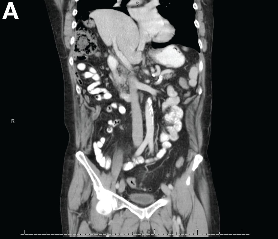
A 67-year-old man presented to the emergency department with complaints of subacute, right-sided flank pain with migratory pain to his right lower quadrant and suprapubic area of increasing intensity for 1 week. He described his pain as cramping in nature and of fluctuating intensity, acutely worse on the day of presentation. However, within 15 minutes of waiting in the emergency department his pain subsided completely. He further denied any associated nausea, vomiting, diarrhea, melena, hematochezia, dysuria, or hematuria. Vital signs and abdominal physical examination were normal. Further, laboratory testing was unremarkable including a normal urinalysis. A bedside ultrasound was negative for gallbladder pathology or nephrolithiasis; however, it revealed an abnormal appearing liver. As further diagnostic work up, an abdominopelvic computed tomography scan revealed the following images (Figures A, B). The patient was discharged from the emergency department with scheduled follow-up in the gastroenterology clinic.
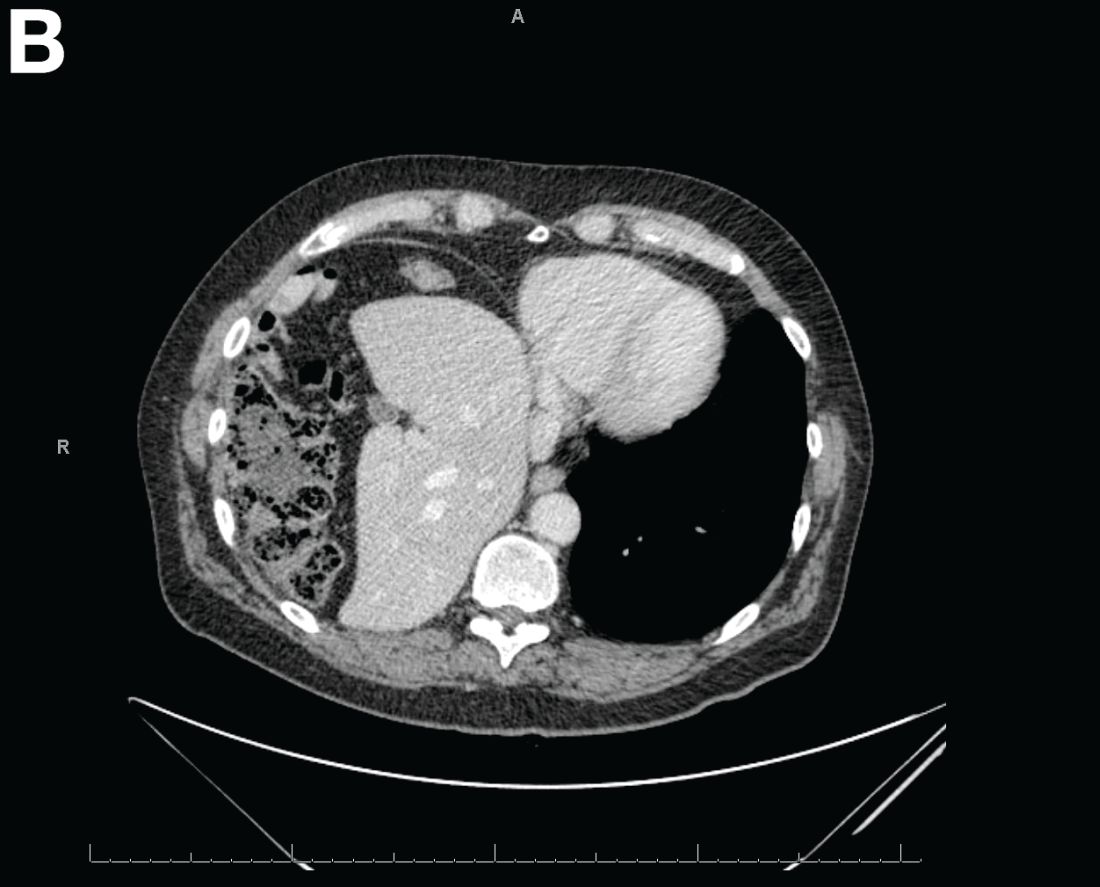
What is your diagnosis and treatment?
Opinions clash over private equity’s effect on dermatology
SAN DIEGO – Although practices that has been snowballing for a little under a decade is often bemoaned by dermatologists, at least some physicians in the field may see it as a positive development.
“I think private equity investment is good for dermatology,” Clifford Perlis, MD, said at the annual meeting of the American Academy of Dermatology. “Dermatologists should be thrilled that private equity is here.” Investment by private equity companies into dermatology practices “adds value to practices, creates more practice options, enhances advocacy, and better manages the complexity” of practices, claimed Dr. Perlis, a dermatologist and Mohs surgeon who practices in King of Prussia, Pa. The debate on this topic was presented during a forum on dermatoethics at the meeting, structured as a pro and con conversation. The organizers, including Dr. Perlis and Jane Grant-Kels, MD., chose the topics and assigned positions to each presenter, which do not necessarily represent their personal views.*

“The challenges to solo practice have nothing to do with private equity investment. It’s narrower insurance networks” that are squeezing out solo practices. He also expressed skepticism that private equity creates a barrier that interferes with the physician-patient relationship.
“Insurers and administrators already do that,” Dr. Perlis said in an interview. It’s “misguided” to place the blame on the financing. Rejection of private equity investment “clings to an outdated image of how medicine is practiced” that had already been disappearing for several decades before entry of private equity into dermatology began, he noted.
But other dermatologists have voiced concern about the changes brought by private equity investment.
“I’m frightened for the future of dermatology,” Jane M. Grant-Kels, MD, said at the meeting. “It shifts control [of patient care] away from dermatologists and into the hands of business and investors who are only interested in profit and not in quality of care,” she added.

“Have we learned nothing from dermatopathology, which has been ruined” by private equity, she asked. “The only ones who benefit are those making the private equity investments.” She cited similar, recently published views from other dermatologists, such as a recently published viewpoint article written by Jack S. Resneck Jr., MD, professor of dermatology, University of California, San Francisco (JAMA Dermatol. 2018 Jan 1;154[1]:13-4).
Dr. Perlis and Dr. Grant-Kels had no disclosures. They spoke during a session on dermatoethics at the AAD annual meeting. In an interview, Dr. Perlis said that his own personal views on this topic are far more nuanced.*
*This article was updated on April 4, 2018.
SAN DIEGO – Although practices that has been snowballing for a little under a decade is often bemoaned by dermatologists, at least some physicians in the field may see it as a positive development.
“I think private equity investment is good for dermatology,” Clifford Perlis, MD, said at the annual meeting of the American Academy of Dermatology. “Dermatologists should be thrilled that private equity is here.” Investment by private equity companies into dermatology practices “adds value to practices, creates more practice options, enhances advocacy, and better manages the complexity” of practices, claimed Dr. Perlis, a dermatologist and Mohs surgeon who practices in King of Prussia, Pa. The debate on this topic was presented during a forum on dermatoethics at the meeting, structured as a pro and con conversation. The organizers, including Dr. Perlis and Jane Grant-Kels, MD., chose the topics and assigned positions to each presenter, which do not necessarily represent their personal views.*
“The challenges to solo practice have nothing to do with private equity investment. It’s narrower insurance networks” that are squeezing out solo practices. He also expressed skepticism that private equity creates a barrier that interferes with the physician-patient relationship.
“Insurers and administrators already do that,” Dr. Perlis said in an interview. It’s “misguided” to place the blame on the financing. Rejection of private equity investment “clings to an outdated image of how medicine is practiced” that had already been disappearing for several decades before entry of private equity into dermatology began, he noted.
But other dermatologists have voiced concern about the changes brought by private equity investment.
“I’m frightened for the future of dermatology,” Jane M. Grant-Kels, MD, said at the meeting. “It shifts control [of patient care] away from dermatologists and into the hands of business and investors who are only interested in profit and not in quality of care,” she added.
“Have we learned nothing from dermatopathology, which has been ruined” by private equity, she asked. “The only ones who benefit are those making the private equity investments.” She cited similar, recently published views from other dermatologists, such as a recently published viewpoint article written by Jack S. Resneck Jr., MD, professor of dermatology, University of California, San Francisco (JAMA Dermatol. 2018 Jan 1;154[1]:13-4).
Dr. Perlis and Dr. Grant-Kels had no disclosures. They spoke during a session on dermatoethics at the AAD annual meeting. In an interview, Dr. Perlis said that his own personal views on this topic are far more nuanced.*
*This article was updated on April 4, 2018.
EXPERT ANALYSIS FROM AAD 18
HDAC inhibition may boost immune therapy efficacy in breast cancer
ORLANDO – The novel combination of entinostat and nivolumab with or without ipilimumab showed encouraging safety, tolerability, and antitumor activity in early results from an ongoing phase 1 trial of patients with advanced breast cancer.
Of 30 patients who were enrolled and treated in the dose-escalation phase of the study as of Feb. 24, 2018, 20 had evaluable responses, and of those, 3 had a partial response for an overall response rate of 15%. An additional 12 had stable disease, and 5 had disease progression, Roisin M. Connolly, MD, reported in a poster at the annual conference of the National Comprehensive Cancer Network.

Responses were seen in all 3 DL1 patients, 12 of 14 DL2 patients, 3 of 4 DL3 patients, and 2 of 9 (with 4 pending first restaging) DL4 patients. Dose-limiting toxicities included one case of pneumonitis at DL2 and an allergic reaction in one DL4 patient, said Dr. Connolly of Johns Hopkins University, Baltimore.
The most common treatment-associated adverse events occurring in 6 or more patients included anemia, fatigue, neutropenia, nausea, and rash, with each occurring in 12 to 22 patients, including grade 3 anemia in 7 patients, grade 3 fatigue in 4 patients, and grade 3 neutropenia in 5 patients. Grade 4 adverse events included lymphopenia in one patient and elevated lipase in one patient, she said.
Possible immune-related adverse events included hypothyroidism in 2 DL2 patients and 3 DL3 patients, hyperthyroidism in 1 DL3 patient, colitis in 1 DL2 and 1 DL3 patient, pneumonitis in 4 DL2 patients, rash in 10 DL2-DL4 patients, and meningoencephalitis and myasthenia gravis in 1 DL3 patient.
Study participants were adults with a mean age of 60 years with metastatic or unresectable solid tumors for which standard treatments did not exist or were no longer effective, or for which treatment with anti–programmed cell death ligand1/cytotoxic T-lymphocyte antigen 4 treatment was appropriate. All had good performance status and adequate organ and pulmonary function, less than 30% liver involvement, and any brain metastases were stable. Those with active autoimmune disease or a history of autoimmune disease that might recur were excluded, as were patients treated within 14 days of enrollment.
“The rationale for the study was based on preclinical work suggesting that epigenetic modifiers might be able to enhance the efficacy of immune therapies, and this would be particularly important for ‘colder’ tumor types like breast cancer that might not have the same sort of responses that we see in other tumor types,” Dr. Connolly explained in an interview. “The lab work suggested, for example, that the [histone deacetylase] inhibitor entinostat might affect myeloid-derived suppressor and regulatory T cells that might prevent cytotoxic T cells from fighting the cancer.”
There may be other mechanisms for this activity as well, she noted.
The run-in period with entinostat alone allowed collection of pre- and posttreatment biopsies to examine the effects on the tissues, such as whether treatment affects T cells, myeloid-derived suppressor cells, or their pathways, she said.
“We’re seeing [the] same types of toxicities seen with combination immune-oncology strategies, and we’re seeing some tumor responses that are of interest. Now we will delve into the tissue biopsies and blood samples we’ve collected to explore the mechanisms in more detail. In the near future we will open our breast cancer expansion cohort to look in more detail at what these drugs might be doing in breast cancer,” she added.
Specifically, she and her colleagues are evaluating the effects of treatment on immune-related biomarkers, measuring tumor-specific mutations and mutant neoantigens recognized by patient T cells in tumor biopsies, evaluating changes in the frequency of T cells recognizing tumor-specific mutant neoantigens in peripheral blood lymphocytes pre- and posttherapy, and looking at epigenetic changes pre- and posttherapy.
These preliminary findings suggest that the combination of entinostat and nivolumab with or without ipilimumab is safe and tolerable, with expected rates of immune-related adverse events, Dr. Connolly said, noting that the recommended phase 2 dose to be used in the dose expansion phase of the study has yet to be determined.
The findings, should they be confirmed as the trial progresses, could have important implications because immune checkpoint inhibitors, which work best in patients with immunogenic cancers that naturally attract T-cell infiltration into their tumor microenvironment, have limited single-agent activity in tumors, such as breast cancer, that are not believed to be immunogenic, she reported. Such cancers have thus far had only modest responses to single-agent immune checkpoint inhibition in advanced triple-negative and HER2+ breast cancer, with overall response rates of 5%-20%.
However, women who do respond to immune checkpoint inhibition tend to have durable and sustainable responses, she said, explaining that suboptimal immune responsiveness is likely a result of a lack of tumor antigen expression and/or recognition, as well as multiple suppressive signals in the tumor microenvironment.
Should the novel strategy tested in this study for converting breast cancers into immune responsive tumors facilitate improved response to immune checkpoint agents, it has the potential to significantly extend survival in breast cancer patients, she concluded.
This study was funded by grants from the National Institutes of Health, Bloomberg Kimmel Institute for Immunotherapy, NCCN, and the Mary Kay Foundation, as well as a V Foundation award. Dr. Connolly reported having no disclosures
SOURCE: Connolly RM et al. NCCN, Poster 3.
ORLANDO – The novel combination of entinostat and nivolumab with or without ipilimumab showed encouraging safety, tolerability, and antitumor activity in early results from an ongoing phase 1 trial of patients with advanced breast cancer.
Of 30 patients who were enrolled and treated in the dose-escalation phase of the study as of Feb. 24, 2018, 20 had evaluable responses, and of those, 3 had a partial response for an overall response rate of 15%. An additional 12 had stable disease, and 5 had disease progression, Roisin M. Connolly, MD, reported in a poster at the annual conference of the National Comprehensive Cancer Network.
Responses were seen in all 3 DL1 patients, 12 of 14 DL2 patients, 3 of 4 DL3 patients, and 2 of 9 (with 4 pending first restaging) DL4 patients. Dose-limiting toxicities included one case of pneumonitis at DL2 and an allergic reaction in one DL4 patient, said Dr. Connolly of Johns Hopkins University, Baltimore.
The most common treatment-associated adverse events occurring in 6 or more patients included anemia, fatigue, neutropenia, nausea, and rash, with each occurring in 12 to 22 patients, including grade 3 anemia in 7 patients, grade 3 fatigue in 4 patients, and grade 3 neutropenia in 5 patients. Grade 4 adverse events included lymphopenia in one patient and elevated lipase in one patient, she said.
Possible immune-related adverse events included hypothyroidism in 2 DL2 patients and 3 DL3 patients, hyperthyroidism in 1 DL3 patient, colitis in 1 DL2 and 1 DL3 patient, pneumonitis in 4 DL2 patients, rash in 10 DL2-DL4 patients, and meningoencephalitis and myasthenia gravis in 1 DL3 patient.
Study participants were adults with a mean age of 60 years with metastatic or unresectable solid tumors for which standard treatments did not exist or were no longer effective, or for which treatment with anti–programmed cell death ligand1/cytotoxic T-lymphocyte antigen 4 treatment was appropriate. All had good performance status and adequate organ and pulmonary function, less than 30% liver involvement, and any brain metastases were stable. Those with active autoimmune disease or a history of autoimmune disease that might recur were excluded, as were patients treated within 14 days of enrollment.
“The rationale for the study was based on preclinical work suggesting that epigenetic modifiers might be able to enhance the efficacy of immune therapies, and this would be particularly important for ‘colder’ tumor types like breast cancer that might not have the same sort of responses that we see in other tumor types,” Dr. Connolly explained in an interview. “The lab work suggested, for example, that the [histone deacetylase] inhibitor entinostat might affect myeloid-derived suppressor and regulatory T cells that might prevent cytotoxic T cells from fighting the cancer.”
There may be other mechanisms for this activity as well, she noted.
The run-in period with entinostat alone allowed collection of pre- and posttreatment biopsies to examine the effects on the tissues, such as whether treatment affects T cells, myeloid-derived suppressor cells, or their pathways, she said.
“We’re seeing [the] same types of toxicities seen with combination immune-oncology strategies, and we’re seeing some tumor responses that are of interest. Now we will delve into the tissue biopsies and blood samples we’ve collected to explore the mechanisms in more detail. In the near future we will open our breast cancer expansion cohort to look in more detail at what these drugs might be doing in breast cancer,” she added.
Specifically, she and her colleagues are evaluating the effects of treatment on immune-related biomarkers, measuring tumor-specific mutations and mutant neoantigens recognized by patient T cells in tumor biopsies, evaluating changes in the frequency of T cells recognizing tumor-specific mutant neoantigens in peripheral blood lymphocytes pre- and posttherapy, and looking at epigenetic changes pre- and posttherapy.
These preliminary findings suggest that the combination of entinostat and nivolumab with or without ipilimumab is safe and tolerable, with expected rates of immune-related adverse events, Dr. Connolly said, noting that the recommended phase 2 dose to be used in the dose expansion phase of the study has yet to be determined.
The findings, should they be confirmed as the trial progresses, could have important implications because immune checkpoint inhibitors, which work best in patients with immunogenic cancers that naturally attract T-cell infiltration into their tumor microenvironment, have limited single-agent activity in tumors, such as breast cancer, that are not believed to be immunogenic, she reported. Such cancers have thus far had only modest responses to single-agent immune checkpoint inhibition in advanced triple-negative and HER2+ breast cancer, with overall response rates of 5%-20%.
However, women who do respond to immune checkpoint inhibition tend to have durable and sustainable responses, she said, explaining that suboptimal immune responsiveness is likely a result of a lack of tumor antigen expression and/or recognition, as well as multiple suppressive signals in the tumor microenvironment.
Should the novel strategy tested in this study for converting breast cancers into immune responsive tumors facilitate improved response to immune checkpoint agents, it has the potential to significantly extend survival in breast cancer patients, she concluded.
This study was funded by grants from the National Institutes of Health, Bloomberg Kimmel Institute for Immunotherapy, NCCN, and the Mary Kay Foundation, as well as a V Foundation award. Dr. Connolly reported having no disclosures
SOURCE: Connolly RM et al. NCCN, Poster 3.
REPORTING FROM THE NCCN ANNUAL CONFERENCE
Key clinical point:
Major finding: Three patients had a partial response, 12 had stable disease, 5 progressed.
Study details: A phase 1 dose-expansion study involving 30 patients.
Disclosures: This study was funded by grants from the National Institutes of Health, Bloomberg Kimmel Institute for Immunotherapy, NCCN, and the Mary Kay Foundation, and by a V Foundation award. Dr. Connolly reported having no disclosures.
Source: Connolly RM et al. NCCN, Poster 3.