User login
From the Editors: The Clinical Congress: Something for everyone
In this issue of ACS Surgery News, you will see articles highlighting the program of the ACS Clinical Congress that took place in Washington, D.C., in mid-October.
This year’s theme, “The Best Surgical Education, All in One Place,” could not be more apt to represent the ACS Clinical Congress. And yet, the Clinical Congress is much, much more than an educational exercise for those who are able to attend. No matter what your age, whether it was your first time attending or, like me, your 41st, there is something at this meeting for everyone. Although the focus is on education, and the meeting does have a dizzying array of educational options, it is also an opportunity to see old friends and make new ones, connect with people with whom you share problems, work with colleagues to devise strategies to solve challenges, put faces with names you’ve only read about, participate in service and governance of our profession, and be inspired by the thoughtful leaders of our profession.
As the years pass by
I remember vividly my earliest ACS Clinical Congress, as a surgical resident at University of California, San Francisco, in October, 1975. It was 3 months into my research year and therefore I was able to attend the congress, which was in San Francisco that year. As one of the few women physicians in attendance at that meeting, I was in strange territory. I watched the opening ceremony with surgical leaders standing on stage, and I thought how homogeneous the group appeared: undeniably brilliant and accomplished, but all white-haired, white, and male. Fast-forward 41 years, and the surgical leaders who appeared on that stage at this year’s meeting were every bit as brilliant and accomplished, but the leaders are now younger and more diverse than in years past. The diversity in the college was evident as I walked the halls of the convention center where Fellows, residents, and guest physicians paused in their rushed transit from Hall D to Ballroom C to converse with an old or new friend.
The program itself has also become far more varied over the years. There is a continued emphasis on basic and clinical research in the scientific forum and scientific sessions. Postgraduate courses still impart new knowledge and skills, and state-of-the-art clinical practice is still taught in panel sessions. But the program now includes numerous nonclinical topics that are of crucial importance to present and future surgeons, such as ethics, end-of-life care, practice management, burnout, deciphering CMS regulations, and global health and humanitarian surgical outreach, to mention only a few. The named lectures continue to feature outstanding speakers who are often inspiring, and even sometimes provocative, and they are well worth attending. I would not want to have missed Past-President Carlos Pellegrini’s profoundly thoughtful John J. Conley Ethics and Philosophy Lecture on “TRUST: The Keystone of the Patient Physician Relationship,” for example.
Even the topics at the scientific forum have expanded over the years. In addition to the traditional basic science and clinical topics, five separate sessions this year on surgical education and on quality, safety, and outcomes attest to the increasing significance of these areas, unheard of when I was a resident. The important topics of ethics, geriatric surgery and palliative care, and global surgery/humanitarian outreach have gained sufficient interest that they now warrant their own sessions. There is something on the program to satisfy everyone’s interests. The forum presentations also provide us aging surgeons a chance to see the impressive contributions that our surgical progeny are making to the future of our profession. That, in itself, is encouraging and comforting.
Serving the profession
Many surgeons attend the congress in part to participate in committees of the ACS or other surgical organizations that meet during the congress to conserve time away from their “real jobs” as surgeons. These meetings offer attendees a chance to “give back” as well as to develop leadership in their profession. Participation in these committees can lead eventually to service on the Board of Governors or Regents, which offer opportunities to help shape the future of our organization and profession.
Profound positive changes in our profession have occurred through the leadership of the college. And some of these initiatives are reflected in the standing committees such as the Women in Surgery Committee, the Committee on Diversity Issues, and the Committee on Health Care Disparities, to name only three. Long-standing groups such as the Committee on Trauma (COT) have evolved greatly through the years and have raised the quality of trauma care and education in trauma across the United States and also globally.
Social and networking opportunities
The educational opportunities are unparalleled at the Clinical Congress, but the opportunities to connect with fellow surgeons is a close second. The chance to meet old friends from residency, recruit new partners for one’s practice, or be introduced to someone whom you have only known through postings on the ACS Communities are all invaluable aspects of the week. An email or telephone conversation is no substitute for these enriching face-to-face activities. And there is no substitute for this unique opportunity to create and extend your network of friends, colleagues, and allies. The Clinical Congress is often the beginning of relationships and professional connections that can last a lifetime.
Perhaps my enjoyment of the ACS Clinical Congress stems in part from how familiar it is after all these years and the comfort of being a member of this amazing organization. But my enthusiasm also comes from seeing how increasingly important and relevant the college has become to all of us – student, resident, Fellow, or guest. For surgeons, there is no substitute for the American College of Surgeons and there is indeed something for everyone at the Clinical Congress.
Dr. Deveney is professor of surgery and vice chair of education in the department of surgery, Oregon Health & Science University, Portland. She is the Coeditor of ACS Surgery News.
Clinical Practice Improvement Activities: The New Reporting Requirement
As was summarized last month, Centers for Medicare and Medicaid Services (CMS) released the final rule pertaining to the Medicare Access and CHIP Reauthorization Act (MACRA) on October 14, 2016. In the ensuing weeks, Division of Advocacy and Health Policy staff had the opportunity to read and further analyze the rule. In general, the initial favorable impression held up under more careful scrutiny. Based on the provisions in the final rule, we continue to make adjustments and modifications to the resources available to Fellows to assist them to prepare for 2017 on the website found at www.facs.org/qpp. By the time this column is printed/released, I anticipate that the video series found on the website will have been updated and expanded upon.
Because change is unsettling, one of the most frequent topics of conversation and question concerning MACRA is the new reporting requirement known as Improvement Activities. Previously, in the proposed rule, this component was known as the Clinical Practice Improvement Activities. In the final rule, the nomenclature was shortened to Improvement Activities.
The reporting mechanism specified by CMS for 2017 is simple attestation. That attestation may be accomplished via any traditional reporting mechanism other than by claims. Accordingly, the use of CMS approved “traditional” registries (registry reporting option) or qualified clinical data registries, such as the ACS’ Surgeon Specific Registry (SSR), will be valid modes by which one may report. Discussions are underway to determine how best to incorporate the Improvement Activities into the SSR. Alternatively, CMS plans to make available a portal on its website where providers will be able to attest to their having satisfied the Improvement Activity requirement.
In reviewing the list of 93 activities, examples of such that likely would or could be applicable to surgeons include:
• Use of a QCDR (qualified clinical data registry) to generate regular performance feedback (20 points)
• Participation in a QCDR, clinical data registries, or other registries run by other government agencies or private entities such as a hospital or medical or surgical society (10 points)
• Provision of episodic care management, including management across transitions and referrals that could include routine and timely follow-up to hospitalizations and ED visits and/or managing care intensively through new diagnoses, injuries and exacerbations of illness (10 points)
• Provision of specialist reports back to referring providers to close the referral loop (10 points)
• Timely communication of test results defined as timely identification of abnormal test results with timely follow-up (10 points)
• Participation in a QCDR, demonstrating performance of activities that promote use of standard practices, tools and processes for quality improvement (10 points)
• Bilateral exchange of necessary patient information to guide patient care that could include participation in a health information exchange or use of structured referral notes (10 points)
• Participation in a QCDR, demonstrating performance of activities that promote implementation of shared clinical decision making capabilities (10 points)
• Use of evidence-based decision aids to support shared decision-making (10 points)
• Participation in Maintenance of Certification Part IV (10 points)
• Annual registration by eligible clinician or group in the prescription drug monitoring program of the state where they practice (10 points)
• Consultation of prescription drug monitoring program prior to the issuance of a Controlled Substance Schedule II opioid prescription that lasts for longer than 3 days (20 points)
• Use of tools that assist specialty practices in tracking specific measures that are meaningful to their practice, such as use of the Surgical Risk Calculator (10 points)
• Seeing new and follow-up Medicaid patients in a timely manner, including individuals dually eligible for Medicaid and Medicare (20 points)
Based upon the list above (and others not included), and because the requirement specified for reporting the Improvement Activities is simple attestation, I am confident that all surgeons will be able to meet the requirement with minimal effort and achieve full credit for this component of the MIPS Composite Performance score. In these last weeks of 2016, I would encourage all Fellows to visit the ACS QPP website at www.facs.org/qpp to map out their overall strategy for success with the new Medicare physician payment system that will become effective beginning in January of 2017.
Until next month ….
Dr. Bailey is a pediatric surgeon, and Medical Director, Advocacy, for the Division of Advocacy and Health Policy in the ACS offices in Washington, D.C.
New Officers–Elect Elected at Annual Business Meeting
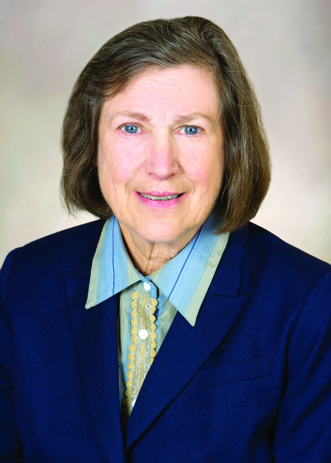
Barbara Lee Bass, MD, FACS, the John F. and Carolyn Bookout Distinguished Endowed Chair and chair, department of surgery, Houston Methodist Hospital, TX, was elected President-Elect of the American College of Surgeons (ACS) at the October 19 Annual Business Meeting of the Members. Dr. Bass is executive director, Houston Methodist Institute for Technology, Innovation and Education (MITIE), a state-of-the-art education and research facility developed to safely train practicing health care professionals in new technologies and procedures. She is professor of surgery at Weill Cornell Medical College, New York, NY, and senior member of the Houston Methodist Hospital Research Institute. A Fellow of the College since 1988, former ACS Regent, and former ACS Governor, Dr. Bass is the recipient of the 2013 ACS Distinguished Service Award—the College’s highest honor.
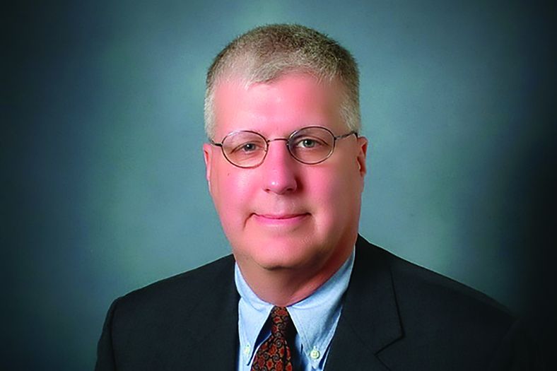
The First Vice-President-Elect is Charles D. Mabry, MD, FACS, a general surgeon from Pine Bluff, AR, and associate professor of surgery and practice management advisor to the chair, department of surgery, University of Arkansas for Medical Sciences, Little Rock. He is medical director of quality, Jefferson Regional Medical Center, Pine Bluff, and serves on the Arkansas Governor’s Trauma Advisory Committee, chairing the Committee’s Quality Improvement Subcommittee. He is Chairman of the Board for the Arkansas Preferred Provider Organization. Dr. Mabry has served on the ACS Young Surgeons Committee, as an ACS representative on the American Medical Association Relative Value Update Committee, as a member of the General Surgery Coding and Reimbursement Committee, and as an ACS Regent.
The Second Vice-President-Elect is Basil A. Pruitt, Jr., MD, FACS, FCCM, MCCM, the Dr. Ferdinand P. Herff Chair in Surgery, clinical professor of surgery, department of surgery, trauma division, University of Texas Health Science Center, San Antonio, and professor of surgery at USUHS. Dr. Pruitt is an esteemed leader in four broad areas: burn, trauma, injury, and critical care surgery; biomedical research and scholarship; organizational leadership and development; and mentorship. He is a former ACS Governor, Scudder Orator, and Excelsior Surgical Society/Edward D. Churchill Lecturer.
Barbara Lee Bass, MD, FACS, the John F. and Carolyn Bookout Distinguished Endowed Chair and chair, department of surgery, Houston Methodist Hospital, TX, was elected President-Elect of the American College of Surgeons (ACS) at the October 19 Annual Business Meeting of the Members. Dr. Bass is executive director, Houston Methodist Institute for Technology, Innovation and Education (MITIE), a state-of-the-art education and research facility developed to safely train practicing health care professionals in new technologies and procedures. She is professor of surgery at Weill Cornell Medical College, New York, NY, and senior member of the Houston Methodist Hospital Research Institute. A Fellow of the College since 1988, former ACS Regent, and former ACS Governor, Dr. Bass is the recipient of the 2013 ACS Distinguished Service Award—the College’s highest honor.
The First Vice-President-Elect is Charles D. Mabry, MD, FACS, a general surgeon from Pine Bluff, AR, and associate professor of surgery and practice management advisor to the chair, department of surgery, University of Arkansas for Medical Sciences, Little Rock. He is medical director of quality, Jefferson Regional Medical Center, Pine Bluff, and serves on the Arkansas Governor’s Trauma Advisory Committee, chairing the Committee’s Quality Improvement Subcommittee. He is Chairman of the Board for the Arkansas Preferred Provider Organization. Dr. Mabry has served on the ACS Young Surgeons Committee, as an ACS representative on the American Medical Association Relative Value Update Committee, as a member of the General Surgery Coding and Reimbursement Committee, and as an ACS Regent.
The Second Vice-President-Elect is Basil A. Pruitt, Jr., MD, FACS, FCCM, MCCM, the Dr. Ferdinand P. Herff Chair in Surgery, clinical professor of surgery, department of surgery, trauma division, University of Texas Health Science Center, San Antonio, and professor of surgery at USUHS. Dr. Pruitt is an esteemed leader in four broad areas: burn, trauma, injury, and critical care surgery; biomedical research and scholarship; organizational leadership and development; and mentorship. He is a former ACS Governor, Scudder Orator, and Excelsior Surgical Society/Edward D. Churchill Lecturer.
New Regents, B/G Executive Committee Members Elected
Two new members of the American College of Surgeons (ACS) Board of Regents (B/R) were elected at the October 19 Annual Business Meeting of Members: Anthony Atala, MD, FACS, and Fabrizio Michelassi, MD, FACS. Dr. Atala is director, Wake Forest Institute for Regenerative Medicine, and W. Boyce Professor and Chair, department of urology, Wake Forest University, Winston Salem, NC. Dr. Michelassi is the Lewis Atterbury Stimson Professor and Chair, Weill Cornell Medical Center, and surgeon-in-chief, New York-Presbyterian/Weill Cornell Medical Center, NY, and Immediate Past-Chair of the Board of Governors (B/G).
The 2016-2017 Chair of the B/R is Michael J. Zinner, MD, FACS, an ACS Regent since 2010 and founding chief executive officer and executive medical director, Miami Cancer Institute, Baptist Health South Florida, Coral Gables. The Vice-Chair is Leigh A. Neumayer, MD, MS, FACS, a Regent since 2009 and professor and chair, department of surgery, University of Arizona, and Margaret and Fenton Maynard Endowed Chair in Breast Cancer Research, University of Arizona College of Medicine, Tucson.
Replacing Dr. Michelassi as Chair of the B/G Executive Committee is Diana L. Farmer, MD, FACS, a pediatric surgeon, Pearl Stamps Stewart Professor of Surgery, and chair, department of surgery, University of California Davis Health System, Sacramento. Steven C. Stain, MD, FACS, a general surgeon and Henry and Sally Schaffer Chair and Professor, department of surgery, Albany Medical Center, NY, has been elected Vice-Chair; and Susan K. Mosier, MD, MBA, FACS, an ophthalmologist, Secretary, Kansas Department of Health and Environment, and State Health Officer for Kansas, Topeka, has been elected Secretary.
In addition, S. Rob Todd, MD, FACS, FCCM, was elected to serve an initial one-year term on the Executive Committee of the B/G. Dr. Todd is professor and chief, section of acute care surgery, department of surgery, and program director, surgical critical care residency, Baylor College of Medicine; and chief, general surgery, and director, Ginni and Richard Mithoff Trauma Center, Ben Taub Hospital, Houston, TX. Elected to an initial two-year term on the B/G Executive Committee was Nicole S. Gibran, MD, FACS, David and Nancy Auth-Washington Research Foundation Endowed Chair for Restorative Burn Surgery, professor, department of surgery, director, UW Medicine Regional Burn Center at Harborview Medical Center, and adjunct professor, department of medicine, division of dermatology, University of Washington, Seattle.
Rheumatology among specialties that continue to see strong Specialty Match Day
Rheumatology saw nearly 97% of its positions filled during the 2016 Specialty Match Day.
The specialty had 217 open slots and ended the Dec. 7 match day with only 7 positions unfilled. There were 312 applicants that selected rheumatology as their primary specialty, with 206 applicants being matched. Six were matched to different specialties and 100 who selected rheumatology as their primary specialty did not get matched to any program.
However, those seven unfilled slots will likely get matched.

Overall, Dr. Bass, who is the program director of the rheumatology fellowship program at the Hospital for Special Surgery, New York, said the numbers reflect strong interest in rheumatology.
“In 2012, there were 240 applicants to rheumatology, and this year there were 312,” she said. “It’s been a linear trend. ... The number of programs has actually gone up but it has not kept pace with demand. In 2012, 63 applicants didn’t match at any program, and this year, the number was 100.”
Dr. Bass said that this speaks to the attractiveness of rheumatology as a field and that there will be a need to create more fellowship slots.
To that end, the Rheumatology Research Foundation, the grant-giving philanthropic arm of ACR that funds research grants and fellowship training awards, is examining that very issue.
“I am leading a working group to try to figure out how to configure this award so that we try to direct them to fellowship programs that can expand or even [create] new fellowship programs,” Dr. Bass said. “The ACR and RRF will continue to fund fellowship programs because many programs have to scramble to fund the salary to support trainees and in particular to try to direct resources that will actually end up creating a new rheumatology fellow where one would not have existed before.”
The group is also looking at ways to encourage programs that have internal funding to increase their capacity if they are able to do so, she added.
Rheumatology was among a number of specialties that were able to fill at least 90% of their open slots, Other areas included allergy/immunology, cardiovascular disease, gastroenterology, hematology and oncology, pulmonary disease and pulmonary/critical care.
On the flip side, nephrology, infectious disease, and geriatric medicine had more positions available than there were applicants who prefer those specialties.
“The results this year mirror prior-year results, with slight improvement in a few specialties but no significant changes,” Mona Singer, president and CEO of the National Resident Match Program, said.
Overall, across all specialties, there were 4,731 slots, with 4,139 positions filled, accounting for about 75% of the 5,512 enrolled applicants.
Rheumatology saw nearly 97% of its positions filled during the 2016 Specialty Match Day.
The specialty had 217 open slots and ended the Dec. 7 match day with only 7 positions unfilled. There were 312 applicants that selected rheumatology as their primary specialty, with 206 applicants being matched. Six were matched to different specialties and 100 who selected rheumatology as their primary specialty did not get matched to any program.
However, those seven unfilled slots will likely get matched.
Overall, Dr. Bass, who is the program director of the rheumatology fellowship program at the Hospital for Special Surgery, New York, said the numbers reflect strong interest in rheumatology.
“In 2012, there were 240 applicants to rheumatology, and this year there were 312,” she said. “It’s been a linear trend. ... The number of programs has actually gone up but it has not kept pace with demand. In 2012, 63 applicants didn’t match at any program, and this year, the number was 100.”
Dr. Bass said that this speaks to the attractiveness of rheumatology as a field and that there will be a need to create more fellowship slots.
To that end, the Rheumatology Research Foundation, the grant-giving philanthropic arm of ACR that funds research grants and fellowship training awards, is examining that very issue.
“I am leading a working group to try to figure out how to configure this award so that we try to direct them to fellowship programs that can expand or even [create] new fellowship programs,” Dr. Bass said. “The ACR and RRF will continue to fund fellowship programs because many programs have to scramble to fund the salary to support trainees and in particular to try to direct resources that will actually end up creating a new rheumatology fellow where one would not have existed before.”
The group is also looking at ways to encourage programs that have internal funding to increase their capacity if they are able to do so, she added.
Rheumatology was among a number of specialties that were able to fill at least 90% of their open slots, Other areas included allergy/immunology, cardiovascular disease, gastroenterology, hematology and oncology, pulmonary disease and pulmonary/critical care.
On the flip side, nephrology, infectious disease, and geriatric medicine had more positions available than there were applicants who prefer those specialties.
“The results this year mirror prior-year results, with slight improvement in a few specialties but no significant changes,” Mona Singer, president and CEO of the National Resident Match Program, said.
Overall, across all specialties, there were 4,731 slots, with 4,139 positions filled, accounting for about 75% of the 5,512 enrolled applicants.
MACRA’s near and potential long-term future outlined for rheumatology
WASHINGTON – Now that implementation of the Medicare Access and CHIP Reauthorization Act is on rheumatologists’ doorsteps, figuring out what is required in 2017 is imperative to avoid future penalties and maximize the chance of earning a bonus.
Recent announcements from the Centers for Medicare & Medicaid Services (CMS) on performance thresholds and how to meet them in 2017 give rheumatologists a great shot at not getting penalized when payment adjustments begin in 2019.
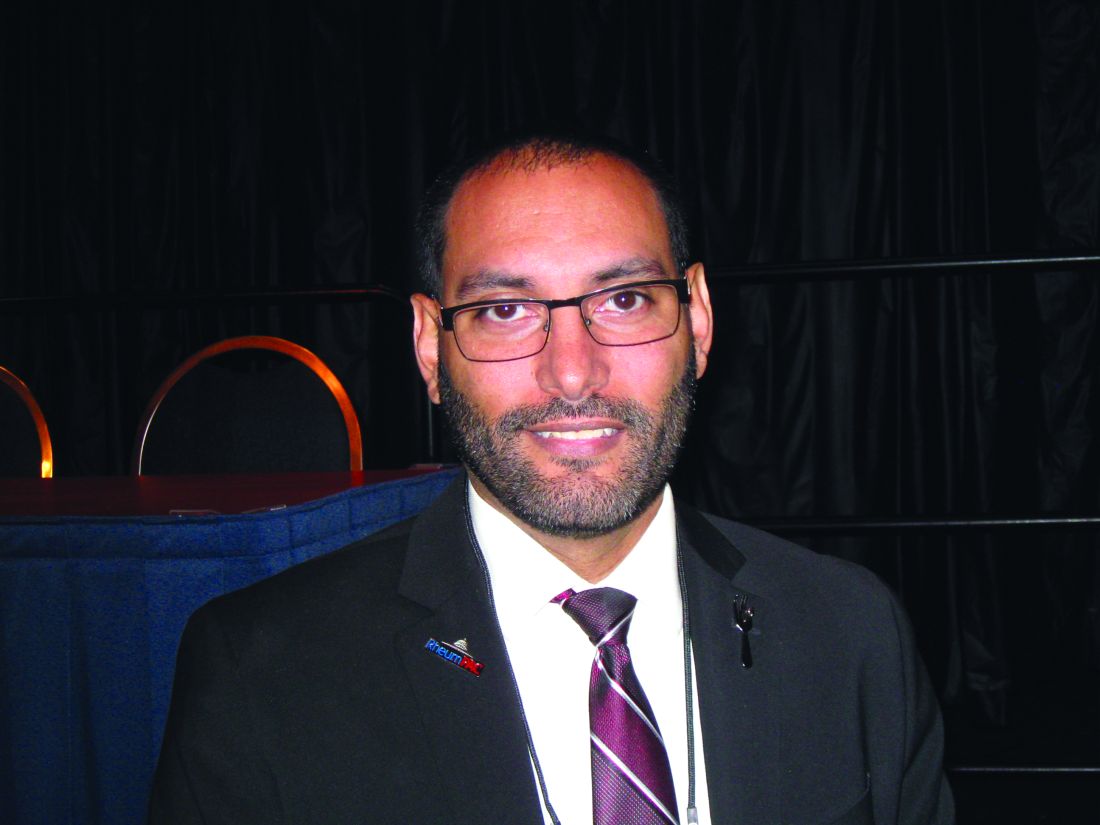
However, efforts by the ACR to create new rheumatology-focused APMs could play a major role in changing that, said Dr. Harvey, clinical director of the Arthritis Treatment Center at Tufts Medical Center, Boston, and former chair of the ACR’s Committee on Government Affairs.
The MIPS option in MACRA is “a repackaging” of old programs, including Meaningful Use (which is now called Advancing Care Information), value-based modifiers, and the Physician Quality Reporting System (PQRS). “One silver lining to this massive piece of legislation is that it’s actually less downside risk than if you had continued with Meaningful Use, PQRS, and value-based modifier,” Dr. Harvey said.
MIPS is onerous by design, in Dr. Harvey’s opinion. “CMS does not want to continue the fee-for-service arrangement and MIPS is basically fee-for-service with a bit of pay-for-performance bolted on top of it. They’ve already stated that they want to push every provider into alternative payment model–type reimbursements going forward.”
Rheumatologists aren’t eligible for MIPS if they see fewer than 100 Medicare patients per year or if they bill less than $30,000 in Medicare fees. For MIPS-eligible physicians, adjustments to Medicare payments begin in 2019 based on 2017 quality reporting data. Physicians will still receive yearly 0.5% increases in Medicare pay in 2017-2019 and 0% each year through 2025 before any adjustments are made based on the MIPS score. Participation in the MIPS pathway gives the potential for payment adjustments starting at plus or minus 4% in 2019, plus or minus 5% in 2020, plus or minus 7% in 2021, and plus or minus 9% in 2022 and beyond, based on how rheumatologists compare against their peers.
Targets for 2017 and adjustments beyond 2017
Each year in November, CMS will announce the base performance threshold goal that physicians will need to reach in the following year in order to avoid negative Medicare payment adjustments 2 years later.
Each year’s threshold will be based on the data collected from the prior year. For the year starting Jan. 1, 2017, in order to avoid a 4% cut in their Medicare Part B base rate payment in 2019, rheumatologists will need to meet a base performance threshold score of just 3 out of 100.
“Basically, if you participate in a program in any way whatsoever, you can hit this target,” Dr. Harvey said.
In 2017, getting a score of 70 or more out of 100 puts rheumatologists into a high performance category that makes them eligible for a piece of the $500 million pool of positive payment adjustments that will be available in each of the first 6 years of MIPS.
Providers who score less than 3 out of 100 will get the maximum penalty – a 4% cut in their 2019 Medicare payment rate. Further, in 2017, only 90 days of reporting is required to qualify for positive adjustments in 2019.
The base performance threshold will certainly go up for 2018 to “probably 30, 40, 50; something like that,” Dr. Harvey said.
Knowing the threshold in advance should help rheumatologists to predict by the middle of each year whether they are going to make it, but how much of any increase (or decrease) in payment adjustment they will have 2 years hence will be very difficult to know, he said, because each year the adjustment must be budget neutral and is dependent on how many physicians are above and below the threshold.
The MIPS score is based on four categories that total up to 100 points:
Quality (60% of 2017 score)
The quality category is determined by six measures that are worth 10 points each. All of the quality domains from PQRS are gone, and out of those six measures providers must have one cross-cutting measure and one outcome measure, or just one high-priority measure. Since rheumatology does not yet have any designated outcome measures, rheumatologists will need to report other designated measures. For each of these six required measures, a provider would earn 3 points for reaching the benchmark, and then the score could be increased to 4-10 points per measure based on a decile system that determines the provider’s performance against others who have met the benchmark for that measure.
Of the 13 quality metrics that are in the rheumatology quality measure set, 7 are not specific to rheumatology (advanced directive documentation, body mass index screening and follow-up, taking a comprehensive medication list, tobacco screening in adolescents, tobacco screening in adults, hypertension screening and follow-up, and sending consult note to referring doctor), whereas 6 are (TB screening within 6 months of starting a biologic, yearly TB screening on biologics, measuring rheumatoid arthritis [RA] disease activity, measuring RA functional assessment, documenting RA prognosis, and RA steroid management).
The quality category will account for 50% of the MIPS score in 2018.
Resource use (0% of 2017 score)
“The fact that the resource use part of the equation has been set to 0% for the 2017 performance year is a really good thing for rheumatologists because we use some of the most expensive drugs around,” Dr. Harvey said. “Part of the reason that CMS did this is that they are having trouble accounting for the fact that they have easy access to Part B cost data – infusion cost data – but they don’t have very good access to Part D self-injectable drug cost data.”
“We are going to make it a key advocacy point that they appropriately take into account cost in future years,” he added.
While the resource use category of MIPS contributes nothing to the 2017 MIPS score, it will increase to 10% in 2018 and by 2021 it’s anticipated to be 30% of the MIPS score, while the quality category is anticipated to drop to 30%.
Clinical practice improvement activities (15% of 2017 score)
This part includes more than 90 proposed activities to count toward the category’s score, including patient engagement activities, care coordination, extended office hours, and participation in a qualified clinical data registry (QCDR), such as the ACR’s RISE Registry, which is already a certified QCDR. A total of 40 points in this category gives full credit for the 15 percentage point value given to it for 2017.
All of the proposed activities for 2017 are weighted as medium (10 points) or high weight (20 points). For example, 40 points could be earned by meeting two medium-weight measures and one high-weight measure.
The clinical practice improvement activities category will stay at 15% of the MIPS score in 2018.
Advancing care information (25% of 2017 score)
“This is another one that gets complicated, but there’s one key take-home message: This is way easier than Meaningful Use has ever been,” Dr. Harvey said. There are 100 points needed in the advancing care information category in order to get the full 25 percentage points that it accounts for in the 2017 MIPS score.
For reporting on five required measures (electronic health record [EHR] security analysis, e-prescribing, patient access to an EHR, and being able to send and accept summary of care documents), a provider will earn 50 points.
Another 90 performance points are available for reporting certain information, and this is based on a decile system just like the quality category in which a rheumatologist’s performance is measured against other rheumatologists who have met the benchmark for each particular measure. These optional measures include providing patient-specific educational information; patients being able to view, download, and transmit their own medical records; secure messaging; acceptance of patient-generated health data; medication reconciliation; and submitting data to a state immunization registry.
Another 15 bonus points are available for additional public health reporting and reporting to CMS using a certified EHR or QCDR.
All these measures together give providers the potential to earn 155 points toward the 100-point threshold.
“If you’re already doing Meaningful Use, chances are you’re going to do really well in this area,” he said.
The advancing care information category will stay at 25% of the MIPS score in 2018.
Problems with current APMs
Under MACRA, participation in an APM means that the physician is exempt from MIPS and will receive a 5% lump sum bonus per year in 2019-2024, a higher annual increase in fee-for-service revenues, and the general benefits of participating in an APM. However, few rheumatologists will qualify for the APM pathway, and there are no rheumatology-specific APMs in existence.
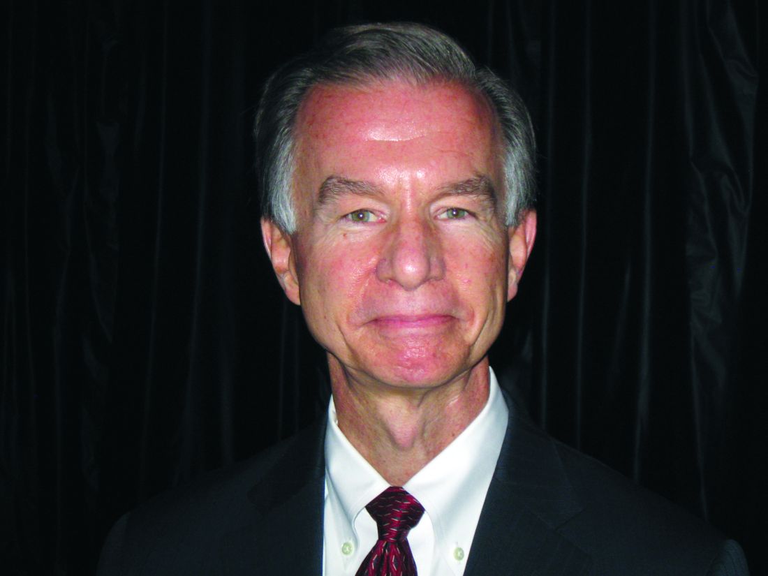
Most current APMs are “shared savings” programs where if the physician or health system can reduce spending below the expected levels, then the provider receives a share of the payer’s savings. However, these shared-savings APMs all still use a fee-for-service structure, which makes them just a different form of pay for performance, according to Mr. Miller.
Physicians still don’t get paid for unfunded or underfunded services, and physicians who are already efficient receive little or no additional revenue and may be forced out of business, while physicians who have been practicing inefficiently or inappropriately are paid more than conservative physicians. There’s also the potential for rewarding the denial of needed care. Physicians in shared-savings APMs are also placed at risk for costs they cannot control and for random variations in spending. And most of all, the shared savings bonuses are temporary and when there are no more savings to be generated, physicians are underpaid, Mr. Miller said.
During 2013-2015, 46%-48% of Medicare shared-savings accountable care organizations (ACOs) increased their Medicare spending rather than reduced it, and only 24%-30% of the ACOs received shared-savings payments. In each year, Medicare spent more than it saved, with net losses ranging from $50 million to $216 million. This happened because physicians in the programs still got paid with fee-for-service payments, Mr. Miller said.
Creating rheumatology-focused APMs
MACRA designated the development of the Physician-Focused Payment Model Technical Advisory Committee, of which Mr. Miller is a member, to solicit and review physician-focused payment models and make recommendations to CMS about which ones to implement.
Physician-focused payment models got their start through pioneering work by physicians who obtained data from insurers to find ways to reach out proactively to patients to address problems before patients are hospitalized. Through these efforts, they have reduced cost and increased patient satisfaction while also increasing payment to physicians by supporting “medical home” services, according to Dr. Miller.
These efforts to create APMs that support high-quality, physician-directed care begin by identifying avoidable spending that varies from specialty to specialty and from condition to condition. Then they address barriers in the current fee-for-service system – such as no payment for many high-value services and insufficient revenue to cover costs when using fewer or low-cost services – by providing flexible, adequate payment while requiring physicians to take responsibility for the things they said could be avoidable when paid in that way. However, this responsibility must be focused on what a particular physician can influence, Mr. Miller noted.
Rheumatologists could be helped by these physician-focused payment models because they receive less than 10% of the total Medicare health care spending per patient whose care was directed by a rheumatologist. In that case, finding ways to drop total spending per patient by 5% could at the same time give rheumatologists a 25% increase in payment while also saving Medicare 3% overall, he said.
Rheumatologists are best set up to take condition-based payments that are geared to keep patients out of the hospital, rather than the hospital episode–based payments that have dominated APMs in existence so far. Central to this condition-based model is getting the right diagnosis at the start, so payment for getting the correct diagnosis would be critical.
APMs designed for rheumatologists could take into account the diseases often seen by a rheumatologist, such as RA, and then identify opportunities to improve care and reduce costs while identifying barriers in the current payment system for achieving those goals, Mr. Miller said. These opportunities and barriers would vary from condition to condition. In some cases, particularly for low-frequency conditions, there may not need to be a new payment model established and it could be done through fee-for-service.
Ideally, these condition-based payment models for specialists such as rheumatologists would exist within the “medical neighborhood” of primary care physicians who could refer to them when necessary. Specialists would be accountable for the aspects of care that they can control, such as avoiding unnecessary tests and procedures and avoiding infections and complications.
“That’s building the ACO from the bottom up, rather than what Medicare is trying to do, which is to create it from the top down,” Mr. Miller said.
Mr. Miller noted that it will be important for specialties such as rheumatology to define the cost data it needs in order to develop condition-based payment models.
Dr. Harvey had no relevant disclosures. Mr. Miller has no financial relationships with any commercial interests.
WASHINGTON – Now that implementation of the Medicare Access and CHIP Reauthorization Act is on rheumatologists’ doorsteps, figuring out what is required in 2017 is imperative to avoid future penalties and maximize the chance of earning a bonus.
Recent announcements from the Centers for Medicare & Medicaid Services (CMS) on performance thresholds and how to meet them in 2017 give rheumatologists a great shot at not getting penalized when payment adjustments begin in 2019.
However, efforts by the ACR to create new rheumatology-focused APMs could play a major role in changing that, said Dr. Harvey, clinical director of the Arthritis Treatment Center at Tufts Medical Center, Boston, and former chair of the ACR’s Committee on Government Affairs.
The MIPS option in MACRA is “a repackaging” of old programs, including Meaningful Use (which is now called Advancing Care Information), value-based modifiers, and the Physician Quality Reporting System (PQRS). “One silver lining to this massive piece of legislation is that it’s actually less downside risk than if you had continued with Meaningful Use, PQRS, and value-based modifier,” Dr. Harvey said.
MIPS is onerous by design, in Dr. Harvey’s opinion. “CMS does not want to continue the fee-for-service arrangement and MIPS is basically fee-for-service with a bit of pay-for-performance bolted on top of it. They’ve already stated that they want to push every provider into alternative payment model–type reimbursements going forward.”
Rheumatologists aren’t eligible for MIPS if they see fewer than 100 Medicare patients per year or if they bill less than $30,000 in Medicare fees. For MIPS-eligible physicians, adjustments to Medicare payments begin in 2019 based on 2017 quality reporting data. Physicians will still receive yearly 0.5% increases in Medicare pay in 2017-2019 and 0% each year through 2025 before any adjustments are made based on the MIPS score. Participation in the MIPS pathway gives the potential for payment adjustments starting at plus or minus 4% in 2019, plus or minus 5% in 2020, plus or minus 7% in 2021, and plus or minus 9% in 2022 and beyond, based on how rheumatologists compare against their peers.
Targets for 2017 and adjustments beyond 2017
Each year in November, CMS will announce the base performance threshold goal that physicians will need to reach in the following year in order to avoid negative Medicare payment adjustments 2 years later.
Each year’s threshold will be based on the data collected from the prior year. For the year starting Jan. 1, 2017, in order to avoid a 4% cut in their Medicare Part B base rate payment in 2019, rheumatologists will need to meet a base performance threshold score of just 3 out of 100.
“Basically, if you participate in a program in any way whatsoever, you can hit this target,” Dr. Harvey said.
In 2017, getting a score of 70 or more out of 100 puts rheumatologists into a high performance category that makes them eligible for a piece of the $500 million pool of positive payment adjustments that will be available in each of the first 6 years of MIPS.
Providers who score less than 3 out of 100 will get the maximum penalty – a 4% cut in their 2019 Medicare payment rate. Further, in 2017, only 90 days of reporting is required to qualify for positive adjustments in 2019.
The base performance threshold will certainly go up for 2018 to “probably 30, 40, 50; something like that,” Dr. Harvey said.
Knowing the threshold in advance should help rheumatologists to predict by the middle of each year whether they are going to make it, but how much of any increase (or decrease) in payment adjustment they will have 2 years hence will be very difficult to know, he said, because each year the adjustment must be budget neutral and is dependent on how many physicians are above and below the threshold.
The MIPS score is based on four categories that total up to 100 points:
Quality (60% of 2017 score)
The quality category is determined by six measures that are worth 10 points each. All of the quality domains from PQRS are gone, and out of those six measures providers must have one cross-cutting measure and one outcome measure, or just one high-priority measure. Since rheumatology does not yet have any designated outcome measures, rheumatologists will need to report other designated measures. For each of these six required measures, a provider would earn 3 points for reaching the benchmark, and then the score could be increased to 4-10 points per measure based on a decile system that determines the provider’s performance against others who have met the benchmark for that measure.
Of the 13 quality metrics that are in the rheumatology quality measure set, 7 are not specific to rheumatology (advanced directive documentation, body mass index screening and follow-up, taking a comprehensive medication list, tobacco screening in adolescents, tobacco screening in adults, hypertension screening and follow-up, and sending consult note to referring doctor), whereas 6 are (TB screening within 6 months of starting a biologic, yearly TB screening on biologics, measuring rheumatoid arthritis [RA] disease activity, measuring RA functional assessment, documenting RA prognosis, and RA steroid management).
The quality category will account for 50% of the MIPS score in 2018.
Resource use (0% of 2017 score)
“The fact that the resource use part of the equation has been set to 0% for the 2017 performance year is a really good thing for rheumatologists because we use some of the most expensive drugs around,” Dr. Harvey said. “Part of the reason that CMS did this is that they are having trouble accounting for the fact that they have easy access to Part B cost data – infusion cost data – but they don’t have very good access to Part D self-injectable drug cost data.”
“We are going to make it a key advocacy point that they appropriately take into account cost in future years,” he added.
While the resource use category of MIPS contributes nothing to the 2017 MIPS score, it will increase to 10% in 2018 and by 2021 it’s anticipated to be 30% of the MIPS score, while the quality category is anticipated to drop to 30%.
Clinical practice improvement activities (15% of 2017 score)
This part includes more than 90 proposed activities to count toward the category’s score, including patient engagement activities, care coordination, extended office hours, and participation in a qualified clinical data registry (QCDR), such as the ACR’s RISE Registry, which is already a certified QCDR. A total of 40 points in this category gives full credit for the 15 percentage point value given to it for 2017.
All of the proposed activities for 2017 are weighted as medium (10 points) or high weight (20 points). For example, 40 points could be earned by meeting two medium-weight measures and one high-weight measure.
The clinical practice improvement activities category will stay at 15% of the MIPS score in 2018.
Advancing care information (25% of 2017 score)
“This is another one that gets complicated, but there’s one key take-home message: This is way easier than Meaningful Use has ever been,” Dr. Harvey said. There are 100 points needed in the advancing care information category in order to get the full 25 percentage points that it accounts for in the 2017 MIPS score.
For reporting on five required measures (electronic health record [EHR] security analysis, e-prescribing, patient access to an EHR, and being able to send and accept summary of care documents), a provider will earn 50 points.
Another 90 performance points are available for reporting certain information, and this is based on a decile system just like the quality category in which a rheumatologist’s performance is measured against other rheumatologists who have met the benchmark for each particular measure. These optional measures include providing patient-specific educational information; patients being able to view, download, and transmit their own medical records; secure messaging; acceptance of patient-generated health data; medication reconciliation; and submitting data to a state immunization registry.
Another 15 bonus points are available for additional public health reporting and reporting to CMS using a certified EHR or QCDR.
All these measures together give providers the potential to earn 155 points toward the 100-point threshold.
“If you’re already doing Meaningful Use, chances are you’re going to do really well in this area,” he said.
The advancing care information category will stay at 25% of the MIPS score in 2018.
Problems with current APMs
Under MACRA, participation in an APM means that the physician is exempt from MIPS and will receive a 5% lump sum bonus per year in 2019-2024, a higher annual increase in fee-for-service revenues, and the general benefits of participating in an APM. However, few rheumatologists will qualify for the APM pathway, and there are no rheumatology-specific APMs in existence.
Most current APMs are “shared savings” programs where if the physician or health system can reduce spending below the expected levels, then the provider receives a share of the payer’s savings. However, these shared-savings APMs all still use a fee-for-service structure, which makes them just a different form of pay for performance, according to Mr. Miller.
Physicians still don’t get paid for unfunded or underfunded services, and physicians who are already efficient receive little or no additional revenue and may be forced out of business, while physicians who have been practicing inefficiently or inappropriately are paid more than conservative physicians. There’s also the potential for rewarding the denial of needed care. Physicians in shared-savings APMs are also placed at risk for costs they cannot control and for random variations in spending. And most of all, the shared savings bonuses are temporary and when there are no more savings to be generated, physicians are underpaid, Mr. Miller said.
During 2013-2015, 46%-48% of Medicare shared-savings accountable care organizations (ACOs) increased their Medicare spending rather than reduced it, and only 24%-30% of the ACOs received shared-savings payments. In each year, Medicare spent more than it saved, with net losses ranging from $50 million to $216 million. This happened because physicians in the programs still got paid with fee-for-service payments, Mr. Miller said.
Creating rheumatology-focused APMs
MACRA designated the development of the Physician-Focused Payment Model Technical Advisory Committee, of which Mr. Miller is a member, to solicit and review physician-focused payment models and make recommendations to CMS about which ones to implement.
Physician-focused payment models got their start through pioneering work by physicians who obtained data from insurers to find ways to reach out proactively to patients to address problems before patients are hospitalized. Through these efforts, they have reduced cost and increased patient satisfaction while also increasing payment to physicians by supporting “medical home” services, according to Dr. Miller.
These efforts to create APMs that support high-quality, physician-directed care begin by identifying avoidable spending that varies from specialty to specialty and from condition to condition. Then they address barriers in the current fee-for-service system – such as no payment for many high-value services and insufficient revenue to cover costs when using fewer or low-cost services – by providing flexible, adequate payment while requiring physicians to take responsibility for the things they said could be avoidable when paid in that way. However, this responsibility must be focused on what a particular physician can influence, Mr. Miller noted.
Rheumatologists could be helped by these physician-focused payment models because they receive less than 10% of the total Medicare health care spending per patient whose care was directed by a rheumatologist. In that case, finding ways to drop total spending per patient by 5% could at the same time give rheumatologists a 25% increase in payment while also saving Medicare 3% overall, he said.
Rheumatologists are best set up to take condition-based payments that are geared to keep patients out of the hospital, rather than the hospital episode–based payments that have dominated APMs in existence so far. Central to this condition-based model is getting the right diagnosis at the start, so payment for getting the correct diagnosis would be critical.
APMs designed for rheumatologists could take into account the diseases often seen by a rheumatologist, such as RA, and then identify opportunities to improve care and reduce costs while identifying barriers in the current payment system for achieving those goals, Mr. Miller said. These opportunities and barriers would vary from condition to condition. In some cases, particularly for low-frequency conditions, there may not need to be a new payment model established and it could be done through fee-for-service.
Ideally, these condition-based payment models for specialists such as rheumatologists would exist within the “medical neighborhood” of primary care physicians who could refer to them when necessary. Specialists would be accountable for the aspects of care that they can control, such as avoiding unnecessary tests and procedures and avoiding infections and complications.
“That’s building the ACO from the bottom up, rather than what Medicare is trying to do, which is to create it from the top down,” Mr. Miller said.
Mr. Miller noted that it will be important for specialties such as rheumatology to define the cost data it needs in order to develop condition-based payment models.
Dr. Harvey had no relevant disclosures. Mr. Miller has no financial relationships with any commercial interests.
EXPERT ANALYSIS FROM THE ACR ANNUAL MEETING
FDA bans powdered gloves
The Food and Drug Administration has banned powdered gloves for use in health care settings, citing “numerous risks to patients and health care workers.” The ban extends to gloves currently in commercial distribution and in the hands of the ultimate user, meaning powdered gloves will have to be pulled from examination rooms and operating theaters.
“A thorough review of all currently available information supports FDA’s conclusion that powdered surgeon’s gloves, powdered patient examination gloves, and absorbable powder for lubricating a surgeon’s glove should be banned,” according to a FDA final rule available now online and scheduled for publication in the Federal Register on Dec. 19, 2016. The ban will become effective 30 days after the document’s publication in the Federal Register.
Notes the final document, “The benefits of powdered gloves appear to only include greater ease of donning and doffing, decreased tackiness, and a degree of added comfort, which FDA believes are nominal when compared to the risks posed by these devices.”
Since viable nonpowdered alternatives exist, the FDA believes that the ban would not have significant economic impact and that shortages should not affect care delivery.
Many nonpowdered gloves, said the FDA, now “have the same level of protection, dexterity, and performance” as powdered gloves.
Powder may still be used in the glove manufacturing process, but the FDA continues to recommend that no more than 2 mg of residual powder per glove remains after the manufacturing process.
Though the final document banning powdered gloves notes that the FDA received many comments asking for a ban of all natural rubber latex (NRL) gloves, the ban applied only to powdered gloves. The FDA noted that NRL gloves already must carry a statement alerting users to the risks of allergic reaction, and also noted that eliminating powder from NRL gloves reduces the risk of latex sensitization.
In explaining its analysis of the costs and benefits of the powdered glove ban, the FDA estimated that the annual net benefits would range between $26.8 million and $31.8 million.
When this ban comes into force, it will be only the second such ban; the first was the 1983 ban of prosthetic hair fibers, which were found to provide no public health benefit.
[email protected]
On Twitter @karioakes
Effective hospitalist roles for NPs, PAs
I’m often asked about effective roles for nurse practitioners (NPs) and physician assistants (PAs), collectively known as advanced practice clinicians (APCs). My first response is always the same: They have much to contribute and can be effective members of hospitalist groups. Most hospital medicine groups (HMGs) should think about having them in their staffing mix if they don’t already.
Yet despite all that NPs/PAs can offer, my experience is that many (even most) hospitalist groups fail to develop roles that optimize their APCs’ skills.
An October 2016 study in the Journal of Clinical Outcomes Management adds additional data to help think about this issue. You may have seen the study mentioned in several news articles and blogs. Most summarized the study along the lines of “using high levels of PA staffing results in lower hospital costs per case.” Framing it this way is awfully misleading, so I’ll go a little deeper here.
Study context
The study, “A Comparison of Conventional and Expanded Physician Assistant Hospitalist Staffing Models at a Community Hospital,” is a retrospective analysis of performance measures from two hospitalist groups at Anne Arundel Medical Center (AAMC) in Annapolis, Md.1 One HMG is employed by the hospital. The other, called MDICS, is a private company that contracts with AAMC as well as approximately 13 other hospitals and 40 rehabilitation facilities. Tim Capstack, MD, is the AAMC medical director for MDICS and lead author of the study (representing a potential conflict of interest acknowledged in the article). Barry Meisenberg, MD, is a coauthor, a hospitalist in the AAMC-employed group, and chair for quality improvement and health care systems research at AAMC.
Tim told me by phone that both groups have practiced at AAMC for more than 10 years and enjoy a collegial relationship. Both groups employ PAs and pair them with a single physician in a dyad arrangement each day. Tim’s MDICS group, the “expanded PA” group, staffs each day shift with three physicians and three PAs, compared with the nine physicians and two PAs in the hospital-employed “conventional” group. The MDICS PAs are responsible for more patients each day than their conventional-group counterparts and, during the January 2012 to July 2013 study period, averaged 14.2 patients versus 8.3, respectively.
Over the course of the study, PAs in the expanded PA group saw and billed 36% of patient visits independently, compared with 5.9% for the conventional group.
Notable study findings
I think the main value of this study is in showing that the expanded PA group had rates of readmission, inpatient mortality, length of stay, and consultant use that weren’t statistically different from the conventional group.
The workloads and years of experience of doctors and PAs in each group were similar. And while there were some differences in the patients each group cared for, they seem unlikely to have a significant influence on outcomes. Clearly, there are many unmeasured variables (e.g., culture, morale, and leadership) in each group that could have influenced the outcomes, so this one study at one hospital doesn’t provide a definitive answer about appropriate APC staffing levels. However, it didn’t uncover big differences in the measured outcomes.
And this study did show that higher levels of PA staffing were associated with lower hospital charges per case. Although the difference was a modest 3%, it was statistically significant (P less than .001). I’m skeptical there is causation here; this more likely is just correlation.
It would be great to see a larger study of this.
Information applications
So does this study support the idea that HMGs can or should increase APC staffing and workload significantly to realize lower hospital cost per case and not harm patient outcomes? Not so fast!
This study only compared two hospitalist groups at one hospital. It’s probably not very generalizable.
And as described in the paper, and stressed by Tim talking with me by phone, the outcomes of their expanded PA model likely have a lot to do with their very careful recruiting and screening of experienced PAs before hiring them, not to mention a lengthy and deliberate on-boarding process (summarized in the article) to support their ability to perform well. Groups that are not as thoughtful and deliberate in how they hire and position APCs to contribute to the practice may not perform as well.
Why study only PAs? What about NPs? Tim told me that his group is agnostic regarding the training background of the APCs they hire; he suspects an identical study with NPs rather than PAs in each hospitalist group would probably yield very similar results. I see this the same way. Although there are differences in background and training between NPs and PAs, I think personal traits like years of experience in various health care settings and the ability to work efficiently are more important than training background.
A practical approach
Any group who thinks this study is evidence that adding more APCs and having them manage a higher number of patients relatively independently will go well in any setting is mistaken. But it does offer a story of one place where, with careful planning and execution, it went OK.
In my view, the real take-home message is to think carefully to ensure any APCs in your group have professionally satisfying roles that position them to contribute effectively. While common, I think configuring APCs and physicians as rounding dyads often ends up underperforming and not working out well because of inefficiency. When well executed, as is apparently the case in this study, it can be fine. But my experience is that positioning APCs to assume primary responsibility for some clinical activities, such as covering the observation unit or serving as an evening admitter/cross-cover provider (all with appropriate physician collaboration and backup), more reliably turns out well.
Reference
Capstack TM, Seguija C, Vollono LM, Moser JD, Meisenberg BR, Michtalik HJ. A comparison of conventional and expanded physician assistant hospitalist staffing models at a community hospital. J Clin Outcomes Manag. 2016;23(10):455-61.
Dr. Nelson has been a practicing hospitalist since 1988. He is co-founder and past president of SHM and principal in Nelson Flores Hospital Medicine Consultants. He is co-director for SHM’s “Best Practices in Managing a Hospital Medicine Program” course. Write to him at [email protected].
Pediatric Dermatology Consult - December 2016
Urticaria pigmentosa
BY CATALINA MATIZ, MD, AND JEREMY UDKOFF
Frontline Medical News
Maculopapular cutaneous mastocytosis (MPCM), colloquially known as urticaria pigmentosa, typically presents as multiple brown lesions on the skin and accounts for the majority of pediatric cutaneous mastocytosis.1 The category of cutaneous mastocytosis also includes diffuse cutaneous mastocytosis (characterized by generalized erythema and thickened skin), solitary mastocytoma (present as a solitary brown or red lesion), and telangiectasia macularis eruptiva perstans (diffuse telangiectatic macules on the skin, usually in adult patients).
Cutaneous manifestations of mastocytosis are caused by collections of mast cells and the local release of their mediators into the skin. Some of these patients can present with pruritus and flushing, and rarely with diarrhea, abdominal pain, or heart palpitations. Alternatively, systemic mastocytosis, where mast cells invade extracutaneous organs such as the bone marrow or the gastrointestinal tract, may present with or without associated skin lesions. These patients usually have more severe systemic symptoms such as fever, weight loss, epigastric pain, neuropsychiatric symptoms, or bone pain.2,3

The pathogenesis of mastocytosis revolves around abnormal mast cell proliferation. These mast cells then accumulate in organs such as the skin. Mechanical stimulations result in a release of cell mediators, such as histamine, eicosanoids, and cytokines, that causes the associated symptoms. Recent studies have shown c-KIT mutations to be present in the majority of mastocytosis cases, and these mutations are thought to contribute to disease progression.6
Differential diagnosis
Because of its varied presentation, the differential diagnosis for MPCM includes café au lait macules, lentigines, nevi, lichen planus, pseudolymphoma, diffuse juvenile xanthogranuloma, and Langerhans cell histiocytosis. Although these diseases may include macules and papules that look similar to MPCM, the Darier’s sign is an extremely powerful tool in differentiating these diseases as it is almost always present in mastocytosis. When there is a history of blistering lesions, the differential diagnosis should include epidermolysis bullosa, herpes simplex, bullous arthropod bites, bullous impetigo, linear IgA dermatosis, and rarely autoimmune bullous conditions. Equivocal cases should be referred to a pediatric dermatologist for evaluation, as conditions such as Langerhans cell histiocytosis and epidermolysis bullosa are usually associated with poor outcomes.
Work-up and treatment
In cases where the lesions are not classic or there is not a clear Darier’s sign, a skin biopsy with special staining, such as Giemsa or toluidine blue, is recommended to help clarify the diagnosis and rule out other worrisome conditions as mentioned above. If the child presents with systemic symptoms, performing a complete blood count with differential, tryptase level, and organ-specific testing based on the symptoms is recommended.
Serum tryptase is a biomarker for mast cell degranulation. It is typically normal in pediatric patients with MPCM but may be positive if there are symptoms of systemic disease such as fever, weight loss, bone pain, epigastric pain, headaches, or anaphylaxis. Similarly, the risk of anaphylactic reactions correlates with the serum tryptase level.4,7 If the tryptase level is elevated, a bone marrow biopsy should be performed to rule out more severe forms of systemic disease.
Potent topical corticosteroids, such as fluocinonide, are typically used to the treat cutaneous lesions when they are inflamed. Other supportive treatments used in MPCM usually revolve around controlling the cutaneous symptoms as there is no cure for the disease. H1 and H2 antihistamines may be used alone or together to control itching and are thought to decrease flushing and blistering associated with MPCM. Other possible treatments include topical cromolyn sodium and leukotriene antagonists. Mastocytoma lesions that are increasing in size or are very symptomatic despite the treatments listed above may benefit from local immunosuppressants, ultraviolet radiation and psoralen application, or excision.8
Lifestyle modifications also are recommended for patients with mastocytosis. They are advised to avoid foods, medications, and environmental triggers that can cause mast cell degranulation. These triggers include aspirin, nonsteroidal anti-inflammatory agents and narcotics, topical polymyxin B sulfate, heat, friction, and certain systemic anesthetic agents.9
References
1. Br J Dermatol. 2015;172(3):642-51.
2. Clin Pediatr. (Phila) 2008;47(8):757-61.
3. “Mastocytosis,” in Dermatology, 3rd ed. (China: Elsevier, 2012).
4. J Allergy Clin Immunol. 2016;137(1):35-45.
5. J Allergy Clin Immunol. 2015 Dec;136(6):1581-90.e1-e3.
6. J Invest Dermatol. 2010;130(3):804-15.
7. Pediatr Dermatol. 2014;31(3):271-5.
8. Eur J Clin Invest. 2007;37(6):435-53.
9. “Histiocytosis and Malignant Skin Diseases,” in Hurwitz Clinical Pediatric Dermatology: A Textbook of Skin Disorders of Childhood and Adolescence, 5th ed. (China: Elsevier, 2016).
Dr. Matiz is assistant professor of dermatology at Rady Children’s Hospital San Diego–University of California, San Diego, and Jeremy Udkoff is a medical student at the university.
Urticaria pigmentosa
BY CATALINA MATIZ, MD, AND JEREMY UDKOFF
Frontline Medical News
Maculopapular cutaneous mastocytosis (MPCM), colloquially known as urticaria pigmentosa, typically presents as multiple brown lesions on the skin and accounts for the majority of pediatric cutaneous mastocytosis.1 The category of cutaneous mastocytosis also includes diffuse cutaneous mastocytosis (characterized by generalized erythema and thickened skin), solitary mastocytoma (present as a solitary brown or red lesion), and telangiectasia macularis eruptiva perstans (diffuse telangiectatic macules on the skin, usually in adult patients).
Cutaneous manifestations of mastocytosis are caused by collections of mast cells and the local release of their mediators into the skin. Some of these patients can present with pruritus and flushing, and rarely with diarrhea, abdominal pain, or heart palpitations. Alternatively, systemic mastocytosis, where mast cells invade extracutaneous organs such as the bone marrow or the gastrointestinal tract, may present with or without associated skin lesions. These patients usually have more severe systemic symptoms such as fever, weight loss, epigastric pain, neuropsychiatric symptoms, or bone pain.2,3
The pathogenesis of mastocytosis revolves around abnormal mast cell proliferation. These mast cells then accumulate in organs such as the skin. Mechanical stimulations result in a release of cell mediators, such as histamine, eicosanoids, and cytokines, that causes the associated symptoms. Recent studies have shown c-KIT mutations to be present in the majority of mastocytosis cases, and these mutations are thought to contribute to disease progression.6
Differential diagnosis
Because of its varied presentation, the differential diagnosis for MPCM includes café au lait macules, lentigines, nevi, lichen planus, pseudolymphoma, diffuse juvenile xanthogranuloma, and Langerhans cell histiocytosis. Although these diseases may include macules and papules that look similar to MPCM, the Darier’s sign is an extremely powerful tool in differentiating these diseases as it is almost always present in mastocytosis. When there is a history of blistering lesions, the differential diagnosis should include epidermolysis bullosa, herpes simplex, bullous arthropod bites, bullous impetigo, linear IgA dermatosis, and rarely autoimmune bullous conditions. Equivocal cases should be referred to a pediatric dermatologist for evaluation, as conditions such as Langerhans cell histiocytosis and epidermolysis bullosa are usually associated with poor outcomes.
Work-up and treatment
In cases where the lesions are not classic or there is not a clear Darier’s sign, a skin biopsy with special staining, such as Giemsa or toluidine blue, is recommended to help clarify the diagnosis and rule out other worrisome conditions as mentioned above. If the child presents with systemic symptoms, performing a complete blood count with differential, tryptase level, and organ-specific testing based on the symptoms is recommended.
Serum tryptase is a biomarker for mast cell degranulation. It is typically normal in pediatric patients with MPCM but may be positive if there are symptoms of systemic disease such as fever, weight loss, bone pain, epigastric pain, headaches, or anaphylaxis. Similarly, the risk of anaphylactic reactions correlates with the serum tryptase level.4,7 If the tryptase level is elevated, a bone marrow biopsy should be performed to rule out more severe forms of systemic disease.
Potent topical corticosteroids, such as fluocinonide, are typically used to the treat cutaneous lesions when they are inflamed. Other supportive treatments used in MPCM usually revolve around controlling the cutaneous symptoms as there is no cure for the disease. H1 and H2 antihistamines may be used alone or together to control itching and are thought to decrease flushing and blistering associated with MPCM. Other possible treatments include topical cromolyn sodium and leukotriene antagonists. Mastocytoma lesions that are increasing in size or are very symptomatic despite the treatments listed above may benefit from local immunosuppressants, ultraviolet radiation and psoralen application, or excision.8
Lifestyle modifications also are recommended for patients with mastocytosis. They are advised to avoid foods, medications, and environmental triggers that can cause mast cell degranulation. These triggers include aspirin, nonsteroidal anti-inflammatory agents and narcotics, topical polymyxin B sulfate, heat, friction, and certain systemic anesthetic agents.9
References
1. Br J Dermatol. 2015;172(3):642-51.
2. Clin Pediatr. (Phila) 2008;47(8):757-61.
3. “Mastocytosis,” in Dermatology, 3rd ed. (China: Elsevier, 2012).
4. J Allergy Clin Immunol. 2016;137(1):35-45.
5. J Allergy Clin Immunol. 2015 Dec;136(6):1581-90.e1-e3.
6. J Invest Dermatol. 2010;130(3):804-15.
7. Pediatr Dermatol. 2014;31(3):271-5.
8. Eur J Clin Invest. 2007;37(6):435-53.
9. “Histiocytosis and Malignant Skin Diseases,” in Hurwitz Clinical Pediatric Dermatology: A Textbook of Skin Disorders of Childhood and Adolescence, 5th ed. (China: Elsevier, 2016).
Dr. Matiz is assistant professor of dermatology at Rady Children’s Hospital San Diego–University of California, San Diego, and Jeremy Udkoff is a medical student at the university.

An 18-month-old girl was referred to our pediatric dermatology clinic for an evaluation of multiple brown macules all over her body. The lesions had been present for about 1 year and were not increasing in size, but she was getting new lesions on her face and scalp. They were not symptomatic and did not seem to bother the patient. The mother noted that some lesions may become red with changes in the weather. She denied symptoms such as wheezing, flushing, or diarrhea. No other family members were affected, and the patient was not taking any medications. She had no family history of neurofibromatosis or other genetic conditions. The physical exam was notable for many scattered, 3- to 5-mm, polymorphic, light brown to orange macules and papules. One of the lesions reddened and wheeled after we stroked it. The patient had no lymphadenopathy or hepatosplenomegaly.
Everything We Say and Do
Editor’s note: “Everything We Say and Do” is an informational series developed by SHM’s Patient Experience Committee to provide readers with thoughtful and actionable communication tactics that have great potential to positively impact patients’ experience of care. Each article will focus on how the contributor applies one or more of the “key communication” tactics in practice to maintain provider accountability for “everything we say and do that affects our patients’ thoughts, feelings, and well-being.”
What I say and do
I inform my patients that I have reviewed their chart and that I am familiar with their diagnosis.
Why I do it
In the hospital setting, in particular, patients are concerned about communication between their various healthcare professionals. Many times, the patient’s primary-care provider works strictly in the outpatient setting, so the hospitalist is the person who assumes total care of the patient throughout hospitalization. This understandably creates anxiety for patients and families because they wonder if the hospitalist really knows their medical history. One way to alleviate this anxiety is to review your patients’ charts prior to speaking with them and to verbally let your patients know you are familiar with their diagnoses.
How I do it
Step 1: Before entering the room, I review my patient’s chart. If I am taking over the service from my colleague, I review all notes from the current hospitalization to ensure I understand everything that has happened. I also review tests, procedures, and radiographic studies. To gain a better understanding of my patient, I read the most recent discharge summary and outpatient clinic note. Likewise, if I am admitting a new patient to the hospital, before entering the room to do the history and physical examination, I review recent hospitalizations, clinic notes, and emergency department visits.
I also like to review the chart to see if I have taken care of the patient before. Patients often remember me even though I may not remember them, so reviewing my prior notes may be helpful. Thankfully, my electronic health record (EHR) has a search function where I can enter my name or any other keyword and it searches for patient records based on this keyword.
Step 2: Even though reading the chart and being informed about my patient is important, it is only the first step. The next step is to let my patient and family know that I have read the chart and that I am up-to-date on my patient’s diagnosis. I feel it is very important for me to verbalize that I have read the chart because without doing this, my patients never really know that I took the time prior to entering the room to learn about them.
I might say:
- “I was reviewing your chart before I came in, and I saw that your daughter brought you to the hospital for chest pain.”
- “I read your chart and saw that you have been to the emergency room twice in the last week.”
- “I read your primary-care doctor’s note, and I saw that she recently treated you for pneumonia.”
- “I read your chart, and I wanted to confirm a few things I read to ensure we are on the same page.”
There are many different ways you can phrase this, but the important point is to make sure your patients know you read the chart by specifically referencing something you learned. This helps your patients feel more confident that you know their medical history.
I know some of the doctors reading this column see patients in the outpatient setting. One way to help yourself remember pertinent facts about a patient’s medical history is to include these facts in a specific place in your clinic note. That way, prior to seeing the patient, you can always review your last note and know the important information about your patient’s medical history will always be in the same place in each note. Another tip is to use your EHR’s note function. My EHR has “sticky notes,” and they provide a place for the PCP to store information about the patient without it becoming part of the permanent medical record.
These notes allow the PCP to record important events that happen between one clinic visit and the next. Thus, when the patient returns to the clinic, the PCP opens the chart, reviews the sticky note, and enters the exam room prepared to discuss significant events in the patient’s recent medical history.
In the end, it does not matter which technique you use. It simply matters that you take time to review your patient’s chart prior to entering the room and that you verbalize what you have learned. In patients, this inspires confidence and trust and helps alleviate concerns that the physician does not know important information in their medical history.
Dr. Dorrah is regional medical director for quality and the patient experience at Baylor Scott & White Health in Round Rock, Tex. She is a member of SHM’s Patient Experience Committee.