User login
Diverticulitis recurs more with observation vs. elective resection
CHICAGO – Observation, compared with elective resection, was associated with significantly increased recurrence rates in a single-center randomized, controlled trial of patients who had successfully recovered via nonoperative management from their first episode of acute sigmoid diverticulitis with extraluminal air/abscess.
Recurrence rates in 111 patients randomized to observation or elective resection were 31% in the observation group and 7% in the resection group, at 15 and 18 months, respectively, Dr. Ryan Bendl of State University of New York, Stony Brook reported at the annual meeting of the American Surgical Association.

Patients in the two groups were comparable with respect to age, sex, body mass index, Colorectal Physiologic and Operative Severity Score for the Enumeration of Mortality and Morbidity (CR-POSSUM), and comorbidities, he noted.
Subjects included in the single-center study were adults admitted for a first episode of acute diverticulitis with abscess or extraluminal air who were managed nonoperatively with intravenous antibiotics, a period of nothing by mouth, drainage, and total parenteral nutrition followed by colonoscopy. They were randomized 3:1 to observation or resection, and 68% of the elective resection patients underwent minimally invasive surgery. The study’s primary endpoint was recurrent diverticulitis defined as an acute episode confirmed by computed tomography and requiring hospitalization with intravenous antibiotics.
Diverticulitis accounted for more than 300,000 hospital admissions in 2010 in the United States alone, and 10%-20% of patients had abscess formation. At one time, most patients were managed with immediate operative intervention, but medical and radiologic advances have led to a shift toward nonoperative management, Dr. Bendl said.
Some prior studies have suggested that recurrence rates are higher with nonoperative management, and the current study supports those data.
However, despite the significant increase in the recurrence rate with observation vs. resection, most patients in the observation group did not experience recurrence, and of those who did, none had peritonitis.
“All those with recurrences were successfully treated again using nonoperative management,” he said.
This study was supported in part by grants from Merck and Covidien. Dr. Bendl reported having no relevant financial disclosures.
CHICAGO – Observation, compared with elective resection, was associated with significantly increased recurrence rates in a single-center randomized, controlled trial of patients who had successfully recovered via nonoperative management from their first episode of acute sigmoid diverticulitis with extraluminal air/abscess.
Recurrence rates in 111 patients randomized to observation or elective resection were 31% in the observation group and 7% in the resection group, at 15 and 18 months, respectively, Dr. Ryan Bendl of State University of New York, Stony Brook reported at the annual meeting of the American Surgical Association.
Patients in the two groups were comparable with respect to age, sex, body mass index, Colorectal Physiologic and Operative Severity Score for the Enumeration of Mortality and Morbidity (CR-POSSUM), and comorbidities, he noted.
Subjects included in the single-center study were adults admitted for a first episode of acute diverticulitis with abscess or extraluminal air who were managed nonoperatively with intravenous antibiotics, a period of nothing by mouth, drainage, and total parenteral nutrition followed by colonoscopy. They were randomized 3:1 to observation or resection, and 68% of the elective resection patients underwent minimally invasive surgery. The study’s primary endpoint was recurrent diverticulitis defined as an acute episode confirmed by computed tomography and requiring hospitalization with intravenous antibiotics.
Diverticulitis accounted for more than 300,000 hospital admissions in 2010 in the United States alone, and 10%-20% of patients had abscess formation. At one time, most patients were managed with immediate operative intervention, but medical and radiologic advances have led to a shift toward nonoperative management, Dr. Bendl said.
Some prior studies have suggested that recurrence rates are higher with nonoperative management, and the current study supports those data.
However, despite the significant increase in the recurrence rate with observation vs. resection, most patients in the observation group did not experience recurrence, and of those who did, none had peritonitis.
“All those with recurrences were successfully treated again using nonoperative management,” he said.
This study was supported in part by grants from Merck and Covidien. Dr. Bendl reported having no relevant financial disclosures.
AT THE ASA ANNUAL MEETING
Key clinical point: Observation vs. elective resection was associated with significantly increased recurrence rates in patients who had recovered via nonoperative management from their first episode of acute sigmoid diverticulitis with extraluminal air/abscess.
Major finding: Recurrence rates in 111 patients randomized to observation or elective resection were 31% in the observation group and 7% in the resection group, at 15 and 18 months, respectively.
Data source: A randomized, controlled trial involving 111 patients.
Disclosures: This study was supported in part by grants from Merck and Covidien. Dr. Bendl reported having no relevant financial disclosures.

Kawasaki disease and infections aren’t mutually exclusive
CHICAGO – Kawasaki disease and concurrent bacterial or viral infection are by no means mutually exclusive, Dr. Cathie-Kim Le cautioned at the annual meeting of the American College of Cardiology.
“Recognizing that both can coexist will ensure timely IVIg [intravenous immunoglobulin] treatment and appropriate containment of coronary artery complications,” said Dr. Le of Sainte-Justine University Hospital in Montreal.
She presented a retrospective study of 128 patients, mean age 3.4 years, admitted to the pediatric academic tertiary care center with a discharge diagnosis of Kawasaki disease. During their hospitalization all of them underwent a work-up for bacterial and viral infectious diseases, which proved positive in 33% of cases. Roughly 40% of subjects had incomplete Kawasaki disease, meaning they lacked a sufficient number of signs of mucocutaneous inflammation to fulfill the epidemiologic case definition; however, their prevalence of concomitant infection was similar to that of the group with classic Kawasaki disease.
Among the most common types of infections in patients with Kawasaki disease were otitis media, which accounted for 17% of the infections; upper respiratory infections, 21%; and group A streptococcal pharyngitis and pneumonia, which accounted for 14% each.
There were no differences in clinical presentation or laboratory values between children with or without concomitant infection. Nor was myocardial profiling useful in differentiating patients with concomitant infection from those without: Ventricular shortening fraction scores and N-terminal probrain natriuretic peptide levels were similar in the two groups of Kawasaki disease patients.
Acute coronary dilatation occurred in 26% of Kawasaki disease patients with concomitant infection and similarly in 30% of those without infection. Coronary aneurysm, the most serious complication of Kawasaki disease, occurred in 14% of patients with infection and an identical proportion of the uninfected.
Almost all patients received IVIg. Resistance to the effects of IVIg occurred in 36% of patients with concomitant infection, a rate twice that seen in the group without infection. Coronary aneurysms and dilatations were significantly more common in IVIg-resistant patients, regardless of their infection status.
Dr. Le reported having no relevant financial conflicts.
AT ACC 16
Key clinical point: One-third of Kawasaki patients have a concomitant common infection that may delay timely diagnosis.
Major finding: Coronary aneurysm, the most serious complication of Kawasaki disease, occurred in 14% of patients with an infection and in 14% of the uninfected.
Data source: A retrospective study of 128 children with Kawasaki disease.
Disclosures: The study presenter reported having no relevant financial conflicts.
Advances in Hematology and Oncology (May 2016)
Table of Contents
- Introduction
- Consensus Statement Supporting the Recommendation for Single-Fraction Palliative Radiotherapy for Uncomplicated, Painful Bone Metastases
- The Availability of Advanced Radiation Oncology Technology Within VHA Radiation Oncology Centers
- γ-δ T-Cell Lymphoma With Disseminated Intravascular Coagulation and Autoimmune Hemolytic Anemia
- Use of Fluorodeoxyglucose-Positron Emission Tomography in the Diagnosis of Intravascular Diffuse Large B-Cell Lymphoma
- Prevalence of Hypogonadism in Low-Risk Prostate Cancer Survivors
- Molecular Profiles Guide Colorectal Cancer Treatment
- Treating VA Patients With Multiple Myeloma

Thrill-Seeking Hospitalist Alleviates Stress Through Scuba Diving
Not much intimidates Jasen Gundersen, MD, president of the acute care services division at TeamHealth, an outsourcer of hospital-based clinical and specialty services based in Knoxville, Tenn. Besides traveling 150,000 miles a year overseeing 2,500 hospitalists at 285 facilities, Dr. Gundersen has climbed frozen waterfalls in Vermont and New Hampshire, raced in bicycle competitions, and skied mountains towering 10,000 feet.

But his love for adventure is now focused below the surface. Over the years, he has spent many weekends diving in open waters surrounding southeast Florida; Cozumel, Mexico; Turks and Caicos; and the Cayman Islands. He believes there’s no place on Earth that is as peaceful, serene, or even magical as under the ocean.
Reclaimed Passion
Growing up in Connecticut, Dr. Gundersen and his family frequently vacationed in the Bahamas, where he was introduced to scuba diving.
“As a teenager, I really loved diving,” he recalls. “Every time we went to the Bahamas, I always tried to go diving or snorkeling.”
However, the harsh Connecticut winters and frigid Atlantic Ocean prevented him from diving. More delays followed, namely medical school. After graduating from the University of Connecticut School of Medicine in 2000, Dr. Gundersen completed his three-year residency in family medicine at the UMass Memorial Medical Center. During the next two years, he worked as a physician and hospitalist at the Family Health Center of Worcester, a federally qualified health center where he did everything from examining sore throats to delivering babies.
In 2005, he launched a small hospital medicine program at the University of Massachusetts that quickly grew and bumped up his title to division chief for hospital medicine. Then in January 2011, he accepted a new position as chief medical officer at TeamHealth, requiring him and his wife, Elizabeth, also a hospitalist, to move to Florida.
Within several weeks, the couple started diving near their home in Pompano Beach. He says Elizabeth, his “diving buddy,” was eager to learn and developed a passion for scuba diving that rivals his own.
“We did 80 to 90 dives in the first year we were down there,” Dr. Gundersen says, explaining that unlike many sports, diving doesn’t require athletic ability, size, or strength. “We normally did recreational diving, where you basically can always swim slowly straight to the surface. You don’t stay down long enough that you build up enough bubbles in your system that you have to stop on the way up.”
Sharks and Shipwrecks
Since then, Dr. Gundersen purchased a 38-foot powerboat, became a PADI (Professional Association of Diving Instructors) open-water scuba instructor, and earned a U.S. Coast Guard 50-ton master captain’s license. He and Elizabeth are certified for advanced nitrox and decompression diving, technical diving that requires the use of different gases to decompress when heading to the surface, and diving in overhead environments, such as caves or shipwrecks.
“One of our favorite wrecks is called the USS Spiegel Gove that sits on the ocean floor in Key Largo,” he says, adding that on occasion, they also swim with hammerhead sharks. “The walls of the ship go 30 feet up on each side. You can swim where they loaded the cargo and see the old crane above you. It’s spectacular.”
Among their favorite spots to dive is Eagle Ray Pass in Grand Cayman, where entire schools of spotted eagle rays live, he says, adding that 17 rays swam and floated around them during one dive.
Fortunately, after some initial costs, he says the sport isn’t too expensive, roughly around $1,500 to get started. Basic scuba gear costs approximately $1,000. Likewise, certifications can run $350 a piece. Boat trips range between $60 and $100, unless you prefer shore diving, where you park at the beach and simply swim into the ocean. Then add a few extra dollars to fill your tank with air.
Scary and Serene
Although the Gundersens are accomplished divers who prefer warm waters and flat seas, Dr. Gundersen says only one moment of one dive actually scared him.
Years ago, he, Elizabeth, and a friend were wreck diving. Diving protocol is based on follow the leader, where divers swim into wrecks one at a time, follow each other, and signal their turns. Somehow, their friend unintentionally swam in between Dr. Gundersen and his wife. Elizabeth and the friend then turned to see something inside the wreck, but the friend failed to signal to Dr. Gundersen that they were turning.
“I went a bit farther and turned around,” Dr. Gundersen recalls. “He and Elizabeth were gone. It gave me a moment of panic. I’m particularly careful about staying with my diving buddy and making sure we don’t get lost. It wasn’t dangerous but broke the cardinal rule of what you’re supposed to do when diving. I swam back and found them.”
While that was a rare experience, he says diving, when done properly, is the most peaceful and serene activity that people may experience. When under water, all you hear are your air bubbles. There are no cellphones ringing, emails or texts to respond to, or work issues to resolve.
“Work-life balance is a really big deal for me and my team to prevent burnout,” Dr. Gundersen says. “It allows me to have my personal time to enjoy and relax so when I’m back at work on Monday, my batteries are recharged. I’m ready to go.” TH
Carol Patton is a freelance writer in Las Vegas.
Not much intimidates Jasen Gundersen, MD, president of the acute care services division at TeamHealth, an outsourcer of hospital-based clinical and specialty services based in Knoxville, Tenn. Besides traveling 150,000 miles a year overseeing 2,500 hospitalists at 285 facilities, Dr. Gundersen has climbed frozen waterfalls in Vermont and New Hampshire, raced in bicycle competitions, and skied mountains towering 10,000 feet.
But his love for adventure is now focused below the surface. Over the years, he has spent many weekends diving in open waters surrounding southeast Florida; Cozumel, Mexico; Turks and Caicos; and the Cayman Islands. He believes there’s no place on Earth that is as peaceful, serene, or even magical as under the ocean.
Reclaimed Passion
Growing up in Connecticut, Dr. Gundersen and his family frequently vacationed in the Bahamas, where he was introduced to scuba diving.
“As a teenager, I really loved diving,” he recalls. “Every time we went to the Bahamas, I always tried to go diving or snorkeling.”
However, the harsh Connecticut winters and frigid Atlantic Ocean prevented him from diving. More delays followed, namely medical school. After graduating from the University of Connecticut School of Medicine in 2000, Dr. Gundersen completed his three-year residency in family medicine at the UMass Memorial Medical Center. During the next two years, he worked as a physician and hospitalist at the Family Health Center of Worcester, a federally qualified health center where he did everything from examining sore throats to delivering babies.
In 2005, he launched a small hospital medicine program at the University of Massachusetts that quickly grew and bumped up his title to division chief for hospital medicine. Then in January 2011, he accepted a new position as chief medical officer at TeamHealth, requiring him and his wife, Elizabeth, also a hospitalist, to move to Florida.
Within several weeks, the couple started diving near their home in Pompano Beach. He says Elizabeth, his “diving buddy,” was eager to learn and developed a passion for scuba diving that rivals his own.
“We did 80 to 90 dives in the first year we were down there,” Dr. Gundersen says, explaining that unlike many sports, diving doesn’t require athletic ability, size, or strength. “We normally did recreational diving, where you basically can always swim slowly straight to the surface. You don’t stay down long enough that you build up enough bubbles in your system that you have to stop on the way up.”
Sharks and Shipwrecks
Since then, Dr. Gundersen purchased a 38-foot powerboat, became a PADI (Professional Association of Diving Instructors) open-water scuba instructor, and earned a U.S. Coast Guard 50-ton master captain’s license. He and Elizabeth are certified for advanced nitrox and decompression diving, technical diving that requires the use of different gases to decompress when heading to the surface, and diving in overhead environments, such as caves or shipwrecks.
“One of our favorite wrecks is called the USS Spiegel Gove that sits on the ocean floor in Key Largo,” he says, adding that on occasion, they also swim with hammerhead sharks. “The walls of the ship go 30 feet up on each side. You can swim where they loaded the cargo and see the old crane above you. It’s spectacular.”
Among their favorite spots to dive is Eagle Ray Pass in Grand Cayman, where entire schools of spotted eagle rays live, he says, adding that 17 rays swam and floated around them during one dive.
Fortunately, after some initial costs, he says the sport isn’t too expensive, roughly around $1,500 to get started. Basic scuba gear costs approximately $1,000. Likewise, certifications can run $350 a piece. Boat trips range between $60 and $100, unless you prefer shore diving, where you park at the beach and simply swim into the ocean. Then add a few extra dollars to fill your tank with air.
Scary and Serene
Although the Gundersens are accomplished divers who prefer warm waters and flat seas, Dr. Gundersen says only one moment of one dive actually scared him.
Years ago, he, Elizabeth, and a friend were wreck diving. Diving protocol is based on follow the leader, where divers swim into wrecks one at a time, follow each other, and signal their turns. Somehow, their friend unintentionally swam in between Dr. Gundersen and his wife. Elizabeth and the friend then turned to see something inside the wreck, but the friend failed to signal to Dr. Gundersen that they were turning.
“I went a bit farther and turned around,” Dr. Gundersen recalls. “He and Elizabeth were gone. It gave me a moment of panic. I’m particularly careful about staying with my diving buddy and making sure we don’t get lost. It wasn’t dangerous but broke the cardinal rule of what you’re supposed to do when diving. I swam back and found them.”
While that was a rare experience, he says diving, when done properly, is the most peaceful and serene activity that people may experience. When under water, all you hear are your air bubbles. There are no cellphones ringing, emails or texts to respond to, or work issues to resolve.
“Work-life balance is a really big deal for me and my team to prevent burnout,” Dr. Gundersen says. “It allows me to have my personal time to enjoy and relax so when I’m back at work on Monday, my batteries are recharged. I’m ready to go.” TH
Carol Patton is a freelance writer in Las Vegas.

May 2016 Digital Edition
Click here to access the May 2016 Digital Edition.
Table of Contents
- Why VA Health Care Is Different
- Anterior Cervical Interbody Fusion Using Polyetheretherketone (PEEK) Cage Device and Local Autograft Bone
- Academic Reasonable Accommodations for Post-9/11 Veterans With Psychiatric Diagnoses, Part 2
- A Real Welcome Home: Permanent Housing for Homeless Veterans
- Veterans’ Satisfaction With Erectile Dysfunction Treatment
- VA PBM Academic Detailing Service: Implementation and Lessons Learned
- Adherence to Disease-Modifying Therapies in Patients With MS: A Retrospective Cohort Study
- The Role of Fidaxomicin in Clostridium difficile Infection

Cell-based strategy curbs constipation
Treatment at the nanomolecular level may become an alternative for various types of constipation, in particular for the opioid-induced constipation that is common after surgery, based on data from a proof of concept study involving mice and a small-molecule activator.
“Activation of the cystic fibrosis transmembrane conductance regulator (CFTR) chloride channel is the primary pathway that drives fluid secretion in the intestine, which maintains lubrication of luminal contents,” wrote Dr. Onur Cil of the University of California, San Francisco, and colleagues. The researchers examined whether direct activation of the CFTR would prompt fluid secretion and reverse stool dehydration when applied to constipated mice. The findings were published online in the May issue of the journal Cellular and Molecular Gastroenterology and Hepatology (2016. doi: 10.1016/j.jcmgh.2015.12.010).
The researchers identified a promising activator, the phenylquinoxalinone CFTRact-J027. Mice received up to 10 mg/kg CFTRact-J027 either orally or intraperitoneally (IP), with doses including 0.1 mg/kg, 0.3 mg/kg, 1 mg/kg, 3 mg/kg, and 10 mg/kg.
Overall, IP doses of CFTRact-J027 at 10 mg/kg normalized stool in the constipated mice, and dose-response studies showed a 50% effective dose of 2 mg/kg in these mice.
When given orally, CFTRact-J027 “normalized stool output and water content in a loperamide-induced mouse model of constipation with a 50% effective dose of approximately 0.5 mg/kg,” that was significantly lower than the IP administration, the researchers noted. An oral dose of 10 mg/kg CFTRact-J027 1 hour before inducing constipation also was effective in normalizing stool output and water content in loperamide-treated mice, with no effect in control nonconstipated mice.
The activator was not effective against constipation in cystic fibrosis mice that were missing a functional CFTR, they added.
The researchers used an in vivo closed loop model to specifically test the effects of CFTRact-J027 on intestinal fluid secretion and absorption and found that CFTRact-J027 caused “a 140% increase in loop weight/length ratio, indicating fluid secretion into the intestinal lumen in wild-type mice.” However, there was no effect in cystic fibrosis mice, further supporting the CFTR-selective mechanism of action, the researchers said.
As for potential toxic effects of the treatment, CFTRact-J027 showed no impact on the major serum chemistry and blood parameters of the mice after 7 days, and had no apparent impact on body weight. No accumulation of fluid (the most significant potential adverse effect) was noted in the airway or lungs of the treated mice.
Additional toxicity data are needed to continue preclinical development, the researchers said. However, “our data provide evidence for the prosecretory action of a CFTR activator in mouse intestine and proof of concept for its use in the treatment of various types of constipation, which could include opioid-induced constipation, chronic idiopathic constipation, and irritable bowel syndrome with constipation predominance,” they wrote. In addition, a CFTR activator similar to that used in this study may have clinical applications for other conditions including asthma, dry eye, cholestatic liver disease, chronic obstructive pulmonary disease and bronchitis, and cigarette smoke–induced lung dysfunction, they added.
Dr. Cil and two coauthors are inventors on a provisional patent filing, with rights owned by the University of California, San Francisco. The study was funded in part by several grants from organizations including the National Institutes of Health and the Cystic Fibrosis Foundation.
FROM CELLULAR AND MOLECULAR GASTROENTEROLOGY AND HEPATOLOGY
Key clinical point: Treatment at the nanomolecular level may become an alternative for various types of constipation.
Major finding: Oral doses of CFTRact-J027 normalized stool output and water content in a loperamide-induced mouse model of constipation with a 50% effective dose of approximately 0.5 mg/kg.
Data source: A proof-of-concept study in which a cell-based screen was performed for 120,000 druglike, synthetic small molecules that were then tested in constipation-induced mice and control mice.
Disclosures: Dr. Cil and two coauthors are inventors on a provisional patent filing, with rights owned by the University of California, San Francisco. The study was funded in part by several grants from organizations including the National Institutes of Health and the Cystic Fibrosis Foundation.
Cirrhosis 30-day readmissions down 40% with quality improvement initiative
Using checklists and electronic decision support in an inpatient liver unit, quality improvement (QI) care protocols reduced 30-day readmissions of patients with cirrhosis by 40%, due mostly to a drop in readmissions for hepatic encephalopathy (HE) according to a report published in the May issue of Clinical Gastroenterology and Hepatology.
For patients initially admitted for overt HE, the 30-day readmission rate was 26.0% (27 of 104), compared with 48.9% (66 of 135) before implementation of QI. The proportion of total readmissions due to HE after QI was 9.6% (14 of 146), compared with 40.7% (79 of 194) before QI. In addition, length of stay for HE patients was significantly reduced (–1.34 days; 95% confidence interval, –2.38 to –0.32; P = .01). There were no significant changes in 90-day mortality.
Source: American Gastroenterological Association
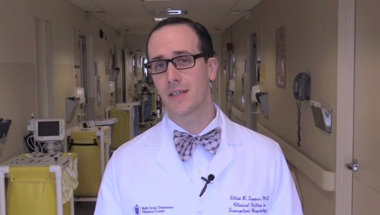
“Our study advances the current literature on QI for patients with cirrhosis by presenting an inexpensive, easy to implement, and generalizable approach,” wrote Dr. Elliot Tapper of Beth Israel Deaconess Medical Center, Boston, and his colleagues. Previous studies have addressed readmission interventions among patients with cirrhosis, but the protocols required costly infrastructure, expertise, and institutional commitments. The current study supports the value of standard checklists and education, according to the investigators, “showing that outcomes improve further when checklist items are hard-wired into the ordering system.” (Clin Gastroenterol Hepatol. 2016 Apr 7. doi: 10.1016/j.cgh.2015.08.041).
The QI initiative encompassed several aspects of care. All HE patients were designated to receive rifaximin, and their lactulose dosing was adjusted to mental status using the Richmond Agitation and Sedation Scale. For patients with spontaneous bacterial peritonitis (SBP), timely administration of the correct dose of antibiotics and albumin was promoted, as were prophylactic measures for all patients, such as variceal hemorrhage prophylaxis and subcutaneous heparin for the prevention of venous thrombosis.
The three-part program entailed a run-in phase for preliminary checklist troubleshooting, a hand-held checklist phase, including the HE protocol, SBP treatment, and prophylactic measures, and a final electronic phase in which checklist items were incorporated into the hospital’s electronic provider order entry system using mandatory preset doses and linked medications.
Individual protocol items were demonstrated to affect the readmission rate. Rifaximin use for HE patients rose from 78.1% to 96.3%, and use of rifaximin was associated with lower adjusted odds of 30-day readmission (OR, 0.39; 95% CI, 0.16-0.87; P = .02). The dose/frequency of lactulose for HE patients increased, and patients who had 6 or more cups of lactulose on the day of their readmission had significantly lower adjusted length of stay (–2.36 days; 95% CI, –3.40 to –1.31; P less than .0001). Patients taking SBP prophylaxis had lower readmission rates (OR, 0.51; 95% CI, 0.31-0.83; P = .007).
The prospective study from 2011 to 2013 evaluated patients with cirrhosis who were admitted to the liver unit of Beth Israel Deaconess Medical Center, Boston. Patients were diagnosed with cirrhosis caused by hepatitis C (44.9%), alcoholic liver disease (34%), hepatitis B (5.4%), and biliary cirrhosis (1.8%). In total, 824 unique patients were admitted 1,720 times; 485 (58.9%) were admitted once, 268 (32.5%) were admitted 2-4 times, and 71 (8.6%) were admitted 5 or more times. The median length of stay for all patients was 4.0 days (interquartile range, 2.0-8.0).
Dr. Tapper and his coauthors reported having no disclosures.
Using checklists and electronic decision support in an inpatient liver unit, quality improvement (QI) care protocols reduced 30-day readmissions of patients with cirrhosis by 40%, due mostly to a drop in readmissions for hepatic encephalopathy (HE) according to a report published in the May issue of Clinical Gastroenterology and Hepatology.
For patients initially admitted for overt HE, the 30-day readmission rate was 26.0% (27 of 104), compared with 48.9% (66 of 135) before implementation of QI. The proportion of total readmissions due to HE after QI was 9.6% (14 of 146), compared with 40.7% (79 of 194) before QI. In addition, length of stay for HE patients was significantly reduced (–1.34 days; 95% confidence interval, –2.38 to –0.32; P = .01). There were no significant changes in 90-day mortality.
Source: American Gastroenterological Association
“Our study advances the current literature on QI for patients with cirrhosis by presenting an inexpensive, easy to implement, and generalizable approach,” wrote Dr. Elliot Tapper of Beth Israel Deaconess Medical Center, Boston, and his colleagues. Previous studies have addressed readmission interventions among patients with cirrhosis, but the protocols required costly infrastructure, expertise, and institutional commitments. The current study supports the value of standard checklists and education, according to the investigators, “showing that outcomes improve further when checklist items are hard-wired into the ordering system.” (Clin Gastroenterol Hepatol. 2016 Apr 7. doi: 10.1016/j.cgh.2015.08.041).
The QI initiative encompassed several aspects of care. All HE patients were designated to receive rifaximin, and their lactulose dosing was adjusted to mental status using the Richmond Agitation and Sedation Scale. For patients with spontaneous bacterial peritonitis (SBP), timely administration of the correct dose of antibiotics and albumin was promoted, as were prophylactic measures for all patients, such as variceal hemorrhage prophylaxis and subcutaneous heparin for the prevention of venous thrombosis.
The three-part program entailed a run-in phase for preliminary checklist troubleshooting, a hand-held checklist phase, including the HE protocol, SBP treatment, and prophylactic measures, and a final electronic phase in which checklist items were incorporated into the hospital’s electronic provider order entry system using mandatory preset doses and linked medications.
Individual protocol items were demonstrated to affect the readmission rate. Rifaximin use for HE patients rose from 78.1% to 96.3%, and use of rifaximin was associated with lower adjusted odds of 30-day readmission (OR, 0.39; 95% CI, 0.16-0.87; P = .02). The dose/frequency of lactulose for HE patients increased, and patients who had 6 or more cups of lactulose on the day of their readmission had significantly lower adjusted length of stay (–2.36 days; 95% CI, –3.40 to –1.31; P less than .0001). Patients taking SBP prophylaxis had lower readmission rates (OR, 0.51; 95% CI, 0.31-0.83; P = .007).
The prospective study from 2011 to 2013 evaluated patients with cirrhosis who were admitted to the liver unit of Beth Israel Deaconess Medical Center, Boston. Patients were diagnosed with cirrhosis caused by hepatitis C (44.9%), alcoholic liver disease (34%), hepatitis B (5.4%), and biliary cirrhosis (1.8%). In total, 824 unique patients were admitted 1,720 times; 485 (58.9%) were admitted once, 268 (32.5%) were admitted 2-4 times, and 71 (8.6%) were admitted 5 or more times. The median length of stay for all patients was 4.0 days (interquartile range, 2.0-8.0).
Dr. Tapper and his coauthors reported having no disclosures.
Key clinical point: Care protocols implemented by electronic decision support reduced 30-day readmissions of patients with cirrhosis by 40% in an inpatient liver unit.
Major finding: The drop was likely driven by fewer readmissions for hepatic encephalopathy (HE): the 30-day HE readmission rate was 26.0% (27 of 104), compared with 48.9% (66 of 135) before implementation of quality improvement.
Data sources: The prospective study evaluated 824 patients who were admitted 1,720 times to the liver unit of Beth Israel Deaconess Medical Center, Boston.
Disclosures: Dr. Tapper and his coauthors reported having no disclosures.
Additional D1 biopsy increased diagnostic yield for celiac disease
Among a large cohort of patients referred for endoscopy for suspected celiac disease as well as all upper gastrointestinal symptoms, a single additional D1 biopsy specimen from any site significantly increased the diagnostic yield for celiac disease, according to researchers.
Of 1,378 patients who had D2 and D1 biopsy specimens taken, 268 were newly diagnosed with celiac disease, and 26 had villous atrophy confined to D1, defined as ultrashort celiac disease (USCD). Compared with a standard D2 biopsy, an additional D1 biopsy increased the diagnostic yield by 9.7% (P less than .0001). Among the 26 diagnosed with USCD, 7 had normal D2 biopsy specimens, and 4 others had negative tests for endomysial antibodies (EMAs), totaling 11 patients for whom celiac disease would have been missed in the absence of a D1 biopsy.
“The addition of a D1 biopsy specimen to diagnose celiac disease may reduce the known delay in diagnosis that many patients with celiac disease experience. This may allow earlier institution of a gluten-free diet, potentially prevent nutritional deficiencies, and reduce the symptomatic burden of celiac disease,” wrote Dr. Peter Mooney of Royal Hallamshire Hospital, Sheffield, England, and his colleagues. (Gastroenterology 2016 April 7. doi: 10.1053/j-gastro.2016.01.029).
The prospective study recruited 1,378 consecutive patients referred to a single teaching hospital for endoscopy from 2008 to 2014. In total, 268 were newly diagnosed with celiac disease, and 26 were diagnosed with USCD.
To investigate the optimal site for targeted D1 sampling, 171 patients underwent quadrantic D1 biopsy, 61 of whom were diagnosed with celiac disease. Biopsy specimens from any topographical area resulted in high sensitivity, a fact that increases the feasibility of a D1 biopsy policy, since no specific target area is required, according to the researchers. Nonceliac abnormalities such as peptic duodenitis or gastric heterotopia have been suggested to impede interpretation of D1 biopsies, but these were rare in the study and did not interfere with the analysis.
USCD may be an early form of conventional celiac disease, an idea supported by the findings. Compared with patients diagnosed with conventional celiac disease, patients diagnosed with USCD were younger and had a much lower rate of diarrhea, which by decision-tree analysis was the single factor discriminating between the two groups. Compared with healthy controls, individuals with conventional celiac disease, but not USCD, were more likely to present with anemia, diarrhea, a family history of celiac disease, lethargy, and osteoporosis. Patients with USCD and conventional disease had similar rates of IgA tissue transglutaminase antibodies (tTG), but USCD patients had lower titers (P less than .001). The USCD group also had fewer ferritin and folate deficiencies.
The researchers suggested that clinical phenotypic differences may be due to minimal loss of absorptive capacity associated with a short segment of villous atrophy. Given the younger average age at diagnosis of USCD and lower tTG titers, USCD may represent an early stage of celiac disease, resulting in fewer nutritional deficiencies observed because of a shorter lead time to diagnosis.
Although USCD patients had a milder clinical phenotype, which has raised concerns that a strict gluten-free diet may be unnecessary, follow-up data demonstrated that a gluten-free diet produced improvement in symptoms and a significant decrease in the tTG titer. These results may indicate that the immune cascade was switched off, according to the researchers, and that early diagnosis may present a unique opportunity to prevent further micronutrient deficiency.
Dr. Mooney and his coauthors reported having no relevant financial disclosures.
FROM GASTROENTEROLOGY
Key clinical point: When added to a standard D2 biopsy, a single D1 biopsy from any site significantly increased the diagnostic yield for celiac disease.
Major finding: In total, 26 of 268 patients diagnosed with celiac disease had villous atrophy confined to D1 (ultrashort celiac disease); an additional D1 biopsy increased the diagnostic yield by 9.7% (P less than .0001), compared with a standard D2 biopsy.
Data source: A prospective study of 1,378 consecutive patients referred to a single teaching hospital for endoscopy from 2008 to 2014, 268 of whom were newly diagnosed with celiac disease and 26 with USCD.
Disclosures: Dr. Mooney and his coauthors reported having no relevant financial disclosures.
Racial disparities in colon cancer survival mainly driven by tumor stage at presentation
Although black patients with colon cancer received significantly less treatment than white patients, particularly for late stage disease, much of the overall survival disparity between black and white patients was explained by tumor presentation at diagnosis rather than treatment differences, according to an analysis of SEER data.
Among demographically matched black and white patients, the 5-year survival difference was 8.3% (P less than .0001). Presentation match reduced the difference to 5.0% (P less than .0001), which accounted for 39.8% of the overall disparity. Additional matching by treatment reduced the difference only slightly to 4.9% (P less than .0001), which accounted for 1.2% of the overall disparity. Black patients had lower rates for most treatments, including surgery, than presentation-matched white patients (88.5% vs. 91.4%), and these differences were most pronounced at advanced stages. For example, significant differences between black and white patients in the use of chemotherapy was observed for stage III (53.1% vs. 64.2%; P less than .0001) and stage IV (56.1% vs. 63.3%; P = .001).

“Our results indicate that tumor presentation, including tumor stage, is indeed one of the most important factors contributing to the racial disparity in colon cancer survival. We observed that, after controlling for demographic factors, black patients in comparison with white patients had a significantly higher proportion of stage IV and lower proportions of stages I and II disease. Adequately matching on tumor presentation variables (e.g., stage, grade, size, and comorbidity) significantly reduced survival disparities,” wrote Dr. Yinzhi Lai of the Department of Medical Oncology at Sidney Kimmel Cancer Center, Philadelphia, and colleagues (Gastroenterology. 2016 Apr 4. doi: 10.1053/j.gastro.2016.01.030).
Treatment differences in advanced-stage patients, compared with early-stage patients, explained a higher proportion of the demographic-matched survival disparity. For example, in stage II patients, treatment match resulted in modest reductions in 2-, 3-, and 5-year survival rate disparities (2.7%-2.8%, 4.1%-3.6%, and 4.6%-4.0%, respectively); by contrast, in stage III patients, treatment match resulted in more substantial reductions in 2-, 3-, and 5-year survival rate disparities (4.5%-2.2%, 3.1%-2.0%, and 4.3%-2.8%, respectively). A similar effect was observed in patients with stage IV disease. The results suggest that, “to control survival disparity, more efforts may need to be tailored to minimize treatment disparities (especially chemotherapy use) in patients with advanced-stage disease,” the investigators wrote.
The retrospective data analysis used patient information from 68,141 patients (6,190 black, 61,951 white) aged 66 years and older with colon cancer identified from the National Cancer Institute SEER-Medicare database. Using a novel minimum distance matching strategy, investigators drew from the pool of white patients to match three distinct comparison cohorts to the same 6,190 black patients. Close matches between black and white patients bypassed the need for model-based analysis.
The primary matching analysis was limited by the inability to control for substantial differences in socioeconomic status, marital status, and urban/rural residence. A subcohort analysis of 2,000 matched black and white patients showed that when socioeconomic status was added to the demographic match, survival differences were reduced, indicating the important role of socioeconomic status on racial survival disparities.
Significantly better survival was observed in all patients who were diagnosed in 2004 or later, the year the Food and Drug Administration approved the important chemotherapy medicines oxaliplatin and bevacizumab. Separating the cohorts into those who were diagnosed before and after 2004 revealed that the racial survival disparity was lower in the more recent group, indicating a favorable impact of oxaliplatin and/or bevacizumab in reducing the survival disparity.
Prior studies have documented racial disparities in the incidence and outcomes of colon cancer in the United States. Black men and women have a higher overall incidence and more advanced stage of disease at diagnosis than white men and women, while being less likely to receive guideline-concordant treatment.

|
| Dr. Jennifer Lund |
To extend this work, the authors evaluated treatment disparities between black and white colon cancer patients aged 66 years and older and examined the impact of a variety of patient characteristics on racial disparities in overall survival using a novel, sequential matching algorithm that minimized the overall distance between black and white patients based on demographic-, tumor specific–, and treatment-related variables. The authors found that differences in overall survival were mainly driven by tumor presentation; however, advanced-stage black colon cancer patients received less guideline concordant-treatment than white patients. While this minimum-distance algorithm provided close black-white matches on prespecified factors, it could not accommodate other factors (for example, socioeconomic, marital, and urban/rural status); therefore, methodologic improvements to this method and comparisons to other commonly used approaches (that is, propensity score matching and weighting) are warranted.
Finally, these results apply to older black and white colon cancer patients with Medicare fee-for-service coverage only. Additional research using similar methods in older Medicare Advantage populations or younger adults may uncover unique drivers of overall survival disparities by race, which may require tailored interventions.
Jennifer L. Lund, Ph.D., is an assistant professor, department of epidemiology, University of North Carolina at Chapel Hill. She receives research support from the UNC Oncology Clinical Translational Research Training Program (K12 CA120780), as well as through a Research Starter Award from the PhRMA Foundation to the UNC Department of Epidemiology.
Prior studies have documented racial disparities in the incidence and outcomes of colon cancer in the United States. Black men and women have a higher overall incidence and more advanced stage of disease at diagnosis than white men and women, while being less likely to receive guideline-concordant treatment.
|
|
| Dr. Jennifer Lund |
To extend this work, the authors evaluated treatment disparities between black and white colon cancer patients aged 66 years and older and examined the impact of a variety of patient characteristics on racial disparities in overall survival using a novel, sequential matching algorithm that minimized the overall distance between black and white patients based on demographic-, tumor specific–, and treatment-related variables. The authors found that differences in overall survival were mainly driven by tumor presentation; however, advanced-stage black colon cancer patients received less guideline concordant-treatment than white patients. While this minimum-distance algorithm provided close black-white matches on prespecified factors, it could not accommodate other factors (for example, socioeconomic, marital, and urban/rural status); therefore, methodologic improvements to this method and comparisons to other commonly used approaches (that is, propensity score matching and weighting) are warranted.
Finally, these results apply to older black and white colon cancer patients with Medicare fee-for-service coverage only. Additional research using similar methods in older Medicare Advantage populations or younger adults may uncover unique drivers of overall survival disparities by race, which may require tailored interventions.
Jennifer L. Lund, Ph.D., is an assistant professor, department of epidemiology, University of North Carolina at Chapel Hill. She receives research support from the UNC Oncology Clinical Translational Research Training Program (K12 CA120780), as well as through a Research Starter Award from the PhRMA Foundation to the UNC Department of Epidemiology.
Although black patients with colon cancer received significantly less treatment than white patients, particularly for late stage disease, much of the overall survival disparity between black and white patients was explained by tumor presentation at diagnosis rather than treatment differences, according to an analysis of SEER data.
Among demographically matched black and white patients, the 5-year survival difference was 8.3% (P less than .0001). Presentation match reduced the difference to 5.0% (P less than .0001), which accounted for 39.8% of the overall disparity. Additional matching by treatment reduced the difference only slightly to 4.9% (P less than .0001), which accounted for 1.2% of the overall disparity. Black patients had lower rates for most treatments, including surgery, than presentation-matched white patients (88.5% vs. 91.4%), and these differences were most pronounced at advanced stages. For example, significant differences between black and white patients in the use of chemotherapy was observed for stage III (53.1% vs. 64.2%; P less than .0001) and stage IV (56.1% vs. 63.3%; P = .001).
“Our results indicate that tumor presentation, including tumor stage, is indeed one of the most important factors contributing to the racial disparity in colon cancer survival. We observed that, after controlling for demographic factors, black patients in comparison with white patients had a significantly higher proportion of stage IV and lower proportions of stages I and II disease. Adequately matching on tumor presentation variables (e.g., stage, grade, size, and comorbidity) significantly reduced survival disparities,” wrote Dr. Yinzhi Lai of the Department of Medical Oncology at Sidney Kimmel Cancer Center, Philadelphia, and colleagues (Gastroenterology. 2016 Apr 4. doi: 10.1053/j.gastro.2016.01.030).
Treatment differences in advanced-stage patients, compared with early-stage patients, explained a higher proportion of the demographic-matched survival disparity. For example, in stage II patients, treatment match resulted in modest reductions in 2-, 3-, and 5-year survival rate disparities (2.7%-2.8%, 4.1%-3.6%, and 4.6%-4.0%, respectively); by contrast, in stage III patients, treatment match resulted in more substantial reductions in 2-, 3-, and 5-year survival rate disparities (4.5%-2.2%, 3.1%-2.0%, and 4.3%-2.8%, respectively). A similar effect was observed in patients with stage IV disease. The results suggest that, “to control survival disparity, more efforts may need to be tailored to minimize treatment disparities (especially chemotherapy use) in patients with advanced-stage disease,” the investigators wrote.
The retrospective data analysis used patient information from 68,141 patients (6,190 black, 61,951 white) aged 66 years and older with colon cancer identified from the National Cancer Institute SEER-Medicare database. Using a novel minimum distance matching strategy, investigators drew from the pool of white patients to match three distinct comparison cohorts to the same 6,190 black patients. Close matches between black and white patients bypassed the need for model-based analysis.
The primary matching analysis was limited by the inability to control for substantial differences in socioeconomic status, marital status, and urban/rural residence. A subcohort analysis of 2,000 matched black and white patients showed that when socioeconomic status was added to the demographic match, survival differences were reduced, indicating the important role of socioeconomic status on racial survival disparities.
Significantly better survival was observed in all patients who were diagnosed in 2004 or later, the year the Food and Drug Administration approved the important chemotherapy medicines oxaliplatin and bevacizumab. Separating the cohorts into those who were diagnosed before and after 2004 revealed that the racial survival disparity was lower in the more recent group, indicating a favorable impact of oxaliplatin and/or bevacizumab in reducing the survival disparity.
FROM GASTROENTEROLOGY
Key clinical point: Tumor stage at diagnosis had a greater effect on survival disparities between black and white patients with colon cancer than treatment differences.
Major finding: Among demographically matched black and white patients, the 5-year survival difference was 8.3% (P less than .0001); matching by presentation reduced the difference to 5.0% (P less than .0001), and additional matching by treatment reduced the difference only slightly to 4.9% (P less than .0001).
Data sources: In total, 68,141 patients (6,190 black, 61,951 white) aged 66 years and older with colon cancer were identified from the National Cancer Institute SEER-Medicare database. Three white comparison cohorts were assembled and matched to the same 6,190 black patients.
Disclosures: Dr. Lai and coauthors reported having no disclosures.

New interventions improve symptoms of GERD
Patients with chronic gastroesophageal reflux disease (GERD) who have failed long-term proton pump inhibitor (PPI) therapy can benefit from surgical intervention with magnetic sphincter augmentation, according to a new study that has validated the long-term safety and efficacy of this procedure.
All 85 patients in the cohort had used PPIs at baseline, but this declined to 15.3% at 5 years. Moderate or severe regurgitation also decreased significantly. It was present in 57% of patients at baseline, but in 1.2% at the 5-year follow-up.
In a second related study, researchers found that compared with patients on esomeprazole therapy, GERD patients who underwent laparoscopic antireflux surgery (LARS), experienced significantly greater reductions in 24-hour esophageal acid exposure after 6 months and at 5 years. Both procedures were effective in achieving and maintaining a reduction in distal esophageal acid exposure down to a normal level, but LARS nearly abolished gastroesophageal acid reflux.

Both studies were published in the May issue of Clinical Gastroenterology and Hepatology (doi: 10.1016/j.cgh.2015.05.028; doi: 10.1016/j.cgh.2015.07.025).
Gastroesophageal reflux disease (GERD) is caused by excessive exposure of esophageal mucosa to gastric acid. Left unchecked, it can lead to chronic symptoms and complications, and is associated with a higher risk for Barrett’s esophagus and esophageal adenocarcinoma.
In the first study, Dr. Robert A. Ganz of Minnesota Gastroenterology PA, Plymouth, Minn., and colleagues, conducted a prospective international study that looked at the safety and efficacy of a magnetic device in adults with GERD.
The Food and Drug Administration approved this magnetic device in 2012, which augments lower esophageal sphincter function in patients with GERD, and the current paper now reports on the final results after 5 years of follow-up.
Quality of life, reflux control, use of PPIs, and side effects were evaluated, and the GERD health-related quality of life (GERD-HRQL) questionnaire was administered at baseline to patients on and off PPIs, and after placement of the device.
A partial response to PPIs was defined as a GERD-HRQL score of 10 or less on PPIs and a score of 15 or higher off PPIs, or a 6-point or more improvement when scores on vs. off PPI were compared.
During the follow-up period, there were no device erosions, migrations, or malfunctions. The median GERD-HRQL score was 27 in patients not taking PPIs and 11 in patients on PPIs at the start of the study. After 5 years with the device in place, this score decreased to 4.
All patients reported that they had the ability to belch and vomit if they needed to. The proportion of patients reporting bothersome swallowing was 5% at baseline and 6% at 5 years (P = .739), and bothersome gas-bloat was present in 52% at baseline but decreased to 8.3% at 5 years.
“Without a procedure to correct an incompetent lower esophageal sphincter, it is unlikely that continued medical therapy would have improved these reflux symptoms, and the severity and frequency of the symptoms may have worsened,” wrote the authors.
In the second study, Dr. Jan G. Hatlebakk of Haukeland University Hospital, Bergen, Norway, and his colleagues analyzed data from a prospective, randomized, open-label trial that compared the efficacy and safety of LARS with esomeprazole (20 or 40 mg/d) over a 5-year period in patients with chronic GERD.
Among patients in the LARS group (n = 116), the median 24-hour esophageal acid exposure was 8.6% at baseline and 0.7% after 6 months and 5 years (P less than .001 vs. baseline).
In the esomeprazole group (n = 151), the median 24-hour esophageal acid exposure was 8.8% at baseline, 2.1% after 6 months, and 1.9% after 5 years (P less than .001, therapy vs. baseline, and LARS vs. esomeprazole).
Gastric acidity was stable in both groups, and patients who needed a dose increase to 40 mg/d experienced more severe supine reflux at baseline, but less esophageal acid exposure (P less than .02) and gastric acidity after their dose was increased. Esophageal and intragastric pH parameters, both on and off therapy, did not seem to long-term symptom breakthrough.
“We found that neither intragastric nor intraesophageal pH parameters could predict the short- and long-term therapeutic outcome, which indicates that response to therapy in patients with GERD is individual and not related directly to normalization of acid reflux parameters alone,” wrote Dr. Hatlebakk and coauthors.
Patients with chronic gastroesophageal reflux disease (GERD) who have failed long-term proton pump inhibitor (PPI) therapy can benefit from surgical intervention with magnetic sphincter augmentation, according to a new study that has validated the long-term safety and efficacy of this procedure.
All 85 patients in the cohort had used PPIs at baseline, but this declined to 15.3% at 5 years. Moderate or severe regurgitation also decreased significantly. It was present in 57% of patients at baseline, but in 1.2% at the 5-year follow-up.
In a second related study, researchers found that compared with patients on esomeprazole therapy, GERD patients who underwent laparoscopic antireflux surgery (LARS), experienced significantly greater reductions in 24-hour esophageal acid exposure after 6 months and at 5 years. Both procedures were effective in achieving and maintaining a reduction in distal esophageal acid exposure down to a normal level, but LARS nearly abolished gastroesophageal acid reflux.
Both studies were published in the May issue of Clinical Gastroenterology and Hepatology (doi: 10.1016/j.cgh.2015.05.028; doi: 10.1016/j.cgh.2015.07.025).
Gastroesophageal reflux disease (GERD) is caused by excessive exposure of esophageal mucosa to gastric acid. Left unchecked, it can lead to chronic symptoms and complications, and is associated with a higher risk for Barrett’s esophagus and esophageal adenocarcinoma.
In the first study, Dr. Robert A. Ganz of Minnesota Gastroenterology PA, Plymouth, Minn., and colleagues, conducted a prospective international study that looked at the safety and efficacy of a magnetic device in adults with GERD.
The Food and Drug Administration approved this magnetic device in 2012, which augments lower esophageal sphincter function in patients with GERD, and the current paper now reports on the final results after 5 years of follow-up.
Quality of life, reflux control, use of PPIs, and side effects were evaluated, and the GERD health-related quality of life (GERD-HRQL) questionnaire was administered at baseline to patients on and off PPIs, and after placement of the device.
A partial response to PPIs was defined as a GERD-HRQL score of 10 or less on PPIs and a score of 15 or higher off PPIs, or a 6-point or more improvement when scores on vs. off PPI were compared.
During the follow-up period, there were no device erosions, migrations, or malfunctions. The median GERD-HRQL score was 27 in patients not taking PPIs and 11 in patients on PPIs at the start of the study. After 5 years with the device in place, this score decreased to 4.
All patients reported that they had the ability to belch and vomit if they needed to. The proportion of patients reporting bothersome swallowing was 5% at baseline and 6% at 5 years (P = .739), and bothersome gas-bloat was present in 52% at baseline but decreased to 8.3% at 5 years.
“Without a procedure to correct an incompetent lower esophageal sphincter, it is unlikely that continued medical therapy would have improved these reflux symptoms, and the severity and frequency of the symptoms may have worsened,” wrote the authors.
In the second study, Dr. Jan G. Hatlebakk of Haukeland University Hospital, Bergen, Norway, and his colleagues analyzed data from a prospective, randomized, open-label trial that compared the efficacy and safety of LARS with esomeprazole (20 or 40 mg/d) over a 5-year period in patients with chronic GERD.
Among patients in the LARS group (n = 116), the median 24-hour esophageal acid exposure was 8.6% at baseline and 0.7% after 6 months and 5 years (P less than .001 vs. baseline).
In the esomeprazole group (n = 151), the median 24-hour esophageal acid exposure was 8.8% at baseline, 2.1% after 6 months, and 1.9% after 5 years (P less than .001, therapy vs. baseline, and LARS vs. esomeprazole).
Gastric acidity was stable in both groups, and patients who needed a dose increase to 40 mg/d experienced more severe supine reflux at baseline, but less esophageal acid exposure (P less than .02) and gastric acidity after their dose was increased. Esophageal and intragastric pH parameters, both on and off therapy, did not seem to long-term symptom breakthrough.
“We found that neither intragastric nor intraesophageal pH parameters could predict the short- and long-term therapeutic outcome, which indicates that response to therapy in patients with GERD is individual and not related directly to normalization of acid reflux parameters alone,” wrote Dr. Hatlebakk and coauthors.