User login
PHM15: How to Make Difficult Conversations Manageable
Summary:
One of PHM15's first sessions was a workshop led by Dr. Rachna May focusing on the role of pediatric hospitalists in end-of-life conversations.
Medically complex pediatric patients are more likely to have end-of-life care in the hospital, placing the pediatric hospitalist in a unique position to address end-of-life issues. Medically complex patients may have waxing and waning courses, with frequent admissions for acute illness. Following these admissions, they may not fully return to their pre-illness baseline, leading to an overall gradual decline in health. An opportunity for discussing quality of life is available with each acute illness and hospitalization.
These conversations, however, can be difficult to initiate and present several barriers to overcome. These barriers include:
- Unknown parental expectations regarding outcome,
- Lack of an established relationship with the patient and family, and
- Lack of readiness of the patient and family to discuss end-of-life decisions.
To overcome these barriers, providers must develop tools for delivery. They must find the right setting for the conversation, limit distractions, and avoid medical jargon. Begin with asking the patient and family's perceptions of the clinical prognosis and be honest when discussing the predicted medical outcomes for the patient. Open discussion of the prognosis allows autonomy in decision making, helps families feel supported, and can help them manage distress surrounding end-of-life care.
Such terminology as "do not resuscitate" can be interpreted as “doing nothing,” and result in feelings of guilt for a family desiring care for their child. Using a phrase such as "allowing a natural death" can alleviate feelings of guilt over end-of-life decisions and help the family actively provide care while optimizing quality of life. TH
Dr. Player is a hospitalist and assistant professor in the Department of Pediatrics at Medical College of Wisconsin, Children’s Hospital of Wisconsin in Milwaukee.
PHM15: Inter-Professional Approach to Patient Safety Training
Summary:
In an era where a majority of the pediatric hospital workforce is just starting to recognize fish bone diagrams, five why questions, root cause analysis, IHI, Lean, six sigma and pareto charts, hospitalists can be daunted as they try to serve as the home for quality improvement and patient safety in hospitals. Hospitalists are expected to know, understand, and practice these models for improvement with limited training and expertise. Beyond being looked at as experts, they are expected to teach residents and other learners when they are unsure of it ourselves. Governing education bodies (i.e., ACGME and CLER) have made it a requirement that residents have these concepts integrated into their curriculums and tracked.
Presented by an inter-professional team from Floating Hospital for Children at Tufts Medical Center in Boston, this PHM15 workshop focused on how to work in multidisciplinary teams to identify, analyze, and create patient-safety solutions, and, therefore, set the stage for systems- or department-based QI projects.
“It is OK to make mistakes, but it is not OK to not learn from them,” stated the presenters.
Starting with a near-miss event that led to a department/resident-led root cause analysis, the importance of system improvement became apparent. Presenters discussed the 12-week curriculum they created for pediatric residents and nursing students, which includes:
- Didactics,
- Online, self-directed learning, and
- An inter-professional, small-group project.
Trainees present their analysis and action items to their departments and, at times, even administration. This helps align hospital goals with resident teaching, while simultaneously providing an environment where discussing errors safely in order to prevent further harms.
Attendees of the workshop walked away with a generalizable, step-by-step toolkit to take home to their home institution.
Key Takeaways:
- Convene a leadership team of nurses and physicians to develop the inter-professional program
- Consider scheduling demands of nurses, physicians and residents.
- Implement administrative support to assist with scheduling of meetings, maintenance of documents and email distribution.
- Program participation must bring value to the staff such as CME credits
- Make the educational experience program flexible in a blended learning environment.
- Recognize staff’s completion of the program with a certificate.
- Provide the opportunity, mentorship and support for staff willing to continue the project as a quality improvement initiative. TH
Dr. Hopkins is a pediatric hospitalist at All Children's Hospital Johns Hopkins Medicine, and an instructor at Johns Hopkins Medicine in St. Petersburg, Fla.
PHM15: Effective Intranasal Sedation
Presenters: Kelly Basaldua, MD; Daniel Sedillo, MD, MBA, and Jason Reynolds, MD
Summary:
In an environment where medicine is becoming ever more specialized and the scope of practice for many is ever narrowing into corridors of expertise, the hospitalist remains a bastion of generalism with an ever-diversifying skill set. One of the skills acquired by many hospitalists to aid in the overall efficiency of the hospital is intranasal (IN) sedation.
Intranasal sedation is becoming more popular given the rapid onset and offset and the relative safety of the sedation of patients without the need for intravenous catheters. This phenomenon is accomplished by avoiding the gut and thus avoiding first-pass metabolism. This allows for greatly increased bioavailability compared with oral administration. In addition, the nasal mucosa is in near direct contact with the CSF via the cribriform plate, allowing for rapid and effective action.
To maximize the effectiveness of intranasal sedation, low volumes with high concentrations, atomization, and minimal nasal occlusion are vital. The ideal volume per nostril is approximately 0.5 ml as using any greater volume results in oversaturation and minimal additional absorption. Thus, concentrating the medications into minimal volumes provides for more efficacious usage. Atomization aids in ensuring thorough surface area coverage and higher absorption. This is a far more efficacious method of delivery than liquid/drop administration.
Because intranasally administered agents have a delayed and widened serum peak compared to IV, IN sedation carries less of a chance to reach serum levels high enough to cause respiratory depression, though monitoring is still necessary. When compared to IV sedation, IN does have a delay in onset, but also provides for a more gentle recovery process, often resulting in a less disorienting recovery for the patient, while also providing for a wider safety profile.
The presenters covered three primary agents:
- midazolam,
- dexmedetomidine, and
- fentanyl
Midazolam is useful for non-painful, minimally invasive procedures. Fentanyl is more useful for painful or more invasive procedures. Dexmedetomidine is off-label use for intranasal sedation at this time, but has promising initial research given its safety profile and longer duration of action compared to most intranasal agents. Also, dexmedetomidine works extremely effectively in combination with other agents, particularly midazolam, to prolong sedations, making it very useful for longer procedures like combination MRIs.
The presenters then provided a practical workshop to practice the delivery of intranasal medication effectively. One of the pearls provided involved proper positioning of the patient in a reclined position as sitting to erect will cause the medication to drip out and having the patient lying flat will result in the medication dripping down the posterior pharynx. This position should be held for 30 seconds after delivery of the medication. Practicing with atomizers to achieve effective aerosolization was discussed. The target of medication should avoid the nasal septum given its poor absorption.
Key Takeaway:
With hospitalists being called to assist in ever-expanding roles within the hospital system while improving efficiency and throughput, intranasal sedation may provide reduced imaging wait times, bedside and ED procedure times in a safe and effective manner. TH
Dr. Crook is a hospitalist in the division of hospitalist medicine, assistant professor of pediatrics, and assistant pediatric clerkship director in the Department of Pediatrics at Vanderbilt University School of Medicine and Monroe Carell Jr. Children's Hospital at Vanderbilt in Nashville.
PHM15: Preparing for Global Health Experiences
Presenters: Gitanjli Arora, Phuc Le, and Christiana Russ
Summary:
Overseas medical missions can be rewarding experiences for both trainees as part of a supervised program and attending physicians. There is substantial inequity in the global distribution of disease versus health care providers with most providers being located in developed countries and higher disease burdens in underdeveloped countries. The goal of global healthcare training is mutual benefit, where the provider gains clinical experience and the host country gains enhanced medical care. Both provider and hosts gain increased cultural awareness.
The American Academy of Pediatrics guidelines for a meaningful international experience recommend 4 components:
- Pre-trip Training. Don’t go without some idea of what to expect
- Pre-travel preparations. Get your vaccines, travel plans, licensure, scope of practice taken care of.
- Preceptorship by host and US faculty
- Post-travel evaluation and feedback
Key Takeaways:
Providers in overseas medical missions will encounter challenging situations—culturally, ethically and medically. Get as much information beforehand. Be respectful of different cultural norms. Get a cultural ambassador. Keep in mind the Serenity Prayer. TH
PHM15: Management of Childhood Severe Acute Malnutrition
Presenters: Sarah White MD, Mark Corden, MD, and Parminder Suchdev, MD, MPH
Summary:
This PHM15 workshop kicked off the Global Health pathway. The speakers explained that they had become interested in nutrition through the international experiences they had in the past with malnourished children around the world.
The learning objectives included reviewing:
- Criteria for admission of malnourished children to feeding centers or inpatient care for severe acute malnutrition (SAM);
- Micronutrient deficiencies in SAM;
- Specific things to look for when treating SAM using rehydration and refeeding protocols;
- Definitions and classification of malnutrition states; and
- The burden disease malnutrition represents in global children’s health, as well as its association with increased mortality.
The presenters used case studies to give specific examples of how malnutrition complicates children’s health. Mid-upper arm circumference (MUAC) was reviewed as a proxy measure to quickly identify children at risk for malnutrition as well as the need for length and weight to fully describe the nutritional/growth state of a child. “Appetite test” was introduced as a way to assess if children have capability to try to increase feeding at home (if access to food is assured) or if they are experiencing a malnutrition state that would benefit from inpatient management. Comorbidities, such as edema, shock, and infections, were considered as reasons to admit the patient for malnutrition rated illness. WHO guidelines for use of specific refeeding formulas [PDF] and therapeutic ready-to-use food (RUTF) to manage stabilization versus transition phases were also reviewed.
In caring for acutely malnourished children, providers also need to be prepared to manage refeeding syndrome. It can be confused with sepsis due to both conditions presenting with acute decompensation of the patient, so it is important to keep in mind. Dehydrated malnourished children have very specialized needs, are sodium sensitive and are at risk of heart failure and pulmonary edema with typical rehydration methods. ReSoMal rehydration solution was described as an oral rehydration solution and lactated ringers IV for use in the management of a dehydrated, malnourished child.
Other topics covered in the workshop included the implications of the presence of edema in a malnourished patient, the use of antibiotics in improving mortality, and need for replenishing micronutrient stores. Overall, the workshop had an effective use of cases to show specific complications that a health care provider may encounter when treating children with SAM. TH
Dr. Hodge is a pediatric hospitalist at Kosair Children’s Hospital in Louisville, Ky., an assistant professor in the department of pediatrics, and director of distinction in global health track at the University of Louisville School of Medicine.
Case volume tied to death rate after LE-DVT treatment

Photo courtesy of the CDC
New research indicates that a higher institutional case volume is associated with lower in-hospital mortality in patients with lower extremity deep vein thrombosis (LE-DVT) who undergo catheter-directed thrombolysis (CDT).
However, a hospital’s CDT case volume did not have a significant impact on other outcomes, such as bleeding events, the incidence of pulmonary embolism, or the need for blood transfusion.
Researchers reported these findings in Circulation.
The team noted that patients who have LE-DVT are increasingly undergoing CDT rather than receiving anticoagulation alone. Recent studies have shown reductions in lifestyle-limiting complications, such as post-thrombotic syndrome, in patients who undergo CDT. However, data have also shown CDT to be associated with an increased risk of bleeding complications.
So Riyaz Bashir, MD, of Temple University Health System in Philadelphia, Pennsylvania, led a study aimed at determining whether the increase in bleeding complications was correlated with the volume of CDT procedures performed at a particular institution.
Dr Bashir and his colleagues used the Nationwide Inpatient Sample database to identify 90,618 patients admitted to US hospitals with an LE-DVT diagnosis from 2005 to 2010.
The researchers further narrowed that group down to 3649 patients treated with CDT. The team then divided the hospitals into 2 groups: high-volume centers, which performed 6 or more CDT procedures per year, and low-volume centers, which performed less than 6 CDT procedures per year.
In-hospital mortality in patients treated with CDT was significantly lower at high-volume centers than low-volume centers—0.6% and 1.5%, respectively (P=0.04).
The rate of intracranial hemorrhage for high-volume centers was less than half that of low-volume centers, but this difference was not statistically significant—0.4% and 1.0%, respectively (P=0.07).
Likewise, there was no significant difference between high-volume centers and low-volume centers with regard to the rate of blood transfusion (10.4% vs 10.8%, P=0.70), gastrointestinal bleeding (1.4% vs 1.8%, P=0.35), or pulmonary embolism (18.4% vs 17.9% P=0.72).
The median length of hospital stay was 6 days in both groups. But hospital charges were higher at high-volume centers than low-volume centers—$75,870 and $65,500, respectively.
“These findings have potentially major future implications for the treatment of deep vein thrombosis,” Dr Bashir said. “For the first time, we have shown a significant inverse relationship between the institutional CDT volumes and adverse outcomes like death . . . .”
“This does not mean that low-volume centers should not perform CDT for patients with LE-DVT. It means that we should focus on standardizing CDT protocols that include careful patient selection as well as peri-procedural patient monitoring. In addition, establishment of centers of excellence in treating venous thromboembolic disease may provide the necessary framework within which bleeding risk to the patient can be minimized.”
Dr Bashir added that patients with LE-DVT, especially young patients, should feel comfortable considering CDT, particularly at a high-volume center, as a viable option to prevent post-thrombotic syndrome.
“Our overall goal is to treat these DVT patients early on and prevent post-thrombotic syndrome and its adverse consequences on the quality of life,” he said. “We feel this research provides more clarity and direction in identifying the best strategies for how to achieve that goal.” ![]()
Malaria vaccine may be on the way to approval
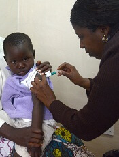
Photo by Caitlin Kleiboer
The European Medicines Agency’s Committee for Medicinal Products for Human Use (CHMP) has adopted a positive opinion of the malaria vaccine RTS,S (also known as RTS,S/AS01 or Mosquirix) for use in children ages 6 weeks to 17 months.
RTS,S is the first candidate vaccine for malaria to reach this milestone. The vaccine was designed to prevent malaria caused by the Plasmodium falciparum parasite.
The CHMP’s opinion on RTS,S is a final stage in the European Medicines Agency’s Article 58 procedure. This allows the CHMP, in cooperation with the World Health Organization (WHO), to give a scientific opinion on a medicinal product intended for markets outside the European Union. This assessment requires products to meet the same standards as those intended for use in the European Union.
Because the CHMP has issued a positive opinion of RTS,S, the WHO is now formulating a policy recommendation on use of the vaccine in national immunization programs.
The vaccine must also pass the WHO pre-qualification process and be approved by national regulatory authorities before it can be used in countries in sub-Saharan Africa, where P falciparum malaria is most prevalent.
About RTS,S
RTS,S aims to trigger the body’s immune system to defend against P falciparum when it first enters the human host’s bloodstream and/or when the parasite infects liver cells.
The vaccine is designed to prevent the parasite from infecting, maturing, and multiplying in the liver, after which time the parasite would re-enter the bloodstream and infect red blood cells, leading to disease symptoms.
The safety and efficacy of RTS,S has been evaluated in a large-scale, phase 3 trial. The CHMP’s recommendation was based mainly on the results of this study. Updated results were published in The Lancet last April.
According to that account, the trial included 15,459 young infants (aged 6 weeks to 12 weeks at first vaccination) and children (5 months to 17 months at first vaccination) from 11 sites across 7 sub-Saharan African countries (Burkina Faso, Gabon, Ghana, Kenya, Malawi, Mozambique, and United Republic of Tanzania) with varying levels of malaria transmission.
The subjects received RTS,S in 3 doses, 1 month apart. Some subjects received an additional booster dose 18 months later. Researchers compared subjects receiving RTS,S to those receiving a control vaccine.
Children who received 3 doses of RTS,S plus a booster had a 36% reduction in the number of clinical episodes of malaria at 4 years. Infants who received 3 doses of RTS,S plus a booster had a 26% reduction in the number of clinical malaria episodes over 3 years.
Children had a significantly lower incidence of severe malaria only if they received the booster dose of RTS,S. The vaccine (with or without a booster dose) did not confer the same benefit in infants.
Subjects who received RTS,S had more adverse events than subjects in the control group. This included meningitis and convulsions.
The road to approval
Two of the WHO’s independent advisory groups, the Strategic Advisory Group of Experts (SAGE) on Immunization and the Malaria Policy Advisory Committee (MPAC), are reviewing the evidence base for RTS,S and will make a joint policy recommendation for how it might be used with other tools to prevent malaria if RTS,S is approved by national regulatory authorities in sub-Saharan Africa.
The WHO has indicated that such a policy recommendation may be possible by end of this year.
Once the WHO policy recommendation is complete, GSK (the company developing RTS,S in partnership with the PATH Malaria Vaccine Initiative) will submit an application to the WHO for pre-qualification of RTS,S.
WHO pre-qualification involves a scientific assessment of the quality, safety, and efficacy of any new vaccine proposed for introduction in the WHO Expanded Programme on Immunization. A pre-qualification decision is used by the United Nations agencies and other large-scale public procurement agencies to help inform vaccine purchasing decisions.
Once a WHO pre-qualification is granted, GSK would then apply for marketing authorization in sub-Saharan Africa on a country-by-country basis. These regulatory and policy decisions would, if positive, enable countries to begin using RTS,S through their universal immunization program.
Both a WHO policy recommendation and WHO pre-qualification are requirements for Gavi, the Vaccine Alliance, to support eligible African countries introducing RTS,S into local immunization programs supported by UNICEF.
GSK has committed to a not-for-profit price for RTS,S. If the vaccine is approved, the price would cover the cost of
manufacturing RTS,S and a return of around 5% to be reinvested in
research and development for second-generation malaria vaccines or
vaccines against other neglected tropical diseases. ![]()
Value-based care poses new legal risks for doctors
The government’s push toward value-based care aims to fix a broken reimbursement system and improve quality of care for patients. But the new payment models also bring new legal risks for physicians, experts and anti-fraud officials warn.
“Novel payment methodologies may present new program integrity vulnerabilities,” Dr. Shantanu Agrawal, director of the Center for Program Integrity at the Centers for Medicare & Medicaid Services, said at a recent American Bar Association meeting. “As they assume financial risk, providers are also assuming program integrity risk. Without adequate controls, provider-run systems may be relatively vulnerable.”

The Department of Health and Human Services plans to have 30% of Medicare payments in value-based payment structures by the end of 2016, and 50% by the end of 2018. The transition will be driven through investments in alternative payment models such as Accountable Care Organizations (ACOs), advanced primary care medical home models, bundled payments models, and integrated care demonstrations for Medicare and Medicaid patients.
At the end of 2014, value-based payments represented 20% of Medicare fee-for-service payments to providers, according to CMS data. The rate was fueled by government programs such as the Medicare Shared Savings Program (MSSP), Pioneer ACOs, the Bundled Payments for Care Improvement Initiative, and the Comprehensive Primary Care Initiative. Meanwhile, HHS is encouraging private payers, marketplace plans, Medicare Advantage plans, and state Medicaid programs to move in the same value-based direction.
With so many new regulations, mandates, and programs coming down the pipeline, physicians are likely not thinking about the legal dangers that may arise with alternative payment structures, said Mark S. Kopson, a health law attorney in Bloomfield Hills, Mich., and chair of the American Health Lawyers Association’s Payers, Plans and Managed Care Practice Group.

Fee-for-service models can involve claims “about excess treatments and unnecessary services to drive up reimbursement,” Mr. Kopson said in an interview. “When you get into these [value-based] types of programs, it’s the exact opposite. The real threat is the withholding of necessary care in order to reduce expenses and therefore drive up those margins for the providers.”
To avoid such claims, physicians should ensure that their charts include the reasoning behind treatment decisions and a thorough record of why certain treatments were chosen and diagnoses were made, Mr. Kopson advised.
“Going forward, your charting better be completely accurate and detailed so that you don’t leave room for the government to make an argument that you should have provided this or that additional treatment,” he said.
Inaccurate reporting of enrollment data or financial information within new payment models could also land doctors in legal trouble, according to CMS officials.
Problematic reports, enrollee data, or other information physicians are required to submit to the government could be considered falsification and lead to False Claims Act violations.
“Providers are responsible for the information reported and should ensure that the appropriate checks and balances are in place that verify data is reported timely and accurately,” Tony A. Salters, a CMS spokesman, said in an interview. “For some models, providers must attest to the accuracy of this data. [To] report inaccurately could result in violations of federal laws.”
Physician-run payment models, such as doctor-led ACOs, may also draw legal scrutiny if physicians fail to prevent bad behavior by de facto partners. Physicians must ensure that all costs claimed by subcontractors, other providers, and suppliers who are paid from or authorized by the provider-run system, have been validated, Mr. Salters said.
“Doctors need to be aware that other entities who become new partners should hold themselves to the same high standards,” he said. “Providers should have basic financial mechanisms in place, with more sophisticated systems requiring more sophisticated methods,” to ensure validation.
CMS officials recommend doctors conduct independent audits of their accounts, manual validation of record system accuracy, and periodic verification of subcontractor claims to confirm the accuracy of claims and costs within new payment models.
These are “all routine steps that practitioners can take in their own offices but which are even more important when the doctor assumes responsibility for a larger scope of services,” Mr. Salters said.
Gaps in documentation surrounding bundled payments can be another legal land mine, Mr. Kopson noted. Adequate records of the care spectrum are essential to prevent accusations that care was not provided during a single episode of care, or over a specific period of time.
“You have to capture and document all the services you are delivering, and have accurate tracking in place for the entire continuum of care,” Mr. Kopson said.
CMS recommends that physicians establish a strong compliance program to assist with anti-fraud controls of new payment systems. When creating or updating a compliance program, government officials said providers should consider the unique characteristics of the model in which they participate.
On Twitter @legal_med
The government’s push toward value-based care aims to fix a broken reimbursement system and improve quality of care for patients. But the new payment models also bring new legal risks for physicians, experts and anti-fraud officials warn.
“Novel payment methodologies may present new program integrity vulnerabilities,” Dr. Shantanu Agrawal, director of the Center for Program Integrity at the Centers for Medicare & Medicaid Services, said at a recent American Bar Association meeting. “As they assume financial risk, providers are also assuming program integrity risk. Without adequate controls, provider-run systems may be relatively vulnerable.”
The Department of Health and Human Services plans to have 30% of Medicare payments in value-based payment structures by the end of 2016, and 50% by the end of 2018. The transition will be driven through investments in alternative payment models such as Accountable Care Organizations (ACOs), advanced primary care medical home models, bundled payments models, and integrated care demonstrations for Medicare and Medicaid patients.
At the end of 2014, value-based payments represented 20% of Medicare fee-for-service payments to providers, according to CMS data. The rate was fueled by government programs such as the Medicare Shared Savings Program (MSSP), Pioneer ACOs, the Bundled Payments for Care Improvement Initiative, and the Comprehensive Primary Care Initiative. Meanwhile, HHS is encouraging private payers, marketplace plans, Medicare Advantage plans, and state Medicaid programs to move in the same value-based direction.
With so many new regulations, mandates, and programs coming down the pipeline, physicians are likely not thinking about the legal dangers that may arise with alternative payment structures, said Mark S. Kopson, a health law attorney in Bloomfield Hills, Mich., and chair of the American Health Lawyers Association’s Payers, Plans and Managed Care Practice Group.
Fee-for-service models can involve claims “about excess treatments and unnecessary services to drive up reimbursement,” Mr. Kopson said in an interview. “When you get into these [value-based] types of programs, it’s the exact opposite. The real threat is the withholding of necessary care in order to reduce expenses and therefore drive up those margins for the providers.”
To avoid such claims, physicians should ensure that their charts include the reasoning behind treatment decisions and a thorough record of why certain treatments were chosen and diagnoses were made, Mr. Kopson advised.
“Going forward, your charting better be completely accurate and detailed so that you don’t leave room for the government to make an argument that you should have provided this or that additional treatment,” he said.
Inaccurate reporting of enrollment data or financial information within new payment models could also land doctors in legal trouble, according to CMS officials.
Problematic reports, enrollee data, or other information physicians are required to submit to the government could be considered falsification and lead to False Claims Act violations.
“Providers are responsible for the information reported and should ensure that the appropriate checks and balances are in place that verify data is reported timely and accurately,” Tony A. Salters, a CMS spokesman, said in an interview. “For some models, providers must attest to the accuracy of this data. [To] report inaccurately could result in violations of federal laws.”
Physician-run payment models, such as doctor-led ACOs, may also draw legal scrutiny if physicians fail to prevent bad behavior by de facto partners. Physicians must ensure that all costs claimed by subcontractors, other providers, and suppliers who are paid from or authorized by the provider-run system, have been validated, Mr. Salters said.
“Doctors need to be aware that other entities who become new partners should hold themselves to the same high standards,” he said. “Providers should have basic financial mechanisms in place, with more sophisticated systems requiring more sophisticated methods,” to ensure validation.
CMS officials recommend doctors conduct independent audits of their accounts, manual validation of record system accuracy, and periodic verification of subcontractor claims to confirm the accuracy of claims and costs within new payment models.
These are “all routine steps that practitioners can take in their own offices but which are even more important when the doctor assumes responsibility for a larger scope of services,” Mr. Salters said.
Gaps in documentation surrounding bundled payments can be another legal land mine, Mr. Kopson noted. Adequate records of the care spectrum are essential to prevent accusations that care was not provided during a single episode of care, or over a specific period of time.
“You have to capture and document all the services you are delivering, and have accurate tracking in place for the entire continuum of care,” Mr. Kopson said.
CMS recommends that physicians establish a strong compliance program to assist with anti-fraud controls of new payment systems. When creating or updating a compliance program, government officials said providers should consider the unique characteristics of the model in which they participate.
On Twitter @legal_med
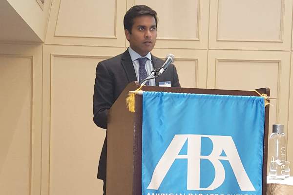
CHMP recommends eltrombopag for SAA

The European Medicines Agency’s Committee for Medicinal Products for Human Use (CHMP) has recommended approval for eltrombopag (Revolade) as a treatment for adults with severe aplastic anemia (SAA) who have had an insufficient response to immunosuppressive therapy (IST) and are not eligible to receive a hematopoietic stem cell transplant (HSCT).
If approved by the European Commission, eltrombopag would be the first treatment option in its class for these patients.
The European Commission will review the CHMP recommendation and is expected to deliver its final decision within 3 months. The decision will be applicable to all 28 European Union member states plus Iceland, Norway, and Liechtenstein.
Phase 2 trial of eltrombopag in SAA
The CHMP’s positive opinion of eltrombopag is mainly based on results of a phase 2 trial (NCT00922883). Results from this ongoing study were published in NEJM in 2012 and Blood in 2013.
The trial has enrolled 43 patients with SAA who had an insufficient response to at least one prior IST and who had a platelet count of 30 x 109/L or less.
At baseline, the median platelet count was 20 x 109/L, hemoglobin was 8.4 g/dL, the absolute neutrophil count was 0.58 x 10⁹/L, and absolute reticulocyte count was 24.3 x 10⁹/L.
Patients had a median age of 45 (range, 17 to 77), and 56% were male. The majority of patients (84%) received at least 2 prior ISTs.
Patients received eltrombopag at an initial dose of 50 mg once daily for 2 weeks. The dose increased over 2-week periods to a maximum of 150 mg once daily.
The study’s primary endpoint was hematologic response, which was initially assessed after 12 weeks of treatment. Treatment was discontinued after 16 weeks in patients who did not exhibit a hematologic response.
Forty percent of patients (n=17) experienced a hematologic response in at least one lineage—platelets, red blood cells (RBCs), or white blood cells—after week 12.
In the extension phase of the study, 8 patients achieved a multilineage response. Four of these patients subsequently tapered off treatment and maintained the response. The median follow up was 8.1 months (range, 7.2 to 10.6 months).
Ninety-one percent of patients were platelet-transfusion-dependent at baseline. Patients who responded to eltrombopag did not require platelet transfusions for a median of 200 days (range, 8 to 1096 days).
Eighty-six percent of patients were RBC-transfusion-dependent at baseline. Patients who responded to eltrombopag did not require RBC transfusions for a median of 208 days (range, 15 to 1082 days).
The most common adverse events (≥20%) associated with eltrombopag were nausea (33%), fatigue (28%), cough (23%), diarrhea (21%), and headache (21%).
Patients were also evaluated for cytogenetic abnormalities. Eight patients had a new cytogenetic abnormality after treatment, including 5 patients who had complex changes in chromosome 7.
Patients who develop new cytogenetic abnormalities while on eltrombopag may need to be taken off treatment.
About eltrombopag
Eltrombopag is already approved to treat SAA in the US and Canada. The drug recently gained approval in the US to treat children age 6 and older who have chronic immune thrombocytopenia and have had an insufficient response to corticosteroids, immunoglobulins, or splenectomy.
Eltrombopag is approved in more than 100 countries to treat adults with chronic immune thrombocytopenia who have had an inadequate response to or are intolerant of other treatments. The drug is approved in more than 45 countries for the treatment of thrombocytopenia in patients with chronic hepatitis C to allow them to initiate and maintain interferon-based therapy.
Eltrombopag is marketed under the brand name Promacta in the US and Revolade in most other countries. For more details on the drug, see the European Medicines Agency’s Summary of Product Characteristics. ![]()
FDA expands approval of carfilzomib
Photo courtesy of Amgen
The US Food and Drug Administration (FDA) has approved carfilzomib (Kyprolis) in combination with lenalidomide and dexamethasone to treat patients with relapsed multiple myeloma (MM) who have received 1 to 3 prior lines of therapy.
Carfilzomib already has accelerated approval from the FDA as monotherapy for MM patients who have received at least 2 prior therapies, including bortezomib and an immunomodulatory agent, and have demonstrated disease progression on or within 60 days of completing their last treatment.
The FDA’s expanded approval was based on results of the phase 3 ASPIRE trial.
The trial enrolled 792 patients with relapsed or refractory MM who had received 1 to 3 prior lines of therapy. The patients were randomized (1:1) to receive lenalidomide and dexamethasone with or without carfilzomib for 18 cycles.
Lenalidomide and dexamethasone were continued thereafter until disease progression. There was no planned cross-over from the control arm to treatment with carfilzomib.
There was a statistically significant prolongation of progression-free survival (PFS), as determined by an independent review committee, in the carfilzomib arm (hazard ratio=0.69, P=0.0001). The median PFS was 26.3 months in the 3-drug arm and 17.6 months in the 2-drug arm.
A treatment effect was observed across all subgroups tested, but the magnitude of the treatment effect was reduced in patients with higher tumor burden at baseline. The improvement in median PFS was 11 months for patients with International Staging System (ISS) Stage I disease, 8 months for ISS Stage II, and 2 months for ISS Stage III.
An interim analysis of overall survival, the key secondary endpoint, was conducted at the same time. The difference in overall survival did not reach the prespecified boundary for statistical significance.
The overall response rate (partial response or better) was 87% in the 3-drug arm and 67% in the 2-drug arm.
The safety profile of carfilzomib in the 3-drug combination was similar to that described on the current label.
Cardiovascular events, venous thromboembolic events (VTE), and thrombocytopenia occurred more frequently in the 3-drug arm than in the 2-drug arm. In cycles 1 to 12, the VTE rate was 13% and 6%, respectively, despite protocol-mandated use of thromboprophylaxis.
The revised labeling for carfilzomib includes new Warnings and Precautions for VTE, cardiac toxicities, acute renal failure, pulmonary toxicities, and hypertension. The increased safety risks, including mortality, for elderly patients is described. Detailed safety information in the prescribing information was also updated for the use of carfilzomib monotherapy.
The recommended dose schedule for carfilzomib has been revised for both monotherapy and combination treatment. For details, see the full prescribing information.
Carfilzomib is marketed as Kyprolis in the US by Onyx Pharmaceuticals, Inc., an Amgen subsidiary. ![]()