User login
CHA2DS2-VASc score of 1 linked to lower stroke risk than previously reported
Patients with atrial fibrillation who had CHA2DS2-VASc scores of 1 were at lower risk of ischemic stroke than previously reported, according to a retrospective analysis of hospital registry data. The research appeared online January 19 in the Journal of American College of Cardiology.
Depending on the definition of stroke used, risk was 0.1% to 0.2% in women and 0.5% to 0.7% in men – so low that oral anticoagulants (OACs) would not be expected to benefit patients of either sex, said Dr. Leif Friberg at the Karolinska Institute in Stockholm and his associates. Past studies had potentially overestimated the risk of stroke in this population, which “may have led to unnecessary, and potentially harmful, OAC treatment of low-risk patients,” they said.

European and U.S. guidelines both recommend using the CHA2DS2-VASc (heart failure, hypertension, age ≥75, diabetes mellitus, prior stroke or transient ischemic attack, vascular disease, age 65-74 years, female) scoring system to assess stroke risk in patients with atrial fibrillation (AF). But past studies have reported a threefold variation (ranging from 0.6% to greater than 2.0%) in stroke risk among AF patients with CHA2DS2-VASc scores of 1 who were not receiving OAC, the researchers noted. Anticoagulation therapy is likely to benefit AF patients whose annual risk of stroke exceeds 1%, but not patients whose risk is only 0.6%, they added.
Their study, which included 140,420 patients in Sweden with nonvalvular AF, assessed the effect of varying definitions of stroke on estimates of stroke risk. Using a broad definition that included ischemic stroke, transient ischemic attack (TIA), and pulmonary embolism led to a 44% greater annual risk of stroke than if only ischemic strokes were considered, the investigators reported. They disagreed with classifying pulmonary embolism events and TIAs as strokes, as some past studies have done. “Primary prevention of pulmonary embolism among patients with AF has, to the best of our knowledge, not been studied and is not an approved indication for OAC treatment,” they said. “We also did not find it relevant to count TIA as an endpoint in studies that describe stroke risk. As a diagnosis, TIA is difficult to validate.”
Several Swedish foundations supported the study. Dr. Friberg reported no relevant financial conflicts of interest.
Given the current state of knowledge, patients with atrial fibrillation who are younger than 65 years but have a CHA2DS2-VASc score of 1 are unlikely to benefit from anticoagulation therapy.
Dr. Friberg and his colleagues make two important observations regarding risk score thresholds for oral anticoagulant therapy. First, they highlight the wide cohort-to-cohort variation in reported CHA2DS2-VASc–stratified rates of stroke for atrial fibrillation patients who are not anticoagulated. Second, they reveal how sensitive estimates of stroke rates are to variations in interrogating administrative databases, which are used repeatedly as sources of “real world” rates of stroke. They conclude that the true stroke rate for patients with a CHA2DS2-VASc score of 1 is less than 0.7% per year, too low for oral anticoagulant therapy to benefit patients with AF.
Going forward, guideline writers should be aware of the drawbacks of the CHA2DS2-VASc score. They should focus on the absolute rates of stroke corresponding to risk prediction point scores and be alert to potential biases in studies reporting these rates. Investigators should work to harmonize methods for analyzing large AF databases. If variation in reported rates cannot be reconciled, then recommendations should reflect this uncertainty.
Dr. Daniel E. Singer is at Harvard Medical School in Boston, and Dr. Michael D. Ezekowitz is at Sidney Kimmel Medical College at Thomas Jefferson University in Philadelphia. Dr. Singer has been a consultant to, advised, and or received research funding from Boehringer Ingelheim, Bristol-Myers Squibb, Johnson & Johnson, Merck, and St. Jude Medical, and Medtronic. Dr. Ezekowitz reported having been a consultant and advisory board member for all those companies and several others. These remarks were taken from their editorial accompanying Dr. Friberg’s report (J. Am. Coll. Cardiol. 2015 Jan. 19 [doi:10.1016/j.jacc.2014.11.013]).
Patients with atrial fibrillation who had CHA2DS2-VASc scores of 1 were at lower risk of ischemic stroke than previously reported, according to a retrospective analysis of hospital registry data. The research appeared online January 19 in the Journal of American College of Cardiology.
Depending on the definition of stroke used, risk was 0.1% to 0.2% in women and 0.5% to 0.7% in men – so low that oral anticoagulants (OACs) would not be expected to benefit patients of either sex, said Dr. Leif Friberg at the Karolinska Institute in Stockholm and his associates. Past studies had potentially overestimated the risk of stroke in this population, which “may have led to unnecessary, and potentially harmful, OAC treatment of low-risk patients,” they said.
European and U.S. guidelines both recommend using the CHA2DS2-VASc (heart failure, hypertension, age ≥75, diabetes mellitus, prior stroke or transient ischemic attack, vascular disease, age 65-74 years, female) scoring system to assess stroke risk in patients with atrial fibrillation (AF). But past studies have reported a threefold variation (ranging from 0.6% to greater than 2.0%) in stroke risk among AF patients with CHA2DS2-VASc scores of 1 who were not receiving OAC, the researchers noted. Anticoagulation therapy is likely to benefit AF patients whose annual risk of stroke exceeds 1%, but not patients whose risk is only 0.6%, they added.
Their study, which included 140,420 patients in Sweden with nonvalvular AF, assessed the effect of varying definitions of stroke on estimates of stroke risk. Using a broad definition that included ischemic stroke, transient ischemic attack (TIA), and pulmonary embolism led to a 44% greater annual risk of stroke than if only ischemic strokes were considered, the investigators reported. They disagreed with classifying pulmonary embolism events and TIAs as strokes, as some past studies have done. “Primary prevention of pulmonary embolism among patients with AF has, to the best of our knowledge, not been studied and is not an approved indication for OAC treatment,” they said. “We also did not find it relevant to count TIA as an endpoint in studies that describe stroke risk. As a diagnosis, TIA is difficult to validate.”
Several Swedish foundations supported the study. Dr. Friberg reported no relevant financial conflicts of interest.
FROM THE JOURNAL OF THE AMERICAN COLLEGE OF CARDIOLOGY
Key clinical point: Stroke risk was low in patients with atrial fibrillation and a CHA2DS2-VASc score of 1.
Major finding: Risk of stroke was 0.1% to 0.2% in women and 0.5% to 0.7% in men.
Data source: Retrospective study of 140,420 patients with nonvalvular AF.
Disclosures: Several Swedish foundations supported the study. Dr. Friberg reported no relevant financial conflicts of interest.

Promising new therapy for critical limb ischemia
CHICAGO– A single set of intramuscular injections of stromal cell–derived factor-1 in patients with critical limb ischemia showed safety as well as evidence of efficacy through 12 months of follow-up in the STOP-CLI trial.
“Patients treated with JVS-100 demonstrated dose-dependent improvement across multiple patient-centered outcomes, including pain, quality of life, and wound healing, with less change in macrovascular objective measures in this small study,” Dr. Melina R. Kibbe reported at the American Heart Association scientific sessions. JVS-100 is a nonviral plasmid encoding human stromal cell–derived factor-1 (SDF-1), a natural chemokine protein that promotes angiogenesis by recruiting endothelial progenitor cells from the bone marrow to ischemic sites, explained Dr. Kibbe, professor and vice chair of surgical research and deputy director of the Simpson Querrey Institute for BioNanotechnology at Northwestern University, Chicago.

STOP-CLI was an exploratory, phase IIa, double-blind, first-in-humans study involving 48 patients with Rutherford classification 4 or 5 critical climb ischemia (CLI). All had an ankle-brachial index of 0.4 or lower, an ankle systolic blood pressure of 70 mm Hg or less or a toe systolic blood pressure of 50 or less, and were poor candidates for surgical revascularization. None had Buerger’s disease.
Participants were randomized to one of four study arms, and within each study arm further randomized 3:1 to stromal cell–derived factor-1 (SDF-1) or placebo injections. The patients received either 8 or 16 injections, each containing either 0.5 or 1.0 mg of SDF-1 or placebo. The injections, given in a single session, were placed at least 0.5 cm apart throughout the ischemic area of the affected limb.
By chance, most patients randomized to the placebo group were Rutherford 4, a category of CLI defined by rest pain, while the majority in the active treatment arms were Rutherford 5, a more severe disease manifestation characterized by ulcers. As a consequence, the SDF-1 recipients also had far larger nonhealing wounds, with an average area of 6.4 cm2, compared with 1.5 cm2, in controls.
The SDF-1 injections proved safe and were well tolerated, with no treatment-related serious adverse events and no safety signals evident in the laboratory results.
Turning to efficacy endpoints, Dr. Kibbe said self-rated visual analog scale pain scores showed clear, dose-dependent improvement over time in the SDF-1 treatment cohorts and no change in controls.
Similarly, the active treatment groups showed improved quality of life scores on all domains of the Short Form-36: physical functioning, bodily pain, general health, social functioning, energy/fatigue, emotional well-being, and overall physical and mental health, the surgeon continued.
Wound area decreased significantly in the SDF-1-treated groups, with the biggest reduction – more than 8 cm2 – being noted in the three patients who received eight 1-mg injections. That was also the group with the largest wounds at baseline, with an average area of 11.4 cm2.
Of note, the major limb amputation rate was “remarkably low” for patients with such severe CLI, according to Dr. Kibbe. The rate was less than 10% over the course of 12 months, with one patient in each of the four active treatment arms having a major amputation at time intervals of 58-112 days post injection. No major limb amputations occurred in the control group.
There was a hint of improvement with SDF-1 therapy over placebo in ankle-brachial index and transcutaneous oxygen pressure, but the between-group differences were too narrow in this study to allow for any conclusions. That must await planned much larger phase III trials, according to Dr. Kibbe.
Audience members, citing the numerous failures of once-promising stem cell therapies for CLI at phase III testing over the last 10-15 years, wondered why Dr. Kibbe thinks SDF-1 will fare any better.
“This is much debated and discussed among all the people involved in these kinds of trials,” she replied. “I’d say, briefly, that a lot of it has to do with patient selection. I think when you have a mixed bag of patients in a trial, including patients with Buerger’s disease, treated in multiple different countries, using different definitions of when to amputate, all those things come into play and could account for why those phase III trials were not successful.”
“Having been involved in lots of the different gene- and cell-based therapy trials, I think one of the unique benefits of this therapy is that it kind of bridges between the two. SDF-1 basically homes your endothelial progenitor cells to the site of ischemic injury for enhanced vasculogenesis. But SDF-1 also has direct effects on endothelial cells, including stimulating proliferation and preventing apoptosis,” she added.
JVS-100 has also successfully completed a phase II clinical trial for the treatment of heart failure. In addition, the agent is being developed as a treatment for acute MI, chronic angina, and for muscle regeneration.
The STOP-CLI study was sponsored by Juventas Therapeutics. Dr. Kibbe reported serving as a consultant to Johnson & Johnson/Cordis and Pluristem.
CHICAGO– A single set of intramuscular injections of stromal cell–derived factor-1 in patients with critical limb ischemia showed safety as well as evidence of efficacy through 12 months of follow-up in the STOP-CLI trial.
“Patients treated with JVS-100 demonstrated dose-dependent improvement across multiple patient-centered outcomes, including pain, quality of life, and wound healing, with less change in macrovascular objective measures in this small study,” Dr. Melina R. Kibbe reported at the American Heart Association scientific sessions. JVS-100 is a nonviral plasmid encoding human stromal cell–derived factor-1 (SDF-1), a natural chemokine protein that promotes angiogenesis by recruiting endothelial progenitor cells from the bone marrow to ischemic sites, explained Dr. Kibbe, professor and vice chair of surgical research and deputy director of the Simpson Querrey Institute for BioNanotechnology at Northwestern University, Chicago.
STOP-CLI was an exploratory, phase IIa, double-blind, first-in-humans study involving 48 patients with Rutherford classification 4 or 5 critical climb ischemia (CLI). All had an ankle-brachial index of 0.4 or lower, an ankle systolic blood pressure of 70 mm Hg or less or a toe systolic blood pressure of 50 or less, and were poor candidates for surgical revascularization. None had Buerger’s disease.
Participants were randomized to one of four study arms, and within each study arm further randomized 3:1 to stromal cell–derived factor-1 (SDF-1) or placebo injections. The patients received either 8 or 16 injections, each containing either 0.5 or 1.0 mg of SDF-1 or placebo. The injections, given in a single session, were placed at least 0.5 cm apart throughout the ischemic area of the affected limb.
By chance, most patients randomized to the placebo group were Rutherford 4, a category of CLI defined by rest pain, while the majority in the active treatment arms were Rutherford 5, a more severe disease manifestation characterized by ulcers. As a consequence, the SDF-1 recipients also had far larger nonhealing wounds, with an average area of 6.4 cm2, compared with 1.5 cm2, in controls.
The SDF-1 injections proved safe and were well tolerated, with no treatment-related serious adverse events and no safety signals evident in the laboratory results.
Turning to efficacy endpoints, Dr. Kibbe said self-rated visual analog scale pain scores showed clear, dose-dependent improvement over time in the SDF-1 treatment cohorts and no change in controls.
Similarly, the active treatment groups showed improved quality of life scores on all domains of the Short Form-36: physical functioning, bodily pain, general health, social functioning, energy/fatigue, emotional well-being, and overall physical and mental health, the surgeon continued.
Wound area decreased significantly in the SDF-1-treated groups, with the biggest reduction – more than 8 cm2 – being noted in the three patients who received eight 1-mg injections. That was also the group with the largest wounds at baseline, with an average area of 11.4 cm2.
Of note, the major limb amputation rate was “remarkably low” for patients with such severe CLI, according to Dr. Kibbe. The rate was less than 10% over the course of 12 months, with one patient in each of the four active treatment arms having a major amputation at time intervals of 58-112 days post injection. No major limb amputations occurred in the control group.
There was a hint of improvement with SDF-1 therapy over placebo in ankle-brachial index and transcutaneous oxygen pressure, but the between-group differences were too narrow in this study to allow for any conclusions. That must await planned much larger phase III trials, according to Dr. Kibbe.
Audience members, citing the numerous failures of once-promising stem cell therapies for CLI at phase III testing over the last 10-15 years, wondered why Dr. Kibbe thinks SDF-1 will fare any better.
“This is much debated and discussed among all the people involved in these kinds of trials,” she replied. “I’d say, briefly, that a lot of it has to do with patient selection. I think when you have a mixed bag of patients in a trial, including patients with Buerger’s disease, treated in multiple different countries, using different definitions of when to amputate, all those things come into play and could account for why those phase III trials were not successful.”
“Having been involved in lots of the different gene- and cell-based therapy trials, I think one of the unique benefits of this therapy is that it kind of bridges between the two. SDF-1 basically homes your endothelial progenitor cells to the site of ischemic injury for enhanced vasculogenesis. But SDF-1 also has direct effects on endothelial cells, including stimulating proliferation and preventing apoptosis,” she added.
JVS-100 has also successfully completed a phase II clinical trial for the treatment of heart failure. In addition, the agent is being developed as a treatment for acute MI, chronic angina, and for muscle regeneration.
The STOP-CLI study was sponsored by Juventas Therapeutics. Dr. Kibbe reported serving as a consultant to Johnson & Johnson/Cordis and Pluristem.
AT THE AHA SCIENTIFIC SESSIONS
Key clinical point: Intramuscular injections of stromal cell–derived factor-1 in patients with critical limb ischemia demonstrated safety and efficacy; the therapy is moving forward to phase III testing.
Major finding: The major limb amputation rate was less than 10% during 12 months of follow-up after a single dose of the novel therapy.
Data source: The STOP-CLI trial was a phase IIa, 12-month, randomized, double-blind, placebo-controlled, six-center trial including 48 patients with critical limb ischemia.
Disclosures: The STOP-CLI trial was sponsored by Juventas Therapeutics. The presenter reported serving as a consultant to Johnson & Johnson/Cordis and Pluristem.

Handheld device illuminates possible routes of melanoma metastases
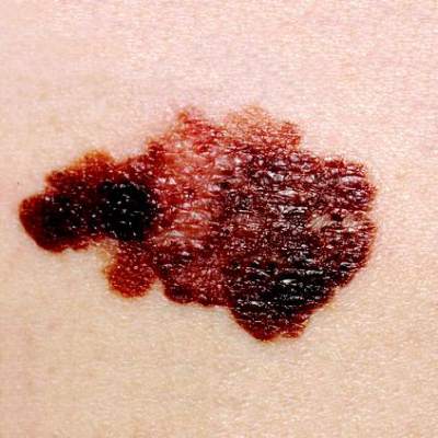
Investigators using a handheld dermoscopy device that allows visualization of colors, structures, and patterns in skin lesions not evident to the naked eye were able to visualize nonblanching blue and red lines in a branched pattern in two patients with in-transit cutaneous melanoma metastases.
Dr. Michael A. Marchetti and his associates at Memorial Sloan Kettering Cancer Center, New York, reported the “intriguing” visualization of dissemination for cutaneous melanoma metastases in a letter to JAMA Dermatology.
In-transit cutaneous melanoma metastases are those located more than 2 cm from the primary melanoma, but not beyond the regional nodal basin.

The first patient had wide local excision of a primary cutaneous melanoma on the forehead, and a year later, received localized irradiation for satellite skin metastases. A year after that, skin examination revealed six blue macules on the scalp more than 2 cm from the excision scar. Dermoscopy revealed nonblanching bluish lines in a branched pattern. Histopathologic examination of a skin biopsy confirmed in-transit metastatic melanoma with atypical melanocytes present in superficial dermal lymphatics, Dr. Marchetti and his associates reported (JAMA Dermatology 2015;103-5)
The second patient had a history of multiple primary melanomas, the most recent being one on the chest treated with wide local excision. At a follow-up visit 5 years later, skin examination revealed eight blue-gray macules on the chest, all more than 2 cm from the excision scar. Dermoscopy revealed nonblanching, red-bluish, fuzzy, branching lines. Histopathologic examination of a skin biopsy confirmed in-transit metastatic melanoma with atypical melanocytes present in superficial dermal blood vessels, the investigators wrote.
Typical dermoscopic features of cutaneous melanoma metastases include peripheral gray spots, atypical vessels, and a blue nevus-like pattern. The histopathologic findings in these two cases suggest that the dermoscopic color differences correspond to unique microanatomic routes of melanoma dissemination, with blue and red-blue lines corresponding to lymphatic and hematogenous dissemination of tumors, respectively, they said.
“While the factors driving lymphatic vs. hematogenous in-transit dissemination of melanoma remain unknown, as do any differences in their biologic significance, our finding is an intriguing clinical/dermoscopic/histopathologic observation,” the investigators concluded.
On Twitter @nikolaideslaura
Investigators using a handheld dermoscopy device that allows visualization of colors, structures, and patterns in skin lesions not evident to the naked eye were able to visualize nonblanching blue and red lines in a branched pattern in two patients with in-transit cutaneous melanoma metastases.
Dr. Michael A. Marchetti and his associates at Memorial Sloan Kettering Cancer Center, New York, reported the “intriguing” visualization of dissemination for cutaneous melanoma metastases in a letter to JAMA Dermatology.
In-transit cutaneous melanoma metastases are those located more than 2 cm from the primary melanoma, but not beyond the regional nodal basin.
The first patient had wide local excision of a primary cutaneous melanoma on the forehead, and a year later, received localized irradiation for satellite skin metastases. A year after that, skin examination revealed six blue macules on the scalp more than 2 cm from the excision scar. Dermoscopy revealed nonblanching bluish lines in a branched pattern. Histopathologic examination of a skin biopsy confirmed in-transit metastatic melanoma with atypical melanocytes present in superficial dermal lymphatics, Dr. Marchetti and his associates reported (JAMA Dermatology 2015;103-5)
The second patient had a history of multiple primary melanomas, the most recent being one on the chest treated with wide local excision. At a follow-up visit 5 years later, skin examination revealed eight blue-gray macules on the chest, all more than 2 cm from the excision scar. Dermoscopy revealed nonblanching, red-bluish, fuzzy, branching lines. Histopathologic examination of a skin biopsy confirmed in-transit metastatic melanoma with atypical melanocytes present in superficial dermal blood vessels, the investigators wrote.
Typical dermoscopic features of cutaneous melanoma metastases include peripheral gray spots, atypical vessels, and a blue nevus-like pattern. The histopathologic findings in these two cases suggest that the dermoscopic color differences correspond to unique microanatomic routes of melanoma dissemination, with blue and red-blue lines corresponding to lymphatic and hematogenous dissemination of tumors, respectively, they said.
“While the factors driving lymphatic vs. hematogenous in-transit dissemination of melanoma remain unknown, as do any differences in their biologic significance, our finding is an intriguing clinical/dermoscopic/histopathologic observation,” the investigators concluded.
On Twitter @nikolaideslaura
FROM JAMA DERMATOLOGY
System can detect Candida better, faster than blood culture
Credit: Jeremy L. Grisham
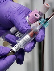
A diagnostic system can detect sepsis pathogens with high sensitivity and specificity in 3 to 5 hours, eliminating the need for blood culture, according to a study published in Clinical Infectious Diseases.
The system consists of the T2Candida Panel and the T2Dx Instrument, and it provides direct detection of 5 yeast pathogens—Candida albicans, Candida tropicalis, Candida parapsilosis, Candida glabrata, and Candida krusei—in whole blood samples.
T2Candida and T2Dx were cleared for use by the US Food and Drug Administration in September. They are the first diagnostic products powered by T2MR, a magnetic resonance-based diagnostic technology platform that does not require blood culture and sample purification or preparation.
“The ability to determine the presence or absence of Candida within hours—compared to days [with blood culture]—is paradigm-changing for patients at risk for these infections,” said study author Eleftherios Mylonakis, MD, PhD, of Rhode Island Hospital and The Miriam Hospital in Providence.
“It will allow us to move from a ‘best-guess’ approach in treating high-risk patients, such as cancer and transplant patients and patients in the intensive care unit, to a more informed approach where we can quickly direct the best course of therapy, potentially improving patient outcomes and saving lives.”
Study findings
For this multicenter study, Dr Mylonakis and his colleagues collected blood specimens from 1801 hospitalized patients between the ages of 18 and 95 who had a blood culture ordered as part of routine care.
T2Candida and T2Dx demonstrated an overall specificity of 99.4% per assay and 98.1% per patient. The system yielded a specificity of 98.9% for C albicans/C tropicalis, 99.3% for C parapsilosis, and 99.9% for C krusei/C glabrata.
The system had an overall sensitivity of 91.1% per assay and 91.0% per patient. It yielded a sensitivity of 92.3% for C albicans/C tropicalis, 94.2% for C parapsilosis, and 88.1% for C krusei/C glabrata.
The mean time to a positive result for T2Candida and T2Dx was 4.4 hours, compared to 129 hours for blood culture and species identification. The mean time to negative result for T2Candida and T2Dx was 4.2 hours, compared to at least 120 hours for blood culture.
In one case described in the paper, T2Candida and T2Dx detected a Candida infection that blood culture missed in 12 successive tests.
Seven days after the T2Candida result was obtained, physicians performed an invasive procedure to obtain a tissue culture, which proved that the T2Candida result accurately identified a case of intra-abdominal candidiasis.
“Blood culture, the current standard of care for the diagnosis of Candida infections, is known to have poor sensitivity overall and has 38% sensitivity in proven and probable cases of invasive candidiasis,” said study author Cornelius J. Clancy, MD, of the University of Pittsburgh in Pennsylvania.
“In our case, the T2Candida Panel has shown that it can rapidly identify intra-abdominal candidiasis where 12 serial blood culture results were negative. In many patients at risk for candidiasis, the collection of tissue samples for diagnosis is not possible due to their underlying medical conditions. Achieving the level of sensitivity demonstrated in this case, without requiring an intra-abdominal sample, has the potential to positively impact the practice of medicine for these patients.” ![]()
Credit: Jeremy L. Grisham
A diagnostic system can detect sepsis pathogens with high sensitivity and specificity in 3 to 5 hours, eliminating the need for blood culture, according to a study published in Clinical Infectious Diseases.
The system consists of the T2Candida Panel and the T2Dx Instrument, and it provides direct detection of 5 yeast pathogens—Candida albicans, Candida tropicalis, Candida parapsilosis, Candida glabrata, and Candida krusei—in whole blood samples.
T2Candida and T2Dx were cleared for use by the US Food and Drug Administration in September. They are the first diagnostic products powered by T2MR, a magnetic resonance-based diagnostic technology platform that does not require blood culture and sample purification or preparation.
“The ability to determine the presence or absence of Candida within hours—compared to days [with blood culture]—is paradigm-changing for patients at risk for these infections,” said study author Eleftherios Mylonakis, MD, PhD, of Rhode Island Hospital and The Miriam Hospital in Providence.
“It will allow us to move from a ‘best-guess’ approach in treating high-risk patients, such as cancer and transplant patients and patients in the intensive care unit, to a more informed approach where we can quickly direct the best course of therapy, potentially improving patient outcomes and saving lives.”
Study findings
For this multicenter study, Dr Mylonakis and his colleagues collected blood specimens from 1801 hospitalized patients between the ages of 18 and 95 who had a blood culture ordered as part of routine care.
T2Candida and T2Dx demonstrated an overall specificity of 99.4% per assay and 98.1% per patient. The system yielded a specificity of 98.9% for C albicans/C tropicalis, 99.3% for C parapsilosis, and 99.9% for C krusei/C glabrata.
The system had an overall sensitivity of 91.1% per assay and 91.0% per patient. It yielded a sensitivity of 92.3% for C albicans/C tropicalis, 94.2% for C parapsilosis, and 88.1% for C krusei/C glabrata.
The mean time to a positive result for T2Candida and T2Dx was 4.4 hours, compared to 129 hours for blood culture and species identification. The mean time to negative result for T2Candida and T2Dx was 4.2 hours, compared to at least 120 hours for blood culture.
In one case described in the paper, T2Candida and T2Dx detected a Candida infection that blood culture missed in 12 successive tests.
Seven days after the T2Candida result was obtained, physicians performed an invasive procedure to obtain a tissue culture, which proved that the T2Candida result accurately identified a case of intra-abdominal candidiasis.
“Blood culture, the current standard of care for the diagnosis of Candida infections, is known to have poor sensitivity overall and has 38% sensitivity in proven and probable cases of invasive candidiasis,” said study author Cornelius J. Clancy, MD, of the University of Pittsburgh in Pennsylvania.
“In our case, the T2Candida Panel has shown that it can rapidly identify intra-abdominal candidiasis where 12 serial blood culture results were negative. In many patients at risk for candidiasis, the collection of tissue samples for diagnosis is not possible due to their underlying medical conditions. Achieving the level of sensitivity demonstrated in this case, without requiring an intra-abdominal sample, has the potential to positively impact the practice of medicine for these patients.” ![]()
Study offers explanation for gender gaps in academia

Credit: Rhoda Baer
New research offers an explanation for the lack of women in certain academic fields.
It isn’t that women don’t want to work long hours or can’t compete in highly selective fields, and it isn’t that they are less analytical than men, the researchers reported.
Instead, it appears that women are underrepresented in academic fields whose practitioners put a lot of emphasis on the importance of being brilliant—a quality some people assume women lack.
Sarah-Jane Leslie, PhD, of Princeton University in New Jersey, and her colleagues reported these findings in Science.
The researchers focused on a broad swath of academic disciplines, including those in the sciences, the humanities, social sciences, and math.
They focused on the culture of different fields, reasoning that stereotypes of women’s inferior intellectual abilities might help explain why women are underrepresented in fields—such as physics or philosophy—that idolize geniuses.
The team surveyed more than 1800 graduate students, post-doctoral researchers, and faculty members in 30 academic disciplines and, among other things, asked them what qualities were required for success in their fields.
Across the board, in the sciences, technology, engineering, and math (STEM) fields, as well as in the humanities and social sciences, women were found to be underrepresented in those disciplines whose practitioners put a premium on brilliance.
“We’re not saying brilliance—or valuing brilliance—is a bad thing,” said study author Andrei Cimpian, PhD, of the University of Illinois at Urbana-Champaign.
“And we’re not saying women are not brilliant or that being brilliant isn’t helpful to one’s academic career. Our data don’t address that. What they suggest is that conveying to your students a belief that brilliance is required for success may have a differential effect on males and females that are looking to pursue careers in your field.”
The team also tested 3 other hypotheses that might help explain women’s underrepresentation in some fields: that women avoid careers that require them to work long hours, that women are less able than men to get into highly selective fields, and that women are outnumbered by men in fields that require analytical, systematical reasoning.
“We found that none of these 3 alternative hypotheses was able to predict women’s representation across the academic spectrum,” Dr Leslie said. “A strong emphasis on brilliance among practitioners of particular fields was the best predictor of women’s underrepresentation in those fields.”
The researchers are still investigating whether women are actively avoiding fields that focus on cultivating brilliant individuals, or if practitioners in those fields are discriminating against women based on their beliefs about women’s aptitudes. A combination of the two is certainly plausible, according to Dr Cimpian.
“There is no convincing evidence in the literature that men and women differ intellectually in ways that would be relevant to their success across the entire range of fields we surveyed,” Dr Cimpian said. “So it is most likely that female underrepresentation is not the result of actual differences in intellectual ability but rather the result of perceived or presumed differences between women and men.” ![]()
Antibiotics may enhance mosquitoes’ ability to transmit malaria

Credit: James Gathany
Mosquitoes’ ability to transmit malaria may be affected by antibiotics in the blood of those they bite, according to research published in Nature Communications.
Feeding on blood that contained the antibiotics penicillin and streptomycin (PS) hindered bacterial growth in mosquitoes’ guts.
The mosquitoes also became more susceptible to malaria infection, exhibited improved fertility, and lived longer than mosquitoes whose blood meals did not contain PS.
Study investigators said these findings do not suggest people should avoid taking antibiotics. But the study does indicate that more research is needed to explore how different combinations of antibiotics affect mosquitoes’ ability to spread malaria.
“Antibiotics are a valuable weapon in the fight against malaria and other diseases,” said study author Mathilde Gendrin, PhD, of Imperial College London in the UK.
“Our study suggests that the presence of antibiotics in people’s blood may have hidden effects on the guts of mosquitoes when mosquitoes ingest that blood, and that these effects could alter the mosquitoes’ ability to transmit malaria.”
Dr Gendrin and her colleagues chose to evaluate the effects of PS because these antibiotics are not used to combat malaria and don’t directly affect malaria parasites.
The team began their research by supplementing blood with therapeutic concentrations of PS and feeding the blood to Anopheles gambiae mosquitoes.
The investigators monitored the bacterial load in the mosquito gut over 3 blood feeds offered every 3 days. And they found that the proliferation of bacteria typically seen at 24 hours after a blood meal was reduced by 70% when mosquitoes received blood containing PS.
Next, the researchers conducted 3 experiments to determine whether receiving blood containing PS would influence the mosquitoes’ susceptibility to malaria infection.
In the first experiment, the team fed mosquitoes blood from rodents infected with Plasmodium berghei before or after the animals received PS.
The investigators assessed infection by counting the proportion of mosquitoes carrying oocysts (prevalence) and the number of oocysts per mosquito (intensity). In the presence of PS, infection prevalence increased by 21% (P=5.10-7), and the median intensity doubled (P=0.041).
In the second experiment, the researchers assessed the effect of PS on infection with human parasites. They fed mosquitoes blood containing Plasmodium falciparum gametocytes (cultured in vitro) and supplemented the blood with PS or buffer.
Again, the team observed higher infection prevalence (P=0.0033) and intensity (P=0.00026) in the presence of PS.
In the third experiment, the investigators fed mosquitoes blood freshly drawn from children carrying P falciparum gametocytes. The blood was supplemented with PS or buffer.
Once more, the intensity of infection significantly increased in the presence of PS (P=0.02). The 6.3% increase in infection prevalence was not statistically significant (P=0.4), but the researchers said this was likely due to the reduced statistical power of the experiment.
The team’s final experiments assessed the effects of PS on mosquitoes’ fertility and survival.
Exposure to PS led to a 32% higher proportion of egg-laying females (P=0.0038) and a 53% higher average number of eggs per female (P=0.016), although the proportion of eggs hatching into larvae was not affected.
Mosquito survival increased significantly after the insects ingested PS-treated human blood. The P value was 0.017 for the first blood meal and 0.0016 for all 3 blood meals.
“We only looked at two antibiotics in our study, so this is early stage research, and we don’t know what it means for other antibiotics,” Dr Gendrin noted.
“It’s possible other antibiotics might have no effect, or that they might make it harder for mosquitoes to transmit malaria. We would like to see much more research carried out to further understand our findings and to explore how other antibiotics might affect bacteria in the guts of mosquitoes.”
“Ultimately, we hope that understanding any hidden effects of different antibiotics would mean we can combat the spread of malaria more effectively,” added study author George Christophides, PhD, also of Imperial College London.
“For example, if particular antibiotics make mosquitoes more able to transmit malaria, the use of these could be combined with supply of bed nets in order to reduce mosquito bites. Additionally, if a doctor prescribed such antibiotics to a malaria patient, they could combine this with a drug such as atovaquone that can both treat malaria and block its transmission.” ![]()
Immunotherapy moves into the breast cancer landscape
Click on the PDF icon at the top of this introduction to read the full article.
Click on the PDF icon at the top of this introduction to read the full article.
Click on the PDF icon at the top of this introduction to read the full article.
Shrink Rap News: National study shows many offenders receive treatment only in prison
In a study published in the December 2014 issue of American Journal of Public Health, Jennifer M. Reingle Gonzalez, Ph.D., and Nadine M. Connell, Ph.D., of the University of Texas Health Science Center at Houston interviewed a nationally representative sample of all state and federal prisoners with psychiatric disorders to determine whether they had been screened for services at intake and to assess whether screening led to continuity of community treatment.
The subjects were chosen first by selecting a random sample of correctional facilities of varying sizes, from diverse geographic regions. From those facilities, 18,185 prisoners were chosen from among all those incarcerated on a single day in September 2002. Each subject was interviewed personally but also given computer-assisted interviews at different times to ensure recollection accuracy and the confidentiality of clinical data (Am. J. Public Health 2014;104:2328-33).
In the direct interview, each prisoner was asked if he or she had ever been diagnosed with a mental health condition such as depression, bipolar disorder, schizophrenia, posttraumatic stress disorder, an anxiety disorder, or a personality disorder. If any condition had ever been diagnosed, the inmates were then asked if they were taking a psychiatric medication upon admission to the facility. If they were in treatment at intake, they were asked if they had ever been on medication since that time. Finally, they were asked if they had had a medical examination while incarcerated.
Almost all prisoners (90%) were screened at intake and received a medical evaluation. The researchers found that 5,207 (26.2%) of the inmates received at least one lifetime mental health diagnosis, with depression being the most common. Eighteen percent reported taking medication for a psychiatric disorder at the time of intake, but of these, only about half were taking medication after incarceration. Medication continuance was twice as likely for schizophrenia as for depression, and those who received an intake screen were significantly more likely to be referred to a physician and receive medication. Notably, 27% of state and 16% of federal prisoners received medication only in prison.
The investigators attributed the discontinuity in treatment after incarceration to a lack of trained mental health professionals to diagnose and treat psychiatric disorders, and to a rise in prison populations out of proportion to available treatment services. The findings of this study supported the investigators’ recommendation for all facilities to employ intake screening to identify and refer prisoners in need of psychiatric care.
This research is important to forensic psychiatrists working in correctional systems and to those working as administrators or external court-appointed monitors, because it highlights the importance and efficacy of intake screening for prisoners with psychiatric disorders.
Unfortunately, traditional media coverage of this research was predictably provocative. Headlines blared: “Mental health care lacking in state and federal prisons.” The researchers themselves also implied that failure to continue medication in prison indicated some systemic deficiency. While an evaluation by a physician was correlated with the prescription of medication, this was not a perfect correlation. The authors did not discuss any of the valid reasons why medication might not be continued in prison.
The most common reason medication is not continued is that inmates are more likely to be abstinent from drugs and alcohol, and thus may require less or even no antidepressant medication after detoxification. This could account for as much as half of the treatment discontinuity, because as many as 20% of the prisoners had been diagnosed with depression. Also, evidence is mounting that indefinite medication may not be necessary for all conditions. Following a clinical assessment, a correctional physician may have determined that the prisoner had successfully completed maintenance therapy. The final reason why medication is not continued is that the inmate may simply have refused the offered treatment; some prisoners do choose to go without medication completely rather than risk a change to a different regimen.
All of this assumes that the subjects accurately reported both their psychiatric history and their mental health service contact following incarceration. Without access to current records or past documentation, this remains an open question. In my prison system, our electronic health record does contain some data provided by the public health care system. In rare cases, this information is consistent with what the patient reports, but this is the exception rather than the rule.
Finally, the most telling aspect of this research is what it reveals about free society care: A quarter of the inmates received treatment only in prison. That is the real finding worthy of a headline.
Dr. Hanson is a forensic psychiatrist and coauthor of Shrink Rap: Three Psychiatrists Explain Their Work. The opinions expressed are those of the author only, and do not represent those of any of Dr. Hanson’s employers or consultees, including the Maryland Department of Health and Mental Hygiene or the Maryland Division of Correction.

A generalist approach to fertility preservation
More than 500,000 women of reproductive age (20-49) are diagnosed with cancer each year. Fortunately, given advances in timely detection and effective therapy, more than 83% of these women will survive at least 5 years (SEER Cancer Statistics Review, 1975-2011). As a result, more women are concerned about their child-bearing potential after undergoing chemotherapy or radiation, and as reproductive technology advances, the issue of fertility preservation becomes even more salient.
Potential candidates for fertility preservation
The importance of considering fertility preservation for any reproductive-aged woman with a new cancer diagnosis cannot be overemphasized. This is especially true for women who may be receiving gonadotoxic therapies, such as alkylating agents or abdominal/pelvic radiation.

To assess the risk of various cancer treatments to fertility, patients and providers can access the Fertility Risk Tool at www.livestrong.org. The Fertility Risk Tool compiles known data about the risk of amenorrhea from specific cancers, chemotherapy agents, and radiation treatments, based on the woman’s age. The risk for infertility is likely higher than the stated incidence of amenorrhea, but the chart provides an initial counseling tool for health care providers and an overview for patients.
Discussions regarding fertility preservation are essential for any woman from menarche through mid-40s. However, as technological advances are rapidly occurring in reproductive medicine, options such as ovarian tissue cryopreservation are becoming available for prepubescent girls. Counseling these young patients with a new cancer diagnosis should not be overlooked.
Fertility preservation options
In vitro fertilization (IVF) with embryo banking is currently the most successful fertility preservation option and the standard of care. The process involves an IVF cycle, including monitored ovarian stimulation, transvaginal oocyte retrieval, fertilization of the oocytes, and cryopreservation of embryos.
The entire process takes a minimum of 12 days, and usually starts shortly after the onset of menses. The embryos can survive for years in liquid nitrogen and the survival rate of frozen embryos is greater than 95%. The pregnancy rate per embryo transfer cycle depends on the age of the woman when the embryos were created, with women under age 35 having higher live-birth rates compared with women older than 42 (live-birth rates 42.4% versus 17.8%, respectively, according to national summary data from the Society for Assisted Reproductive Technology).

Another option, which may be more attractive for women without a committed male partner, involves egg banking. This process also involves ovarian stimulation and egg retrieval, but fertilization is not performed. Instead the oocytes are cryopreserved, often by way of a vitrification technique shown to have a higher percentage of oocytes that survive the thaw (Fertil. Steril. 2011;96:277-85).
While the data regarding live birth after egg banking are limited, studies have shown reassuring birth outcomes for more than 900 babies from this technology (Reprod. Biomed. Online 2009;18:769-76).
Timing of treatment must be coordinated with the help of an oncologist. For example, many women with breast cancer opt to have their oncological surgery and undergo ovarian stimulation during the 4-6 week recovery period before initiating chemotherapy.
Ovarian tissue banking is considered experimental, but may be the only option available for women who must initiate treatment immediately, or for prepubescent girls. This technology involves surgical removal of part of an ovary, which is divided into small sections and frozen. The options for reproductive potential may include in vitro maturation of the immature oocytes in the strips of ovarian tissue with subsequent fertilization in the laboratory versus transplantation of the segments of ovarian tissue with the goal of some restoration of ovarian function (Hum. Reprod. 2014;29:1931-40).
Another option is the use of a gonadotropin-releasing hormone (GnRH) agonist during therapy to induce a prepubertal state, with the hypothetical goal to decrease damage to immature oocytes. A recent meta-analysis of randomized trials found that suppression with a GnRH agonist during chemotherapy significantly decreased premature ovarian failure in young women (Cancer Treat. Rev. 2014;40:675-83).
To date, the available literature does not address the effect of GnRH agonist use on rates of infertility in cancer survivors.
Special considerations for hormone sensitive cancers
Women with hormone sensitive cancers, such as breast cancer, often have understandable concerns about preserving their fertility and the impact that ovarian stimulation and future pregnancy may have on their prognosis.
For breast cancer patients, an effective adjuvant treatment during ovarian stimulation is an aromatase inhibitor, which lowers the peak estrogen levels compared to a standard ovarian stimulation cycle, with a similar oocyte yield (J. Clin. Endocrinol. Metab. 2006;91:3885-90).
There does not appear to be a difference in recurrence of breast cancer in women who pursued egg or embryo banking versus those who did not (J. Clin. Oncol. 2008;26:2630-5). Even after subsequent successful pregnancies, recurrence risk in hormone sensitive cancers, including breast cancer, is not increased (Cancer 2004;100:465-9). It is important to note that many women with breast cancer are placed on tamoxifen for many years. These women may consider a surrogate or a “tamoxifen break” after consultation with their oncologists.
When to refer
In any pediatric or reproductive-aged woman with a new cancer diagnosis, it is important to have a conversation exploring fertility options as soon as possible after the diagnosis is made. An early referral yields more options prior to initiating treatment and more time for the woman to discuss and consider all approaches to preserving her fertility.
Dr. Mersereau is an associate professor at the University of North Carolina at Chapel Hill, and director of UNC’s Fertility Preservation Program. Dr. Hoff is a clinical instructor and a fellow in reproductive endocrinology and infertility at UNC-Chapel Hill.
More than 500,000 women of reproductive age (20-49) are diagnosed with cancer each year. Fortunately, given advances in timely detection and effective therapy, more than 83% of these women will survive at least 5 years (SEER Cancer Statistics Review, 1975-2011). As a result, more women are concerned about their child-bearing potential after undergoing chemotherapy or radiation, and as reproductive technology advances, the issue of fertility preservation becomes even more salient.
Potential candidates for fertility preservation
The importance of considering fertility preservation for any reproductive-aged woman with a new cancer diagnosis cannot be overemphasized. This is especially true for women who may be receiving gonadotoxic therapies, such as alkylating agents or abdominal/pelvic radiation.
To assess the risk of various cancer treatments to fertility, patients and providers can access the Fertility Risk Tool at www.livestrong.org. The Fertility Risk Tool compiles known data about the risk of amenorrhea from specific cancers, chemotherapy agents, and radiation treatments, based on the woman’s age. The risk for infertility is likely higher than the stated incidence of amenorrhea, but the chart provides an initial counseling tool for health care providers and an overview for patients.
Discussions regarding fertility preservation are essential for any woman from menarche through mid-40s. However, as technological advances are rapidly occurring in reproductive medicine, options such as ovarian tissue cryopreservation are becoming available for prepubescent girls. Counseling these young patients with a new cancer diagnosis should not be overlooked.
Fertility preservation options
In vitro fertilization (IVF) with embryo banking is currently the most successful fertility preservation option and the standard of care. The process involves an IVF cycle, including monitored ovarian stimulation, transvaginal oocyte retrieval, fertilization of the oocytes, and cryopreservation of embryos.
The entire process takes a minimum of 12 days, and usually starts shortly after the onset of menses. The embryos can survive for years in liquid nitrogen and the survival rate of frozen embryos is greater than 95%. The pregnancy rate per embryo transfer cycle depends on the age of the woman when the embryos were created, with women under age 35 having higher live-birth rates compared with women older than 42 (live-birth rates 42.4% versus 17.8%, respectively, according to national summary data from the Society for Assisted Reproductive Technology).
Another option, which may be more attractive for women without a committed male partner, involves egg banking. This process also involves ovarian stimulation and egg retrieval, but fertilization is not performed. Instead the oocytes are cryopreserved, often by way of a vitrification technique shown to have a higher percentage of oocytes that survive the thaw (Fertil. Steril. 2011;96:277-85).
While the data regarding live birth after egg banking are limited, studies have shown reassuring birth outcomes for more than 900 babies from this technology (Reprod. Biomed. Online 2009;18:769-76).
Timing of treatment must be coordinated with the help of an oncologist. For example, many women with breast cancer opt to have their oncological surgery and undergo ovarian stimulation during the 4-6 week recovery period before initiating chemotherapy.
Ovarian tissue banking is considered experimental, but may be the only option available for women who must initiate treatment immediately, or for prepubescent girls. This technology involves surgical removal of part of an ovary, which is divided into small sections and frozen. The options for reproductive potential may include in vitro maturation of the immature oocytes in the strips of ovarian tissue with subsequent fertilization in the laboratory versus transplantation of the segments of ovarian tissue with the goal of some restoration of ovarian function (Hum. Reprod. 2014;29:1931-40).
Another option is the use of a gonadotropin-releasing hormone (GnRH) agonist during therapy to induce a prepubertal state, with the hypothetical goal to decrease damage to immature oocytes. A recent meta-analysis of randomized trials found that suppression with a GnRH agonist during chemotherapy significantly decreased premature ovarian failure in young women (Cancer Treat. Rev. 2014;40:675-83).
To date, the available literature does not address the effect of GnRH agonist use on rates of infertility in cancer survivors.
Special considerations for hormone sensitive cancers
Women with hormone sensitive cancers, such as breast cancer, often have understandable concerns about preserving their fertility and the impact that ovarian stimulation and future pregnancy may have on their prognosis.
For breast cancer patients, an effective adjuvant treatment during ovarian stimulation is an aromatase inhibitor, which lowers the peak estrogen levels compared to a standard ovarian stimulation cycle, with a similar oocyte yield (J. Clin. Endocrinol. Metab. 2006;91:3885-90).
There does not appear to be a difference in recurrence of breast cancer in women who pursued egg or embryo banking versus those who did not (J. Clin. Oncol. 2008;26:2630-5). Even after subsequent successful pregnancies, recurrence risk in hormone sensitive cancers, including breast cancer, is not increased (Cancer 2004;100:465-9). It is important to note that many women with breast cancer are placed on tamoxifen for many years. These women may consider a surrogate or a “tamoxifen break” after consultation with their oncologists.
When to refer
In any pediatric or reproductive-aged woman with a new cancer diagnosis, it is important to have a conversation exploring fertility options as soon as possible after the diagnosis is made. An early referral yields more options prior to initiating treatment and more time for the woman to discuss and consider all approaches to preserving her fertility.
Dr. Mersereau is an associate professor at the University of North Carolina at Chapel Hill, and director of UNC’s Fertility Preservation Program. Dr. Hoff is a clinical instructor and a fellow in reproductive endocrinology and infertility at UNC-Chapel Hill.

Doctors are easy targets for threats and attacks
Buried under news of the terrible Charlie Hebdo terrorism murders was another serious attack. A doctor was shot and killed by an angry patient at a Texas VA hospital, who then took his own life.
I’m not trying to belittle the tragedy in Paris, but instead point out that medicine can be more hazardous than many realize.
We don’t intentionally try to offend, but in a field like this, it’s impossible to please everyone. People get upset that I can’t cure them or find a cause for their (medically unexplainable) symptoms, or won’t give them as many narcotics as they want. The unhappy ones never come back, or post an angry review on Yelp, or send a nasty letter, or some combination of the above.
But, occasionally, we get threats. They’re rare in an office practice, though I suspect surprisingly common in emergency department work. Most are empty threats to sue, but occasionally my staff and I get threatened with physical harm. While most are simply words, there’s really no easy way of knowing who will or won’t actually snap and carry them out.
We live in a society where guns are common, easily obtained, and affordable. So anyone might have one. Unless your office has a metal detector or does pat downs, you’re at risk (at least hypothetically). Putting up a sign that says “no guns allowed” isn’t going to stop anyone. Neither do laws to protect health professionals. Those who have decided to harm others don’t worry about such things.
For that matter, I have several patients who usually have a gun on them. Sometimes concealed, sometimes obvious. Does it bother me? Not at all. They’re all polite and pleasant, and I understand their reason for keeping one on hand.
But doctors, unfortunately, are easy targets for the irrational and armed. The shooting in El Paso occurred in a government hospital with armed security, and that certainly didn’t make a difference. We generally keep predictable hours, park in the same spaces, and our offices aren’t locked up. We do a job where trust is assumed, because people are coming to us for help and we’re here for their benefit.
Is there an answer? I know doctors who keep a handgun under their coats, or in their desks. In a perfect world, they wouldn’t need it, but our world is far from it. Being a doctor, whether you’re on the front line in the emergency department or hidden in a nameless medical plaza, can still be a dangerous business.
Medicine is a surprising field to think of as a hazardous one, but these days, sadly, it is.
Dr. Block has a solo neurology practice in Scottsdale, Ariz.
