User login
CLINICAL UPDATE: The Treatment of Atopic Dermatitis
Click here to view the supplement
- Introduction
- Nonpharmacologic Management of AD
- Pharmacologic Management of AD
- Conclusions
Clinical Professor of Dermatology
University of Louisville, KY
Click here to view the supplement
- Introduction
- Nonpharmacologic Management of AD
- Pharmacologic Management of AD
- Conclusions
Clinical Professor of Dermatology
University of Louisville, KY
Click here to view the supplement
- Introduction
- Nonpharmacologic Management of AD
- Pharmacologic Management of AD
- Conclusions
Clinical Professor of Dermatology
University of Louisville, KY
ATX-101 Nearing End of Pipeline
DANA POINT, CALIF. – No longer just a pipe dream, a novel injectable treatment effective at reducing fat is approaching the end of the drug development process.
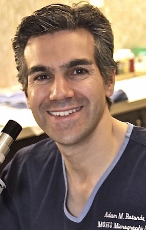
The product is a non–animal derived, pharmaceutical grade sodium deoxycholate that "acts much like a detergent. It attacks the cell membranes of fat, disrupting and disintegrating its membranes. Thereafter, fat cell contents are released and the subsequent host response, including inflammation, macrophage scavenging, and subclinical fibrosis, may confer a long-lasting fat-removal effect. It doesn’t shrink the fat cell; it ablates the fat it encounters," said, Dr. Adam M. Rotunda.
Phase III trials of an injectable form of sodium deoxycholate for the reduction of submental fat have begun in the United States, building on the previous success of four phase I, three phase II, and two phase III European studies of the product, known as ATX-101, noted Dr. Rotunda at the Summit in Aesthetic Medicine sponsored by the Skin Disease Education Foundation (SDEF).
In the 1990s, compounded phosphatidylcholine/deoxycholate injections (PC/DC), or lipodissolve, caused harm to some patients because "essentially everything about that experience was wrong: the wrong dose (too much volume, too high concentration); wrong depth (injections at times were too superficial. For example, cellulite, which was erroneously believed to improve from the injections, was injected intradermally); wrong indication (injections in places that shouldn’t have been injected); and wrong formulation," said Dr. Rotunda, who practices dermatology in Newport Beach, Calif. "These compounds were [derived] from animal sources and their sterility and purity could be called into question."
As a result, the FDA and other regulatory agencies around the world warned against or banned the use of unapproved PC/DC for aesthetic purposes. But in 2003, sodium deoxycholate was identified as the major component producing fat cell lysis in compounded PC/DC formulations, said Dr. Rotunda, who is also with the department of dermatology at the University of California, Los Angeles. "It’s taken many years to get that message across," he said.
ATX-101, which is being developed by Kythera Biopharmaceuticals, "is very different from the DC in compounded formulations, which was derived from cow bile," he said. "ATX-101 is being studied for very small volumes of fat relatively small for the submental area, which is really an ideal area to observe the desired effect of this product."

Phase I histology studies revealed that ATX-101 causes rapid adipocytolysis on day 1. On day 28 "there’s septal thickening and macrophage infiltration, which removes cellular debris and triglycerides, cleared via the lymphatic system," Dr. Rotunda said, adding that before and after MRIs have quantitatively confirmed the volume reductions.
Tissue surrounding the fatty treatment area, he continued, "has relatively high protein content. This protein (such as albumin) binds and inactivates deoxycholate, making subcutaneous injections relatively safe and fat specific. There’s an inverse relationship: the higher the protein content of certain tissue, such as muscle, tendon, and dermis, the lower the lytic activity of the deoxycholate. Less activity means less damage to that tissue. In fat, however, we want maximal damage, and it works out well because fat has relatively low protein content."
The administration of ATX-101 involves the use of a 30g needle, a 1-mL syringe, and placement of a temporary tattoo grid to the submental fat to control spacing of injections. "The distribution of the grid and how much is injected depends on the patient’s configuration and neck fat volume," Dr. Rotunda said. During the clinical trials, up to 10 mL of medication was used in each session, with up to four monthly treatments.
Most adverse events in clinical trials to date have been mild to moderate. "This is not a lunchtime procedure," he said. "It will be associated with local swelling and tenderness that may last days to several weeks, depending on the amount of ATX-101 injected or the volume of fat treated."
He concluded his remarks by noting that ATX-101 may become a novel approach that complements but does not compete with other fat-reduction therapies.
Dr. Rotunda disclosed that he, along with Dr. Michael S. Kolodney, are coinventors of ATX-101. He is a consultant to Kythera and holds stock in the company. He also is a consultant to Lithera and Allergan.
SDEF and this news organization are owned by Elsevier.
DANA POINT, CALIF. – No longer just a pipe dream, a novel injectable treatment effective at reducing fat is approaching the end of the drug development process.
The product is a non–animal derived, pharmaceutical grade sodium deoxycholate that "acts much like a detergent. It attacks the cell membranes of fat, disrupting and disintegrating its membranes. Thereafter, fat cell contents are released and the subsequent host response, including inflammation, macrophage scavenging, and subclinical fibrosis, may confer a long-lasting fat-removal effect. It doesn’t shrink the fat cell; it ablates the fat it encounters," said, Dr. Adam M. Rotunda.
Phase III trials of an injectable form of sodium deoxycholate for the reduction of submental fat have begun in the United States, building on the previous success of four phase I, three phase II, and two phase III European studies of the product, known as ATX-101, noted Dr. Rotunda at the Summit in Aesthetic Medicine sponsored by the Skin Disease Education Foundation (SDEF).
In the 1990s, compounded phosphatidylcholine/deoxycholate injections (PC/DC), or lipodissolve, caused harm to some patients because "essentially everything about that experience was wrong: the wrong dose (too much volume, too high concentration); wrong depth (injections at times were too superficial. For example, cellulite, which was erroneously believed to improve from the injections, was injected intradermally); wrong indication (injections in places that shouldn’t have been injected); and wrong formulation," said Dr. Rotunda, who practices dermatology in Newport Beach, Calif. "These compounds were [derived] from animal sources and their sterility and purity could be called into question."
As a result, the FDA and other regulatory agencies around the world warned against or banned the use of unapproved PC/DC for aesthetic purposes. But in 2003, sodium deoxycholate was identified as the major component producing fat cell lysis in compounded PC/DC formulations, said Dr. Rotunda, who is also with the department of dermatology at the University of California, Los Angeles. "It’s taken many years to get that message across," he said.
ATX-101, which is being developed by Kythera Biopharmaceuticals, "is very different from the DC in compounded formulations, which was derived from cow bile," he said. "ATX-101 is being studied for very small volumes of fat relatively small for the submental area, which is really an ideal area to observe the desired effect of this product."
Phase I histology studies revealed that ATX-101 causes rapid adipocytolysis on day 1. On day 28 "there’s septal thickening and macrophage infiltration, which removes cellular debris and triglycerides, cleared via the lymphatic system," Dr. Rotunda said, adding that before and after MRIs have quantitatively confirmed the volume reductions.
Tissue surrounding the fatty treatment area, he continued, "has relatively high protein content. This protein (such as albumin) binds and inactivates deoxycholate, making subcutaneous injections relatively safe and fat specific. There’s an inverse relationship: the higher the protein content of certain tissue, such as muscle, tendon, and dermis, the lower the lytic activity of the deoxycholate. Less activity means less damage to that tissue. In fat, however, we want maximal damage, and it works out well because fat has relatively low protein content."
The administration of ATX-101 involves the use of a 30g needle, a 1-mL syringe, and placement of a temporary tattoo grid to the submental fat to control spacing of injections. "The distribution of the grid and how much is injected depends on the patient’s configuration and neck fat volume," Dr. Rotunda said. During the clinical trials, up to 10 mL of medication was used in each session, with up to four monthly treatments.
Most adverse events in clinical trials to date have been mild to moderate. "This is not a lunchtime procedure," he said. "It will be associated with local swelling and tenderness that may last days to several weeks, depending on the amount of ATX-101 injected or the volume of fat treated."
He concluded his remarks by noting that ATX-101 may become a novel approach that complements but does not compete with other fat-reduction therapies.
Dr. Rotunda disclosed that he, along with Dr. Michael S. Kolodney, are coinventors of ATX-101. He is a consultant to Kythera and holds stock in the company. He also is a consultant to Lithera and Allergan.
SDEF and this news organization are owned by Elsevier.
EXPERT ANALYSIS FROM THE SDEF SUMMIT IN AESTHETIC MEDICINE
CLINICAL UPDATE: The Treatment of Atopic Dermatitis
- Introduction
- Nonpharmacologic Management of AD
- Pharmacologic Management of AD
- Conclusions
- Introduction
- Nonpharmacologic Management of AD
- Pharmacologic Management of AD
- Conclusions
- Introduction
- Nonpharmacologic Management of AD
- Pharmacologic Management of AD
- Conclusions

The EHR Report Podcast: The Employed Physician
Physicians who are employed by a hospital or health system may believe they have no power over which EHR their health system chooses or how that EHR will be used. But employed physicians actually can have tremendous impact on how their organizations choose and use EHRs. In this installment, Dr. Skolnik and Dr. Notte talk about what you can do when you're not in charge.
To download the podcast, right-click here.
To read the related column, click here.
To listen via this Web page, click on the player below:
Pop Warner Football Aims to Limit Concussions
Pop Warner youth football programs have issued new rules to restrict the amount of contact players can have during practice in an attempt to limit head injuries to young players.
Recent research from Virginia Tech led to this change as the organization – which has more than 250,000 children ages 5 to 15 in its leagues – tries to limit concussions.

The study headed by Stefan Duma placed sensors in the helmets of seven youth football players aged 6-8 years during their 2011 season. He found that 95% of impacts were between 15-20 g’s, what he likened to an “aggressive pillow fight.” But the other 5% were 50-100 g’s, what he said was force like a “car accident,” and the majority of these hits took place during practice.
Research has shown that damage from concussions can be cumulative, so Pop Warner is trying to lessen the number of impacts by reducing incidents in practice.
Read the news story in the NY Times.
Pop Warner youth football programs have issued new rules to restrict the amount of contact players can have during practice in an attempt to limit head injuries to young players.
Recent research from Virginia Tech led to this change as the organization – which has more than 250,000 children ages 5 to 15 in its leagues – tries to limit concussions.
The study headed by Stefan Duma placed sensors in the helmets of seven youth football players aged 6-8 years during their 2011 season. He found that 95% of impacts were between 15-20 g’s, what he likened to an “aggressive pillow fight.” But the other 5% were 50-100 g’s, what he said was force like a “car accident,” and the majority of these hits took place during practice.
Research has shown that damage from concussions can be cumulative, so Pop Warner is trying to lessen the number of impacts by reducing incidents in practice.
Read the news story in the NY Times.
BEST PRACTICES IN: Helping Patients Eat More Seafood
A supplement to Family Practice News. This supplement was sponsored by National Fisheries Institute.

Click here to download PDF
Topics
- Introduction
- Survey Reveals Knowledge Gap That Inhibits Patient Dialogue
- Barriers to Eating More Seafood
- Conclusions
Faculty/Faculty Disclosure
Jeffrey D. Fisher, MD
Cardiology and Internal Medicine
Weill Cornell Medical College
New York, New York
William Goodnight, MD
Maternal and Fetal Medicine University of North Carolina
Chapel Hill Chapel Hill, North Carolina
Laura Jana, MD
Pediatrician, Health Communicator
Omaha, Nebraska
John La Puma, MD
C.H.E.F. Clinic
Santa Barbara, California
Drs Goodnight, Jana and La Puma have nothing to disclose. Dr Fisher is a consultant to, and has received funding for, clinical grants from National Fisheries Institute.
Copyright © 2012 Elsevier Inc.
A supplement to Family Practice News. This supplement was sponsored by National Fisheries Institute.
Click here to download PDF
Topics
- Introduction
- Survey Reveals Knowledge Gap That Inhibits Patient Dialogue
- Barriers to Eating More Seafood
- Conclusions
Faculty/Faculty Disclosure
Jeffrey D. Fisher, MD
Cardiology and Internal Medicine
Weill Cornell Medical College
New York, New York
William Goodnight, MD
Maternal and Fetal Medicine University of North Carolina
Chapel Hill Chapel Hill, North Carolina
Laura Jana, MD
Pediatrician, Health Communicator
Omaha, Nebraska
John La Puma, MD
C.H.E.F. Clinic
Santa Barbara, California
Drs Goodnight, Jana and La Puma have nothing to disclose. Dr Fisher is a consultant to, and has received funding for, clinical grants from National Fisheries Institute.
Copyright © 2012 Elsevier Inc.
Pediatric Migraine Often Responsive to Treatment
A pediatric migraine diagnosis starts with a thorough patient history. I start by having the child or adolescent characterize their headache. What is its location, what does it feel like, how long does it last, and is there associated light or noise sensitivity or nausea? For the young children, this is ascertained by asking if they want to be in a dark or quiet room, or if they complain of stomach upset. I also find out about the onset and temporal course of the headache: When did the headaches start, have they become more frequent over time, and has there been a progression in the intensity of the headaches?
It is also quite important to identify headache triggers. I always ask about sleep schedule, eating habits, and fluid intake, as well as potential stress triggers, as these frequently impact on headaches.

It is appropriate to manage children in the primary care setting when they respond to fairly benign, over-the-counter medications, such as ibuprofen or acetaminophen, or triptan medications in the older kids. Refer to a specialist when your patient is not responding to these types of medications, when the headaches are becoming more frequent or severe, or if you have concerns about your patient’s neurologic status.
Also check family history because migraine is strongly genetically based. A family history of migraine headaches coupled with a typical headache character and normal neurologic exam can support your diagnostic suspicion.
In addition to taking a good history, it is critical to perform a detailed neurological examination to exclude any abnormalities that might suggest a more serious underlying cause for the headaches. It is especially important to look at their optic disks to rule out any evidence of increased intracranial pressure or papilledema. If you are unable to perform this type of exam, it is best to refer your patient to an ophthalmologist for a complete ophthalmologic exam. Any focal neurologic abnormalities should prompt a neuroimaging evaluation such as an MRI.
Evaluate for other headache types. Ask about stress triggers. Kids get stress- or tension-type headaches just like adults. When I see children who report frequent headaches that occur predominantly at school and infrequently on weekends, I’m more suspicious of a stress trigger. If headaches occur shortly before mealtimes, they could be caused by transient hypoglycemia and may be prevented by adding a snack or changing the child’s eating schedule.
Another scenario is headaches that occur after football or soccer practice or other vigorous activities. Here, I consider fatigue, dehydration, or perhaps excessive sun exposure as potential triggers. Ask about fluid intake – particularly how much water, not soft drinks, the child drinks. Sodas do not help with dehydration and are frequently loaded with caffeine. Educate them about hydration and how drinking enough fluids can make a huge difference in their headache frequency and severity.
Headaches that occur infrequently or that do not disrupt the child’s typical activities are less worrisome. For example, I am much less concerned when a child or adolescent reports headaches, but they still go outside to play, or go about their regular routine, and stay engaged in family activities.
A headache calendar filled out by the patient, preferably over weeks or months, is very helpful to your headache specialist. This helps us to better characterize the frequency and severity of episodes, what time of day they occur, and any potential precipitating triggers.
I become concerned when headaches get progressively more severe over time, or become more frequent over a short period. Headaches that awaken kids in the middle of the night, or those associated with nausea and vomiting on awakening, may point to a more serious condition, such as a tumor or other expanding mass inside the head causing increased intracranial pressure.
Probably the most over-ordered tests in the children I see with headaches are neuroimaging studies. The majority of young kids with headaches do not require an expensive MRI scan. Most require only a good history and neurologic exam for appropriate diagnosis.
However, a CT or MRI scan is indicated if you suspect a more serious etiology and/or the patient is younger. For example, I am much more likely to get an imaging study when a 3- or 4-year-old child complains of frequent or severe headaches. Historical information will be more limited in the preschoolers because they frequently can’t tell you as much about their headaches, and your examination might be less reliable as well – as the younger kids may be less cooperative and more difficult to examine. I also perform blood tests on some headache patients, looking for infectious, inflammatory, or metabolic derangements as a cause for headaches, but those are infrequently helpful.
Also keep in mind that some headache complaints may be functional in nature. For example, if a particular child gets significant attention with their headaches, there may be some associated secondary gain. A child also might be mimicking adult behavior. If the parents complain frequently about headaches, you might find the kid also complains about headaches.
Although pediatric and adult migraines share many of the same features, the good news is pediatric migraines are frequently not as severe or as protracted in children as they are for adults, and are often highly responsive to treatment. It can be very rewarding managing children with headaches because so many do well.
Dr. Berenson is a pediatric neurologist and section chief of neurology at Children’s Healthcare of Atlanta at Scottish Rite. He is also in private practice at Atlanta Headache Specialists and Pediatric and Adolescent NeuroDevelopmental Associates (PANDA) Neurology. He said he had no relevant financial disclosures.
A pediatric migraine diagnosis starts with a thorough patient history. I start by having the child or adolescent characterize their headache. What is its location, what does it feel like, how long does it last, and is there associated light or noise sensitivity or nausea? For the young children, this is ascertained by asking if they want to be in a dark or quiet room, or if they complain of stomach upset. I also find out about the onset and temporal course of the headache: When did the headaches start, have they become more frequent over time, and has there been a progression in the intensity of the headaches?
It is also quite important to identify headache triggers. I always ask about sleep schedule, eating habits, and fluid intake, as well as potential stress triggers, as these frequently impact on headaches.
It is appropriate to manage children in the primary care setting when they respond to fairly benign, over-the-counter medications, such as ibuprofen or acetaminophen, or triptan medications in the older kids. Refer to a specialist when your patient is not responding to these types of medications, when the headaches are becoming more frequent or severe, or if you have concerns about your patient’s neurologic status.
Also check family history because migraine is strongly genetically based. A family history of migraine headaches coupled with a typical headache character and normal neurologic exam can support your diagnostic suspicion.
In addition to taking a good history, it is critical to perform a detailed neurological examination to exclude any abnormalities that might suggest a more serious underlying cause for the headaches. It is especially important to look at their optic disks to rule out any evidence of increased intracranial pressure or papilledema. If you are unable to perform this type of exam, it is best to refer your patient to an ophthalmologist for a complete ophthalmologic exam. Any focal neurologic abnormalities should prompt a neuroimaging evaluation such as an MRI.
Evaluate for other headache types. Ask about stress triggers. Kids get stress- or tension-type headaches just like adults. When I see children who report frequent headaches that occur predominantly at school and infrequently on weekends, I’m more suspicious of a stress trigger. If headaches occur shortly before mealtimes, they could be caused by transient hypoglycemia and may be prevented by adding a snack or changing the child’s eating schedule.
Another scenario is headaches that occur after football or soccer practice or other vigorous activities. Here, I consider fatigue, dehydration, or perhaps excessive sun exposure as potential triggers. Ask about fluid intake – particularly how much water, not soft drinks, the child drinks. Sodas do not help with dehydration and are frequently loaded with caffeine. Educate them about hydration and how drinking enough fluids can make a huge difference in their headache frequency and severity.
Headaches that occur infrequently or that do not disrupt the child’s typical activities are less worrisome. For example, I am much less concerned when a child or adolescent reports headaches, but they still go outside to play, or go about their regular routine, and stay engaged in family activities.
A headache calendar filled out by the patient, preferably over weeks or months, is very helpful to your headache specialist. This helps us to better characterize the frequency and severity of episodes, what time of day they occur, and any potential precipitating triggers.
I become concerned when headaches get progressively more severe over time, or become more frequent over a short period. Headaches that awaken kids in the middle of the night, or those associated with nausea and vomiting on awakening, may point to a more serious condition, such as a tumor or other expanding mass inside the head causing increased intracranial pressure.
Probably the most over-ordered tests in the children I see with headaches are neuroimaging studies. The majority of young kids with headaches do not require an expensive MRI scan. Most require only a good history and neurologic exam for appropriate diagnosis.
However, a CT or MRI scan is indicated if you suspect a more serious etiology and/or the patient is younger. For example, I am much more likely to get an imaging study when a 3- or 4-year-old child complains of frequent or severe headaches. Historical information will be more limited in the preschoolers because they frequently can’t tell you as much about their headaches, and your examination might be less reliable as well – as the younger kids may be less cooperative and more difficult to examine. I also perform blood tests on some headache patients, looking for infectious, inflammatory, or metabolic derangements as a cause for headaches, but those are infrequently helpful.
Also keep in mind that some headache complaints may be functional in nature. For example, if a particular child gets significant attention with their headaches, there may be some associated secondary gain. A child also might be mimicking adult behavior. If the parents complain frequently about headaches, you might find the kid also complains about headaches.
Although pediatric and adult migraines share many of the same features, the good news is pediatric migraines are frequently not as severe or as protracted in children as they are for adults, and are often highly responsive to treatment. It can be very rewarding managing children with headaches because so many do well.
Dr. Berenson is a pediatric neurologist and section chief of neurology at Children’s Healthcare of Atlanta at Scottish Rite. He is also in private practice at Atlanta Headache Specialists and Pediatric and Adolescent NeuroDevelopmental Associates (PANDA) Neurology. He said he had no relevant financial disclosures.
New antibody achieves high CR rate in relapsed/refractory adult ALL

Credit: ASCO/Scott Morgan
CHICAGO—A new monoclonal antibody, blinatumomab, achieves an “exceptionally high complete remission rate” as a single agent in acute lymphoblastic leukemia (ALL), according to investigators.
They reported that about 70% of adult patients with relapsed/refractory B-precursor ALL achieved a hematologic complete remission (CR).
Max Topp, MD, of the Wuerzburg University Medical Center in Germany, presented the findings as abstract 6500 at the 2012 ASCO Annual Meeting held here recently.
Outcomes are poor for adult patients with relapsed/refractory ALL following frontline therapy. Several clinical trials have shown that most patients fail to achieve CR. Response rates are typically 20% to 30%.
“Treatment-related mortality is high, CR is not durable, and overall survival is dismal at a median of 4-6 months after relapse,” Dr Topp said. “[A]llogeneic stem cell transplant (alloSCT) is only really available to patients who reach a CR, and few patients make it to SCT.”
After alloSCT, the overall survival rate at 1 year is about 20%, regardless of CR status. “There is a need for something completely different,” Dr Topp said.
Enter blinatumomab. Blinatumomab is a bispecific T-cell engaging antibody that directs cytotoxic T cells to CD19-expressing target cells.
Dr Topp and colleagues designed an open-label, multicenter, exploratory phase 2 study and enrolled 36 relapsed/refractory ALL patients. The patients were a median age of 31 years and had a high blast count. Some 40% had relapsed after alloSCT.
Patients received blinatumomab by continuous intravenous infusion for 4 weeks, with 2 weeks off, for up to 5 cycles. The investigators determined the safest dose to be 5 µg/m²/day in week 1, followed by 15 µg/m²/day for the remaining treatment. Twenty-three patients entered the extension phase at this dose.
Of the 36 patients enrolled, 26 (72%) achieved a hematologic CR, as did 17 of 23 patients (74%) in the extension phase.
The duration of hematologic CR was 8.9 months after a median observation time of 4.5 months.
Median overall survival was 9 months after a median follow-up of 10.7 months. This compares favorably with historical data, Dr Topp pointed out.
“Most importantly,” he added, “almost every patient who achieved CR had a molecular remission. Only 2 out of 26 patients didn’t reach this endpoint.”
He noted that 13 patients who reached CR had an alloSCT.
Early on, several patients developed cytokine release syndrome. So the researchers developed a prevention strategy of giving corticosteroids upfront. As a result, there were no cases of cytokine release syndrome in the extension phase.
The most common adverse events in the safest dosing schedule were pyrexia and headache. There were few grade 3 events, and all adverse events were reversible.
Dr Topp said the data support further investigation of blinatumomab in adult patients with relapsed/refractory ALL.
He noted that a global phase 2 study in this setting has already been initiated in the United States and Europe. ![]()
Wave of Pertussis Cases Raises Questions About Diagnoses, Testing
Robert Gould, MD, a hospitalist in suburban Seattle, knows that his HM colleagues don't immediately think of pertussis as a diagnosis. But as an epidemic of whooping cough rolls through Washington state, he urges they keep the disease in mind.
"I'm thinking about it more," says Dr. Gould, a hospitalist at Swedish/Edmonds Hospital in Edmonds, Wash., who has treated one patient who tested positive for the illness. "One thing I think about is if someone comes in with a primary respiratory issue and they have underlying COPD and they're having a cough. Do you test for it? Do you consider it? It's just so hard, because do you test everyone who comes in with one week of cough?"
The topic is timely. The Washington State Department of Health reports that through May 26, the state reported 1,947 cases of whooping cough, up from just 154 cases for the same time period last year.
Dr. Gould says the outbreak of pertussis brings up an interesting question for hospitalists. HM physicians don't want to order unnecessary tests—particularly in light of recent initiatives to combat the practice—but not testing can leave a person vulnerable to the disease's progression. When suspicions are high that whooping cough is the diagnosis, one solution is simply to order one of the most common therapies: azithromycin. That eliminates the testing cost, which can run up to several hundred dollars, while giving the patient a medication not greatly associated with Clostridium difficile or other negative outcomes, Dr. Gould says.
"Thinking about it is the biggest thing," he adds.
ITL: Physician Reviews of HM-Relevant Research
Clinical question: With the current use of warfarin for stroke prophylaxis in patients with nonvalvular atrial fibrillation, what do the most recent data show with regard to time spent in the therapeutic window, stroke risk, and bleeding risk?
Background: Historically, warfarin has been shown to decrease stroke risk in nonvalvular atrial fibrillation by 62% compared with placebo, balanced by a significant risk of bleeding. Despite the availability of multiple new antithrombotic agents, warfarin will likely continue to be widely used given its lower cost. As a result, physicians need an accurate estimate of warfarin’s efficacy and safety as currently used in practice.
Study design: Meta-analysis of randomized controlled trials (RCTs).
Setting: RCTs comparing warfarin to an alternative antithrombotic agent from 2001 to 2011.
Synopsis: Eight RCTs of nonvalvular atrial fibrillation were included, yielding data on 32,053 patients with a mean age range of 70 to 82 years and widely variable CHADS2 scores. The time spent at a therapeutic INR was found to be improved when compared to historical rates, ranging from 55% to 68%. The rate of stroke or non-central-nervous-system embolism ranged from 1.2% to 2.3% per year, with a pooled event rate of 1.66% per year, compared with 2.09% per year in earlier trials.
Major bleeding was defined differently across studies, with a reported incidence of 1.4% to 3.4% per year, a pooled event rate of intracranial hemorrhage of 0.61%, and a cumulative adverse event rate of 3.0% to 7.64%. Stroke rates were highest in patients older than 75 years, women, those with a history of transient ischemic attack or stroke, those new to warfarin, and those with higher CHADS2 scores.
Bottom line: Warfarin as currently used is associated with an annual rate of stroke or systemic embolism of 1.66% and an annual rate of major bleeding ranging from 1.4% to 3.4%.
Citation: Agarwal S, Hachamovitch R, Menon V. Current trial-associated outcomes with warfarin in prevention of stroke in patients with nonvalvular atrial fibrillation: a meta-analysis. Arch Intern Med. 2012;172:623-631.