User login
Can reversing T-cell exhaustion benefit in B-cell lymphoma relapse?
Durable remissions have been obtained in around 30%-40% of relapsed/refractory large B-cell lymphomas (BCL) through the use of CD19-directed chimeric antigen receptor-modified T-cell (CAR T-cell) therapy. However, T cell exhaustion and/or an immunosuppressive tumor environment may contribute to CAR T cell failure and BCL relapse.
To counter this failure, researchers assessed the use PD1 blockade with pembrolizumab after CD19-directed CAR T-cell therapy. Such treatment appeared safe and was able to achieve clinical responses in some patients with B-cell lymphomas refractory to or relapsed after CAR T-cell therapy, according to the results of a small study (NCT02650999) reported in Blood.
Success for some
Twelve patients with BCL who were either refractory to (nine patients) or relapsed after (three patients) CD19-directed CAR T-cell therapy were treated with pembrolizumab at 200 mg IV every 3 weeks, according to Elise A. Chong, MD, of the University of Pennsylvania, Philadelphia, and colleagues.
Overall, 3 of the 12 patients showed a response after pembrolizumab: One complete response; two partial responses. In addition, 1 patient had stable disease; thus, 4 of the 12 patients showed clinical benefit, according to the researchers. After pembrolizumab, these four patients with clinical benefit showed an increase in the percentage of CAR T cells as assessed by mass cytometry, and three out of the four also showed increases in CAR19 transgene levels as determined by qPCR. In addition, immune profiling using mass cytometry revealed increased CAR T-cell activation and proliferation and less T-cell exhaustion in these clinical responders.
In terms of safety, pembrolizumab appeared to be well tolerated and the only ≥ grade 3 adverse events related to pembrolizumab were neutropenia in three patients, the researchers added.
Looking forward
“Although patient numbers are small, these data suggest potential differences in the biology of CAR T cells or in the overall immune landscape of responders and nonresponders that influence the clinical efficacy of PD-1 blockade administered in this setting. Future work aimed at improving immune health after CAR T-cell infusion, as well as work aimed at decreasing CD8+ CAR T-cell exhaustion in CAR T-cell products, may serve as potential platforms for enhancing the efficacy of immune checkpoint blockade in patients treated with CAR T cells,” the researchers concluded.
The study was sponsored by the Abramson Cancer Center of the University of Pennsylvania. The authors reported serving on advisory boards and receiving research funding from a variety of pharmaceutical and biotechnology companies.
FROM BLOOD
Evans’ Syndrome in Undiagnosed Small Lymphocytic Lymphoma: Case Report and Literature Review
Background
Evans’ syndrome is a rare entity characterized by concomitant or sequential multilineage cytopenia particularly autoimmune hemolytic anemia, ITP and very rarely autoimmune neutropenia. Although more common in young adults, it can occur in elderly usually associated with malignancies like CLL.
Case Report
A 74 years old Veteran presented with complaints of fatigue and worsening dyspnea on exertion. His physical exam was unremarkable except jaundice. His labs were significant for macrocytic anemia with Hemoglobin of 7.4g/dl compared to 11.7g/dl 6 months prior, MCV 106.9 fL, LDH 809U/L, indirect bilirubin 4.1mg/dl, absolute reticulocyte 0.16M/uL, Haptoglobin <15mg/dl and Positive DAT. Platelets were mildly decreased at 111K/ul. No lymphocytosis was noted. Initially, the hemolysis was thought to be cephalosporin- related given that the patient had taken cephalexin recently for cellulitis. As part of the workup for anemia, the patient underwent EGD and colonoscopy which was initially unrevealing. However, random biopsies from the descending colon and terminal ileum returned with a small lymphocytic infiltrate consistent with SLL/CLL. Cytogenetics showed trisomy-12 which is associated with intermediate prognosis for CLL. PET scan done subsequently revealed only a reactive marrow and an enlarged 15.8cm non-hypermetabolic spleen. This veteran having anemia, positive DAT, thrombocytopenia, and splenomegaly got diagnosed with Evans’s syndrome. This syndrome was the initial manifestation of his underlying CLL. We started the patient on a prednisone taper for 4 weeks to which anemia and thrombocytopenia barely responded, ultimately Rituximab 375mg/m2 x4 weekly doses was started which led to complete resolution of anemia and thrombocytopenia. We closely followed the patient and monitored CBC and hemolytic markers. The patient relapsed in two years which was subsequently managed with another course of Rituximab 375mg/m2 x4 weekly doses.
Conclusions
This case report aims to call attention to this relatively rare entity which is difficult to treat and often associated with frequent relapses. Though rare, physicians should maintain high suspicion for this syndrome in patients with multi-lineage cytopenia which are usually not even responding well to the common treatment for cytopenia. Furthermore, there is room for improvement in Evans’ syndrome management since mortality remains higher in these patients than in those with isolated autoimmuce cytopenias.
FDA approves zanubrutinib in Waldenström’s macroglobulinemia
The Food and Drug Administration has approved zanubrutinib (Brukinsa) capsules for use in the treatment of adult patients with Waldenström’s macroglobulinemia (WM), a rare non-Hodgkin lymphoma, according to an approval letter from the agency to BeiGene, the drug’s maker.
The FDA stipulated that the company conduct an additional clinical trial (rather than an observational study) to assess the “known serious risk of second primary malignancies” associated with use of zanubrutinib. The study should further characterize the clinical benefit and safety of zanubrutinib for the treatment of patients with newly diagnosed WM with MYD88 mutation, the agency said.
The drug, which is a small-molecule inhibitor of Bruton’s tyrosine kinase (BTK), previously received accelerated approval for use in patients with mantle cell lymphoma who have received one prior therapy. It is also being studied for the treatment of chronic lymphocytic leukemia.
The new approval is primarily based on results from ASPEN, a randomized, active control, open-label trial that compared zanubrutinib and ibrutinib.
The ASPEN trial provided “compelling evidence” that zanubrutinib is a highly active BTK inhibitor in WM and that it showed improved tolerability across a number of clinically important side effects in comparison with the first-generation BTK inhibitor ibrutinib, said study investigator Steven Treon, MD, PhD, director of the Bing Center for Waldenström’s Macroglobulinemia Research at the Dana-Farber Cancer Institute, Boston. “The approval of [zanubrutinib] provides an important new option for targeted therapy in Waldenström’s macroglobulinemia,” he added in a company press statement.
The recommended dosage is 160 mg orally twice daily or 320 mg orally once; the drug should be swallowed whole with water with or without food.
In ASPEN, all patients had MYD88 mutation WM. Patients in cohort 1 (n = 201) were randomly assigned in a 1:1 ratio to receive zanubrutinib 160 mg twice daily or ibrutinib 420 mg once daily until disease progression or unacceptable toxicity. A total of 82% of patients had relapsed/refractory disease.
The major efficacy outcome was the response rate, defined as partial response or better (i.e., partial response, very good partial response, and complete response), as determined on the basis of standard consensus response criteria from the International Workshop on Waldenström’s Macroglobulinemia (IWWM-6) criteria.
The drugs had nearly identical response rates (roughly 77%). There were no complete responses with either drug. However, zanubrutinib had twice the rate of very good partial responses compared with ibrutinib (15.7% vs. 7.1%). In addition, on the basis of modified IWWM-6 criteria, the very good partial response rate was 28% with zanubrutinib, compared to 19% with ibrutinib.
An additional efficacy outcome measure was duration of response, which was measured by the percentage of patients who were event free at 12 months. Zanubrutinib bested ibrutinib in this measure (94.4% vs. 87.9%).
The safety of zanubrutinib was also investigated in the ASPEN trial. Among patients who received zanubrutinib, 93% were exposed for 6 months or longer, and 89% were exposed for longer than 1 year. In cohort 1, serious adverse reactions occurred in 44% of patients who received zanubrutinib. Serious adverse reactions that occurred in > 2% of patients included influenza (3%), pneumonia (4%), neutropenia and decreased neutrophil count (3%), hemorrhage (4%), pyrexia (3%), and febrile neutropenia (3%).
In the FDA’s prescribing information for the drug, which includes approved indications and pooled safety data, the most common adverse reactions for zanubrutinib (≥ 20%) are listed as decreased neutrophil count, upper respiratory tract infection, decreased platelet count, rash, hemorrhage, musculoskeletal pain, decreased hemoglobin, bruising, diarrhea, pneumonia, and cough.
A version of this article first appeared on Medscape.com.
FDA warns of higher death risk with Pepaxto in multiple myeloma
participating in the ongoing OCEAN clinical trial.

The drug was granted accelerated approval in February 2021 for use in combination with dexamethasone in the treatment of adults with relapsed or refractory multiple myeloma who had received at least four prior lines of therapy and whose disease was refractory to at least one proteasome inhibitor, one immunomodulator, and one CD38-directed monoclonal antibody.
As a condition of the accelerated approval, the manufacturer, Oncopeptides, was required to conduct a confirmatory clinical trial and launched the OCEAN trial.
Enrollment in OCEAN as well as other ongoing trials of the drug have now been halted, according to the FDA alert.
The warning comes in the wake of OCEAN trial findings showing worse survival among patients in the experimental group, who were receiving melphalan plus low-dose dexamethasone, compared with patients in the control group, who were receiving pomalidomide plus low-dose dexamethasone (hazard ratio for overall survival, 1.104). Median overall survival in the treatment and control groups was 19.7 and 25.0 months, respectively.
Health care professionals should “review patients’ progress on Pepaxto and discuss the risks of continued administration with each patient in the context of other treatments,” and patients currently receiving the drug should discuss the risks and benefits with their health care professional, the FDA advises. “Patients receiving clinical benefit from Pepaxto may continue treatment in the OCEAN trial provided they are informed of the risks and sign a revised written informed consent.”
The FDA also hinted at “a future public meeting to discuss the safety findings and explore the continued marketing of Pepaxto,” which has a price tag of $19,000 per treatment course.
Accelerated approval data
Melphalan flufenamide was initially evaluated in combination with low-dose dexamethasone in the multicenter, single-arm HORIZON trial of adults with relapsed or refractory multiple myeloma who received at least four prior lines of therapy and whose disease was refractory to at least one proteasome inhibitor, one immunomodulator, and one CD38-directed monoclonal antibody.
Patients received melphalan flufenamide at a dose of 40 mg intravenously on day 1 along with oral dexamethasone at a dose of 40 mg (or 20 mg for those over age 75 years) on days 1, 8, 15, and 22 of each 28-day cycle until disease progression or unacceptable toxicity.
The most common adverse reactions, occurring in at least 20% of patients, were fatigue, nausea, diarrhea, pyrexia, and respiratory tract infection. The most common laboratory abnormalities, occurring in at least 50% of patients, were decreased leukocytes, platelets, lymphocytes, neutrophils, and hemoglobin, and increased creatinine.
Accelerated approval was granted after the HORIZON trial showed an overall response rate of 23.7% and median duration of response of 4.2 months. The application by Oncopeptides received priority review and orphan drug status.
Confirmatory trial data
The confirmatory OCEAN trial compared melphalan flufenamide plus low-dose dexamethasone to pomalidomide plus low-dose dexamethasone in patients with relapsed or refractory multiple myeloma following 2-4 lines of therapy and in patients who were resistant to lenalidomide in the last line of therapy.
The FDA conducted an efficacy and safety evaluation of the OCEAN trial using a data cutoff date of February 3, 2021. At a median follow-up of 19.1 months, 117 of 246 patients (48%) in the melphalan flufenamide group had died, compared with 108 of 249 patients (43%) in the pomalidomide control group.
“Patient safety is paramount to Oncopeptides,” the company said in a press statement, which also notes that “dialogue with the FDA” is ongoing and that updated information will be provided as it becomes available.
The company plans to submit complete data from the OCEAN study to the International Myeloma Workshop meeting in Vienna being held September 8-11, 2021.
Health care professionals and patients should report adverse events or quality issues experienced with melphalan flufenamide or any other medication to the FDA MedWatch Adverse Event Reporting program, either online or by downloading and completing a reporting form and submitting via fax at 1-800-FDA-0178.
A version of this article first appeared on Medscape.com.
participating in the ongoing OCEAN clinical trial.
The drug was granted accelerated approval in February 2021 for use in combination with dexamethasone in the treatment of adults with relapsed or refractory multiple myeloma who had received at least four prior lines of therapy and whose disease was refractory to at least one proteasome inhibitor, one immunomodulator, and one CD38-directed monoclonal antibody.
As a condition of the accelerated approval, the manufacturer, Oncopeptides, was required to conduct a confirmatory clinical trial and launched the OCEAN trial.
Enrollment in OCEAN as well as other ongoing trials of the drug have now been halted, according to the FDA alert.
The warning comes in the wake of OCEAN trial findings showing worse survival among patients in the experimental group, who were receiving melphalan plus low-dose dexamethasone, compared with patients in the control group, who were receiving pomalidomide plus low-dose dexamethasone (hazard ratio for overall survival, 1.104). Median overall survival in the treatment and control groups was 19.7 and 25.0 months, respectively.
Health care professionals should “review patients’ progress on Pepaxto and discuss the risks of continued administration with each patient in the context of other treatments,” and patients currently receiving the drug should discuss the risks and benefits with their health care professional, the FDA advises. “Patients receiving clinical benefit from Pepaxto may continue treatment in the OCEAN trial provided they are informed of the risks and sign a revised written informed consent.”
The FDA also hinted at “a future public meeting to discuss the safety findings and explore the continued marketing of Pepaxto,” which has a price tag of $19,000 per treatment course.
Accelerated approval data
Melphalan flufenamide was initially evaluated in combination with low-dose dexamethasone in the multicenter, single-arm HORIZON trial of adults with relapsed or refractory multiple myeloma who received at least four prior lines of therapy and whose disease was refractory to at least one proteasome inhibitor, one immunomodulator, and one CD38-directed monoclonal antibody.
Patients received melphalan flufenamide at a dose of 40 mg intravenously on day 1 along with oral dexamethasone at a dose of 40 mg (or 20 mg for those over age 75 years) on days 1, 8, 15, and 22 of each 28-day cycle until disease progression or unacceptable toxicity.
The most common adverse reactions, occurring in at least 20% of patients, were fatigue, nausea, diarrhea, pyrexia, and respiratory tract infection. The most common laboratory abnormalities, occurring in at least 50% of patients, were decreased leukocytes, platelets, lymphocytes, neutrophils, and hemoglobin, and increased creatinine.
Accelerated approval was granted after the HORIZON trial showed an overall response rate of 23.7% and median duration of response of 4.2 months. The application by Oncopeptides received priority review and orphan drug status.
Confirmatory trial data
The confirmatory OCEAN trial compared melphalan flufenamide plus low-dose dexamethasone to pomalidomide plus low-dose dexamethasone in patients with relapsed or refractory multiple myeloma following 2-4 lines of therapy and in patients who were resistant to lenalidomide in the last line of therapy.
The FDA conducted an efficacy and safety evaluation of the OCEAN trial using a data cutoff date of February 3, 2021. At a median follow-up of 19.1 months, 117 of 246 patients (48%) in the melphalan flufenamide group had died, compared with 108 of 249 patients (43%) in the pomalidomide control group.
“Patient safety is paramount to Oncopeptides,” the company said in a press statement, which also notes that “dialogue with the FDA” is ongoing and that updated information will be provided as it becomes available.
The company plans to submit complete data from the OCEAN study to the International Myeloma Workshop meeting in Vienna being held September 8-11, 2021.
Health care professionals and patients should report adverse events or quality issues experienced with melphalan flufenamide or any other medication to the FDA MedWatch Adverse Event Reporting program, either online or by downloading and completing a reporting form and submitting via fax at 1-800-FDA-0178.
A version of this article first appeared on Medscape.com.
FDA approves new asparaginase product for leukemia
The new product is Jazz Pharmaceutical’s Rylaze (asparaginase erwinia chrysanthemi [recombinant]-rywn), and it is approved for use in the treatment of acute lymphoblastic leukemia and lymphoblastic lymphoma.
Asparaginase, an enzyme that helps kill blood cancer cells, is a key component of chemotherapy for both conditions.
The problem is that about 20% of patients become allergic to the standard option, asparaginase derived from Escherichia coli.
The only alternative until now has been Erwinaze (also distributed by Jazz Pharmaceuticals), which, like Rylaze, is derived from Erwinia chrysanthemi, a plant pathogen related to Escherichia coli.
However, Erwinaze has been bedeviled by manufacturing problems and has been in short supply since 2016.
The situation has been “extremely disconcerting to patients, families and providers,” and the hope is that Rylaze will “provide a consistently sourced alternative,” Gregory Reaman, MD, the FDA’s associate director of pediatric oncology, said in a press release.
Rylaze will hit the U.S. market in mid-July. Jazz has been a distributor of Erwinaze as well, but it released its last batch in May, according to a spokesperson.
The key difference between the two products is that the asparaginase in Erwinaze is derived directly from Erwinia chrysanthemi, whereas the asparaginase in Rylaze is a recombinant product produced by different bacteria that have been genetically altered with Erwinia chrysanthemi DNA.
The approval for Rylaze was based on a study involving 102 patients (median age, 10 years) who developed hypersensitivity to Escherichia coli–derived enzyme or “silent inactivation” from neutralizing antibodies. In the study, almost 94% of patients achieved asparaginase target activity levels at the approved dosage of 25 mg/m2 IM every 48 hours. The study is ongoing, and investigators are currently evaluating intravenous dosing.
The most common side effects are hypersensitivity reactions, blood clots, hemorrhage, and pancreatic and liver toxicity. There is also a risk for fetal harm, so labeling advises women to use effective nonhormonal contraception during treatment and for 3 months afterward.
A version of this article first appeared on Medscape.com.
Real-world CAR T outcomes for DLBCL mimic clinical trials
Data from a large French registry on a multicenter experience with chimeric antigen receptor T-cell (CAR T) therapy for aggressive lymphoma suggests that the favorable outcomes seen in clinical trials can be replicated in the real world.
Among 481 patients with relapsed or refractory diffuse large B-cell lymphoma (DLBCL) treated with either of two commercially available CAR T products – tisagenlecleucel (Kymriah) or axicabtagene ciloleucel (Yescarta) – the duration of responses, progression-free survival (PFS), and overall survival (OS) rates at 6 months mirror those seen in clinical trials, reported Steven LeGouill, MD, PhD, from the University of Nantes (France), on behalf of colleagues in the DESCAR-T (Dispositive d’Evaluation et de Suivi des CAR-T) registry.
“CAR T has now become a standard of care in a lot of French centers, with more than 640 patients treated with CAR T in less than 2 years. The DESCAR-T real-life experience mimics the experience that had been previously by other real-life registries but also in clinical trials. We didn’t see new emerging toxicity signals in real life,” he said in an oral abstract session during the European Hematology Association annual congress (Abstract S216).
“I am convinced that a population registry about CAR T–treated patients is needed,” commented Pieter Sonneveld, MD, from Erasmus Medical Center in Rotterdam, the Netherlands, who was not involved in the study.
“Selection criteria for CAR T trials have been incredibly restrictive, and academic trials have not gained ground yet. It is important to collect and analyze more data, include non-trial patients and analyze long-term follow-up in order to determine the real effects of this innovative treatment in lymphoma and other diseases,” he said.
Dr. Sonneveld, EHA past president, was the moderator a briefing where Dr. LeGouill presented the DESCAR-T study findings.
Natalie Sophia Grover, MD, a leukemia and lymphoma specialist at the University of North Carolina at Chapel Hill, noted in an interview that “there have been several publications recently that have shown that these promising outcomes for these really refractory, high-risk patients with diffuse large B-cell lymphoma seem to be similar with what we’ve seen in trials, which is definitely exciting.”
She noted that the median time from CAR T order to treatment in the study, 50 days, was longer than in her experience.
“Generally, from collection to treatment is less than a month. Looking at that, I would have expected more patients not to make it CAR T, but nearly 90% of patients who had collections got treatment, which is pretty good. Those patients that didn’t make it to treatment had really poor outcomes,” she said.
Dr. Grover was not involved in the study.
More data, s’il vous plait
The DESCAR-T registry was created in response to a request from French health authorities for data beyond that provided in the EBMT patient registry. The health authorities asked for characterization of the CAR T–eligible population in an intention to treat, 15-year follow-up of both CAR T recipients and candidates who were not treated for whatever reasons, and a full accounting of previous lines of therapy.
Dr. LeGouill presented the first analysis of data from the registry involving 19 enrolling site and 647 patients with DLBCL for whom CAR T cells were ordered from January 2018 to March 2021.
Of the 647 candidates, 10 did not have CAR T ordered for reasons that included patient deaths or disease progression, infection, and patient refusal. An additional 30 patients either had leukapheresis performed or pending, and 607 had CAR T ordered.
Of the 607 patients, 53 did not receive CAR T infusions because of disease progression, death before product administration, manufacturing or leukapheresis failures, uncontrolled infections, patient choice, or progression of other malignancies.
That left 550 patients (85%) who received a CAR T product, either tisagenlecleucel (200 patients) or axicabtagene ciloleucel (350 patients).
Among all patients, the median age at CAR T order was 63 for patients who received tisagenlecleucel, and 65 for patients receiving axicabtagene ciloleucel. Patients 65 and older comprised 44% and 51% of the population, respectively.
Patients treated with each CAR T product had a median of three prior lines of therapy.
Manageable toxicities
Toxicities within 10 days of CAR T infusion included 418 cases among 515 patients (81.2%) of cytokine release syndrome, with most being grade 1 or 2 in severity; 44 patients had grade 3 or 4 CRS.
Any-grade neurotoxicity was seen in 184 patients (35.7%), primarily grade 1 or 2 in severity; 50 patients had grade 3 or greater neurotoxicity.
Of 427 patients with at least one CAR T–specific toxicity within 10 days, 139 (32.8%) required ICU admission, 325 (76,1%) were treated for CAR T–specific toxicities, 278 (65.1%) received tocilizumab, 13 (3%) received siltuximab, and 176 (41.2%) received corticosteroids.
Favorable outcomes
Overall response rates, at 1, 3, and 6 months post infusions were 70.6%, 56.3%, and 60%, respectively, with the majority of response at each time point being complete responses (CR).
The 6-month overall survival (OS) rate among all patients who were treated was 83.7%, compared with 5.5% for patients who did not receive CAR T infusions.
Progression-free survival (PFS) at 6 months was 44.5%, and 57.7% of patients had an ongoing response at the same time point.
Among patients who received bridging therapy between leukapheresis and CAR T infusion, the 6-month PFS was 58.4% for patients with either a CR, partial response, or stable disease, compared with 63.3% for patients who did not receive bridging therapy, and 29.8% for those with disease progression.
The respective 6 months OS rates were 87.4%, 82.3%, and 65.5%.
The results showed that patients who do not have at least stable disease at the time of CAR T infusion are at risk for early relapse, but approximately 30% of these patients still had long-term disease control, Dr. LeGouill said.
He acknowledged that longer follow-up will be need to see whether the plateaus in the PFS and OS curves the investigators observed can be maintained over time. Questions that still need to be answered include the impact of bridging therapy or disease status at the start of treatment on outcomes, and how to improve CAR T efficacy based on individual patient characteristics.
The registry will be extended to include data on patients treated with CAR T for mantle cell lymphoma and multiple myeloma, investigators announced.
The study is supported by participating centers and Gilead/Kite and Novartis. Dr. LeGouill disclosed advisory board activity and honoraria from the companies and others. Dr. Grover disclosed advisory board participating for Kite and others. Dr. Sonneveld has disclosed research grants and honoraria from several companies, not including Kite or Novartis.
FROM EHA 2021
Experimental antibody-drug conjugate shown active against r/r DLBCL
Patients with relapsed or refractory B-cell non-Hodgkin lymphomas who are not candidates for hematopoietic stem cell transplant have a generally poor prognosis and few treatment options, but an experimental combination of the antibody-drug conjugate naratuximab with rituximab showed promising efficacy and acceptable safety in these patients in a phase 2 trial.
Among patients with relapsed/refractory diffuse large B-cell lymphoma (DLBCL) the combination was associated with a 44.7% overall response rate, including 31.6% complete responses, and two-thirds of patients had responses lasting more than 12 months, reported Moshe Yair Levy, MD, from Texas Oncology–Baylor Charles A Sammons Cancer Center in Dallas.
“This is, in my viewpoint, very exciting therapy,” he said in a question-and-answer session following his presentation of the data in a late-breaking abstract session during the European Hematology Association annual congress. (Abstract LB1903).
Naratuximab emtansine is an investigational antibody-drug conjugate (ADC) consisting of a humanized monoclonal antibody against CD37, a surface marker on B lymphocytes that is highly expressed in non-Hodgkin lymphoma (NHL), conjugated to a cytotoxic derivative of maitansine.
CD37 is also an internalizable cell-surface antigen, making it an attractive candidate for an ADC approach.
In a phase 1 trial, naratuximab monotherapy showed a good safety profile and a 22% overall response rate, Dr. Levy noted.
“What they found is that, if you coadminister this ADC with rituximab, you’re actually going to get more internalization of the CD37 monoclonal, therefore more payload delivered to your target cells,” he said.
He reported results of a multicenter, adaptive phase 2 study of the combination in patients with DLBCL and other relapsed/refractory NHL.
DLBCL and others
The trial was divided into two parts, with the first consisting of a safety run-in phase with expansion in patients with confirmed diagnoses of relapsed/refractory NHL, including DLBCL, follicular lymphoma, mantle cell lymphoma, and marginal zone lymphoma.
Patients with double- or triple-hit disease (with translocations in MYC plus either BCL2 and/or BCL6), bulky disease, or transformed lymphoma were eligible.
The second part consisted of two cohorts of patients with DLBCL treated with naratuximab and rituximab either weekly or every 3 weeks.
All patients in the study had received one to six prior lines of therapy, and had Eastern Cooperative Oncology Group performance status of 0-2. Patients with CNS lymphomas or prior anti-CD37 targeting therapy were excluded.
The safety population included 50 patients with DLBCL assigned to therapy every 3 weeks, 30 assigned to weekly therapy, and 20 patients with other NHL.
DLBCL efficacy
A total of 76 patients with DLBCL were evaluable for efficacy.
The ORR was 44% for patients in both the weekly and every 3 week cohorts, with 31.6% having complete responses.
Among 61 patients with nonbulky disease (longest diameter 7.5 cm or less), the ORR was 50.8%, and among 28 patients who had three or more prior lines of therapy the ORR was 46.4%, with 32.1% having a complete response.
Among responders followed for a median of 15 months, the median duration of response was not reached, and 66% had responses lasting beyond 12 months.
In the weekly dosing DLBCL cohort, 53.3% of patients discontinued treatment of both study drugs because of disease progression, as did 58% of those in the every 3 week cohort, and 30% of patients with other lymphomas. Only eight patients discontinued the combination because of treatment-emergent adverse events. Six patients had treatment-emergent adverse events leading to naratuximab dose reduction.
The most common grade 3 or 4 adverse events were neutropenia, leukopenia, lymphopenia and thrombocytopenias. Dr. Levy commented that the use of granulocyte colony-stimulating factor, which was not mandatory in the study, would likely have lowered the incidence of cytopenias.
There were 10 deaths during the study, 2 of which were considered to be treatment related, occurring in 1 patient each in the DLBCL dosing cohorts; 1 of the patients died from pneumonitis, and the other from left ventricular heart failure.
Other patients deaths were attributed to non–treatment-related cardiac arrest, acute renal failure, exacerbation of chronic heart failure, respiratory failure, multiorgan failure, lung infection, or colon adenocarcinoma.
Q 3 weeks suffices
In the question-and-answer session following the presentation, Kenny Lei, MD, from the Chinese University of Hong Kong asked Dr. Levy what the half-life of naratuximab is, and what was the investigator’s rationale for testing a weekly dosing schedule.
“I think the reason they checked the two different regimens, the Q week and the Q 3-week group, is that they noted that [naratuximab] was cleared relatively quickly, and they wanted to see whether or not, by giving Q weekly, when you get a continuous CD37 site occupancy if they would have a better outcome. But as you saw, in the groups there was really no clinically relevant difference in outcome,” Dr. Levy said.
Andrew Davies, MD, PhD, from the University of Southampton (England), asked whether the neutropenia seen in the study was related to myeloid expression of the target of from the off-target deconjugated payload.
“I don’t know that I necessarily have the answer to that,” Dr. Levy replied. “Remember there is the CD20 monoclonal rituximab which we know can cause neutropenia, as well as the CD37 and the target payload. I don’t know if we have enough information to attribute it to one specific component of the therapy,” he said.
The study was funded by Debiopharm International. Dr. Levy disclosed speaker activities for multiple companies, not including Debiopharm. Dr. Lei and Dr. Davies had no disclosures relevant to the study.
FROM EHA 2021
Choosing the right R-CHOP dosage for elderly patients with DLBCL
Physicians often face the choice of whether to treat elderly patients with diffuse large B-cell lymphoma (DLBCL) with a full or reduced dose intensity (DI) of R-CHOP (cyclophosphamide, doxorubicin, vincristine, and prednisolone + rituximab), according to Edward J. Bataillard of the Imperial College Healthcare National Health Service Trust, London, and colleagues.
To address this issue, the researchers conducted a systematic review assessing the impact of R-CHOP DI on DLBCL survival outcomes, according to Preferred Reporting Items for Systematic Reviews and Meta-Analyses for Protocols (PRISMA-P) guidelines. They found that greater than 80 years of age is an important cutoff for treating patients with a reduced R-CHOP dosage, according to their results, published in Blood Advances (2021;5[9]:2426-37).
Cutoff at 80 years of age
Their final review comprised 13 studies including 5,188 patients. Overall, the lower DI (intended or relative) was associated with inferior survival in seven of nine studies reporting crude survival analyses. In addition, most studies and those larger studies of higher quality showed poorer outcomes associated with reduced R-CHOP DI.
However, in subgroups of patients aged 80 years or more, survival was not consistently affected by the use of lower dosage R-CHOP, according to the researchers.
“We found evidence of improved survival with higher RDIs (up to R-CHOP-21) in those aged < 80 years, but the literature to date does not support full-dose intensity in those 80 years [or older],” they stated.
However, the researchers concluded that: “In the absence of improved options beyond R-CHOP in DLBCL over the past 20 years, prospective studies of DI are warranted, despite the recognized challenges involved.”
Two of the authors reported being previously employed by Roche. A third served as a consultant and adviser and received honoraria from Roche and other pharmaceutical companies. Several authors reported disclosures related to multiple other pharmaceutical companies.
FROM BLOOD ADVANCES
High-dose methotrexate of no CNS benefit for patients with high-risk DLBCL
Patients with high-risk diffuse large B-cell lymphoma (DLBCL) have a greater than 10% risk of central nervous system (CNS) relapse, and the use of prophylactic high-dose methotrexate (HD-MTX) has been proposed as a preventative measure.
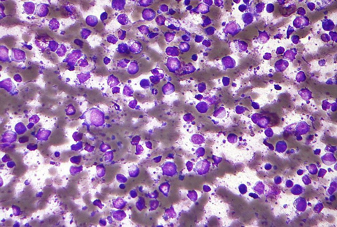
However, the use of prophylactic HD-MTX did not improve CNS or survival outcomes of patients with high-risk DLBCL, but instead was associated with increased toxicities, according to the results of a retrospective study by Hyehyun Jeong, MD, of University of Ulsan College of Medicine, Seoul, Republic of Korea, and colleagues.
The researchers evaluated the effects of prophylactic HD-MTX on CNS relapse and survival outcomes in newly diagnosed R-CHOP (rituximab, cyclophosphamide, doxorubicin, vincristine, and prednisone)–treated patients with high-risk DLBCL. The assessment was based on the initial treatment intent (ITT) of the physician on the use of prophylactic HD-MTX.
A total of 5,130 patients were classified into an ITT HD-MTX group and an equal number into a non-ITT HD-MTX group, according to the report, published online in Blood Advances.
Equivalent results
The study showed that the CNS relapse rate was not significantly different between the two groups, with 2-year CNS relapse rates of 12.4% and 13.9%, respectively (P = .96). Three-year progression-free survival and overall survival rates in the ITT HD-MTX and non-ITT HD-MTX groups were 62.4% vs. 64.5% (P = .94) and 71.7% vs. 71.4% (P = .7), respectively. In addition, the propensity score–matched analyses showed no significant differences in the time-to-CNS relapse, progression-free survival, or overall survival, according to the researchers.
One key concern, however, was the increase in toxicity seen in the HD-MTX group. In this study, the ITT HD-MTX group had a statistically higher incidence of grade 3/4 oral mucositis and elevated alanine aminotransferase (ALT) levels, a marker for liver damage. In addition, the ITT HD-MTX group tended to have a higher incidence of elevated creatinine levels during treatment compared with the non-ITT HD-MTX group.
The HD-MTX group also showed a more common treatment delay or a dose reduction in R-CHOP, which might be attributable to toxicities related to intercalated HD-MTX treatments between R-CHOP cycles, the researchers suggested, potentially resulting in a reduced dose intensity of R-CHOP that could play a role in the lack of an observed survival benefit with additional HD-MTX.
“Another vital issue to consider is that HD-MTX treatment requires hospitalization because intensive hydration and leucovorin rescue is needed, which increases the medical costs,” the authors added.
“This real-world experience, which is unique in its scope and analytical methods, should provide insightful information on the role of HD-MTX prophylaxis to help guide current practice, given the lack of prospective clinical evidence in this patient population,” the researchers concluded.
The authors reported that they had no competing financial interests.
Patients with high-risk diffuse large B-cell lymphoma (DLBCL) have a greater than 10% risk of central nervous system (CNS) relapse, and the use of prophylactic high-dose methotrexate (HD-MTX) has been proposed as a preventative measure.
However, the use of prophylactic HD-MTX did not improve CNS or survival outcomes of patients with high-risk DLBCL, but instead was associated with increased toxicities, according to the results of a retrospective study by Hyehyun Jeong, MD, of University of Ulsan College of Medicine, Seoul, Republic of Korea, and colleagues.
The researchers evaluated the effects of prophylactic HD-MTX on CNS relapse and survival outcomes in newly diagnosed R-CHOP (rituximab, cyclophosphamide, doxorubicin, vincristine, and prednisone)–treated patients with high-risk DLBCL. The assessment was based on the initial treatment intent (ITT) of the physician on the use of prophylactic HD-MTX.
A total of 5,130 patients were classified into an ITT HD-MTX group and an equal number into a non-ITT HD-MTX group, according to the report, published online in Blood Advances.
Equivalent results
The study showed that the CNS relapse rate was not significantly different between the two groups, with 2-year CNS relapse rates of 12.4% and 13.9%, respectively (P = .96). Three-year progression-free survival and overall survival rates in the ITT HD-MTX and non-ITT HD-MTX groups were 62.4% vs. 64.5% (P = .94) and 71.7% vs. 71.4% (P = .7), respectively. In addition, the propensity score–matched analyses showed no significant differences in the time-to-CNS relapse, progression-free survival, or overall survival, according to the researchers.
One key concern, however, was the increase in toxicity seen in the HD-MTX group. In this study, the ITT HD-MTX group had a statistically higher incidence of grade 3/4 oral mucositis and elevated alanine aminotransferase (ALT) levels, a marker for liver damage. In addition, the ITT HD-MTX group tended to have a higher incidence of elevated creatinine levels during treatment compared with the non-ITT HD-MTX group.
The HD-MTX group also showed a more common treatment delay or a dose reduction in R-CHOP, which might be attributable to toxicities related to intercalated HD-MTX treatments between R-CHOP cycles, the researchers suggested, potentially resulting in a reduced dose intensity of R-CHOP that could play a role in the lack of an observed survival benefit with additional HD-MTX.
“Another vital issue to consider is that HD-MTX treatment requires hospitalization because intensive hydration and leucovorin rescue is needed, which increases the medical costs,” the authors added.
“This real-world experience, which is unique in its scope and analytical methods, should provide insightful information on the role of HD-MTX prophylaxis to help guide current practice, given the lack of prospective clinical evidence in this patient population,” the researchers concluded.
The authors reported that they had no competing financial interests.
FROM BLOOD ADVANCES
Pediatric cancer survivors at risk for opioid misuse
Survivors of childhood cancers are at increased risk for prescription opioid misuse compared with their peers, a review of a claims database revealed.
Among more than 8,000 patients age 21 or younger who had completed treatment for hematologic, central nervous system, bone, or gonadal cancers, survivors were significantly more likely than were their peers to have an opioid prescription, longer duration of prescription, and higher daily doses of opioids, and to have opioid prescriptions overlapping for a week or more, reported Xu Ji, PhD, of Emory University in Atlanta.
Teenage and young adult patients were at higher risk than were patients younger than 12, and the risk was highest among patients who had been treated for bone malignancies, as well as those who had undergone any hematopoietic stem cell transplant.
“These findings suggest that health care providers who regularly see survivors should explore nonopioid options to help prevent opioid misuse, and screen for potential misuse in those who actually receive opioids,” she said in an oral abstract presented during the annual meeting of the American Society of Pediatric Hematology/Oncology.
“This is a really important topic, and something that’s probably been underinvestigated and underexplored in our patient population,” said session comoderator Sheri Spunt, MD, Endowed Professor of Pediatric Cancer at Stanford (Calif.) University.
Database review
Dr. Ji and colleagues used the IBM MarketScan Commercial Claims and Encounters database from 2009 to 2018 to examine prescription opioid use, potential misuse, and substance use disorders in pediatric cancer survivors in the first year after completion of therapy, and to identify factors associated with risk for misuse or substance use disorders. Specifically, the period of interest was the first year after completion of all treatments, including surgery, chemotherapy, radiation, and stem cell transplant (Abstract 2015).
They looked at deidentified records on any opioid prescription and for treatment of any opioid use or substance use disorder (alcohol, psychotherapeutic drugs, marijuana, or illicit drug use disorders).
They defined indicators of potential misuse as either prescriptions for long-acting or extended-release opioids for acute pain conditions; opioid and benzodiazepine prescriptions overlapping by a week or more; opioid prescriptions overlapping by a week or more; high daily opioid dosage (prescribed daily dose of 100 or greater morphine milligram equivalent [MME]; and/or opioid dose escalation (an increase of at least 50% in mean MMEs per month twice consecutively within 1 year).
They compared outcomes between a total of 8,635 survivors and 44,175 controls, matched on a 1:5 basis with survivors by age, sex, and region, and continuous enrollment during the 1-year posttherapy period.
In each of three age categories – 0 to 11 years, 12 to 17 years, and 18 years and older – survivors were significantly more likely to have received an opioid prescription, at 15% for the youngest survivors vs. 2% of controls, 25% vs. 8% for 12- to 17-year-olds, and 28% vs. 12% for those 18 and older (P < .01 for all three comparisons).
Survivors were also significantly more likely to have any indicator of potential misuse (1.6% vs. 0.1%, 4.6% vs. 0.5%, and 7.4% vs. 1.2%, respectively, P < .001 for all) and both the youngest and oldest groups (but not 12- to 17-year-olds) were significantly more like to have opioid or substance use disorder (0.4% vs. 0% for 0-11 years, 5.76% vs. 4.2% for 18 years and older, P < .001 for both).
Among patients with any opioid prescription, survivors were significantly more likely than were controls of any age to have indicators for potential misuse. For example, 13% of survivors aged 18 years and older had prescriptions for high opioid doses, compared with 5% of controls, and 12% had prescription overlap, vs. 2%.
Compared with patients with leukemia, patients treated for bone malignancies had a 6% greater risk for having any indicator of misuse, while patients with other malignancies were at slightly lower risk for misuse than those who completed leukemia therapy.
Patients who received any stem cell transplant had an 8.4% greater risk for misuse compared with patients who had surgery only.
Opioids pre- and posttreatment?
“Being someone who takes care of a lot of bone cancer patients, I do see patients with these issues,” Dr. Spunt said.
Audience member Jack H. Staddon, MD, PhD, of the Billings (Montana) Clinic, noted the possibility that opioid use during treatment may have been carried on into the posttreatment period, and asked whether use of narcotics during treatment was an independent risk factor for posttreatment narcotic use or misuse.
The researchers plan to investigate this question in future studies, Dr. Ji replied.
They did not report a study funding source. Dr. Ji and coauthors and Dr. Staddon reported no relevant disclosures.
FROM 2021 ASPHO CONFERENCE