User login
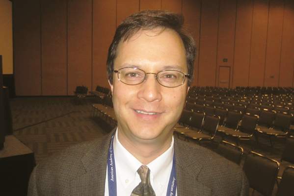
Merkel cell carcinoma responds to first-line pembrolizumab
First-line therapy with pembrolizumab is associated with frequent and durable responses in patients with advanced Merkel cell carcinoma (MCC), according to a study presented at the annual meeting of the American Association for Cancer Research.
The objective response rate wad 56% among 25 patients with MCC who had received at least one dose of pembrolizumab in a phase II study, Dr. Paul Nghiem said at a press conference during the meeting.
This rare but aggressive skin cancer has been notoriously hard to treat with typical chemotherapy of platinum compounds and etoposide, with half of tumors progressing by 3 months and 90% progressing by 10 months. MCC is about three times more likely to kill a patient compared with melanoma.
Given that about 40% of the 2,000 cases per year develop advanced disease, pembrolizumab has the potential to be an advance over chemotherapy although at this point no therapy is approved by the Food and Drug Administration for MCC advanced disease. The disease may be caused by ultraviolet light exposure in older individuals or with immune suppression. In about 80% of cases, Merkel cell polyomavirus (MCPyV) is present, but the virus is ubiquitous in the environment, with widespread exposure in the population without ill effects for most people.
Pembrolizumab acts to block the interaction of PD-1 on tumor-specific or MCPyV-specific T cells with its ligand, PD-L1, on tumor cells or antigen-presenting cells, thus allowing the T cells to recognize the tumors and enhance tumor cell killing.
In a phase II multicenter, single-arm, open-label trial, patients with unresectable or metastatic disease who had never before received any systemic therapy received pembrolizumab 2 mg/kg IV every 3 weeks for up to 2 years. Tumor evaluations were done every 9 or 12 weeks with a median follow-up of 7.6 months.
Of the 25 patients who had received at least one dose and had at least one radiological assessment, there was an objective response rate of 56% (14/25), including 4 complete, 10 partial, and 1 unconfirmed partial response. One patient had stable disease, and nine had progressive disease.
“By the first scan at 3 months most patients have found where they were going to go,” said Dr. Nghiem, professor of medicine in dermatology at the University of Washington, Seattle. “Several had failed early, but by the time of the first scan at 3 months there were often very profound and complete responses, and a lot of the partial responses just remain very steady over time.”
With the caveat that this was not a randomized trial, he said the progression-free survival (PFS) among responders was 67% at 6 months and the median PFS was 9 months, compared to a historical median overall survival with chemotherapy of 9.5 months after metastatic diagnosis. Among responders, 86% continue to have excellent disease control more than 6 months after starting therapy.
Objective responses in this small study appeared to be associated with positive viral status of the tumors. Among patients with virus-positive tumors, 62% (10/16) had objective responses vs. 44% (4/9) in whom the tumors were virus negative. “That was not a statistically significant difference … but maybe suggests, like head and neck [cancers], there may be a better story there for virus positive,” Dr. Nghiem said. PD-L1 expression in the tumors did not predict responses.
In response to a question during the news conference about the high response rate compared to responses to therapy of most other solid tumors, Dr. Nghiem pointed out, “Historically, this has been a very immune-associated cancer … If you had CD8 T cells infiltrating into this cancer at any reasonable number, among 300 patients not one died of the disease.” Unleashing the immune system in this trial through PD-1 blockade may therefore help to explain the good outcomes.
Adverse events were managed with corticosteroid treatment and discontinuing the drug. Two patients who had severe drug-related toxicities improved after steroids and stopping the drug. But, nonetheless, they have ongoing antitumor responses several months after stopping pembrolizumab.
The investigators are currently expanding the trial and recruiting more patients.
The study was simultaneously published online in the New England Journal of Medicine (2016 April 19. doi: 10.1056/NEJMoa1603702).
The study was supported in part by Merck. Dr. Nghiem is a consultant to EMD Serono and has grant/research support from Bristol-Myers Squibb.
FROM THE AACR ANNUAL MEETING
Key clinical point: Rare Merkel cell carcinoma responds well to immunotherapy with pembrolizumab.
Major finding: The response rate was 56%, all occurring by 3 months.
Data source: Phase II multicenter, single-arm, open-label trial of 25 patients (NCT02267603).
Disclosures: The study was supported in part by Merck. Dr. Nghiem is a consultant to EMD Serono and has grant/research support from Bristol-Myers Squibb.

Pembrolizumab: 33% response rate in advanced melanoma
Immunotherapy with the PD-1 inhibitor pembrolizumab yielded a 33% overall response rate, a 35% progression-free survival rate at 1 year, and a median overall survival of 23 months in 655 patients with advanced melanoma, according to a report published online in JAMA.
The agent generally was well tolerated, with grade 3 or 4 treatment-related toxicities occurring in 14% of patients. Only 4% of patients discontinued pembrolizumab because of adverse events, including colitis, pyrexia, pneumonitis, and thyroid abnormalities, said Dr. Antoni Ribas of University of California, Los Angeles, and his associates.

The investigators analyzed data pooled from 8 open-label phase I cohorts, some of which have been reported previously, in this manufacturer-sponsored study. All the participants were adults (median age, 61 years) with advanced unresectable melanoma who resided in Australia, Canada, France, and the U.S. This pooled study population was heterogeneous, as the various cohorts had different eligibility criteria; some patients were treatment-naïve, some had received ipilimumab, and some were treated concomitantly with BRAF or MEK inhibitors.
The estimated median duration of response was 28.2 months, and 44% of patients who responded to pembrolizumab had a response duration of longer than 1 year. Approximately half of the patients were still alive at 2 years.
Treatment benefit was consistent regardless of whether or not patients had previously received ipilimumab and regardless of the dose or schedule of pembrolizumab they were given. Patients who were treatment-naïve showed the greatest response to pembrolizumab: an overall response rate of 45%, a 1-year progression-free survival rate of 52%, and a median overall survival of 31 months, the investigators said (JAMA. 2016 Apr 19. doi: 10.1001/jama.2016.4059). “Collectively, these data suggest that the majority of patients with melanoma treated with pembrolizumab will experience lasting objective responses,” they added.
The encouraging results of Ribas et al add to the burgeoning literature on the potential of PD-1 blockade to improve outcomes in metastatic melanoma. One finding in particular – the fact that half of the patients in these cohorts were alive at 2 years – is noteworthy because the median overall survival in most previous clinical trials of the disease was consistently shorter than 1 year.
However, the results also highlight the limitations of PD-1 blockade. Median progression-free survival was only 4 months, and the 1-year progression-free survival rate was only 35%. This suggests that the host immune system fails to adequately control the malignancy in two-thirds of patients within the first year. And even among treatment responders, disease progression occurred in 26%.
Dr. Shailender Bhatia and Dr. John A. Thompson are at the University of Washington and the Fred Hutchinson Cancer Research Center, both in Seattle. Dr. Bhatia and Dr. Thompson made these remarks in an editorial accompanying Dr. Ribas’s report (JAMA. 2016 Apr 19;315:1573-4).
Immunotherapy with the PD-1 inhibitor pembrolizumab yielded a 33% overall response rate, a 35% progression-free survival rate at 1 year, and a median overall survival of 23 months in 655 patients with advanced melanoma, according to a report published online in JAMA.
The agent generally was well tolerated, with grade 3 or 4 treatment-related toxicities occurring in 14% of patients. Only 4% of patients discontinued pembrolizumab because of adverse events, including colitis, pyrexia, pneumonitis, and thyroid abnormalities, said Dr. Antoni Ribas of University of California, Los Angeles, and his associates.
The investigators analyzed data pooled from 8 open-label phase I cohorts, some of which have been reported previously, in this manufacturer-sponsored study. All the participants were adults (median age, 61 years) with advanced unresectable melanoma who resided in Australia, Canada, France, and the U.S. This pooled study population was heterogeneous, as the various cohorts had different eligibility criteria; some patients were treatment-naïve, some had received ipilimumab, and some were treated concomitantly with BRAF or MEK inhibitors.
The estimated median duration of response was 28.2 months, and 44% of patients who responded to pembrolizumab had a response duration of longer than 1 year. Approximately half of the patients were still alive at 2 years.
Treatment benefit was consistent regardless of whether or not patients had previously received ipilimumab and regardless of the dose or schedule of pembrolizumab they were given. Patients who were treatment-naïve showed the greatest response to pembrolizumab: an overall response rate of 45%, a 1-year progression-free survival rate of 52%, and a median overall survival of 31 months, the investigators said (JAMA. 2016 Apr 19. doi: 10.1001/jama.2016.4059). “Collectively, these data suggest that the majority of patients with melanoma treated with pembrolizumab will experience lasting objective responses,” they added.
FROM JAMA
Key clinical point: Immunotherapy with pembrolizumab yielded a 33% overall response rate, a 35% 1-year progression-free survival rate, and a median overall survival of 23 months in 655 patients with advanced melanoma.
Major finding: The estimated median duration of response was 28.2 months, and half of the study population was still alive at 2 years.
Data source: A manufacturer-sponsored pooled analysis of data from 8 open-label phase I cohorts involving 655 patients followed for a median of 15 months.
Disclosures: This study was supported by Merck, maker of pembrolizumab, which also participated in the design and conduct of the study; the collection, analysis, and interpretation of the data; and preparation of the report. Dr. Ribas reported owning stock in several pharmaceutical companies. His associates reported ties to numerous industry sources.

Engineered helper T cells targeting MAGE-A3 produced some clinical responses
CD4+ T cells genetically engineered to recognize MAGE-A3, a protein found on many types of tumor cells but not on normal cells, were safely given to patients with metastatic cancers, and some of the patients had clinical responses.
Whereas the majority of current adoptive T-cell therapies have used genetically modified unpurified T cells or CD8+ (cytotoxic) T cells, Yong-Chen William Lu, Ph.D., of the surgery branch of the National Cancer Institute conducted a phase I trial of CD4+ (helper) T cells to evaluate their contribution to tumor killing. Preclinical and clinical studies indicated that CD4+ T cells can induce tumor regression.
Dr. Lu explained the individualized process of making the T cells for infusion, during a news conference at the annual meeting of the American Association for Cancer Research. First, T cells are harvested from the peripheral blood of a patient, from which CD4+ T cells are isolated. Using a retroviral vector, scientists insert the gene for a T-cell receptor that recognizes MAGE-A3 in conjunction with the HLA molecule DPB1*0401. In this way, the cells can recognize the tumor antigen in anyone of that HLA type. Then the cells carrying the engineered T-cell receptor are expanded in vitro and infused into the patient.
DPB1*0401 was chosen as the HLA genotype because “it is present in 60% of the Caucasian population. It is the highest one,” Dr. Lu said. MAGE-A3 is expressed on cells during fetal development and is later lost on normal tissues. But several tumor types, including some melanomas and some urothelial, esophageal, and cervical cancers reexpress this protein, making it a good target for immunotherapy.
The trial included patients of the HLA DPB1*0401 type with metastatic cancer that is MAGE-A3 positive. Patients had received at least one first-line treatment for metastatic disease and had been nonresponders or had had a recurrence of the tumor.
This phase I trial involved 14 patients in nine different dose cohorts, receiving from 10 million engineered T cells at the lowest dose to 78-103 billion cells in the highest cohort. Three patients in the middle and highest dosing cohorts achieved partial responses, one each with cervical, esophageal, and urothelial tumors. The partial response lasted 4 months for the patient with esophageal cancer and is ongoing at 7 months for the urothelial and 15 months for the cervical cancer patients.
In response to a question from the audience about adverse effects, Dr. Lu said patients had short-term elevations of circulating cytokines, “which only lasted for a few days,” and a fever lasting 1-2 weeks.
In summary, he said, “We think this first [CD4+] engineered T-cells therapy targeting MAGE-A3 is safe and has shown early clinical response.” Phase II trials are now beginning.
The approach of this immunotherapy method is different from that involved in using checkpoint inhibitors, “which essentially release the brakes of the immune system so the T cells can do their job of attacking the cancer,” commented Dr. Louis Weiner, director of the Georgetown Lombardi Cancer Center in Washington, D.C. “This is actually not trying to release brakes but essentially press on the accelerator, if you will, by using an expanded tumor-specific population of T cells that will then be able to hone in and attack the cancer.” He envisioned a time in the future when this approach could be combined with checkpoint inhibitors for enhanced antitumor effects.
Dr, Weiner also noted that the T cells with receptors engineered to recognize MAGE-A3 are more specific for tumors, compared with approaches using chimeric antigen receptor T cells to treat certain hematologic malignancies. In that setting the chimeric antigen receptor T cells recognize and eliminate, for example, not only malignant B cells bearing a pan-B cell marker but also all normal B cells.
However, in the question and answer period, Dr. Lu said one problem with his approach is the possibility that not all cells in a tumor may express MAGE-A3.
There was no commercial funding of the study. Dr. Lu had no relevant financial disclosures. Dr. Weiner is an adviser, review panel member, or consultant, or has other relationships, with numerous pharmaceutical companies.
FROM THE AACR ANNUAL MEETING
Key clinical point: Engineered helper T cells safely target tumors and produce clinical responses.
Major finding: Three of 14 patients with different tumor types had partial tumor responses when given T cells with receptors engineered to recognize a common tumor antigen, MAGE-A3.
Data source: A phase I dose-escalating study of 14 patients.
Disclosures: There was no commercial funding of the study. Dr. Lu had no relevant financial disclosures. Dr. Weiner is an adviser, review panel member, or consultant, or has other relationships, with numerous pharmaceutical companies.
Study found long-term benefit of nivolumab for advanced melanoma patients
One-third of a group of heavily pretreated patients with advanced melanoma who received nivolumab monotherapy were alive 5 years after starting the drug, a phase I trial showed. Survival plateaued at 34% by 48 months, after which time disease control was maintained.
“What it does demonstrate is the importance of the durability of clinical benefit to patients [who] now we’re measuring in terms of years, as well as the memory aspect of how the immunologic memory can translate into benefit,” said Dr. Stephen Hodi, director of the melanoma center at Dana-Farber Cancer Institute in Boston during a news conference at the annual meeting of the American Association for Cancer Research.

Nivolumab is an immune checkpoint inhibitor that blocks PD-1, allowing enhanced antitumor immune responses. It is approved as a first-line agent for advanced melanoma, alone or in combination with ipilimumab. The approved dose for monotherapy is 3 mg/kg every 2 weeks.
The study randomized 107 patients (median age, 61 years; 67% male) to one of five doses ranging from 0.1 mg/kg to 10 mg/kg IV given every 2 weeks for up to 96 weeks. The patients had advanced melanoma at any site and had been on one to five lines of systemic therapy prior to entering the trial but could not have received any immune checkpoint inhibitors. They had treated and stable brain metastases and an ECOG (Eastern Cooperative Oncology Group) performance status of 0, 1, or 2. The minimum follow-up was 45 months.
Among all the patients, the median overall survival was 17.3 months (95% confidence interval, 12.5-37.8 months), and for those patients receiving 3-mg/kg doses, the median survival was 20.3 months (95% CI, 7.2 months and no upper confidence level reached). The Kaplan-Meier survival curves for all patients and those receiving the 3-mg/kg dose began to level off at about 36 months, and were essentially flat at 34% for all patients as a group from 48 months out to 5 years.
Five patients who achieved disease control but later had progressive disease were retreated with their original doses of nivolumab after being off the drug for more than 100 days. “In all five patients, disease control was obtained again, and this disease control was again durable,” Dr. Hodi reported.
Nivolumab monotherapy was safe and well tolerated. No study deaths or new safety signals were observed. The most common drug-related adverse effects were any grade of fatigue (29.9% of all patients; 47.1% of patients at the 3-mg/kg dose) and any grade of rash (23.4% and 11.8%, respectively). Although there were some grade 3-4 adverse events, adverse reactions led to study discontinuation for only 10.3% of the total patient group and 5.9% of patients on the 3-mg/kg dose.
Calling the study findings “very compelling,” conference moderator Dr. Louis Weiner, director of the Georgetown Lombardi Comprehensive Cancer Center in Washington, D.C., identified what he said is a key point of the study – the length of benefit. “What distinguishes immunotherapy at this point when it’s effective from other forms of cancer treatment is the durability of the benefit,” he said. “People who have good responses really seem to be protected against the disease returning in many cases.”
He asked Dr. Hodi if he would expect even better disease control results if nivolumab were combined with a different checkpoint inhibitor, such as ipilimumab. Dr. Hodi replied that it is reasonable to think that agents working through different mechanisms of action should produce even better results.
A reporter in the audience asked if it was Dr. Hodi’s practice to test tumors for PD-1 expression before using nivolumab. He replied that he and his colleagues do not routinely test for PD-1 because there have been observations that some tumors that test negative for the biomarker do respond to the drug.
One-third of a group of heavily pretreated patients with advanced melanoma who received nivolumab monotherapy were alive 5 years after starting the drug, a phase I trial showed. Survival plateaued at 34% by 48 months, after which time disease control was maintained.
“What it does demonstrate is the importance of the durability of clinical benefit to patients [who] now we’re measuring in terms of years, as well as the memory aspect of how the immunologic memory can translate into benefit,” said Dr. Stephen Hodi, director of the melanoma center at Dana-Farber Cancer Institute in Boston during a news conference at the annual meeting of the American Association for Cancer Research.
Nivolumab is an immune checkpoint inhibitor that blocks PD-1, allowing enhanced antitumor immune responses. It is approved as a first-line agent for advanced melanoma, alone or in combination with ipilimumab. The approved dose for monotherapy is 3 mg/kg every 2 weeks.
The study randomized 107 patients (median age, 61 years; 67% male) to one of five doses ranging from 0.1 mg/kg to 10 mg/kg IV given every 2 weeks for up to 96 weeks. The patients had advanced melanoma at any site and had been on one to five lines of systemic therapy prior to entering the trial but could not have received any immune checkpoint inhibitors. They had treated and stable brain metastases and an ECOG (Eastern Cooperative Oncology Group) performance status of 0, 1, or 2. The minimum follow-up was 45 months.
Among all the patients, the median overall survival was 17.3 months (95% confidence interval, 12.5-37.8 months), and for those patients receiving 3-mg/kg doses, the median survival was 20.3 months (95% CI, 7.2 months and no upper confidence level reached). The Kaplan-Meier survival curves for all patients and those receiving the 3-mg/kg dose began to level off at about 36 months, and were essentially flat at 34% for all patients as a group from 48 months out to 5 years.
Five patients who achieved disease control but later had progressive disease were retreated with their original doses of nivolumab after being off the drug for more than 100 days. “In all five patients, disease control was obtained again, and this disease control was again durable,” Dr. Hodi reported.
Nivolumab monotherapy was safe and well tolerated. No study deaths or new safety signals were observed. The most common drug-related adverse effects were any grade of fatigue (29.9% of all patients; 47.1% of patients at the 3-mg/kg dose) and any grade of rash (23.4% and 11.8%, respectively). Although there were some grade 3-4 adverse events, adverse reactions led to study discontinuation for only 10.3% of the total patient group and 5.9% of patients on the 3-mg/kg dose.
Calling the study findings “very compelling,” conference moderator Dr. Louis Weiner, director of the Georgetown Lombardi Comprehensive Cancer Center in Washington, D.C., identified what he said is a key point of the study – the length of benefit. “What distinguishes immunotherapy at this point when it’s effective from other forms of cancer treatment is the durability of the benefit,” he said. “People who have good responses really seem to be protected against the disease returning in many cases.”
He asked Dr. Hodi if he would expect even better disease control results if nivolumab were combined with a different checkpoint inhibitor, such as ipilimumab. Dr. Hodi replied that it is reasonable to think that agents working through different mechanisms of action should produce even better results.
A reporter in the audience asked if it was Dr. Hodi’s practice to test tumors for PD-1 expression before using nivolumab. He replied that he and his colleagues do not routinely test for PD-1 because there have been observations that some tumors that test negative for the biomarker do respond to the drug.
FROM THE AACR ANNUAL MEETING
Key clinical point: Heavily pretreated patients with advanced melanoma had long-term responses on nivolumab monotherapy.
Major finding: Nivolumab monotherapy resulted in a 34% overall survival rate at 5 years for melanoma patients who had received extensive prior therapy. Data source: A phase I randomized dose-ranging study involving 107 patients with advanced melanoma.
Disclosures: The study was funded by Bristol-Myers Squibb. Dr. Hodi has received grant/research support from and is a nonpaid consultant to Bristol-Myers. Dr. Weiner is an adviser, review panel member, or consultant or has other relationships with numerous pharmaceutical companies.

For BCC, consider curettage only
WAIKOLOA, HAWAII – Curettage alone for the treatment of basal cell carcinomas offers several distinct advantages over the widely accepted technique of curettage followed by electrodesiccation, Dr. David L. Swanson said at the Hawaii Dermatology Seminar provided by the Global Academy for Medical Education/Skin Disease Education Foundation.
When he asked for an audience show of hands as to who treats BCCs using curettage without electrodesiccation, the packed auditorium full of dermatologists remained hands-down. But there is good evidence that curettage alone provides cure rates similar to published rates for curettage and electrodesiccation, along with improved cosmesis, faster healing, and fewer problems with scarring and hypopigmentation, according to Dr. Swanson, a dermatologist at Mayo Clinic Arizona, Scottsdale.

He cited a persuasive study by other dermatologists at Mayo Clinic Arizona (J Am Acad Dermatol. 2006 Jun;54[6]:1039-45).
“This article was transformative for my practice,” said Dr. Swanson, who wasn’t involved in the study.
The investigators reviewed the records of 302 biopsy-proven BCCs in 136 patients. All were amenable to curettage and electrodesiccation but were instead treated by curettage alone during 1993-1998. The BCCs lacked histopathologic high-risk features; most were superficial or nodular lesions. Nearly one-quarter of the BCCs, however, were located on high-risk anatomic sites.
The 5-year cure rate was 96%. The authors’ review of 10 published series of BCCs treated using curettage and electrodesiccation showed recurrence rates mostly in the 3%-7% range.
Among the advantages cited by the investigators of going with curettage alone were its simplicity, reduced equipment requirement, cost effectiveness, lack of electrical interference with implantable cardiac devices, the avoidance of potentially hazardous viral smoke plumes, and reduced rates of hypopigmentation and scarring.
This was a retrospective study. Its other significant limitation was that all 302 BCCs were treated by the same dermatologist: Dr. Mark J. Zalla, who Dr. Swanson considers to be a dermatologic surgeon of exceptional talent.
“I’m not Mark Zalla. Nobody else in this room is Mark Zalla, so we can’t necessarily extrapolate from this study what your outcomes are going to be. That said, I’ve adopted this technique, and most of my colleagues do not do electrodesiccation anymore. I take off several basal cell carcinomas per day with curettage, and it’s my sense that I don’t have higher recurrence rates and that it does heal better with less scarring,” Dr. Swanson said.
He reported having no financial conflicts of interest regarding his presentation. SDEF and this news organization are owned by the same parent company.
WAIKOLOA, HAWAII – Curettage alone for the treatment of basal cell carcinomas offers several distinct advantages over the widely accepted technique of curettage followed by electrodesiccation, Dr. David L. Swanson said at the Hawaii Dermatology Seminar provided by the Global Academy for Medical Education/Skin Disease Education Foundation.
When he asked for an audience show of hands as to who treats BCCs using curettage without electrodesiccation, the packed auditorium full of dermatologists remained hands-down. But there is good evidence that curettage alone provides cure rates similar to published rates for curettage and electrodesiccation, along with improved cosmesis, faster healing, and fewer problems with scarring and hypopigmentation, according to Dr. Swanson, a dermatologist at Mayo Clinic Arizona, Scottsdale.
He cited a persuasive study by other dermatologists at Mayo Clinic Arizona (J Am Acad Dermatol. 2006 Jun;54[6]:1039-45).
“This article was transformative for my practice,” said Dr. Swanson, who wasn’t involved in the study.
The investigators reviewed the records of 302 biopsy-proven BCCs in 136 patients. All were amenable to curettage and electrodesiccation but were instead treated by curettage alone during 1993-1998. The BCCs lacked histopathologic high-risk features; most were superficial or nodular lesions. Nearly one-quarter of the BCCs, however, were located on high-risk anatomic sites.
The 5-year cure rate was 96%. The authors’ review of 10 published series of BCCs treated using curettage and electrodesiccation showed recurrence rates mostly in the 3%-7% range.
Among the advantages cited by the investigators of going with curettage alone were its simplicity, reduced equipment requirement, cost effectiveness, lack of electrical interference with implantable cardiac devices, the avoidance of potentially hazardous viral smoke plumes, and reduced rates of hypopigmentation and scarring.
This was a retrospective study. Its other significant limitation was that all 302 BCCs were treated by the same dermatologist: Dr. Mark J. Zalla, who Dr. Swanson considers to be a dermatologic surgeon of exceptional talent.
“I’m not Mark Zalla. Nobody else in this room is Mark Zalla, so we can’t necessarily extrapolate from this study what your outcomes are going to be. That said, I’ve adopted this technique, and most of my colleagues do not do electrodesiccation anymore. I take off several basal cell carcinomas per day with curettage, and it’s my sense that I don’t have higher recurrence rates and that it does heal better with less scarring,” Dr. Swanson said.
He reported having no financial conflicts of interest regarding his presentation. SDEF and this news organization are owned by the same parent company.
EXPERT ANALYSIS FROM SDEF HAWAII DERMATOLOGY SEMINAR

New research institute to advance cancer immunotherapy
A new research institute launched by tech billionaire Sean Parker aims to advance the field of cancer immunotherapy by uniting the country’s top scientists, including Dr. Carl June, Dr. Jedd Wolchok, and Dr. James Allison, who are leaders in the field.
Best known for founding music-sharing service Napster, Mr. Parker has provided a $250 million grant to develop the Parker Institute for Cancer Immunotherapy, a collaboration between researchers, clinicians, and other stakeholders that focuses on expanding immunotherapy research efforts, according to an April announcement by the Parker Foundation.

“We are at an inflection point in cancer research and now is the time to maximize immunotherapy’s unique potential to transform all cancers into manageable diseases, saving millions of lives,” Mr. Parker said in a statement. “We believe that the creation of a new funding and research model can overcome many of the obstacles that currently prevent research breakthroughs. Working closely with our scientists and more than 30 industry partners, the Parker Institute is positioned to broadly disseminate discoveries and, most importantly, more rapidly deliver treatments to patients.”
The Parker Institute for Cancer Immunotherapy will include 40 laboratories and more than 300 researchers from leading cancer centers, including Memorial Sloan Kettering Cancer Center; Stanford Medicine; the University of California, Los Angeles; the University of California, San Francisco; the University of Pennsylvania; and the University of Texas MD Anderson Cancer Center. Each research center will receive comprehensive funding, clinical resources, and key technologies needed to accelerate research discoveries, according to the foundation. In a unique agreement, the administration of intellectual property will be shared between the research centers, allowing scientists to have immediate access to the broad spectrum of core discoveries.
Scientists involved with the institute call immunotherapy “one of the most important medical advances of our time,” and the first approach that could generate long-lasting remissions for all cancer types and stages. The relatively new treatment area mobilizes the body’s own defense systems to fight cancer cells.
“Immunotherapy represents a fundamentally new, breakthrough treatment paradigm in the fight against cancer – it harnesses the body’s own powerful immune system to mobilize its highly refined disease-fighting arsenal to engage and eliminate the cancer cells,” Parker Institute CEO and President Jeff Bluestone, Ph.D., said in a statement. “Our scientists are leaders in the field and will now work together to make discoveries to treat and potentially cure cancer.”
In the 1990s, Dr. Bluestone of the University of California, San Francisco, was one of two scientists who discovered that, to prevent autoimmunity and overreactions, a molecule called CTLA-4 acts as a “brake,” or checkpoint, on the immune response. The finding fueled a new class of checkpoint-blockade treatments for multiple cancers.
The Parker Institute will focus on several initial objectives, including developing novel approaches to modify T-cells to enhance function and creating a new generation of more effective T-cell therapies. Researchers also will work to improve the rates of durable responses and broaden the use of checkpoint-blocking agents, alone or in combination to treat more types of cancer. Finally, scientists will conduct research to advance DNA sequencing, antigenic peptide discovery efforts, and immune monitoring technologies. Their goal is to understand how to more effectively target tumors and ultimately develop new vaccines and T-cell therapies.
A major key of the institute is to remove competition among researchers that silo breakthroughs and instead enhance teamwork, said James P. Allison, Ph.D., of the University of Texas MD Anderson Cancer Center, Houston.
“One of the goals is to try to remove that competitive edge and have everybody bring their skills together,” Dr. Allison said in a video. “Still, you’re trying to do more than the other guy, but you’re doing it with them. So you use that competitive edge to an advantage.”
On Twitter @legal_med
A new research institute launched by tech billionaire Sean Parker aims to advance the field of cancer immunotherapy by uniting the country’s top scientists, including Dr. Carl June, Dr. Jedd Wolchok, and Dr. James Allison, who are leaders in the field.
Best known for founding music-sharing service Napster, Mr. Parker has provided a $250 million grant to develop the Parker Institute for Cancer Immunotherapy, a collaboration between researchers, clinicians, and other stakeholders that focuses on expanding immunotherapy research efforts, according to an April announcement by the Parker Foundation.
“We are at an inflection point in cancer research and now is the time to maximize immunotherapy’s unique potential to transform all cancers into manageable diseases, saving millions of lives,” Mr. Parker said in a statement. “We believe that the creation of a new funding and research model can overcome many of the obstacles that currently prevent research breakthroughs. Working closely with our scientists and more than 30 industry partners, the Parker Institute is positioned to broadly disseminate discoveries and, most importantly, more rapidly deliver treatments to patients.”
The Parker Institute for Cancer Immunotherapy will include 40 laboratories and more than 300 researchers from leading cancer centers, including Memorial Sloan Kettering Cancer Center; Stanford Medicine; the University of California, Los Angeles; the University of California, San Francisco; the University of Pennsylvania; and the University of Texas MD Anderson Cancer Center. Each research center will receive comprehensive funding, clinical resources, and key technologies needed to accelerate research discoveries, according to the foundation. In a unique agreement, the administration of intellectual property will be shared between the research centers, allowing scientists to have immediate access to the broad spectrum of core discoveries.
Scientists involved with the institute call immunotherapy “one of the most important medical advances of our time,” and the first approach that could generate long-lasting remissions for all cancer types and stages. The relatively new treatment area mobilizes the body’s own defense systems to fight cancer cells.
“Immunotherapy represents a fundamentally new, breakthrough treatment paradigm in the fight against cancer – it harnesses the body’s own powerful immune system to mobilize its highly refined disease-fighting arsenal to engage and eliminate the cancer cells,” Parker Institute CEO and President Jeff Bluestone, Ph.D., said in a statement. “Our scientists are leaders in the field and will now work together to make discoveries to treat and potentially cure cancer.”
In the 1990s, Dr. Bluestone of the University of California, San Francisco, was one of two scientists who discovered that, to prevent autoimmunity and overreactions, a molecule called CTLA-4 acts as a “brake,” or checkpoint, on the immune response. The finding fueled a new class of checkpoint-blockade treatments for multiple cancers.
The Parker Institute will focus on several initial objectives, including developing novel approaches to modify T-cells to enhance function and creating a new generation of more effective T-cell therapies. Researchers also will work to improve the rates of durable responses and broaden the use of checkpoint-blocking agents, alone or in combination to treat more types of cancer. Finally, scientists will conduct research to advance DNA sequencing, antigenic peptide discovery efforts, and immune monitoring technologies. Their goal is to understand how to more effectively target tumors and ultimately develop new vaccines and T-cell therapies.
A major key of the institute is to remove competition among researchers that silo breakthroughs and instead enhance teamwork, said James P. Allison, Ph.D., of the University of Texas MD Anderson Cancer Center, Houston.
“One of the goals is to try to remove that competitive edge and have everybody bring their skills together,” Dr. Allison said in a video. “Still, you’re trying to do more than the other guy, but you’re doing it with them. So you use that competitive edge to an advantage.”
On Twitter @legal_med

FDA Proposes New Rule to Ban Use of Indoor Tanning Devices by Minors
The US Food and Drug Administration (FDA) has proposed 2 new rules to protect consumers from health risks associated with indoor tanning by banning use of indoor tanning devices by minors and imposing safety measures.
The first proposed rule restricts the use of indoor tanning devices to adults 18 years and older. It also requires indoor tanning facilities to inform adult users about the health risks of indoor tanning and obtain a signed risk acknowledgement from consumers before their first tanning session and every 6 months thereafter.
“Exposure to UV radiation from indoor tanning is a preventable cause of skin cancer,” explained Markham C. Luke, MD, PhD, deputy office director for the Office of Device Evaluation at the FDA’s Center for Devices and Radiological Health. “The FDA is committed to protecting public health by informing consumers of the risks of indoor tanning.”
The second proposed rule addresses performance standards, requiring manufacturers and indoor tanning facilities to take measures to improve the overall safety of tanning devices. Key changes would include:
- Make consumer warnings more prominent and easier to read on tanning devices.
- Require an easily accessible emergency shutoff switch (or panic button) on all tanning devices.
- Add requirements to limit the amount of light allowed through protective eyewear to protect the eyes.
- Improve labeling on replacement bulbs to ensure tanning facility operators use the proper bulbs to reduce risk for accidental burns.
- Prohibit tanning facilities from making dangerous device modifications (eg, installing stronger bulbs) without recertifying and reidentifying the device with the FDA.
The FDA reports that more than 1 million minors use indoor tanning facilities each year. According to the American Academy of Dermatology, consumers younger than 35 years who use indoor tanning facilities are 59% more likely to develop melanoma than those who have never tanned indoors. Because the effects of UV exposure are cumulative and add up over the course of one’s lifetime, minors who use indoor tanning devices are at an increased risk for developing melanoma and nonmelanoma skin cancers later in life.
In 2014 the FDA began requiring tanning devices to be labeled with a visible warning stating that individuals younger than 18 years should not use them. Additionally, several states have already passed laws prohibiting minors from indoor tanning; in Connecticut, New Jersey, New York, and Pennsylvania, tanning devices are banned in individuals younger than 17 years.
Dermatologists are in the position to discuss the health risks of indoor tanning with all patients regardless of age. Patients should be reminded that failure to wear appropriate protective eyewear can lead to short-term and long-term eye injury and that long exposures can lead to burning that may not be recognized until it is too late. It also is important to warn patients that tanning while using certain medications or cosmetics may cause increased sensitivity to UV radiation. Patients can be referred to the FDA website for more consumer updates about indoor tanning and the proposed rules.

Guidelines highlight revolution in advanced melanoma therapies
HOLLYWOOD, FLA. –The remarkable changes that have occurred in the treatment of advanced malignant melanoma over the last several years are reflected in revised National Comprehensive Cancer Network guidelines.
“Gone are the days where our active agents were interferon and dacarbazine,” said Dr. John A. Thompson of the Fred Hutchinson Cancer Research Center, Seattle.

“We’ve seen the approval by the FDA of the PD-1-targeted agents and also molecular-targeted agents to treat patients with mutations in the BRAF gene, and the rate of change continues apace,” he said at the annual conference of the National Comprehensive Cancer Network.
Among the changes in the 2016 guidelines are the addition of high-dose ipilimumab (Yervoy) as an option for stage III (node-positive) disease, and intralesional talimogene laherparepvec (T-VEC) for primary treatment of stage III in-transit disease.
Guidelines for first-line systemic therapy for metastatic or unresectable disease recommend treatment based on evaluation of individual patients and include:
• Monotherapy with an anti-PD-1 (programmed death-1) agent, either pembrolizumab (Keytruda) or nivolumab (Opdivo) (category 1 recommendation).
• Targeted therapy for BRAF-mutated disease with combinations (preferred) of either dabrafenib (Tafinlar) and trametinib (Mekinist) or vemurafenib (Zelboraf)and cobimetinib (Cotellic); both are category 1 recommendations.
• Single-agent therapy with either vemurafenib or dabrafenib (category 1), or clinical trial.
For patients with disease progression or those who have had their maximum clinical benefit from BRAF-targeted therapy and good performance status (0-2), the guidelines recommend second-line or subsequent therapy with anti-PD-1 agents as above, ipilimumab, or a combination of nivolumab and ipilimumab.
The guidelines note, however, that “nivolumab/ipilimumab combination therapy is associated with improved relapse-free survival compared with single-agent nivolumab or ipilimumab, at the expense of significantly increased toxicity. Compared to single-agent therapy, the impact of nivolumab/ipilimumab combination therapy on overall survival is not known.”
Also recommended are targeted therapy with the BRAF-inhibitor combinations listed above (preferred) or single-agent therapy with vemurafenib or dabrafenib.
“In previously untreated patients with unresectable stage IIIC or stage IV disease, the combination of vemurafenib/cobimetinib was associated with improved PFS [progression-free survival] and response rate when compared to vemurafenib alone. The impact on overall survival compared to single-agent vemurafenib is unknown,” the guidelines state.
Other second-line options for patients with good performance status include high-dose interleukin-2 (not suitable for patients with inadequate organ reserve, poor performance status, or untreated or active brain metastases), biochemotherapy and cytotoxic agents (dacarbazine or temozolomide, and cisplatin or carboplatin, with or without vinblastine or nitrosourea, and IL-2 and interferon alfa-2b; paclitaxel, albumin-bound paclitaxel).
For patients with poor performance status (3-4) the guidelines call for best supportive care according to guidelines for palliative care.
Oncolytic immunotherapy
For treatment of recurrent disease, the guidelines newly recommend use of T-VEC, a genetically modified herpes simplex-1 virus that is injected intralesionally into accessible tumors. The attenuated virus does not kill healthy cells, but replicates in tumors and causes the cells to secrete granulocyte macrophage–colony stimulating factor (GM-CSF), leading to lysis of the tumor cells.
“It is hypothesized that the GM-CSF works with the immune system to boost a resident immune response against the destroyed melanoma cells and antigens that are released from this process,” Dr. Thompson said.
In a recent clinical trial (J Clin Oncol. 2015;33:2780-8), patients with injectable melanoma that was not surgically resectable were randomized to either intralesional T-VEC injections on week 0, 3, and then every 2 weeks (295 patients), or subcutaneous GM-CSF 125 mcg/m2 on days 1 to 14 for every 28 days (141 patients).
T-VEC was associated with significantly more durables responses (16% vs. 2.1% for GM-CSF, P less than .001) and a significantly higher overall response rate (26.4% vs. 5.7%, respectively, P less than .001).
In the primary analysis in an intent-to-treat population, T-VEC had a borderline significant association with better overall survival (median 23.2 vs. 18.9 months, hazard ratio [HR] 0.70, log-rank P = .051).
In subanalyses of outcomes in patient subgroups, however, the benefits of T-VEC were seen in patients with stage IIIB IIIC, and IV M1a disease (HR 0.57, P less than .001), but not in patients with IV M1b or M1c disease. In addition, patients treated with T-VEC in first-line therapy had significantly better overall survival compared with GM-CSF (HR 0.50, P less than .001), but those who received it in second or subsequent lines of therapy saw no survival benefit from intralesional injection.
Future directions for the treatment of advanced melanoma include identification of predictive biomarkers to guide the choice of therapy, new immune agonist antibodies or checkpoint inhibitors to act in concert with PD-1 inhibitors, adaptive T-cell therapies, and lymphokines, cytokines, vaccines, and targeted therapies that can overcome mechanisms of resistance and work in combination with immunomodulators, Dr. Thompson said.
HOLLYWOOD, FLA. –The remarkable changes that have occurred in the treatment of advanced malignant melanoma over the last several years are reflected in revised National Comprehensive Cancer Network guidelines.
“Gone are the days where our active agents were interferon and dacarbazine,” said Dr. John A. Thompson of the Fred Hutchinson Cancer Research Center, Seattle.
“We’ve seen the approval by the FDA of the PD-1-targeted agents and also molecular-targeted agents to treat patients with mutations in the BRAF gene, and the rate of change continues apace,” he said at the annual conference of the National Comprehensive Cancer Network.
Among the changes in the 2016 guidelines are the addition of high-dose ipilimumab (Yervoy) as an option for stage III (node-positive) disease, and intralesional talimogene laherparepvec (T-VEC) for primary treatment of stage III in-transit disease.
Guidelines for first-line systemic therapy for metastatic or unresectable disease recommend treatment based on evaluation of individual patients and include:
• Monotherapy with an anti-PD-1 (programmed death-1) agent, either pembrolizumab (Keytruda) or nivolumab (Opdivo) (category 1 recommendation).
• Targeted therapy for BRAF-mutated disease with combinations (preferred) of either dabrafenib (Tafinlar) and trametinib (Mekinist) or vemurafenib (Zelboraf)and cobimetinib (Cotellic); both are category 1 recommendations.
• Single-agent therapy with either vemurafenib or dabrafenib (category 1), or clinical trial.
For patients with disease progression or those who have had their maximum clinical benefit from BRAF-targeted therapy and good performance status (0-2), the guidelines recommend second-line or subsequent therapy with anti-PD-1 agents as above, ipilimumab, or a combination of nivolumab and ipilimumab.
The guidelines note, however, that “nivolumab/ipilimumab combination therapy is associated with improved relapse-free survival compared with single-agent nivolumab or ipilimumab, at the expense of significantly increased toxicity. Compared to single-agent therapy, the impact of nivolumab/ipilimumab combination therapy on overall survival is not known.”
Also recommended are targeted therapy with the BRAF-inhibitor combinations listed above (preferred) or single-agent therapy with vemurafenib or dabrafenib.
“In previously untreated patients with unresectable stage IIIC or stage IV disease, the combination of vemurafenib/cobimetinib was associated with improved PFS [progression-free survival] and response rate when compared to vemurafenib alone. The impact on overall survival compared to single-agent vemurafenib is unknown,” the guidelines state.
Other second-line options for patients with good performance status include high-dose interleukin-2 (not suitable for patients with inadequate organ reserve, poor performance status, or untreated or active brain metastases), biochemotherapy and cytotoxic agents (dacarbazine or temozolomide, and cisplatin or carboplatin, with or without vinblastine or nitrosourea, and IL-2 and interferon alfa-2b; paclitaxel, albumin-bound paclitaxel).
For patients with poor performance status (3-4) the guidelines call for best supportive care according to guidelines for palliative care.
Oncolytic immunotherapy
For treatment of recurrent disease, the guidelines newly recommend use of T-VEC, a genetically modified herpes simplex-1 virus that is injected intralesionally into accessible tumors. The attenuated virus does not kill healthy cells, but replicates in tumors and causes the cells to secrete granulocyte macrophage–colony stimulating factor (GM-CSF), leading to lysis of the tumor cells.
“It is hypothesized that the GM-CSF works with the immune system to boost a resident immune response against the destroyed melanoma cells and antigens that are released from this process,” Dr. Thompson said.
In a recent clinical trial (J Clin Oncol. 2015;33:2780-8), patients with injectable melanoma that was not surgically resectable were randomized to either intralesional T-VEC injections on week 0, 3, and then every 2 weeks (295 patients), or subcutaneous GM-CSF 125 mcg/m2 on days 1 to 14 for every 28 days (141 patients).
T-VEC was associated with significantly more durables responses (16% vs. 2.1% for GM-CSF, P less than .001) and a significantly higher overall response rate (26.4% vs. 5.7%, respectively, P less than .001).
In the primary analysis in an intent-to-treat population, T-VEC had a borderline significant association with better overall survival (median 23.2 vs. 18.9 months, hazard ratio [HR] 0.70, log-rank P = .051).
In subanalyses of outcomes in patient subgroups, however, the benefits of T-VEC were seen in patients with stage IIIB IIIC, and IV M1a disease (HR 0.57, P less than .001), but not in patients with IV M1b or M1c disease. In addition, patients treated with T-VEC in first-line therapy had significantly better overall survival compared with GM-CSF (HR 0.50, P less than .001), but those who received it in second or subsequent lines of therapy saw no survival benefit from intralesional injection.
Future directions for the treatment of advanced melanoma include identification of predictive biomarkers to guide the choice of therapy, new immune agonist antibodies or checkpoint inhibitors to act in concert with PD-1 inhibitors, adaptive T-cell therapies, and lymphokines, cytokines, vaccines, and targeted therapies that can overcome mechanisms of resistance and work in combination with immunomodulators, Dr. Thompson said.
AT THE NCCN ANNUAL CONFERENCE
Key clinical point: Immunotherapies and targeted agents have revolutionized the treatment of advanced melanoma.
Major finding: Current therapies are associated with response rates ranging from 60% to 75%.
Data source: Review of NCCN melanoma guideline changes and evidence supporting the changes.
Disclosures: Dr. Thompson disclosed consulting fees or honoraria from Eisai, Genentech, and Seattle Genetics, and “other financial benefit” from Celidex Therapeutics.
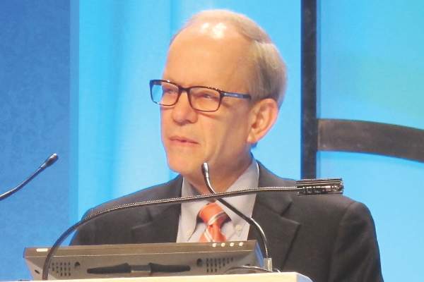
Nevi, Melanoma, and the Ongoing Argument on Atypia
In a case study published online on March 2 in JAMA Dermatology, Geller et al examined the relationship between total nevi, atypical nevi, and melanoma thickness. The study included 566 patients with melanoma. They were administered written surveys and underwent skin examinations at academic centers in Michigan and California within 3 months of diagnosis, measuring current total nevus count and atypical nevus count in addition to cataloguing melanoma thickness, histologic subtype, patient age, sex, marital status, skin self-examination and physician skin examination tendency, other health care visits, and mode of melanoma discovery.
Many epidemiologic trends were noted, but in summary, most melanoma patients had 0 to 20 total nevi (66.4%) and no atypical nevi (73.3%), a trend most pronounced in older patients (≥60 years). In patients younger than 60 years, higher nevus count (>50) was associated with thinner melanomas (≤2.0 mm), and the presence of more than 5 atypical nevi was associated with thicker melanomas (>2.0 mm).
What’s the issue?Studies clarifying the overall clinical characteristics of patients with aggressive melanomas appear every month in reputable journals, touting that concurrent total nevus count is important; or nevus size is important; or atypia is important; or clinical stigmata, medical history, and family history are important. Who is correct? Is everyone correct? On the pathology arm of the argument, Rosendahl et al (J Am Acad Dermatol. 2015;73:507-512) highlighted the same dilemma in which clinicians do not agree on the histopathologic features of nevi that consistently put patients at risk for individual lesion or de novo melanoma.
For me, each clinic day involves performing many total-body skin examinations, and many of these patients have innumerable nevi and various scars from lesions removed over the years with “atypical mole,” “premelanoma,” “precancer,” and various other self-reported labels. Some lesions may have documented pathology reports, but many do not. Some reports refer to dysplasia as a gradient, some do not. Some reports include molecular testing or clinical markers to grade lesions, and each can vary between institutions and pathologists. On the macroscopic level, clinically atypical nevi do not have a widely agreed upon set of criteria or threshold for biopsy; some clinicians use dermoscopic markers, and others utilize some version of the ABCDE (a=asymmetry; b=border; c=color; d=diameter; e=evolving) features.
The Geller et al study supports that these melanoma patients did not necessarily have more total nevi, and younger patients with aggressive melanoma may have a tendency toward more clinically atypical nevi. Although the study establishes what those institutions and clinicians determined to be atypical, I’m not sure that this is something that most clinicians widely agree upon. Additionally, these features were not paired with histopathologic dysplasia because the lesions were not biopsied.
What I find in conversation with colleagues is that some agree with what Geller et al defined as atypical, but some clinicians do not even refer to nevi as clinically atypical in a medical record unless they have pathology evidence of atypia (or the term their pathologist may use), which may be to avoid controversy regarding legal implications of atypia or “open-note” misunderstanding that the patient may have about this term, likening it to Papanicolaou test premalignancy verbiage.
I am not aware of one dermatologist or dermatopathologist who does not find this quandary to be frustrating. How do any of us really know which patients to follow more often for melanoma surveillance? How does your practice or institution report atypia in the clinical and histopathologic setting, and what do you find are the most important markers for development of melanoma?

Targeted therapy, immunotherapy may benefit patients with leptomeningeal metastases from melanoma
Patients with leptomeningeal metastases (LM) from melanoma have a poor prognosis, but treatment with targeted therapy and immune checkpoint inhibitors increased overall survival and resulted in long-term remission in a few patients, according to researchers.
The median survival for 21 patients who were treated with a BRAF inhibitor and/or ipilimumab was 21.7 weeks (range 2 to 235+ weeks); 24.9 weeks for treatment including a BRAF inhibitor (with and without a MEK inhibitor); 15.8 weeks for treatment including ipilimumab; and 4.3 weeks for radiotherapy (RT) only (Annals of Oncology 2016 March 8. doi: 10.1093/annonc/mdw134).
“A remarkable and encouraging new finding in our study is the long-term survival of patients who were treated with targeted treatment or immunotherapy. Moreover, the median survival of 22 weeks following these new therapies compares favorably to reported results of [intrathecal] chemotherapy for LM from melanoma,” wrote Dr. Marnix Geukes Foppen of Netherlands Cancer Institute, Amsterdam, and colleagues.
The retrospective analysis evaluated 39 consecutive patients treated at the Netherlands Cancer Institute during 2010-2015. Because of rapid disease progression or poor performance status, 14 patients did not receive treatment for LM, and their median survival was 2.9 weeks. Among 25 patients who were treated for LM, 15 received cranial or spinal RT and 21 received systemic therapy, and the median survival was 16.9 weeks.
Patients with performance status of 2 or 3 had worse survival than did those with performance status of 0 or 1 (3.6 vs. 18.8 weeks, P less than .001), and treatment did not significantly improve survival in this group (median 1.9 weeks for untreated vs. 3.9 weeks for treated). Patients with increased serum levels of lactate dehydrogenase and/or S100B had poorer survival compared with patients with normal levels.
The investigators noted that ipilimumab increases anti-tumor T cell activation in the lymph nodes, and activated T cells can cross the blood-brain or blood-CSF barrier. RT combined with ipilimumab may increase responses. Among 10 patients whose treatment included ipilimumab, median survival was 15.8 weeks; median survival was 47 weeks for ipilimumab with RT, and 6 weeks for ipilimumab without RT. A patient who had lymph node and lung metastasis, and LM without brain metastasis, was treated with whole brain RT and four cycles of ipilimumab, resulting in a complete radiological and clinical remission beyond 54 months.
Combining systemic targeted therapy and immunotherapy may result in long-term remissions, according the investigators.
“Especially in LM patients with a good performance score and low serum LDH and S100B levels these treatment options should be considered,” they wrote.
FROM ANNALS OF ONCOLOGY
Key clinical point: Leptomeningeal metastases from melanoma yield a poor prognosis, but patients may benefit from systemic targeted therapy and immunotherapy.
Major finding: Median survival for patients treated with systemic targeted therapy and/or immunotherapy (n = 21) was 21.7 weeks (range 2 to 235+ weeks).
Data source: Retrospective analysis of 39 consecutive patients treated at the Netherlands Cancer Institute during 2010-2015.
Disclosures: Dr. Geukes Foppen and coauthors reported having no disclosures.