User login
Specialty drug coupons - a double-edged sword of improved adherence, increased costs
While the availability of coupons can help defray out-of-pocket costs and improve access and adherence to high-cost specialty drugs, it may also result in higher costs to the health system.
Patients used drug coupons to pay for $21.2 million of $35.3 million in out-of-pocket costs, according to an analysis of 265,000 prescriptions worth $911 million in 2013. The coupons generally dropped a patient’s out-of-pocket costs to less than $250 per prescription, a point below which patients were “far less likely” to abandon prescriptions in cases of specialty anti-inflammatory drugs and treatments for multiple sclerosis (Health Affairs 2014;33:1761-9 [doi:10.1377/hlthaff.2014.0497]).
But while the use of coupons demonstrated that the lower cost led to lower prescription abandonment rates, it also allowed patients to circumvent cost-containment measures that pharmacy benefit managers employ.

“We found that drug coupons were extremely effective in lowering patients’ costs to less than $50 per prescription, thus eliminating the incentive to select a preferred drug,” wrote Catherine I. Starner, Pharm.D., senior health outcomes researcher at pharmacy benefit manager Prime Therapeutics, and her colleagues.
To control costs at the health system level, tighter controls could be placed on specialty drugs at the pharmacy benefit manager level, according to Dr. Starner and her colleagues. Such controls could include prior authorization or the use of step therapies,as well as excluding coverage of nonpreferred drugs. The latter strategy, however, could make drug access a problem, especially if physicians are not aware of an insurance plan taking these steps.
Excluding specialty drugs “makes the patient responsible for the entire cost of the drug,” the authors noted. “If prescribers are unaware of such exclusions, or if patients require specific therapies, such exclusions may impose considerable costs on patients” and potentially lead to access issues.
While the availability of coupons can help defray out-of-pocket costs and improve access and adherence to high-cost specialty drugs, it may also result in higher costs to the health system.
Patients used drug coupons to pay for $21.2 million of $35.3 million in out-of-pocket costs, according to an analysis of 265,000 prescriptions worth $911 million in 2013. The coupons generally dropped a patient’s out-of-pocket costs to less than $250 per prescription, a point below which patients were “far less likely” to abandon prescriptions in cases of specialty anti-inflammatory drugs and treatments for multiple sclerosis (Health Affairs 2014;33:1761-9 [doi:10.1377/hlthaff.2014.0497]).
But while the use of coupons demonstrated that the lower cost led to lower prescription abandonment rates, it also allowed patients to circumvent cost-containment measures that pharmacy benefit managers employ.
“We found that drug coupons were extremely effective in lowering patients’ costs to less than $50 per prescription, thus eliminating the incentive to select a preferred drug,” wrote Catherine I. Starner, Pharm.D., senior health outcomes researcher at pharmacy benefit manager Prime Therapeutics, and her colleagues.
To control costs at the health system level, tighter controls could be placed on specialty drugs at the pharmacy benefit manager level, according to Dr. Starner and her colleagues. Such controls could include prior authorization or the use of step therapies,as well as excluding coverage of nonpreferred drugs. The latter strategy, however, could make drug access a problem, especially if physicians are not aware of an insurance plan taking these steps.
Excluding specialty drugs “makes the patient responsible for the entire cost of the drug,” the authors noted. “If prescribers are unaware of such exclusions, or if patients require specific therapies, such exclusions may impose considerable costs on patients” and potentially lead to access issues.
FROM HEALTH AFFAIRS

Psoriasis for Seniors
We are always conscious of the use of systemic medications in older patients, as they may have the potential for more toxicity. For dermatologists, use of systemic medications in psoriasis represents a large share of our total use. Theoretically, we are concerned that older patients may be more likely to develop serious infections and malignancies. Older psoriasis patients also represent a growing demographic, and they are underrepresented in clinical trials.
In an article published online on September 3, 2014, in the Journal of the European Academy of Dermatology and Venereology, Medina et al performed an analysis to evaluate the safety of systemic psoriasis therapy in patients older than 65 years compared to younger patients. Patients registered in Biobadaderm, a Spanish national registry of psoriasis patients treated with systemic therapy, were divided into 2 categories: elderly (≥65 years) and younger patients (<65 years). Rates of adverse events (AEs) were described by severity and type. The risks were compared in both groups, taking into account exposure to classic or biologic drugs using Cox regression.
In the overall study population, 175 (9.8%) of 1793 patients were elderly. The authors found that overall risk for AEs was not higher in elderly patients; the drug group–adjusted hazard ratio (HR) was 1.09 (95% confidence interval [CI], 0.93-1.3). However, serious AEs were more common in older patients, with a drug group–adjusted HR of 3.2 (95% CI, 2.0-5.1). Age-adjusted HR of all AEs was lower for patients exposed to biologics compared to classic drugs in the whole sample (HR, 0.7 [95% CI, 0.6-0.7]). Age did not seem to modify the effect of therapy (biologic vs classic) in the risk for AEs (likelihood ratio test for interaction: P=.12 for all AEs; P=.09 for serious AEs).
The authors concluded that serious AEs were more common in elderly patients. They argued, however, that these serious AEs might be related to other factors associated with this age group and not due to the treatment itself. Use of biologics was associated with lower risk for AEs in the whole group. There were no differences in this association between young and old. These results are reassuring, though uncontrolled confounding could not be excluded as an explanation for these findings. The power of the study to detect differences was low.
What’s the issue?
The results indicate that overall there is no increase in AEs in older patients. More data will need to be evaluated to see if the incidence of serious AEs is due to medication use or endogenous health factors. How do you approach the use of systemic medications in older patients?

Reviewing the Medication List With Psoriasis Patients
For more information, access Dr. Weinberg's editorial from the September 2014 issue, "Under Pressure."
For more information, access Dr. Weinberg's editorial from the September 2014 issue, "Under Pressure."
For more information, access Dr. Weinberg's editorial from the September 2014 issue, "Under Pressure."
Apremilast approval expanded to include plaque psoriasis
The oral phosphodiesterase-4 inhibitor apremilast is now indicated for the treatment of moderate to severe plaque psoriasis.
On Sept. 23, the manufacturer, Celgene, announced that the Food and Drug Administration had approved the expanded indication for apremilast, which was initially approved in March 2014 for treating psoriatic arthritis. The new indication is for the treatment of patients with moderate to severe plaque psoriasis who are candidates for phototherapy or systemic therapy.
Approval was primarily based on the results of two multicenter, randomized, double-blind, placebo-controlled studies of adults with moderate to severe plaque psoriasis, according to Celgene. At 16 weeks, 33% and 29% of those randomized to the 30-mg, twice daily dose of apremilast had achieved at least a 75% reduction in the Psoriasis Area and Severity Index (PASI 75), compared with 5%-6% of those on placebo, according to the prescribing information.
The most common adverse events reported in studies, which affected at least 1% of treated patients and were more common than in patients on placebo, included diarrhea in 17% and nausea in 17%; other adverse events included upper respiratory infection, tension headache, and headache. The warnings and precautions section of the label includes the recommendation to be alert for the emergence or worsening of depression, suicidal thoughts, or other mood changes in patients treated with the drug, and to monitor weight regularly for significant weight loss. In studies, treatment with apremilast has been associated with an increase in reports of depression and significant weight loss.
Celgene markets apremilast as Otezla.
Serious adverse events associated with treatment should be reported to the FDA at 800-332-1088 or www.fda.gov/medwatch.
Information about the pregnancy registry of women exposed to the drug during pregnancy can be obtained at 877-311-8972.
The updated prescribing information is available at http://www.celgene.com/content/uploads/2014/09/psoriasis-pi.pdf
Recent Findings About Pustular Psoriasis
Therapies for Palmoplantar Pustular Psoriasis
There is limited evidence on therapies for palmoplantar pustular psoriasis (PPPP) and maintaining long-term remission. Sevrain et al (J Eur Acad Dermatol Venereol. 2014;28[suppl 5]:13-16) conducted a systematic literature search to identify trials in patients with PPPP that assessed therapeutic interventions (not including systemic biotherapy). They found that oral retinoid therapy (acitretin), photochemotherapy, or a combination of both; low-dose cyclosporine; or topical corticosteroids under occlusion appeared to help relieve symptoms of PPPP. Newer studies have noted the benefits of psoralen plus UVA, with better efficacy compared to UVB therapy. A 308-nm excimer laser has been shown to improve PPPP.
Practice Point: Although phototherapy, cyclosporine, and topical corticosteroids seem to be able to control PPPP, more randomized controlled trials are needed to better define treatment strategies for PPPP.
>>Read more at Journal of the European Academy of Dermatology and Venereology
Pediatric Pustular Psoriasis Cleared With Systemic Medications
Treatment of pediatric pustular psoriasis is challenging. Posso-De Los Rios and colleagues (Pediatr Dermatol. 2014;31:430-439) conducted a systematic review of systemic interventions for pediatric pustular psoriasis with a focus on clinical response and treatment outcomes. The most common therapies used for generalized pustular psoriasis were acitretin, cyclosporine, and methotrexate.
Practice Point: Acitretin, methotrexate, and cyclosporine seem to control generalized pustular psoriasis in the pediatric population; however, long-term data are lacking and physicians may encounter disease regression after discontinuing or tapering the medications.
>>Read more at Pediatric Dermatology
Biologics for Erythrodermic and Pustular Psoriasis
Levin et al (J Drugs Dermatol. 2014;13:342-354) sought to evaluate the efficacy and safety of biologics in the treatment of erythrodermic and generalized pustular psoriasis by reviewing the literature. In both psoriasis subtypes, infliximab was used to treat more than half of the reported cases. Other biologics that were successfully used included etanercept, ustekinumab, adalimumab, and anakinra.
Practice Point: Patients with erythrodermic or generalized pustular psoriasis may experience disease improvement with biologics.
>>Read more at Journal of Drugs in Dermatology
Minimal disease activity in psoriatic arthritis is a realistic target
LAS VEGAS – A number of drugs are showing promise for the treatment of psoriatic arthritis, but broader improvements are needed with respect to treatment strategies, according to Dr. Iain McInnes.
Earlier diagnosis, appropriate therapies, and treating to target are among the topics he covered in a presentation on maximizing patient outcomes at the annual Perspectives in Rheumatic Diseases.
A recent study demonstrated that even a 6-month delay in the diagnosis of psoriatic arthritis is associated with worse long-term outcomes on several measures. In that study of 283 patients (Ann. Rheum. Dis. 2014 Feb. 13 [doi: 10.1136/annrheumdis-2013-204858]), the median lag time from disease onset to first rheumatological assessment was 1 year, with only 30% of patients being seen by a rheumatologist within 6 months.

Multiple stepwise regression analysis showed that late consulters had a greater risk of developing peripheral joint erosions (odds ratio, 4.25) and had worse Health Assessment Questionnaire scores (odds ratio, 2.2).
One finding that could help with earlier diagnosis is detection of early entheseal involvement, said Dr. McInnes, professor of medicine and director of the Institute of Infection, Immunity and Immunology at the University of Glasgow (Scotland).
Subclinical entheseal involvement appears to predict psoriatic arthritis in patients presenting with psoriasis, he said at the conference.
In one study, the mean Glasgow Ultrasound Enthesitis Scoring System (GUESS) score was 7.9 in patients with psoriasis, compared with 2.9 in controls, and in another study, thickness of the quadriceps tendon was a significant predictor of the development of psoriatic arthritis in those with psoriasis.
A key challenge in maximizing outcomes in psoriatic arthritis is that rheumatoid arthritis and psoriatic arthritis are typically treated as homogeneous for the purposes of drug development, but the two diseases have distinct clinical phenotypes and time course, tissue distribution, tissue cell/molecular features, genetic susceptibility, and probably impact of immune senescence.
In fact, while disease-modifying antirheumatic drugs are included in European League Against Rheumatsm (EULAR) treatment guidelines for psoriatic arthritis, the Methotrexate in Psoriatic Arthritis (MIPA) trial, a 6-month, double-blind, randomized, placebo-controlled trial of methotrexate, showed no benefit for key psoriatic arthritis endpoints at 3 and 6 months (Rheumatology 2012;51:1368-77).
Data on methotrexate combination therapy is limited, but existing clinical trials and registry data show no clear evidence of benefit. Methotrexate also has long-term toxicity issues.
In studies of patients who received anti–tumor necrosis factor therapy, 59%-69% of patients achieved 20% improvement (ACR 20 response), 37%-50% achieved 50% improvement (ACR 50 response), and up to 52% of patients achieved minimal disease activity at various time points. Also, radiographic progression was improved with anti-TNF therapy vs. placebo. However, TNF inhibitors lose efficacy over time in some patients, and are associated with an increased risk of infection.
Other disappointments or unmet needs in psoriatic arthritis are dichotomous skin and joint responses to biologics, lack of established predictors of response to biologics, lack of a cure, and difficulties in achieving sustained remission, Dr. McInnes said.
Future therapeutic options, including several drugs in development, include drugs that target cytokines and their receptors, such as ustekinumab, which targets IL-12/23; secukinumab and ixekizumab, which target IL-17; and brodalumab, which targets IL-17 receptor A. Other potential targets include IL-6 and the IL-6 receptor and IL-23 and the IL-23 receptor.
Studies of ustekinumab, for example, demonstrate beneficial effects on enthesitis, dactylitis, and modified van der Heijde-Sharp score.
Drugs that target intracellular signaling pathways are also being studied in psoriatic arthritis, including JAK family agents such as tofacitinib and phosphodiesterase inhibitors like apremilast.
As for strategies to improve outcomes, the Tight Control in Psoriatic Arthritis (TICOPA) study provided a rationale for treating to target. Current recommendations call for a target of remission or low disease activity.
The availability of highly effective biologics makes minimal level of disease activity a realistic treatment target, Dr. McInnes said.
The conference was held by Global Academy for Medical Education. Global Academy for Medical Education and this news organization are owned by Frontline Medical Communications.
Dr. McInnes reported receiving research/grant support from Pfizer, Bristol-Myers Squibb, AstraZeneca, and Novo Nordisk, and serving as a consultant to Novartis, Bristol-Myers Squibb, Pfizer, and Galapagos. He is a member of scientific advisory boards for AstraZeneca, Crescendo Bioscience, and Janssen Biotech.
LAS VEGAS – A number of drugs are showing promise for the treatment of psoriatic arthritis, but broader improvements are needed with respect to treatment strategies, according to Dr. Iain McInnes.
Earlier diagnosis, appropriate therapies, and treating to target are among the topics he covered in a presentation on maximizing patient outcomes at the annual Perspectives in Rheumatic Diseases.
A recent study demonstrated that even a 6-month delay in the diagnosis of psoriatic arthritis is associated with worse long-term outcomes on several measures. In that study of 283 patients (Ann. Rheum. Dis. 2014 Feb. 13 [doi: 10.1136/annrheumdis-2013-204858]), the median lag time from disease onset to first rheumatological assessment was 1 year, with only 30% of patients being seen by a rheumatologist within 6 months.
Multiple stepwise regression analysis showed that late consulters had a greater risk of developing peripheral joint erosions (odds ratio, 4.25) and had worse Health Assessment Questionnaire scores (odds ratio, 2.2).
One finding that could help with earlier diagnosis is detection of early entheseal involvement, said Dr. McInnes, professor of medicine and director of the Institute of Infection, Immunity and Immunology at the University of Glasgow (Scotland).
Subclinical entheseal involvement appears to predict psoriatic arthritis in patients presenting with psoriasis, he said at the conference.
In one study, the mean Glasgow Ultrasound Enthesitis Scoring System (GUESS) score was 7.9 in patients with psoriasis, compared with 2.9 in controls, and in another study, thickness of the quadriceps tendon was a significant predictor of the development of psoriatic arthritis in those with psoriasis.
A key challenge in maximizing outcomes in psoriatic arthritis is that rheumatoid arthritis and psoriatic arthritis are typically treated as homogeneous for the purposes of drug development, but the two diseases have distinct clinical phenotypes and time course, tissue distribution, tissue cell/molecular features, genetic susceptibility, and probably impact of immune senescence.
In fact, while disease-modifying antirheumatic drugs are included in European League Against Rheumatsm (EULAR) treatment guidelines for psoriatic arthritis, the Methotrexate in Psoriatic Arthritis (MIPA) trial, a 6-month, double-blind, randomized, placebo-controlled trial of methotrexate, showed no benefit for key psoriatic arthritis endpoints at 3 and 6 months (Rheumatology 2012;51:1368-77).
Data on methotrexate combination therapy is limited, but existing clinical trials and registry data show no clear evidence of benefit. Methotrexate also has long-term toxicity issues.
In studies of patients who received anti–tumor necrosis factor therapy, 59%-69% of patients achieved 20% improvement (ACR 20 response), 37%-50% achieved 50% improvement (ACR 50 response), and up to 52% of patients achieved minimal disease activity at various time points. Also, radiographic progression was improved with anti-TNF therapy vs. placebo. However, TNF inhibitors lose efficacy over time in some patients, and are associated with an increased risk of infection.
Other disappointments or unmet needs in psoriatic arthritis are dichotomous skin and joint responses to biologics, lack of established predictors of response to biologics, lack of a cure, and difficulties in achieving sustained remission, Dr. McInnes said.
Future therapeutic options, including several drugs in development, include drugs that target cytokines and their receptors, such as ustekinumab, which targets IL-12/23; secukinumab and ixekizumab, which target IL-17; and brodalumab, which targets IL-17 receptor A. Other potential targets include IL-6 and the IL-6 receptor and IL-23 and the IL-23 receptor.
Studies of ustekinumab, for example, demonstrate beneficial effects on enthesitis, dactylitis, and modified van der Heijde-Sharp score.
Drugs that target intracellular signaling pathways are also being studied in psoriatic arthritis, including JAK family agents such as tofacitinib and phosphodiesterase inhibitors like apremilast.
As for strategies to improve outcomes, the Tight Control in Psoriatic Arthritis (TICOPA) study provided a rationale for treating to target. Current recommendations call for a target of remission or low disease activity.
The availability of highly effective biologics makes minimal level of disease activity a realistic treatment target, Dr. McInnes said.
The conference was held by Global Academy for Medical Education. Global Academy for Medical Education and this news organization are owned by Frontline Medical Communications.
Dr. McInnes reported receiving research/grant support from Pfizer, Bristol-Myers Squibb, AstraZeneca, and Novo Nordisk, and serving as a consultant to Novartis, Bristol-Myers Squibb, Pfizer, and Galapagos. He is a member of scientific advisory boards for AstraZeneca, Crescendo Bioscience, and Janssen Biotech.
EXPERT ANALYSIS FROM THE ANNUAL PERSPECTIVES IN RHEUMATIC DISEASES

Calcipotriene–Betamethasone Dipropionate Topical Suspension in the Management of Psoriasis: A Status Report on Available Data With an Overview of Practical Clinical Application
Psoriasis is a common inflammatory skin disorder that appears to be induced by multifactorial pathophysiologic processes associated with immunologic dysregulation.1 It can affect patients of any age, gender, and ethnicity, and it presents clinically with a variety of visible manifestations. The disease course and severity of psoriasis varies among affected patients.1 Chronic plaque psoriasis (PP), also referred to as psoriasis vulgaris, is the most common clinical presentation.1,2 Although many patients are affected by psoriasis that is widespread and in some cases severe, the majority of affected patients exhibit localized involvement that usually affects less than 2% to 5% of the body surface area. Although the skin at any anatomic location can be affected, commonly involved sites are described by the mnemonic term SNAKES (scalp, nails, anogenital region, knees, elbows, sacral region).2,3
Because the majority of patients with PP present with localized disease, topical therapy is the foundation of treatment in most cases. Topical corticosteroids (TCs) are the most commonly utilized agents, supported by a long track record of favorable efficacy and safety over approximately 6 decades.4,5 However, optimal management of PP with TCs requires use of a formulation that is of adequate potency, is adaptable for application to the affected body sites, and is properly monitored and adjusted to avoid potential TC-induced adverse effects.4-6 Nonsteroidal topical therapies such as vitamin D analogues (eg, calcipotriene) and retinoids (eg, tazarotene) are commonly integrated into topical regimens to reduce the application frequency and duration of TC use as well as to sustain efficacy.5,7,8 Plaque psoriasis is characteristically a chronic disease associated with periods of persistence and episodes of flaring; therefore, intermittent use of TC therapy along with concurrent or sequential use of a nonsteroidal topical agent are commonly employed to achieve and sustain control of the disorder.7-9
In the last decade, several advances have revolutionized the management of psoriasis, especially for PP patients with extensive involvement who require systemic therapy and/or phototherapy as well as for those with psoriatic arthritis.10,11 The availability of biologic agents such as tumor necrosis factor a inhibitors and certain interleukin inhibitors (eg, IL-12/IL-23) have been at the forefront of major advances in PP treatment, with some agents also blocking the progression of joint destruction associated with psoriatic arthritis.10-12 However, even when patients with PP respond favorably to biologic therapy, it is not uncommon for them to still be affected by some persistent PP. In these cases, although much of the chronic PP may clear with use of the biologic agent, persistence of psoriatic plaques may involve the lower extremities, scalp, and/or trunk, with topical therapy often added to augment the therapeutic response.13-15
This article provides a review of a patented topical suspension combination formulation that contains calcipotriene hydrate 0.005%, a vitamin D analogue, and betamethasone dipropio-nate (Bd) 0.064%, a high-potency TC. In 2008, the US Food and Drug Administration approved the once-daily application of calcipotriene 0.005%–Bd 0.064% topical suspension (C/Bd-TS) for the treatment of PP; this formulation is approved for use on the scalp and body in patients 18 years of age and older. According to the product insert, the recommended maximum duration of treatment with C/Bd-TS once daily is 8 weeks, and patients may not exceed a maximum weekly dose of 100 g.16 It is important to note that the terms calcipotriene and calcipotriol refer to the same molecule and are used interchangeably in the literature. Formulation characteristics of C/Bd-TS, perspectives on modes of action, outcomes from pivotal trials, and efficacy and safety data reported from additional studies are discussed in this article.
What are the formulation characteristics of C/Bd-TS?
Each gram of C/Bd-TS contains 52.18 mg of calcipotriene hydrate (equivalent to 50 µg of calcipotriene) and 0.643 µg of Bd (equivalent to 0.5 mg of betamethasone), formulated together in a viscous, nearly odorless, almost clear to slightly off-white suspension. The excipients are hydrogenated castor oil, polypropylene glycol 11 stearyl ether, α-tocopherol, butylhydroxytoluene, and mineral oil, collectively producing a gel base in which both active ingredients are suspended.16 Although the viscous quality of the suspension warrants some additional effort for removal during hair washing, the tenacious gel-like viscosity assists in removing scale on psoriatic plaques, which is often adherent, especially on the scalp. Additionally, it is important that C/Bd-TS be shaken well before use.16 Initially, C/Bd-TS was studied and marketed in the United States for treatment of scalp psoriasis; however, the indication was expanded to include treatment of PP on the rest of the body, supported by evidence from randomized controlled trials (RCTs).16-23
Vitamin D analogues (eg, calcipotriene/calcitriol) have been shown to be photolabile when exposed to UV light, especially UVA. They also have been shown to be chemically incompatible and less stable when admixed with a variety of other active ingredients and/or vehicles used to treat PP, including hydrocortisone valerate ointment 0.2%, ammonium lactate lotion 12%, and salicylic acid compound ointment 6%.24-26 As a result, it is important for clinicians to consider avoidance of concomitant topical calcipotriene application with use of a TC unless the stability of the active ingredients has been tested when the formulations are combined. Calcipotriene 0.005%/Bd 0.064% topical suspension utilizes vehicle technology that maintains the stability and activity of both calcipotriene and Bd within the suspension formulation.16,26
What is the rationale behind combining calcipotriene and Bd in a single formulation for the treatment of PP?
The potential advantages of C/Bd-TS include the combined modes of action of 2 different active ingredients used for treatment of PP, complementary immunomodulatory effects as compared to use of a TC or vitamin D analogue alone, ease of use with a single product applied once daily, adaptability of the vehicle for use on scalp and/or body skin, and improvement in quality-of-life (QOL) measures.27-34
Combined Modes of Action
Calcipotriene 0.005%–Bd 0.064% topical suspension combines the modes of action of a high-potency topical suspension and a vitamin D analogue for the treatment of PP in a single stable gel formulation that is approved in the United States for treatment of PP in adults.16 The multiple anti-inflammatory properties of corticosteroids as well as the efficacy and safety of TC therapy for psoriasis have been well described.4,6,7,9,27 The antiproliferative and anti-inflammatory properties of vitamin D analogues that appear to correlate with therapeutic effects in the treatment of PP also have been discussed in the literature.28
Complementary Immunomodulatory Effects
More recent studies using various research assays have provided further evidence supporting relevant immunomodulatory properties of calcipotriene alone and in combination with Bd that favorably modify immune dysregulation pathways described more recently in the pathogenesis of PP.1,29,30 Treatment of psoriatic plaques with calcipotriene has been shown to suppress the increased production of peptide alarmins (psoriasin and koebnerisin) in psoriatic skin and their TH17-mediated regulation in epidermal ke-ratinocytes, thus interfering with the S100 amplification loop that appears to produce inflammation in psoriasis.29 In T-lymphocyte cultures evaluating exposure to calcipotriene and Bd both alone and as a combined therapy, calcipotriene inhibited IFN-g, IL-8, IL-17, and IL-22 expression, and it reversed the corticosteroid-induced suppression of IL-4, IL-5, IL-10, and IL-13; Bd inhibited both IL-6 and tumor necrosis factor α expression. The outcomes demonstrated that the combination of calcipotriene and Bd inhibited the endogenous release of TH1- and TH17-associated cytokines that are associated with psoriatic inflammation and together induced a more favorable anti-inflammatory cytokine profile.30 Although the broad range of anti-inflammatory effects provided by a TC of adequate potency, such as Bd, can clear or markedly improve PP, the concurrent use of calcipotriene was shown to provide additional immunomodulatory effects that suppressed the key TH17/TH1 pathophysiologic mediators of psoriatic inflammation and simultaneously induced a TH2/T regulatory response that is believed to provide therapeutic benefit.29,30
Ease of Use and Vehicle Adaptability
A once-daily regimen and a vehicle formulation adaptable for use on both the scalp and body are advantageous in enhancing the potential for greater patient adherence.31,32 The adaptability of the C/Bd-TS for use on the scalp and/or body is supported by several studies encompassing a large number of actively treated subjects. Calcipotriene 0.005%–Bd 0.064% topical suspension has been extensively studied in patients with PP on the scalp and/or body as evidenced by a pooled analysis of 9 eight-week RCTs (scalp, n=6; body, n=3) that encompassed 2777 total subjects treated once daily for PP (scalp, n=1953; body, n=824).23 Additionally, C/Bd-TS applied once daily was evaluated in an open-label, single-arm, 8-week, phase 2 study of adolescents (N=78; age range, 12–17 years [mean age, 14.6 years]) with scalp psoriasis (mean affected scalp area, 43.7%). The investigator global assessment of treatment success (clear or almost clear) and the patient global assessment of treatment success (clear or very mild) were essentially identical among participants and investigators with 85% and 87% reported after 8 weeks, respectively; approximately 50% of participants achieved treatment success after 2 weeks based on both the investigator global assessment and patient global assessment.33
Improvement in QOL Measures
Quality-of-life measures were compared in an 8-week RCT of participants with at least moderate scalp psoriasis treated with C/Bd-TS once daily (n=207) or calcipotriene solution twice daily (n=107). Significantly greater improvement in QOL scores compared to baseline were noted at all time points using the Skindex-16 questionnaire in participants treated with C/Bd-TS compared to calcipotriene solution (total score, P<.001 at weeks 2 and 4 and P=.008 at week 8; symptoms score, P<.001 at weeks 2 and 4 and P=.004 at week 8; emotions score, P<.001 at weeks 2 and 4 and P=.005 at week 8).34 A 4-week, open-label, noninterventional cohort, postmarketing (“real life”) study of 721 patients treated at 333 dermatology centers with C/Bd-TS showed a 69.5% improvement in the scalp life quality index score compared to baseline (P<.0001), with 89.5% and 90.4% of participants reporting that C/Bd-TS was better/much better than previously used therapies for scalp psoriasis and easy/very easy to use, respectively.35 An 8-week RCT trial evaluated C/Bd-TS once daily compared to calcipotriene alone, betamethasone dipropionate alone, and vehicle in 1152 participants with mild to moderate PP involving the trunk and extremities. Participants treated with C/Bd-TS (n=442) demonstrated superior reductions in QOL scores using the dermatology life quality index at weeks 4 and 8 compared to those treated with Bd alone (n=418) or vehicle (n=77) but not compared to calcipotriene alone (n=80).19
What data are available on the efficacy and safety of C/Bd-TS?
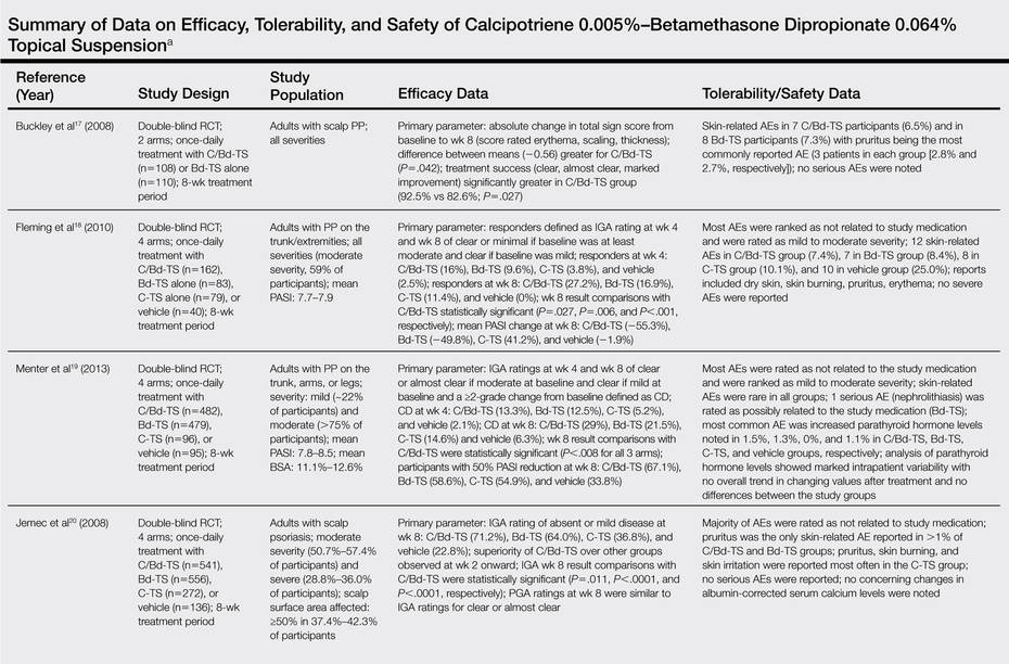
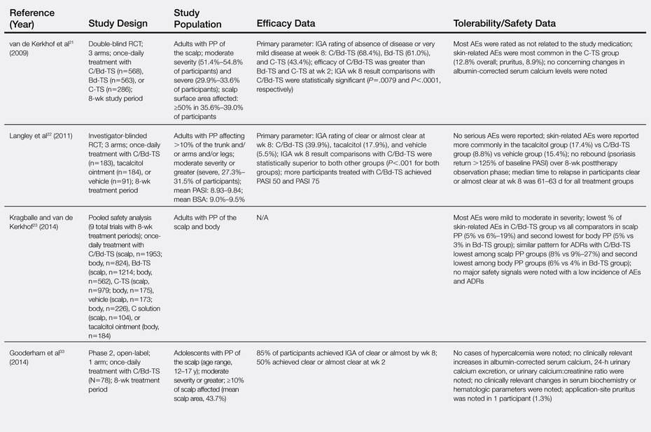
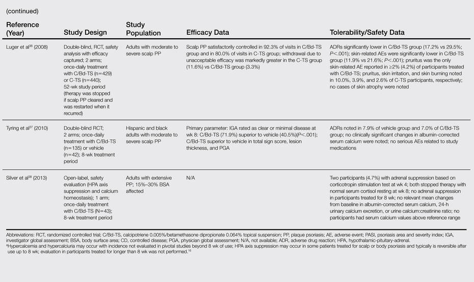
Several clinical studies have evaluated the efficacy, tolerability, and safety of C/Bd-TS applied once daily for PP of the scalp and body (ie, trunk, extremities). Most studies were completed over a duration of 8 weeks in adults16-23; however, studies also have been performed in adolescents,33 in adults treated for up to 52 weeks,36 and in a subgroup of Hispanic/Latino and black/African American patients with scalp psoriasis.37 The Table provides a detailed summary of primary efficacy data along with important tolerability and safety considerations based on study outcomes.
What practical recommendations can be made regarding the use of C/Bd-TS for PP?
Calcipotriene 0.005%–Bd 0.064% topical suspension applied once daily provides a formulation that allows for treatment of PP involving both the scalp and body using a single product, which provides an element of convenience and is likely to enhance compliance and reduce costs compared with the use of 2 separate products. The efficacy and safety of C/Bd-TS has been well established in several studies,16-23,37 including a 52-week trial in patients with scalp psoriasis.36 The combination of calcipotriene and Bd appears to favorably address the pathophysiologic pathways involved in psoriasis.29,30 Calcipotriene 0.005%–Bd 0.064% topical suspension is a rational option for the treatment of PP in patients with localized disease or in patients treated systemically or with phototherapy for more extensive disease who exhibit persistence or recurrence of scattered areas of PP.6-9,14,15 Appropriate use of C/Bd-TS is likely to achieve favorable efficacy with a low risk of tolerability reactions and a very low risk of major adverse drug reactions.16-23,36-38
1. Guttman-Yassky E, Krueger JG. Psoriasis: evolution of pathogenic concepts and new therapies through phases of translational research. Br J Dermatol. 2007;157:1103-1115.
2. Menter MA. An overview of psoriasis. In: Koo JYM, Levin EC, Leon A, et al, eds. Moderate to Severe Psoriasis. 4th ed. Boca Raton, FL: CRC Press Taylor & Francis Group; 2014:1-21.
3. Sandoval LF, Feldman SR. General approach to psoriasis treatment. In: Koo JYM, Levin EC, Leon A, et al, eds. Moderate to Severe Psoriasis. 4th ed. Boca Raton, FL: CRC Press Taylor & Francis Group; 2014:3-8.
4. Zeichner JA, Lebwohl MG, Menter A, et al. Optimizing topical therapies for treating psoriasis: a consensus conference. Cutis. 2010;(3 suppl):5-31.
5. American Academy of Dermatology Work Group, Menter A, Korman NJ, et al. Guidelines of care for the management of psoriasis and psoriatic arthritis: section 6. guidelines of care for the treatment of psoriasis and psoriatic arthritis: case-based presentations and evidence-based conclusions [published online ahead of print February 8, 2011]. J Am Acad Dermatol. 2011;65:137-174.
6. Del Rosso J, Friedlander SF. Corticosteroids: options in the era of steroid-sparing therapy. J Am Acad Dermatol. 2005;53(1, suppl 1):S50-S58.
7. Menter A, Korman NJ, Elmets CA, et al; American Academy of Dermatology. Guidelines of care for the management of psoriasis and psoriatic arthritis. section 3. guidelines of care for the management and treatment of psoriasis with topical therapies [published online ahead of print February 13, 2009]. J Am Acad Dermatol. 2009;60:643-659.
8. Koo J. New developments in topical sequential therapy for psoriasis. Skin Therapy Lett. 2005;10:1-4.
9. Mason AR, Mason J, Cork M, et al. Topical treatments for chronic plaque psoriasis. Cochrane Database Syst Rev. 2009;2:CD005028.
10. Belge K, Brück J, Ghoreschi K. Advances in treating psoriasis. F1000Prime Rep. 2014;6:4.
11. Mease PJ, Armstrong AW. Managing patients with psoriatic disease: the diagnosis and pharmacologic treatment of psoriatic arthritis in patients with psoriasis. Drugs. 2014;74:423-441.
12. Boehncke WH, Qureshi A, Merola JF, et al. Diagnosing and treating psoriatic arthritis: an update. Br J Dermatol. 2014;170:772-786.
13. Feldman SR. Effectiveness of clobetasol propionate spray 0.05% added to other stable treatments: add-on therapy in the COBRA trial. Cutis. 2007;80(suppl 5):20-28.
14. Feldman SR, Gelfand JM, Stein Gold L, et al. The role of topical therapy for patients with extensive psoriasis. Cutis. 2007;79(suppl 1[ii]):18-31.
15. Kircik L. Topical calcipotriene 0.005% and betamethasone dipropionate 0.064% maintains efficacy of etanercept after step-down dose in patients with moderate-to-severe plaque psoriasis: results of an open label trial. J Drugs Dermatol. 2011;10:878-882.
16. Taclonex [product insert]. Parsippany, NJ: LEO Pharma Inc; 2014.
17. Buckley C, Hoffmann V, Shapiro J, et al. Calcipotriol plus betamethasone dipropionate scalp formulation is effective and well tolerated in the treatment of scalp psoriasis: a phase II study. Dermatology. 2008;217:107-113.
18. Fleming C, Ganslandt C, Guenther L, et al. Calcipotriol plus betamethasone dipropionate gel compared with its active components in the same vehicle and the vehicle alone in the treatment of psoriasis vulgaris: a randomized, parallel group, double-blind, exploratory study. Eur J Dermatol. 2010;20:465-471.
19. Menter A, Stein Gold L, Bukhalo M, et al. Calcipotriene plus betamethasone dipropionate topical suspension for the treatment of mild to moderate psoriasis vulgaris on the body: a randomized, double-blind, vehicle-controlled trial. J Drugs Dermatol. 2013;12:92-98.
20. Jemec GBE, Ganslandt C, Ortonne JP, et al. A new scalp formulation of calcipotriene plus betamethasone compared with its active ingredients and the vehicle in the treatment of scalp psoriasis: a randomized, double-blind, controlled trial. J Am Acad Dermatol. 2008;59:455-463.
21. van de Kerkhof PC, Hoffmann V, Anstey A, et al. A new scalp formulation of calcipotriol plus betamethasone dipropionate compared with each of its active ingredients in the same vehicle for the treatment of scalp psoriasis: a randomized, double-blind, controlled trial. Br J Dermatol. 2009;160:170-176.
22. Langley RG, Gupta A, Papp K, et al. Calcipotriol plus betamethasone dipropionate gel compared with tacalcitol ointment and the gel vehicle alone in patients with psoriasis vulgaris: a randomized, controlled clinical trial. Dermatology. 2011;222:148-156.
23. Kragballe K, van de Kerkhof P. Pooled safety analysis of calcipotriol plus betamethasone dipropionate gel for the treatment of psoriasis on the body and scalp. J Eur Acad Dermatol Venereol. 2014;28:10-21.
24. Lebwohl MG, Corvari L. Compatibility of topical therapies for psoriasis: challenges and innovations. Cutis. 2007;79(suppl 1[ii]):5-10.
25. Patel B, Siskin S, Krazmien R, et al. Compatibility of calcipotriene with other topical medications. J Am Acad Dermatol. 1998;38(6, pt 1):1010-1011.
26. Traulsen J. Bioavailability of betamethasone dipropionate when combined with calcipotriol. Int J Dermatol. 2004;43:611-617.
27. Sandoval LF, Feldman SR. Topical corticosteroids. In: Koo JYM, Levin EC, Leon A, et al, eds. Moderate to Severe Psoriasis. 4th ed. Boca Raton, FL: CRC Press Taylor & Francis Group; 2014:21-36.
28. deShazo R, Krueger GG, Duffin KC. Topical agents. In: Koo JYM, Levin EC, Leon A, et al, eds. Moderate to Severe Psoriasis. 4th ed. Boca Raton, FL: CRC Press Taylor & Francis Group; 2014:41-65.
29. Hegyi Z, Zwicker S, Bureik D, et al. Vitamin D analog calcipotriol suppresses the Th17 cytokine-induced proinflammatory S100 “alarmins” psoriasin (S100A7) and koebnerisin (S100A15) in psoriasis. J Invest Dermatol. 2012;132:1416-1424.
30. Lovato P, Norsgaard H, Ropke M. Key immunomodulatory effects exerted by calcipotriol in combination with corticosteroid on human cells. Poster presented at: 21st European Academy of Dermatology and Venereology Congress; September 27-30, 2012; Prague, Czech Republic.
31. Renton C. Diagnosis and treatment of adults with scalp psoriasis. Nurs Stand. 2014;28:35-39.
32. Feldman SR, Housman TS. Patients’ vehicle preference for corticosteroid treatments of scalp psoriasis. Am J Clin Dermatol. 2003;4:221-224.
33. Gooderham M, Debarre JM, Keddy-Grant J, et al. Safety and efficacy of calcipotriene plus betamethasone dipropionate topical suspension in adolescents with scalp psoriasis: an open, non-controlled, 8-week trial. Poster presented at: American Academy of Dermatology 72nd Annual Meeting; March 21-25, 2014; Denver, CO.
34. Ortonne JP, Tan J, Nordin P, et al. Quality of life of patients with scalp psoriasis treated with calcipotriene plus betamethasone dipropionate gel compared to calcipotriene solution. J Am Academy Dermatol. 2008;58(2, suppl 2):AB134.
35. Mrowietz U, Macheleidt O, Eicke C. Effective treatment and improvement of quality of life in patients with scalp psoriasis by topical use of calcipotriol/betamethasone (Xamiol®-gel): results [in German]. J Dtsch Dermatol Ges. 2011;9:825-831.
36. Luger TA, Cambazard F, Larsen FG, et al. A study of the safety and efficacy of calcipotriol and betamethasone dipropionate scalp formulation in the long-term management of scalp psoriasis. Dermatology. 2008;217:321-328.
37. Tyring S, Mendoza N, Appell M, et al. A calcipotriene/betamethasone dipropionate two-compound scalp formulation in the treatment of scalp psoriasis in Hispanic/Latino and Black/African American patients: results of the randomized, 8-week, double-blind phase of a clinical trial. Int J Dermatol. 2010;49:1328-1333.
38. Silver S, Tuppal R, Gupta AK, et al. Effect of calcipotriene plus betamethasone dipropionate topical suspension on the hypothalamic-pituitary-adrenal axis and calcium homeostasis in subjects with extensive psoriasis vulgaris: an open, non-controlled, 8-week trial. J Drugs Dermatol. 2013;12:882-888.
Psoriasis is a common inflammatory skin disorder that appears to be induced by multifactorial pathophysiologic processes associated with immunologic dysregulation.1 It can affect patients of any age, gender, and ethnicity, and it presents clinically with a variety of visible manifestations. The disease course and severity of psoriasis varies among affected patients.1 Chronic plaque psoriasis (PP), also referred to as psoriasis vulgaris, is the most common clinical presentation.1,2 Although many patients are affected by psoriasis that is widespread and in some cases severe, the majority of affected patients exhibit localized involvement that usually affects less than 2% to 5% of the body surface area. Although the skin at any anatomic location can be affected, commonly involved sites are described by the mnemonic term SNAKES (scalp, nails, anogenital region, knees, elbows, sacral region).2,3
Because the majority of patients with PP present with localized disease, topical therapy is the foundation of treatment in most cases. Topical corticosteroids (TCs) are the most commonly utilized agents, supported by a long track record of favorable efficacy and safety over approximately 6 decades.4,5 However, optimal management of PP with TCs requires use of a formulation that is of adequate potency, is adaptable for application to the affected body sites, and is properly monitored and adjusted to avoid potential TC-induced adverse effects.4-6 Nonsteroidal topical therapies such as vitamin D analogues (eg, calcipotriene) and retinoids (eg, tazarotene) are commonly integrated into topical regimens to reduce the application frequency and duration of TC use as well as to sustain efficacy.5,7,8 Plaque psoriasis is characteristically a chronic disease associated with periods of persistence and episodes of flaring; therefore, intermittent use of TC therapy along with concurrent or sequential use of a nonsteroidal topical agent are commonly employed to achieve and sustain control of the disorder.7-9
In the last decade, several advances have revolutionized the management of psoriasis, especially for PP patients with extensive involvement who require systemic therapy and/or phototherapy as well as for those with psoriatic arthritis.10,11 The availability of biologic agents such as tumor necrosis factor a inhibitors and certain interleukin inhibitors (eg, IL-12/IL-23) have been at the forefront of major advances in PP treatment, with some agents also blocking the progression of joint destruction associated with psoriatic arthritis.10-12 However, even when patients with PP respond favorably to biologic therapy, it is not uncommon for them to still be affected by some persistent PP. In these cases, although much of the chronic PP may clear with use of the biologic agent, persistence of psoriatic plaques may involve the lower extremities, scalp, and/or trunk, with topical therapy often added to augment the therapeutic response.13-15
This article provides a review of a patented topical suspension combination formulation that contains calcipotriene hydrate 0.005%, a vitamin D analogue, and betamethasone dipropio-nate (Bd) 0.064%, a high-potency TC. In 2008, the US Food and Drug Administration approved the once-daily application of calcipotriene 0.005%–Bd 0.064% topical suspension (C/Bd-TS) for the treatment of PP; this formulation is approved for use on the scalp and body in patients 18 years of age and older. According to the product insert, the recommended maximum duration of treatment with C/Bd-TS once daily is 8 weeks, and patients may not exceed a maximum weekly dose of 100 g.16 It is important to note that the terms calcipotriene and calcipotriol refer to the same molecule and are used interchangeably in the literature. Formulation characteristics of C/Bd-TS, perspectives on modes of action, outcomes from pivotal trials, and efficacy and safety data reported from additional studies are discussed in this article.
What are the formulation characteristics of C/Bd-TS?
Each gram of C/Bd-TS contains 52.18 mg of calcipotriene hydrate (equivalent to 50 µg of calcipotriene) and 0.643 µg of Bd (equivalent to 0.5 mg of betamethasone), formulated together in a viscous, nearly odorless, almost clear to slightly off-white suspension. The excipients are hydrogenated castor oil, polypropylene glycol 11 stearyl ether, α-tocopherol, butylhydroxytoluene, and mineral oil, collectively producing a gel base in which both active ingredients are suspended.16 Although the viscous quality of the suspension warrants some additional effort for removal during hair washing, the tenacious gel-like viscosity assists in removing scale on psoriatic plaques, which is often adherent, especially on the scalp. Additionally, it is important that C/Bd-TS be shaken well before use.16 Initially, C/Bd-TS was studied and marketed in the United States for treatment of scalp psoriasis; however, the indication was expanded to include treatment of PP on the rest of the body, supported by evidence from randomized controlled trials (RCTs).16-23
Vitamin D analogues (eg, calcipotriene/calcitriol) have been shown to be photolabile when exposed to UV light, especially UVA. They also have been shown to be chemically incompatible and less stable when admixed with a variety of other active ingredients and/or vehicles used to treat PP, including hydrocortisone valerate ointment 0.2%, ammonium lactate lotion 12%, and salicylic acid compound ointment 6%.24-26 As a result, it is important for clinicians to consider avoidance of concomitant topical calcipotriene application with use of a TC unless the stability of the active ingredients has been tested when the formulations are combined. Calcipotriene 0.005%/Bd 0.064% topical suspension utilizes vehicle technology that maintains the stability and activity of both calcipotriene and Bd within the suspension formulation.16,26
What is the rationale behind combining calcipotriene and Bd in a single formulation for the treatment of PP?
The potential advantages of C/Bd-TS include the combined modes of action of 2 different active ingredients used for treatment of PP, complementary immunomodulatory effects as compared to use of a TC or vitamin D analogue alone, ease of use with a single product applied once daily, adaptability of the vehicle for use on scalp and/or body skin, and improvement in quality-of-life (QOL) measures.27-34
Combined Modes of Action
Calcipotriene 0.005%–Bd 0.064% topical suspension combines the modes of action of a high-potency topical suspension and a vitamin D analogue for the treatment of PP in a single stable gel formulation that is approved in the United States for treatment of PP in adults.16 The multiple anti-inflammatory properties of corticosteroids as well as the efficacy and safety of TC therapy for psoriasis have been well described.4,6,7,9,27 The antiproliferative and anti-inflammatory properties of vitamin D analogues that appear to correlate with therapeutic effects in the treatment of PP also have been discussed in the literature.28
Complementary Immunomodulatory Effects
More recent studies using various research assays have provided further evidence supporting relevant immunomodulatory properties of calcipotriene alone and in combination with Bd that favorably modify immune dysregulation pathways described more recently in the pathogenesis of PP.1,29,30 Treatment of psoriatic plaques with calcipotriene has been shown to suppress the increased production of peptide alarmins (psoriasin and koebnerisin) in psoriatic skin and their TH17-mediated regulation in epidermal ke-ratinocytes, thus interfering with the S100 amplification loop that appears to produce inflammation in psoriasis.29 In T-lymphocyte cultures evaluating exposure to calcipotriene and Bd both alone and as a combined therapy, calcipotriene inhibited IFN-g, IL-8, IL-17, and IL-22 expression, and it reversed the corticosteroid-induced suppression of IL-4, IL-5, IL-10, and IL-13; Bd inhibited both IL-6 and tumor necrosis factor α expression. The outcomes demonstrated that the combination of calcipotriene and Bd inhibited the endogenous release of TH1- and TH17-associated cytokines that are associated with psoriatic inflammation and together induced a more favorable anti-inflammatory cytokine profile.30 Although the broad range of anti-inflammatory effects provided by a TC of adequate potency, such as Bd, can clear or markedly improve PP, the concurrent use of calcipotriene was shown to provide additional immunomodulatory effects that suppressed the key TH17/TH1 pathophysiologic mediators of psoriatic inflammation and simultaneously induced a TH2/T regulatory response that is believed to provide therapeutic benefit.29,30
Ease of Use and Vehicle Adaptability
A once-daily regimen and a vehicle formulation adaptable for use on both the scalp and body are advantageous in enhancing the potential for greater patient adherence.31,32 The adaptability of the C/Bd-TS for use on the scalp and/or body is supported by several studies encompassing a large number of actively treated subjects. Calcipotriene 0.005%–Bd 0.064% topical suspension has been extensively studied in patients with PP on the scalp and/or body as evidenced by a pooled analysis of 9 eight-week RCTs (scalp, n=6; body, n=3) that encompassed 2777 total subjects treated once daily for PP (scalp, n=1953; body, n=824).23 Additionally, C/Bd-TS applied once daily was evaluated in an open-label, single-arm, 8-week, phase 2 study of adolescents (N=78; age range, 12–17 years [mean age, 14.6 years]) with scalp psoriasis (mean affected scalp area, 43.7%). The investigator global assessment of treatment success (clear or almost clear) and the patient global assessment of treatment success (clear or very mild) were essentially identical among participants and investigators with 85% and 87% reported after 8 weeks, respectively; approximately 50% of participants achieved treatment success after 2 weeks based on both the investigator global assessment and patient global assessment.33
Improvement in QOL Measures
Quality-of-life measures were compared in an 8-week RCT of participants with at least moderate scalp psoriasis treated with C/Bd-TS once daily (n=207) or calcipotriene solution twice daily (n=107). Significantly greater improvement in QOL scores compared to baseline were noted at all time points using the Skindex-16 questionnaire in participants treated with C/Bd-TS compared to calcipotriene solution (total score, P<.001 at weeks 2 and 4 and P=.008 at week 8; symptoms score, P<.001 at weeks 2 and 4 and P=.004 at week 8; emotions score, P<.001 at weeks 2 and 4 and P=.005 at week 8).34 A 4-week, open-label, noninterventional cohort, postmarketing (“real life”) study of 721 patients treated at 333 dermatology centers with C/Bd-TS showed a 69.5% improvement in the scalp life quality index score compared to baseline (P<.0001), with 89.5% and 90.4% of participants reporting that C/Bd-TS was better/much better than previously used therapies for scalp psoriasis and easy/very easy to use, respectively.35 An 8-week RCT trial evaluated C/Bd-TS once daily compared to calcipotriene alone, betamethasone dipropionate alone, and vehicle in 1152 participants with mild to moderate PP involving the trunk and extremities. Participants treated with C/Bd-TS (n=442) demonstrated superior reductions in QOL scores using the dermatology life quality index at weeks 4 and 8 compared to those treated with Bd alone (n=418) or vehicle (n=77) but not compared to calcipotriene alone (n=80).19
What data are available on the efficacy and safety of C/Bd-TS?
Several clinical studies have evaluated the efficacy, tolerability, and safety of C/Bd-TS applied once daily for PP of the scalp and body (ie, trunk, extremities). Most studies were completed over a duration of 8 weeks in adults16-23; however, studies also have been performed in adolescents,33 in adults treated for up to 52 weeks,36 and in a subgroup of Hispanic/Latino and black/African American patients with scalp psoriasis.37 The Table provides a detailed summary of primary efficacy data along with important tolerability and safety considerations based on study outcomes.
What practical recommendations can be made regarding the use of C/Bd-TS for PP?
Calcipotriene 0.005%–Bd 0.064% topical suspension applied once daily provides a formulation that allows for treatment of PP involving both the scalp and body using a single product, which provides an element of convenience and is likely to enhance compliance and reduce costs compared with the use of 2 separate products. The efficacy and safety of C/Bd-TS has been well established in several studies,16-23,37 including a 52-week trial in patients with scalp psoriasis.36 The combination of calcipotriene and Bd appears to favorably address the pathophysiologic pathways involved in psoriasis.29,30 Calcipotriene 0.005%–Bd 0.064% topical suspension is a rational option for the treatment of PP in patients with localized disease or in patients treated systemically or with phototherapy for more extensive disease who exhibit persistence or recurrence of scattered areas of PP.6-9,14,15 Appropriate use of C/Bd-TS is likely to achieve favorable efficacy with a low risk of tolerability reactions and a very low risk of major adverse drug reactions.16-23,36-38
1. Guttman-Yassky E, Krueger JG. Psoriasis: evolution of pathogenic concepts and new therapies through phases of translational research. Br J Dermatol. 2007;157:1103-1115.
2. Menter MA. An overview of psoriasis. In: Koo JYM, Levin EC, Leon A, et al, eds. Moderate to Severe Psoriasis. 4th ed. Boca Raton, FL: CRC Press Taylor & Francis Group; 2014:1-21.
3. Sandoval LF, Feldman SR. General approach to psoriasis treatment. In: Koo JYM, Levin EC, Leon A, et al, eds. Moderate to Severe Psoriasis. 4th ed. Boca Raton, FL: CRC Press Taylor & Francis Group; 2014:3-8.
4. Zeichner JA, Lebwohl MG, Menter A, et al. Optimizing topical therapies for treating psoriasis: a consensus conference. Cutis. 2010;(3 suppl):5-31.
5. American Academy of Dermatology Work Group, Menter A, Korman NJ, et al. Guidelines of care for the management of psoriasis and psoriatic arthritis: section 6. guidelines of care for the treatment of psoriasis and psoriatic arthritis: case-based presentations and evidence-based conclusions [published online ahead of print February 8, 2011]. J Am Acad Dermatol. 2011;65:137-174.
6. Del Rosso J, Friedlander SF. Corticosteroids: options in the era of steroid-sparing therapy. J Am Acad Dermatol. 2005;53(1, suppl 1):S50-S58.
7. Menter A, Korman NJ, Elmets CA, et al; American Academy of Dermatology. Guidelines of care for the management of psoriasis and psoriatic arthritis. section 3. guidelines of care for the management and treatment of psoriasis with topical therapies [published online ahead of print February 13, 2009]. J Am Acad Dermatol. 2009;60:643-659.
8. Koo J. New developments in topical sequential therapy for psoriasis. Skin Therapy Lett. 2005;10:1-4.
9. Mason AR, Mason J, Cork M, et al. Topical treatments for chronic plaque psoriasis. Cochrane Database Syst Rev. 2009;2:CD005028.
10. Belge K, Brück J, Ghoreschi K. Advances in treating psoriasis. F1000Prime Rep. 2014;6:4.
11. Mease PJ, Armstrong AW. Managing patients with psoriatic disease: the diagnosis and pharmacologic treatment of psoriatic arthritis in patients with psoriasis. Drugs. 2014;74:423-441.
12. Boehncke WH, Qureshi A, Merola JF, et al. Diagnosing and treating psoriatic arthritis: an update. Br J Dermatol. 2014;170:772-786.
13. Feldman SR. Effectiveness of clobetasol propionate spray 0.05% added to other stable treatments: add-on therapy in the COBRA trial. Cutis. 2007;80(suppl 5):20-28.
14. Feldman SR, Gelfand JM, Stein Gold L, et al. The role of topical therapy for patients with extensive psoriasis. Cutis. 2007;79(suppl 1[ii]):18-31.
15. Kircik L. Topical calcipotriene 0.005% and betamethasone dipropionate 0.064% maintains efficacy of etanercept after step-down dose in patients with moderate-to-severe plaque psoriasis: results of an open label trial. J Drugs Dermatol. 2011;10:878-882.
16. Taclonex [product insert]. Parsippany, NJ: LEO Pharma Inc; 2014.
17. Buckley C, Hoffmann V, Shapiro J, et al. Calcipotriol plus betamethasone dipropionate scalp formulation is effective and well tolerated in the treatment of scalp psoriasis: a phase II study. Dermatology. 2008;217:107-113.
18. Fleming C, Ganslandt C, Guenther L, et al. Calcipotriol plus betamethasone dipropionate gel compared with its active components in the same vehicle and the vehicle alone in the treatment of psoriasis vulgaris: a randomized, parallel group, double-blind, exploratory study. Eur J Dermatol. 2010;20:465-471.
19. Menter A, Stein Gold L, Bukhalo M, et al. Calcipotriene plus betamethasone dipropionate topical suspension for the treatment of mild to moderate psoriasis vulgaris on the body: a randomized, double-blind, vehicle-controlled trial. J Drugs Dermatol. 2013;12:92-98.
20. Jemec GBE, Ganslandt C, Ortonne JP, et al. A new scalp formulation of calcipotriene plus betamethasone compared with its active ingredients and the vehicle in the treatment of scalp psoriasis: a randomized, double-blind, controlled trial. J Am Acad Dermatol. 2008;59:455-463.
21. van de Kerkhof PC, Hoffmann V, Anstey A, et al. A new scalp formulation of calcipotriol plus betamethasone dipropionate compared with each of its active ingredients in the same vehicle for the treatment of scalp psoriasis: a randomized, double-blind, controlled trial. Br J Dermatol. 2009;160:170-176.
22. Langley RG, Gupta A, Papp K, et al. Calcipotriol plus betamethasone dipropionate gel compared with tacalcitol ointment and the gel vehicle alone in patients with psoriasis vulgaris: a randomized, controlled clinical trial. Dermatology. 2011;222:148-156.
23. Kragballe K, van de Kerkhof P. Pooled safety analysis of calcipotriol plus betamethasone dipropionate gel for the treatment of psoriasis on the body and scalp. J Eur Acad Dermatol Venereol. 2014;28:10-21.
24. Lebwohl MG, Corvari L. Compatibility of topical therapies for psoriasis: challenges and innovations. Cutis. 2007;79(suppl 1[ii]):5-10.
25. Patel B, Siskin S, Krazmien R, et al. Compatibility of calcipotriene with other topical medications. J Am Acad Dermatol. 1998;38(6, pt 1):1010-1011.
26. Traulsen J. Bioavailability of betamethasone dipropionate when combined with calcipotriol. Int J Dermatol. 2004;43:611-617.
27. Sandoval LF, Feldman SR. Topical corticosteroids. In: Koo JYM, Levin EC, Leon A, et al, eds. Moderate to Severe Psoriasis. 4th ed. Boca Raton, FL: CRC Press Taylor & Francis Group; 2014:21-36.
28. deShazo R, Krueger GG, Duffin KC. Topical agents. In: Koo JYM, Levin EC, Leon A, et al, eds. Moderate to Severe Psoriasis. 4th ed. Boca Raton, FL: CRC Press Taylor & Francis Group; 2014:41-65.
29. Hegyi Z, Zwicker S, Bureik D, et al. Vitamin D analog calcipotriol suppresses the Th17 cytokine-induced proinflammatory S100 “alarmins” psoriasin (S100A7) and koebnerisin (S100A15) in psoriasis. J Invest Dermatol. 2012;132:1416-1424.
30. Lovato P, Norsgaard H, Ropke M. Key immunomodulatory effects exerted by calcipotriol in combination with corticosteroid on human cells. Poster presented at: 21st European Academy of Dermatology and Venereology Congress; September 27-30, 2012; Prague, Czech Republic.
31. Renton C. Diagnosis and treatment of adults with scalp psoriasis. Nurs Stand. 2014;28:35-39.
32. Feldman SR, Housman TS. Patients’ vehicle preference for corticosteroid treatments of scalp psoriasis. Am J Clin Dermatol. 2003;4:221-224.
33. Gooderham M, Debarre JM, Keddy-Grant J, et al. Safety and efficacy of calcipotriene plus betamethasone dipropionate topical suspension in adolescents with scalp psoriasis: an open, non-controlled, 8-week trial. Poster presented at: American Academy of Dermatology 72nd Annual Meeting; March 21-25, 2014; Denver, CO.
34. Ortonne JP, Tan J, Nordin P, et al. Quality of life of patients with scalp psoriasis treated with calcipotriene plus betamethasone dipropionate gel compared to calcipotriene solution. J Am Academy Dermatol. 2008;58(2, suppl 2):AB134.
35. Mrowietz U, Macheleidt O, Eicke C. Effective treatment and improvement of quality of life in patients with scalp psoriasis by topical use of calcipotriol/betamethasone (Xamiol®-gel): results [in German]. J Dtsch Dermatol Ges. 2011;9:825-831.
36. Luger TA, Cambazard F, Larsen FG, et al. A study of the safety and efficacy of calcipotriol and betamethasone dipropionate scalp formulation in the long-term management of scalp psoriasis. Dermatology. 2008;217:321-328.
37. Tyring S, Mendoza N, Appell M, et al. A calcipotriene/betamethasone dipropionate two-compound scalp formulation in the treatment of scalp psoriasis in Hispanic/Latino and Black/African American patients: results of the randomized, 8-week, double-blind phase of a clinical trial. Int J Dermatol. 2010;49:1328-1333.
38. Silver S, Tuppal R, Gupta AK, et al. Effect of calcipotriene plus betamethasone dipropionate topical suspension on the hypothalamic-pituitary-adrenal axis and calcium homeostasis in subjects with extensive psoriasis vulgaris: an open, non-controlled, 8-week trial. J Drugs Dermatol. 2013;12:882-888.
Practice Points
- Calcipotriene 0.005%–betamethasone dipropionate 0.064% topical suspension (C/Bd-TS) applied once daily has been shown in multiple studies to be effective, well tolerated, and safe for the treatment of plaque psoriasis (PP) involving the scalp and/or other body sites such as the trunk and extremities. Studies have included all severities of PP, with both investigator and subject assessments shown to be favorable overall.
- Most studies were completed in adults over a duration of 8 weeks; however, clinical trials also have been performed with C/Bd-TS in adults treated for up to 52 weeks for scalp psoriasis, in a subgroup of Hispanic/Latino and black/African American adult patients with scalp psoriasis, and in adolescents with scalp psoriasis.
- Studies evaluating application of C/Bd-TS once daily for PP affecting nonscalp sites have primarily involved use on the trunk and extremities.
- The adaptability for scalp and body application allows for use in many cases of a single topical product without needing to prescribe a second leave-on medication specifically for use on the scalp.
Psoriatic arthritis patients often changed or stopped treatment
Patients with psoriatic arthritis who began taking disease-modifying antirheumatic drugs often changed regimens soon afterward, especially if they had started with nonbiologic treatments, researchers reported online in Arthritis Research and Therapy.
Only 31% of patients who started methotrexate or another nonbiologic disease-modifying antirheumatic drug (DMARD) for psoriatic arthritis stayed with their initial treatment for the next year, compared with 54% of patients who started on a biologic DMARD such as etanercept, infliximab, or golimumab, said Dr. Huabin F. Zhang at Celgene in Summit, N.J., and his associates.
The researchers analyzed U. S. health care claims data for 1,698 adults with psoriatic arthritis who were started on oral nonbiologic DMARDs and for 3,263 patients started on biologic DMARDs. In all, 69% of patients who started nonbiologic DMARDs and 46% of those who started biologic DMARDs discontinued, switched, or added on to their treatment within a year of starting therapy, they found. Patients most often switched to or added a biologic as opposed to a nonbiologic DMARD, the investigators said. Those who started on nonbiologics primarily took methotrexate, and "patient persistence with treatment was generally low and relatively brief," they said. Other studies have shown that use of nonbiologic DMARDs "erodes rapidly and progressively over time," they said (Arthritis Res. Ther. 2014 Aug. 22 [doi:10.1186/s13075-014-0420-5]).
Health insurance companies often require patients to take one or two nonbiologics before they will reimburse for a biologic DMARD, which could partially explain these findings, the researchers said. But the retrospective observational study did not capture data on side effects, safety concerns, or other reasons for treatment changes, they noted.
Celgene funded the study and employs Dr. Zhang. The other three coauthors reported receiving consultancy fees from Celgene.
FROM ARTHRITIS RESEARCH AND THERAPY
Key clinical point: Patients with psoriatic arthritis who started taking a nonbiologic disease-modifying antirheumatic drug often changed therapies within a year, frequently by switching to or adding on a biologic DMARD.
Major finding: In all, 69% of patients who started nonbiologic DMARDs and 46% of those who started biologic DMARDs discontinued, switched, or added on to their treatment within a year of starting therapy.
Data source: Retrospective observational study of health claims data from 1,698 adults with psoriatic arthritis who started oral nonbiologic DMARDs and 3,263 patients who started biologic DMARDs.
Disclosures: Celgene funded the study and employs Dr. Zhang. The other three coauthors reported receiving consultancy fees from Celgene.
Jury still out on effect of systemic psoriasis meds on MI risk
VANCOUVER, B.C. – Results from some studies have concluded that treatment with conventional systemic or biologic therapy improves the elevated risk of cardiovascular disease in patients with psoriasis, but the association is not yet definitive, according to Dr. Robert Kalb.
"Current evidence is suggestive, but certainly our patients with psoriasis need intensive management of cardiovascular risk factors and appropriate psoriasis therapy, which may also produce benefits from a cardiovascular standpoint," Dr. Kalb said at the annual meeting of the Pacific Dermatologic Association.

Researchers who conducted the earliest study to explore the association between psoriasis and increased risk for cardiovascular death found that when they controlled for risk factors including age, sex, smoking, diabetes, hypertension, and hyperlipidemia, having psoriasis led to a hazard ratio of 1.57 for cardiovascular death (Eur. Heart J. 2010;31:1000-6). Since that time, six meta-analyses have appeared in the medical literature showing that psoriasis is linked to an increased risk of cardiovascular disease. Most of these studies defined severe psoriasis as patients who had received a systemic agent. Body surface area or other objective measures of psoriasis were not part of the definition, said Dr. Kalb, a dermatologist in group practice in Buffalo.
Emerging evidence supports the idea that increased risk factors are associated with the severity of psoriasis. As part of the landmark Incident Health Outcomes and Psoriasis Events (iHope) study, investigators found that patients with disease affecting 10% or more body surface area had an increased risk of myocardial infarction. One study defined psoriasis severity by body surface area (JAMA Dermatol. 2013;149:1173-9). The investigators found that the burdens of MI and other comorbid diseases increase with increasing disease severity, particularly in those with 10% body surface area or more affected.
Investigators are also working to determine if systemic therapy reduces the risk of MI in psoriasis patients. The Consortium of Rheumatology Researchers of North America (CORRONA) registry database found that in patients with rheumatoid arthritis, anti–tumor necrosis factor (TNF) agents reduced the risk of cardiovascular events (HR, 0.39), compared with nonbiologic disease-modifying antirheumatic drugs. Methotrexate did not reduce the risk of cardiovascular events, while the use of prednisone increased the risk (Ann. Rheum. Dis. 2011;70:576-82). Another review showed that anti-TNF therapy decreased the risk, but the use of methotrexate decreased the risk slightly more (Rheumatology 2011;50:518-31). "Reassuringly, there was no increased risk of congestive heart failure, which has always been [included] in the label of [anti-]TNF agents," Dr. Kalb said.
A cohort study of 25,554 patients with moderate to severe psoriasis used U.S. administrative and pharmacy claims data to examine the risk of acute myocardial infarction in those who underwent systemic therapy, compared with those who underwent phototherapy. The investigators found a trend toward an increased risk of MI in those who received systemic therapy, but there were no significant differences in risk between the two treatment groups (Br. J. Dermatol. 2011;165:1066-73). On the other hand, a retrospective, longitudinal cohort study of 2,400 patients with severe psoriasis in Denmark showed that the use of biologic agents and methotrexate was associated with fewer cardiovascular events (hazard ratios, 0.48 and 0.50, respectively), but the use of other antipsoriatic therapies – including cyclosporine, retinoids, phototherapy, and topicals – were not (J. Intern Med. 2013;273:197-204).
A retrospective cohort study of 8,845 Kaiser Permanente psoriasis patients set out to determine whether treatment with TNF inhibitors was associated with a decreased risk of MI, compared with those who did not receive TNF inhibitors (Arch. Dermatol. 2012;148:1244-50) . After adjusting for MI risk factors, the researchers found that those who received TNF inhibitors had a significantly lower risk of MI, compared with those who received topical therapy (adjusted HR, 0.50).
"Why do different studies reach different conclusions?" Dr. Kalb asked. "Different populations were studied, and there were differences in terms of how reference groups were defined and in the methods for categorizing therapy and severity of disease. Data from population studies are not yet sufficient to determine whether [anti-]TNF agents will reduce the incidence of MI."
One conundrum for researchers is how to measure this decreased risk. "What surrogate markers should we use?" Dr. Kalb asked. "One study showed a significant decrease in CRP [C-reactive protein] and ESR [erythrocyte sedimentation rate] in patients receiving therapy. The gold standard of decrease in mortality in this type of study will not be done because it requires thousands of patients studied over at least 10 years." However, in cardiology, 18-fluorodeoxyglucose PET scanning shows decreased inflammation in patients taking statins. Whether the same effect can be shown in psoriasis patients taking systemic medications for their disease remains unknown, but prospective studies are ongoing.
Dr. Kalb disclosed that he is an investigator and/or consultant for AbbVie, Amgen, Celgene, Dermira, Janssen, Leo, Merck, Pfizer, and Taro. He is also on the dermatology safety monitoring board for ApoPharma and Eli Lilly.
On Twitter @dougbrunk
VANCOUVER, B.C. – Results from some studies have concluded that treatment with conventional systemic or biologic therapy improves the elevated risk of cardiovascular disease in patients with psoriasis, but the association is not yet definitive, according to Dr. Robert Kalb.
"Current evidence is suggestive, but certainly our patients with psoriasis need intensive management of cardiovascular risk factors and appropriate psoriasis therapy, which may also produce benefits from a cardiovascular standpoint," Dr. Kalb said at the annual meeting of the Pacific Dermatologic Association.
Researchers who conducted the earliest study to explore the association between psoriasis and increased risk for cardiovascular death found that when they controlled for risk factors including age, sex, smoking, diabetes, hypertension, and hyperlipidemia, having psoriasis led to a hazard ratio of 1.57 for cardiovascular death (Eur. Heart J. 2010;31:1000-6). Since that time, six meta-analyses have appeared in the medical literature showing that psoriasis is linked to an increased risk of cardiovascular disease. Most of these studies defined severe psoriasis as patients who had received a systemic agent. Body surface area or other objective measures of psoriasis were not part of the definition, said Dr. Kalb, a dermatologist in group practice in Buffalo.
Emerging evidence supports the idea that increased risk factors are associated with the severity of psoriasis. As part of the landmark Incident Health Outcomes and Psoriasis Events (iHope) study, investigators found that patients with disease affecting 10% or more body surface area had an increased risk of myocardial infarction. One study defined psoriasis severity by body surface area (JAMA Dermatol. 2013;149:1173-9). The investigators found that the burdens of MI and other comorbid diseases increase with increasing disease severity, particularly in those with 10% body surface area or more affected.
Investigators are also working to determine if systemic therapy reduces the risk of MI in psoriasis patients. The Consortium of Rheumatology Researchers of North America (CORRONA) registry database found that in patients with rheumatoid arthritis, anti–tumor necrosis factor (TNF) agents reduced the risk of cardiovascular events (HR, 0.39), compared with nonbiologic disease-modifying antirheumatic drugs. Methotrexate did not reduce the risk of cardiovascular events, while the use of prednisone increased the risk (Ann. Rheum. Dis. 2011;70:576-82). Another review showed that anti-TNF therapy decreased the risk, but the use of methotrexate decreased the risk slightly more (Rheumatology 2011;50:518-31). "Reassuringly, there was no increased risk of congestive heart failure, which has always been [included] in the label of [anti-]TNF agents," Dr. Kalb said.
A cohort study of 25,554 patients with moderate to severe psoriasis used U.S. administrative and pharmacy claims data to examine the risk of acute myocardial infarction in those who underwent systemic therapy, compared with those who underwent phototherapy. The investigators found a trend toward an increased risk of MI in those who received systemic therapy, but there were no significant differences in risk between the two treatment groups (Br. J. Dermatol. 2011;165:1066-73). On the other hand, a retrospective, longitudinal cohort study of 2,400 patients with severe psoriasis in Denmark showed that the use of biologic agents and methotrexate was associated with fewer cardiovascular events (hazard ratios, 0.48 and 0.50, respectively), but the use of other antipsoriatic therapies – including cyclosporine, retinoids, phototherapy, and topicals – were not (J. Intern Med. 2013;273:197-204).
A retrospective cohort study of 8,845 Kaiser Permanente psoriasis patients set out to determine whether treatment with TNF inhibitors was associated with a decreased risk of MI, compared with those who did not receive TNF inhibitors (Arch. Dermatol. 2012;148:1244-50) . After adjusting for MI risk factors, the researchers found that those who received TNF inhibitors had a significantly lower risk of MI, compared with those who received topical therapy (adjusted HR, 0.50).
"Why do different studies reach different conclusions?" Dr. Kalb asked. "Different populations were studied, and there were differences in terms of how reference groups were defined and in the methods for categorizing therapy and severity of disease. Data from population studies are not yet sufficient to determine whether [anti-]TNF agents will reduce the incidence of MI."
One conundrum for researchers is how to measure this decreased risk. "What surrogate markers should we use?" Dr. Kalb asked. "One study showed a significant decrease in CRP [C-reactive protein] and ESR [erythrocyte sedimentation rate] in patients receiving therapy. The gold standard of decrease in mortality in this type of study will not be done because it requires thousands of patients studied over at least 10 years." However, in cardiology, 18-fluorodeoxyglucose PET scanning shows decreased inflammation in patients taking statins. Whether the same effect can be shown in psoriasis patients taking systemic medications for their disease remains unknown, but prospective studies are ongoing.
Dr. Kalb disclosed that he is an investigator and/or consultant for AbbVie, Amgen, Celgene, Dermira, Janssen, Leo, Merck, Pfizer, and Taro. He is also on the dermatology safety monitoring board for ApoPharma and Eli Lilly.
On Twitter @dougbrunk
EXPERT ANALYSIS AT PDA 2014

Under Pressure
One of the most important elements of a psoriasis patient’s medical history is the medication list. We are aware of the possible association between induction or exacerbation of psoriasis and intake of drugs including beta-blockers, calcium channel blockers, angiotensin-converting enzyme inhibitors, lithium, and nonsteroidal anti-inflammatory drugs. However, we are lacking comprehensive data on these putative relationships.
Wu et al1 evaluated the association of hypertension and antihypertensive medications with risk for psoriasis. The authors pointed out that psoriasis patients have an increased risk for hypertension, and medications for hypertension, especially beta-blockers, have been associated with the development of psoriasis. They noted, however, that there has been no prospective assessment of the association of existing hypertension and antihypertensive medications with risk for incident psoriasis.1
The authors performed a prospective cohort study (June 1996 to June 1998) of 77,728 women from the Nurses’ Health Study who provided biennially updated data on hypertension and antihypertensive medications.1 They documented 843 incidents of psoriasis during 1,066,339 person-years of follow-up. Women with hypertension tended to be older and had a higher body mass index. In addition, they had proportionately higher prevalence rates of cardiovascular disease, type 2 diabetes mellitus, and hypercholesterolemia. They also were less physically active than subjects without hypertension. Compared with those with normal blood pressure, women with hypertension lasting 6 years or more were at higher risk for development of psoriasis (hazard ratio [HR], 1.27; 95% confidence interval [CI], 1.03-1.57). In stratified analysis, the risk for psoriasis was higher among hypertensive women without medication use (HR, 1.49; 95% CI, 1.15-1.92) and among hypertensive women with current medication use (HR, 1.31; 95% CI, 1.10-1.55) when compared with those without hypertension and without medication use.1
Among the individual antihypertensive drugs, only beta-blockers were associated with an increased risk for psoriasis.1 Of interest, this association persisted in a duration-dependent manner, with a higher risk for psoriasis found among women who regularly used beta-blockers with a duration of use of 6 years or more (HR, 1.39; 95% CI, 1.11-1.73; P for trend=.009). No association was found between any other individual hypertension medication and the development of psoriasis.1
The authors concluded that their study provided evidence that a history of long-term hypertension was associated with an increased risk for psoriasis.1 Among the individual medications analyzed in the study, only beta-blockers were linked to an increased risk for psoriasis after long-term regular use (≥6 years). They noted that these findings provided insights into the relationship between hypertension, medications for the condition, and psoriasis. However, further work is necessary to confirm these findings and clarify the biological mechanisms that may explain these links.1
As we further evaluate the associations between psoriasis and systemic comorbidities, we are learning more about the complex interrelationship between these conditions. The findings reported by Wu et al1 serve as another reminder that clinicians should be proactive in having psoriasis patients actively monitor their blood pressure, either with the dermatologist or with the primary care physician. This type of novel prospective information serves as another piece of the puzzle in our comprehensive management of psoriasis.
- Wu S, Han J, Li WQ, et al. Hypertension, antihypertensive medication use, and risk of psoriasis [published online ahead of print July 2, 2014]. JAMA Dermatol. doi:10.1001/jamadermatol.2013.9957.
One of the most important elements of a psoriasis patient’s medical history is the medication list. We are aware of the possible association between induction or exacerbation of psoriasis and intake of drugs including beta-blockers, calcium channel blockers, angiotensin-converting enzyme inhibitors, lithium, and nonsteroidal anti-inflammatory drugs. However, we are lacking comprehensive data on these putative relationships.
Wu et al1 evaluated the association of hypertension and antihypertensive medications with risk for psoriasis. The authors pointed out that psoriasis patients have an increased risk for hypertension, and medications for hypertension, especially beta-blockers, have been associated with the development of psoriasis. They noted, however, that there has been no prospective assessment of the association of existing hypertension and antihypertensive medications with risk for incident psoriasis.1
The authors performed a prospective cohort study (June 1996 to June 1998) of 77,728 women from the Nurses’ Health Study who provided biennially updated data on hypertension and antihypertensive medications.1 They documented 843 incidents of psoriasis during 1,066,339 person-years of follow-up. Women with hypertension tended to be older and had a higher body mass index. In addition, they had proportionately higher prevalence rates of cardiovascular disease, type 2 diabetes mellitus, and hypercholesterolemia. They also were less physically active than subjects without hypertension. Compared with those with normal blood pressure, women with hypertension lasting 6 years or more were at higher risk for development of psoriasis (hazard ratio [HR], 1.27; 95% confidence interval [CI], 1.03-1.57). In stratified analysis, the risk for psoriasis was higher among hypertensive women without medication use (HR, 1.49; 95% CI, 1.15-1.92) and among hypertensive women with current medication use (HR, 1.31; 95% CI, 1.10-1.55) when compared with those without hypertension and without medication use.1
Among the individual antihypertensive drugs, only beta-blockers were associated with an increased risk for psoriasis.1 Of interest, this association persisted in a duration-dependent manner, with a higher risk for psoriasis found among women who regularly used beta-blockers with a duration of use of 6 years or more (HR, 1.39; 95% CI, 1.11-1.73; P for trend=.009). No association was found between any other individual hypertension medication and the development of psoriasis.1
The authors concluded that their study provided evidence that a history of long-term hypertension was associated with an increased risk for psoriasis.1 Among the individual medications analyzed in the study, only beta-blockers were linked to an increased risk for psoriasis after long-term regular use (≥6 years). They noted that these findings provided insights into the relationship between hypertension, medications for the condition, and psoriasis. However, further work is necessary to confirm these findings and clarify the biological mechanisms that may explain these links.1
As we further evaluate the associations between psoriasis and systemic comorbidities, we are learning more about the complex interrelationship between these conditions. The findings reported by Wu et al1 serve as another reminder that clinicians should be proactive in having psoriasis patients actively monitor their blood pressure, either with the dermatologist or with the primary care physician. This type of novel prospective information serves as another piece of the puzzle in our comprehensive management of psoriasis.
- Wu S, Han J, Li WQ, et al. Hypertension, antihypertensive medication use, and risk of psoriasis [published online ahead of print July 2, 2014]. JAMA Dermatol. doi:10.1001/jamadermatol.2013.9957.
- Wu S, Han J, Li WQ, et al. Hypertension, antihypertensive medication use, and risk of psoriasis [published online ahead of print July 2, 2014]. JAMA Dermatol. doi:10.1001/jamadermatol.2013.9957.