User login
Atezolizumab plus bevacizumab benefits subset of HCC patients as first-line therapy
Key clinical point: A combination of atezolizumab plus bevacizumab (ATZ+BV) caused less fatigue in HCC patients as a first-line therapy than when given as a later line therapy, based on the IMbrave150 criteria.
Major finding: Treatment interruption due to fatigue was significantly higher in Child-Pugh B patients who received later treatment compared to Child-Pugh A patients who received early treatment (P = 0.030). In a multivariate analysis, neutrophil lymphocyte ratios and B-IMbrave150 criteria were independent predictors of objective response to the combination therapy (hazard ratios 4.591 and 4.108, respectively).
Study details: The data come from 94 adults with unresectable HCC treated with ATZ+BV at multiple centers. Of these, 46 Child-Pugh A patients received treatment early (B-IMbrave150-in) and 48 Child-Pugh B patients received the combination as first-line or later line therapy (B-IMbrave150-out).
Disclosures: The study received no outside funding. The researchers had no financial conflicts to disclose.
Source: Chuma M et al. Hepatol Res. 2021 Nov 10. doi: 10.1111/hepr.13732.
Complete response after TACE predicts survival for new HCC patients
Key clinical point: Complete response after initial treatment with TACE was significantly associated with improved survival in adults with newly diagnosed hepatocellular carcinoma.
Major finding: After their first treatment with TACE, 22.3% of the patients achieved complete response, and these patients had a better overall survival rate over a median follow-up period of 26.6 months than those who did not achieve complete response (35.8 months vs 24.0 months, P < 0.001).
Study details: The data come from 699 adults with newly diagnosed hepatocellular carcinoma who were initially treated with transarterial chemoembolization (TACE) between 2010 and 2013.
Disclosures: The study was supported by the Chang Gung Medical Research Fund and the National Science Council, Taiwan. The researchers had no financial conflicts to disclose.
Source: Peng C-W et al. Am J Cancer Res. 2021 Oct 15. 11(10): 4956–4965. PMID: 34765303. PMCID: PMC8569367.
Neutrophil ratios predict survival in HCC patients after SBRT
Key clinical point: Baseline neutrophil-to-lymphocyte ratio (NLR), and delta NLR were predictors of overall survival in HCC patients treated with stereotactic body radiation therapy; however, neither platelet-to-lymphocyte ratio (PLR) nor delta PLR were predictive of survival.
Major finding: Elevations in NLR and delta-NLR (dNLR) prior to SBRT in hepatocellular carcinoma patients were significant predictors of worse overall survival (P <0.001 and P = 0.011, respectively).
Study details: The data come from a retrospective study of 93 adult HCC patients treated with stereotactic body radiation therapy (SBRT), with a median follow-up of 10.7 months.
Disclosures: The study was supported by the Tri-Service General Hospital. The researchers had no financial conflicts to disclose.
Source: Hsiang C-W et al. J Hepatocell Carcinoma. 2021 Oct 29. doi: 10.2147/JHC.S334933.
Fevers following radiofrequency ablation for HCC strike soon after procedure
Key clinical point: Fever following radiofrequency ablation in hepatocellular carcinoma patients was independently associated with younger age, low serum albumin level, general anesthesia, tumor size, and tumor number.
Major finding: The adjusted odds ratios for the independent predictors of fever in these patients were 0.96 for younger age, 0.49 for low serum albumin level, 2.06 for general anesthesia, 1.52 for tumor size, and 1.71 for tumor number. HCC patients who developed fevers after radiofrequency ablation also had significantly longer hospital stays than those without fevers (9.06 days vs 5.50 days).
Study details: The data come from a retrospective study of 272 adults with new or recurrent hepatocellular carcinoma who underwent ultrasonography-guided radiofrequency ablation (RFA) between April 2014 and February 2019.
Disclosures: The study received no outside funding. The researchers had no financial conflicts to disclose.
Source: Chen P-Y et al. Cancers (Basel). 2021 Oct 22. doi: 10.3390/cancers13215303.
AI analysis predicts post-surgery recurrence of hepatocellular carcinoma
Key clinical point: A multi-dimensional “combined index” model using artificial intelligence and SHG/TPEF microscopy was a stronger predictor of HCC recurrence than alpha fetoprotein levels in HCC patients who underwent curative hepatectomy.
Major finding: The sensitivity and specificity of the combined index model was 81.8% and 90.5%, respectively, compared to 68.2% and 47.6%, respectively, for alpha fetoprotein level as a predictor of recurrent HCC.
Study details: The data come from a study of 81 adults with hepatocellular carcinoma who underwent curative intent hepatectomy. Researchers used fibrotic features of resected tissue to create a recurrence prediction model.
Disclosures: The study was supported by the Taiwan Ministry of Health. The researchers had no financial conflicts to disclose.
Source: Liu I-T et al. Cancers (Basel). 2021 Oct 23. doi: 10.3390/cancers13215323.
Selective internal radiation therapy (SIRT) triggers late liver decompensation in HCC
Key clinical point: Liver decompensation is a common late complication in HCC patients treated with SIRT, although overall survival rates surpass those in patients treated with sorafenib.
Major finding: Liver decompensation, based on a Child-Pugh (CP) score of B7 or higher, occurred in significantly more HCC patients treated with SIRT compared to those treated with sorafenib (62% vs 27%) in a case-matched analysis. However, the median overall survival was significantly longer in SIRT-treated patients compared with sorafenib patients (16 months vs 9 months).
Study details: The data come from 69 adults with advanced HCC who underwent selective internal radiation therapy (SIRT) and had not developed radioembolization-induced liver disease (REILD).
Disclosures: The study received no outside funding. The researchers had no financial conflicts to disclose.
Source: van Doorn DJ et al. Cancers (Basel). 2021 Oct 29. doi: 10.3390/cancers13215427.
Laparoscopic liver resection yields better outcomes after HCC
Key clinical point: Patients who underwent laparoscopic liver resection for HCC had fewer complications and similar long-term outcomes to those who had open resection procedures.
Major finding: Patients in the laparoscopic group had significantly fewer complications of Clavien-Dindo grade III or higher compared to those in the open group (14% vs 29%, P = .01). Overall survival rates at 1, 3, and 5 years were similar between the laparoscopic group and the open group, 90.9%, 79.3%, 70.5% vs 91.3%, 88.5%, 83.1%, respectively, and cumulative recurrence rates were not significantly different between the groups.
Study details: The data come from a retrospective study of 149 adults who underwent either laparoscopic or open liver resection between January 2008 and December 2019. Primary outcomes of overall survival and cumulative incidence of recurrence were assessed after propensity score matching.
Disclosures: The study received no outside funding. The researchers had no financial conflicts to disclose.
Source: Ivanics T et al. Surgery. 2021 Nov 3. doi: 10.1016/j.surg.2021.10.017.
For EGPA vasculitis, rituximab comparable with cyclophosphamide for inducing remission
Rituximab didn’t perform any worse or better at inducing remission after a year in patients with eosinophilic granulomatosis with polyangiitis (EGPA) than did conventional treatment with cyclophosphamide in the phase 3 REOVAS trial conducted in France.

The B-cell–depleting agent also had a safety profile similar to cyclophosphamide during that window of time, Benjamin Terrier, MD, PhD, said in presenting results of the trial at the virtual annual meeting of the American College of Rheumatology. He is a professor of rheumatology at Cochin Hospital in Paris, the University of Paris, and vice president of the French Vasculitis Study Group.
“So some of the major adverse events that we can consider with this treatment, especially with cyclophosphamide and specifically the fertility issues and the cancer issues, the follow-up of this study does not allow us to evaluate the potential benefit of rituximab over cyclophosphamide,” Dr. Terrier said in an interview.
“But on the short-term study of 1 year, for which the major adverse events are infections, there is almost no difference between the two treatments,” he said. A total of 11 patients taking rituximab and 9 patients taking cyclophosphamide had infections in the study period.
The REOVAS trial randomly assigned 105 adult patients with EPGA, otherwise known as Churg-Strauss syndrome, to either rituximab (52 patients) or the conventional strategy using cyclophosphamide (53). The patients had either newly diagnosed or relapsing disease. There was no significant differences in average daily glucocorticoid use between the two arms.
Patient characteristics were similar in both arms, including the percentages of patients with severe disease. Overall, 60% in each group had a Five-Factor Score (FFS) of 0, while the remainder had an FFS greater than 1. The FFS is calculated by giving 1 point for the presence of each of the following: proteinuria greater than 1 g/day, serum creatinine greater than 140 micromol/L, GI involvement, cardiomyopathy, and CNS involvement. The presence of each FFS component was similar between the groups at baseline, although 25% of the rituximab group and 30.2% of the conventional arm had myeloperoxidase-positive antineutrophilic cytoplasmic antibodies, and the absolute eosinophil count was 180 per mm3 in the former and 300 per mm3 in the latter.
Treatment outcomes at 6 months and 1 year were similar in both groups. At 1 year, remission occurred in 59.6% with rituximab and 64.2% with cyclophosphamide, Birmingham Vasculitis Activity Score equaled 0 in 85.7% with rituximab and 86.5% with cyclophosphamide, and prednisone dose was less than 7.5 mg/day in 77.1% with rituximab and 71.2% with cyclophosphamide.
The cumulative total weeks of complete remission was about 16 weeks in each group. Relapse-free survival, prednisone dosage, quality of life, and disease sequelae patterns also were similar.
Tailor treatment choice to the patient
Despite the equivocal findings between the two treatments, Dr. Terrier said there may be individual patients for whom rituximab would be preferred to cyclophosphamide. “It’s not dependent on the disease by itself but more on the patients we want to treat,” he said. “And clearly if there is a young female patient who wants to get pregnant, knowing that rituximab is not superior but does not appear to be inferior to cyclophosphamide can be interesting.”
The researchers opted to test the superiority rather than noninferiority of rituximab versus cyclophosphamide because a noninferiority design would have required a larger patient population, “which is not possible for a disease like this one,” he said.
“Ostensibly, [the trial] failed to show superiority, but given its favorable side-effect profile, it may be sufficient to show it’s more or less the same,” Michael Putman, MD, MSc, an associate professor and director of the vasculitis program at the Medical College of Wisconsin, Milwaukee, said in an interview. However, he noted that the study didn’t include some phenotypes seen in patients with EGPA, “in particular patients with neurological involvement.”
Dr. Putman added that the French REOVAS findings support recommendations in the 2021 ACR/Vasculitis Foundation guideline for using rituximab as a possible first-line agent to induce remission in severe granulomatosis with polyangiitis and microscopic polyangiitis. “I was somewhat surprised by that,” he said of the ACR/VF recommendation. “These are the best data to date comparing rituximab to cyclophosphamide in EGPA. I would feel more comfortable using rituximab in cases where previously I would have reached for cyclophosphamide.”
He also concurred with Dr. Terrier’s comment that rituximab may be preferred in people of child-bearing age, even extending that to men. “Cyclophosphamide can be quite toxic in terms of ovarian toxicity and premature ovarian failure,” he said. “Young men or women of child-bearing age are a group in whom I would strongly consider using rituximab.”
The French REOVAS investigators’ future research into rituximab for EGPA will include long-term follow-up for relapse and the induction of remission, Dr. Terrier said.
Dr. Terrier disclosed relationships with Roche, Chugai, Vifor, LFB, Grifols, GlaxoSmithKline, AstraZeneca, Bristol-Myers Squibb, Octapharma, and Janssen. Dr. Putnam has no relevant relationships to disclose
Rituximab didn’t perform any worse or better at inducing remission after a year in patients with eosinophilic granulomatosis with polyangiitis (EGPA) than did conventional treatment with cyclophosphamide in the phase 3 REOVAS trial conducted in France.
The B-cell–depleting agent also had a safety profile similar to cyclophosphamide during that window of time, Benjamin Terrier, MD, PhD, said in presenting results of the trial at the virtual annual meeting of the American College of Rheumatology. He is a professor of rheumatology at Cochin Hospital in Paris, the University of Paris, and vice president of the French Vasculitis Study Group.
“So some of the major adverse events that we can consider with this treatment, especially with cyclophosphamide and specifically the fertility issues and the cancer issues, the follow-up of this study does not allow us to evaluate the potential benefit of rituximab over cyclophosphamide,” Dr. Terrier said in an interview.
“But on the short-term study of 1 year, for which the major adverse events are infections, there is almost no difference between the two treatments,” he said. A total of 11 patients taking rituximab and 9 patients taking cyclophosphamide had infections in the study period.
The REOVAS trial randomly assigned 105 adult patients with EPGA, otherwise known as Churg-Strauss syndrome, to either rituximab (52 patients) or the conventional strategy using cyclophosphamide (53). The patients had either newly diagnosed or relapsing disease. There was no significant differences in average daily glucocorticoid use between the two arms.
Patient characteristics were similar in both arms, including the percentages of patients with severe disease. Overall, 60% in each group had a Five-Factor Score (FFS) of 0, while the remainder had an FFS greater than 1. The FFS is calculated by giving 1 point for the presence of each of the following: proteinuria greater than 1 g/day, serum creatinine greater than 140 micromol/L, GI involvement, cardiomyopathy, and CNS involvement. The presence of each FFS component was similar between the groups at baseline, although 25% of the rituximab group and 30.2% of the conventional arm had myeloperoxidase-positive antineutrophilic cytoplasmic antibodies, and the absolute eosinophil count was 180 per mm3 in the former and 300 per mm3 in the latter.
Treatment outcomes at 6 months and 1 year were similar in both groups. At 1 year, remission occurred in 59.6% with rituximab and 64.2% with cyclophosphamide, Birmingham Vasculitis Activity Score equaled 0 in 85.7% with rituximab and 86.5% with cyclophosphamide, and prednisone dose was less than 7.5 mg/day in 77.1% with rituximab and 71.2% with cyclophosphamide.
The cumulative total weeks of complete remission was about 16 weeks in each group. Relapse-free survival, prednisone dosage, quality of life, and disease sequelae patterns also were similar.
Tailor treatment choice to the patient
Despite the equivocal findings between the two treatments, Dr. Terrier said there may be individual patients for whom rituximab would be preferred to cyclophosphamide. “It’s not dependent on the disease by itself but more on the patients we want to treat,” he said. “And clearly if there is a young female patient who wants to get pregnant, knowing that rituximab is not superior but does not appear to be inferior to cyclophosphamide can be interesting.”
The researchers opted to test the superiority rather than noninferiority of rituximab versus cyclophosphamide because a noninferiority design would have required a larger patient population, “which is not possible for a disease like this one,” he said.
“Ostensibly, [the trial] failed to show superiority, but given its favorable side-effect profile, it may be sufficient to show it’s more or less the same,” Michael Putman, MD, MSc, an associate professor and director of the vasculitis program at the Medical College of Wisconsin, Milwaukee, said in an interview. However, he noted that the study didn’t include some phenotypes seen in patients with EGPA, “in particular patients with neurological involvement.”
Dr. Putman added that the French REOVAS findings support recommendations in the 2021 ACR/Vasculitis Foundation guideline for using rituximab as a possible first-line agent to induce remission in severe granulomatosis with polyangiitis and microscopic polyangiitis. “I was somewhat surprised by that,” he said of the ACR/VF recommendation. “These are the best data to date comparing rituximab to cyclophosphamide in EGPA. I would feel more comfortable using rituximab in cases where previously I would have reached for cyclophosphamide.”
He also concurred with Dr. Terrier’s comment that rituximab may be preferred in people of child-bearing age, even extending that to men. “Cyclophosphamide can be quite toxic in terms of ovarian toxicity and premature ovarian failure,” he said. “Young men or women of child-bearing age are a group in whom I would strongly consider using rituximab.”
The French REOVAS investigators’ future research into rituximab for EGPA will include long-term follow-up for relapse and the induction of remission, Dr. Terrier said.
Dr. Terrier disclosed relationships with Roche, Chugai, Vifor, LFB, Grifols, GlaxoSmithKline, AstraZeneca, Bristol-Myers Squibb, Octapharma, and Janssen. Dr. Putnam has no relevant relationships to disclose
FROM ACR 2021
In diabetes, fast-growing pancreatic cysts may be a red flag
LAS VEGAS – New results from a single center, retrospective analysis suggest that individuals with diabetes and pancreatic cysts have larger cyst sizes at diagnosis, and a faster subsequent cyst growth rate. Smoking was independently associated with faster growth rate.
Most pancreatic cancer patients were previously diagnosed with hyperglycemia and diabetes, and pancreatic cancer can cause diabetes. “This sort of dual causality raises questions as to whether or not hyperglycemia, or the new diagnosis of diabetes itself, could be a harbinger of cancer or precancer. And should these patients be more closely monitored?” David Robbins, MD, said in an interview.
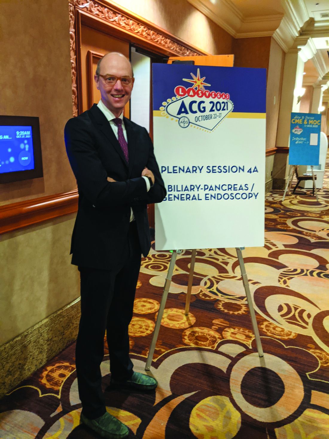
Dr. Robbins, associate professor of medicine and program director in gastroenterology in the Northwell Health System, New York, presented the study at the annual meeting of the American College of Gastroenterology.
Faster growth rates of pancreatic cysts in the presence of diabetes are important because they represent a potential mark for cyst aggressiveness. “So the question really is, in the setting of diabetes, are there factors perhaps circulating in the bloodstream, or other intrinsic factors, that make these cysts more dangerous and require a different surveillance approach than someone who doesn’t have diabetes? We have (surveillance) guidelines that address the average population, but they don’t really hone in on what do you do with (individuals with diabetes),” Dr. Robbins said during the presentation.
The study could have implications for screening, said session moderator Dayna Early, MD, professor of medicine at Washington University and director of endoscopy at Barnes Jewish Hospital, both in St. Louis. “I think this is important information to guide us to look more closely at patients with diabetes who do have pancreatic cysts,” she said in an interview.
The study included 177 adults with pancreatic cysts or abnormal imaging results between 2013 and 2020. Sixty-five percent were female, and the mean age was 65.4 years; 64% were White, 10% were Black, and 8.5% were Asian. Among the participants, 24.8% were smokers and 32.2% had type 2 diabetes.
Patients with diabetes had larger cyst sizes (2.23 cm versus 2.76 cm), as well as a higher annual cyst growth rate (1.90 cm versus 1.30 cm). Cyst size and growth rate were similar between patients with controlled and uncontrolled diabetes. Smoking was associated with a larger cyst size overall (2.2 cm versus 1.81 cm), and were larger still among patients with diabetes who smoked (2.35 cm).
Seventy-one patients went on to have pathologic confirmation by endoscopic ultrasound-guided fine needle aspiration. “In the diabetic group, two developed adenocarcinoma, six of the nondiabetics developed adenocarcinoma, and there was no difference in CEA or serum CA 19-9,” Dr. Robbins said during his presentation.
Of 28 patients diagnosed with pancreatic cancer, 13 had type 2 diabetes.
Defining danger
There remains uncertainty about what cyst growth rate is most dangerous. Some guidelines recommend that individuals with new-onset or worsening diabetes and intraductal papillary mucinous neoplasm or mucinous cystic neoplasm cysts, or cysts alone that are growing faster than 3 mm per year, may be at significantly increased risk of pancreatic cancer. These guidelines recommend that they be screened with short-interval magnetic resonance imaging or endoscopic ultrasound (EUS) fine needle aspiration. However, this recommendation is conditional and is backed by a very low level of evidence.
Other reports have shown varying risks at different growth rates. “It’s not really clear at this point. And that’s why I think, while our study is small and exploratory, this is a particular area that is relatively easy to evaluate. We have huge databases of pancreatic cyst evolution, and we know that 30 million Americans have diabetes. So, the next obvious study is to do a more systematic look at that, and work towards refining and making sense of these divergent guidelines, all of which are saying the same thing but using different threshold numbers,” said Dr. Robbins.
The next step is do larger, multicenter studies in the context of other risk factors such as family history and smoking, but the current finding represents an opportunity to catch at least some pancreatic cancers earlier, according to Dr. Robbins. He suggested that individuals with diabetes who are diagnosed with a pancreatic cyst should be referred to a gastroenterologist or another specialist to track cyst growth. “That is going to miss a lot of folks who didn’t get imaging for whatever reason (and so don’t have a cyst identified), but it is an early opportunity, and it’s better than what we’re doing now.”
During the talk, Dr. Robbins said, “Given the ease, availability and low cost of diabetes screening in the general clinic population, we encourage the inclusion of HbA1c and fasting glucose in algorithms for pancreatic cyst surveillance.”
Dr. Early found the suggestion intriguing, but wasn’t ready to lend full support. “I think looking at the suggestion of possibly monitoring hemoglobin A1c levels was novel. I don’t know that we’ll necessarily adopt that as standard practice, but that’s something I think that could be looked at in the future as a way to help risk stratify whether patients need to be surveyed more frequently,” she said.
Dr. Robbins and Dr. Early have no relevant financial disclosures.
LAS VEGAS – New results from a single center, retrospective analysis suggest that individuals with diabetes and pancreatic cysts have larger cyst sizes at diagnosis, and a faster subsequent cyst growth rate. Smoking was independently associated with faster growth rate.
Most pancreatic cancer patients were previously diagnosed with hyperglycemia and diabetes, and pancreatic cancer can cause diabetes. “This sort of dual causality raises questions as to whether or not hyperglycemia, or the new diagnosis of diabetes itself, could be a harbinger of cancer or precancer. And should these patients be more closely monitored?” David Robbins, MD, said in an interview.
Dr. Robbins, associate professor of medicine and program director in gastroenterology in the Northwell Health System, New York, presented the study at the annual meeting of the American College of Gastroenterology.
Faster growth rates of pancreatic cysts in the presence of diabetes are important because they represent a potential mark for cyst aggressiveness. “So the question really is, in the setting of diabetes, are there factors perhaps circulating in the bloodstream, or other intrinsic factors, that make these cysts more dangerous and require a different surveillance approach than someone who doesn’t have diabetes? We have (surveillance) guidelines that address the average population, but they don’t really hone in on what do you do with (individuals with diabetes),” Dr. Robbins said during the presentation.
The study could have implications for screening, said session moderator Dayna Early, MD, professor of medicine at Washington University and director of endoscopy at Barnes Jewish Hospital, both in St. Louis. “I think this is important information to guide us to look more closely at patients with diabetes who do have pancreatic cysts,” she said in an interview.
The study included 177 adults with pancreatic cysts or abnormal imaging results between 2013 and 2020. Sixty-five percent were female, and the mean age was 65.4 years; 64% were White, 10% were Black, and 8.5% were Asian. Among the participants, 24.8% were smokers and 32.2% had type 2 diabetes.
Patients with diabetes had larger cyst sizes (2.23 cm versus 2.76 cm), as well as a higher annual cyst growth rate (1.90 cm versus 1.30 cm). Cyst size and growth rate were similar between patients with controlled and uncontrolled diabetes. Smoking was associated with a larger cyst size overall (2.2 cm versus 1.81 cm), and were larger still among patients with diabetes who smoked (2.35 cm).
Seventy-one patients went on to have pathologic confirmation by endoscopic ultrasound-guided fine needle aspiration. “In the diabetic group, two developed adenocarcinoma, six of the nondiabetics developed adenocarcinoma, and there was no difference in CEA or serum CA 19-9,” Dr. Robbins said during his presentation.
Of 28 patients diagnosed with pancreatic cancer, 13 had type 2 diabetes.
Defining danger
There remains uncertainty about what cyst growth rate is most dangerous. Some guidelines recommend that individuals with new-onset or worsening diabetes and intraductal papillary mucinous neoplasm or mucinous cystic neoplasm cysts, or cysts alone that are growing faster than 3 mm per year, may be at significantly increased risk of pancreatic cancer. These guidelines recommend that they be screened with short-interval magnetic resonance imaging or endoscopic ultrasound (EUS) fine needle aspiration. However, this recommendation is conditional and is backed by a very low level of evidence.
Other reports have shown varying risks at different growth rates. “It’s not really clear at this point. And that’s why I think, while our study is small and exploratory, this is a particular area that is relatively easy to evaluate. We have huge databases of pancreatic cyst evolution, and we know that 30 million Americans have diabetes. So, the next obvious study is to do a more systematic look at that, and work towards refining and making sense of these divergent guidelines, all of which are saying the same thing but using different threshold numbers,” said Dr. Robbins.
The next step is do larger, multicenter studies in the context of other risk factors such as family history and smoking, but the current finding represents an opportunity to catch at least some pancreatic cancers earlier, according to Dr. Robbins. He suggested that individuals with diabetes who are diagnosed with a pancreatic cyst should be referred to a gastroenterologist or another specialist to track cyst growth. “That is going to miss a lot of folks who didn’t get imaging for whatever reason (and so don’t have a cyst identified), but it is an early opportunity, and it’s better than what we’re doing now.”
During the talk, Dr. Robbins said, “Given the ease, availability and low cost of diabetes screening in the general clinic population, we encourage the inclusion of HbA1c and fasting glucose in algorithms for pancreatic cyst surveillance.”
Dr. Early found the suggestion intriguing, but wasn’t ready to lend full support. “I think looking at the suggestion of possibly monitoring hemoglobin A1c levels was novel. I don’t know that we’ll necessarily adopt that as standard practice, but that’s something I think that could be looked at in the future as a way to help risk stratify whether patients need to be surveyed more frequently,” she said.
Dr. Robbins and Dr. Early have no relevant financial disclosures.
AT ACG 2021
Hantavirus 5-Point Screen Tool Identifies Patients With Deadly Infection
In May 2016, Tarrah Oliver had been working as a bench tech in hematology at Tséhootsooí Medical Center (TMC) for 4 years when the hospital launched a 5-point screen for detecting hantavirus cardiopulmonary syndrome (HCPS), a potentially fatal disease that disproportionately affects Native Americans.
Developed at the University of New Mexico, the 5-point screen was particularly useful for facilities like TMC, a 56-bed hospital in Arizona serving Navajo Nation patients. Although the Navajo make up only 1.7% of the population, they account for 18% of all reported HCPS cases in the US.
But Oliver, who was the laboratory Safety and Training supervisor, her department supervisor, and his assistant were the only people who knew how to do the screen. “I was storing completed worksheets in a folder,” Oliver said, “and soon I had to transfer them to a 3-inch binder because we had so many being ordered. I started saving slides that scored high so that I could show my fellow lab mates what certain characteristics looked like under the microscope.” She also created case studies to train staff using analyzer printouts and slides from the patients. “It was my little side project to take care of while I was working and training new employees in the hematology department.”
The screen was to become much more than a side project very quickly.
In 2017, there was a hantavirus outbreak in one of the nearby communities. A team from the Centers for Disease Control and Prevention (CDC) came out to visit the local hospitals to educate the medical staff on what to look for in their patients with suspected hantavirus. Among those: the TMC staff. The visit would prove both serendipitous and synergistic.
Mary Choi, MD, MPH, medical officer with the CDC Viral Special Pathogens Branch, said, “Members of my branch were asked to come to Navajo Nation [Lukachukai chapter, Arizona] because of a cluster of hantaviruses cases that had been occurring in the area over the course of a few years. During a meeting with medical providers at TMC, our team met Tarrah and she told us that TMC had implemented the screen. We were really intrigued and impressed by the initiative of this small community hospital.”
“We were excited that the CDC even knew we existed!” Oliver recalled. “This little lab in the middle of the reservation.”
HCPS is rare but severe. It can quickly progress from nonspecific initial symptoms, such as fever, body aches, and shortness of breath, to severe respiratory distress. Without immediate intervention, patients usually die within 24 to 48 hours of the onset of cardiopulmonary symptoms. “What makes the disease even more challenging,” said Choi, “is that some of the critical lifesaving measures that physicians normally employ to save a critically-ill patient can actually make the situation worse.
“For example, when HCPS patients reach the cardiopulmonary phase of the illness, their blood pressure will drop. The normal response to this is to give IV fluids (IVF). But in hantavirus, giving IVF can actually make the signs and symptoms of the disease worse and only judicious use of IVF is recommended.”
She gave other examples of treatment challenges: “Physicians are taught to intubate critically ill patients. However, intubation of hanta patients during the cardiopulmonary phase can actually be detrimental. Also, in general, extracorporeal membrane oxygenation (ECMO) is an intervention of last resort for most critically-ill patients. As the procedure essentially involves hooking the patient up to a heart-lung machine, which has a lot of complications, doctors do not do this unless they have to. But in hantavirus disease, early initiation of ECMO has been shown to significantly improve survival.”
“Another tricky thing about ECMO is that not all hospitals have an ECMO machine… oftentimes patients have to be transferred from one hospital to another because they need ECMO and it is not available where they are.” The ECMO-equipped facility closest to TMC is 170 miles away, at the University of New Mexico Hospital in Albuquerque.
For all those reasons, Dr. Choi said, it is critically important for physicians to know if the patient they are treating has hantavirus or not. The problem is, there is no rapid test for hantavirus disease. The tests have to be sent out to a laboratory and it can take days before physicians receive a test result. “This is where the 5-point screen is such a valuable tool. Although it is not a diagnostic test, it is a very accurate screening tool. Even better, it is readily and widely available, because to perform the screen, you only need a complete blood count (CBC) and a peripheral blood smear…which can be performed in the vast majority of laboratories and clinics. In addition, the CBC and peripheral blood smear are very commonly ordered tests.”
The screen could be particularly crucial for small community hospitals and clinics. “[TMC] took a screen developed by hematopathologists for hematopathologists,” Choi said, “and adapted it so that it could be performed by clinical laboratorians—because hematopathologists don’t exactly grow on trees! Once we heard about the work that TMC was doing, we knew that this was something we wanted to support and advocate for.”
“We started to throw around the idea of teaching this method to other laboratories on the reservation,” Oliver said. She and Mary Choi teamed up to “knock on everyone’s door to see who was utilizing the screening or if they had their own methods of detecting hantavirus in their patients.”
Two years later, they began holding training sessions at a nearby community college; they also trained an entire laboratory at their hospital in late 2019. Plans were to set up other training sessions towards the border towns before spring of 2020.
Then SARS-CoV-2 showed up.
Hantavirus or coronavirus?
COVID-19 hit the Navajo Nation like a sledgehammer. Native Americans in rural areas, as many Navajo are, may have multigenerational and extended families living in tight quarters, with limited access to running water, miles from the nearest healthcare facility. They were particularly vulnerable to a virus that thrives on close contact, at a time when the most common preventives were distancing, frequent handwashing, and testing. By May 2020, the Navajo Nation had surpassed the epicenters—New York and New Jersey—in cases of infection. Even as late as November 2021, the Navah Nation has reported > 37,000 positive cases, and nearly 1,500 confirmed deaths, despite reaching a 70% vaccination rate.
Making things ever more challenging: How do you know whether the patient has hantavirus or COVID-19? The distinction is critical because the disease courses of the 2 infections differ greatly. “The importance of [TMC’s] work was really brought home by the COVID pandemic,” Mary Choi said. “The signs and symptoms of early COVID and hantavirus are really indistinguishable. But the problem is that most people with COVID will live, while most people with hantavirus will die without critical care.”
The overall US case fatality rates are drastically different: 36% to 38% for hantavirus, 1.6% for COVID-19. In Arizona alone, of 81 patients who developed hantavirus (as of 2019), 27 died. In New Mexico, of 117 patients, 51 died.
“The scary part,” said Tarrah Oliver, “was that when patients were flooding our ER with symptoms exactly like those of hantavirus, I kept thinking that it was now up to us in the lab to scrutinize CBC results for any characteristics of hantavirus because our locum providers and permanent providers would likely not have hanta on their minds right now to suspect it.”
“Once COVID hit Navajo Nation, we really worried about how clinicians were going to distinguish between the two disease entities and worried about possible excess deaths of hanta because they were mistaken for COVID-19,” Choi says. “Unfortunately, in the spring of 2020, 2 fatal cases of hantavirus were reported. Their deaths were initially thought to be due to COVID, but later testing showed that they both died of hantavirus. We knew that we had to study whether the 5-point screen could distinguish between hantavirus disease and COVID.”
They conducted the comparison study at TMC and Emory, in Atlanta. TMC, as a small hospital, might not get many COVID cases while Emory, a large hospital system, had many COVID cases but rarely hantavirus cases (Box).
The researchers found that the screen did indeed work as they had hoped. No matter who performed the screen, the demographics of the population screened, or when in the COVID-19 disease course the sample was taken, individuals positive for COVID-19 received a low score on the screen. None of the participants who were positive for COVID-19 demonstrated all 5 hallmarks of hantavirus infection, and only 3 patients received a score of 4.
The screen was most accurate when the specimen was collected during the cardiopulmonary phase. Before the cardiopulmonary phase [BOX], there is a short febrile prodromal phase in which the platelets are starting to drop but aren’t low enough to be considered thrombocytopenic. Hematocrit and hemoglobin levels are still normal, and immunoblasts or plasma cells haven’t been released into the blood yet. “A patient’s score is likely to be pretty low or intermediate,” Tarrah Oliver said. “In the febrile prodromal phase, the patient will have chills, fever, headache and myalgia lasting 3 to 6 days, which might be written off as the flu or even COVID-19, especially if a provider is not familiar with the endemic regions of the Navajo Nation.”
“When we found that the screen could distinguish between the 2 diseases,” Choi said, “we worked frantically to write up the findings and publish so that healthcare providers would be aware of the value of this tool.”
“I couldn’t be more excited that the word is getting out in the medical community,” Oliver said. They plan to share the information as widely as possible, booking local radio spots, for instance. “We’re trying to disseminate our research across our partners on the reservation.”
The results of the research were recently published in the American Journal of Clinical Pathology. “Our next follow-up research will be an inter-rater reliability between TMC’s 2-year and 4-year-degree medical laboratorians and Emory’s pathologists performing the 5-point hantavirus screen on the same slides. We want to show that rural hospitals without the expertise of pathologists can perform this screen for their patients in a timely manner and still get the same outcomes.”
Lastly, and the most exciting, Oliver said, is putting that training manual she created to wider use. In addition to all the case studies she collected, analyzer printouts, and PowerPoint slides about the epidemiology, the manual includes a procedure, billing codes, and layout of what it looks like on certain medical record software that most Indian Health Service hospitals use. “With the manual, we may be able to expand our reach, so I won’t need to travel,” Oliver said. “We could use Zoom to reach further and I could explain it all while they have the manual right in front them.”
So far, nearly 200 screens have been performed at TMC. Four cases of hantavirus disease have been identified. The screen has proved successful, but for Oliver, there’s a personal reward. “I heard stories as a kid of relatives and friends succumbing to this mystery flu in the 1990s and how our parents protected us from it,” she said. “Now I feel like it’s my turn to take care of my family and friends from both hantavirus and COVID, our current ‘mystery flu.’ … I’ve realized that now I’m doing this for our people, the Diné (Navajo) people.”
This article offers some background and follow-up to Hantavirus Disease and COVID-19: Evaluation of the Hantavirus 5-Point Screen in 139 COVID-19 Patients