User login
Circumferential urethral diverticulum: A surgical conundrum

Communicating serious news over video. Bringing protocols to the forefront. Sleep and burnout in health care workers. Lung cancer screening.
Palliative and end-of-life care
Communicating serious news over video
Critical care consultation using telemedicine is increasingly prevalent. Having serious conversations regarding end-of life care over video can be extremely challenging. Here are some suggestions and sample phrases to make palliative-focused conversations more successful
Prior to initiating the conversation, communicate with the bedside team. Ensure they want you to discuss palliative options and make an outline of discussion topics together. Identify and include all important decision-makers. Family may need to be connected over a digital meeting platform such as Zoom© or WEBex© and arrange for interpreter services if needed.
Prepare the virtual meeting place ahead of time. Test the connection, and make sure the audiovisual quality is clear. Have the camera centrally positioned, and ensure adequate lighting to easily see facial expressions. Remove distracting background furniture, and clear your space of confidential material. Have a quiet area planned to avoid interruptions (J Gen Intern Med. 2020 Oct 27:1-4. doi: 10.1007/s11606-020-06278-z. Online ahead of print).
Open the conversation with introductions, and explore perceptions with open-ended questions: “So I know where to begin, tell me about your understanding of what has been happening?” Get a sense of the patient’s previous function, quality of life, and their values as an individual. Maintain good eye contact throughout. When ready to give an update, use simple language and avoid details: “Unfortunately, your condition is worse. You have not been responding to treatments as hoped. Your lungs are needing much more support, and I’m worried they are not going to get better.”Anticipate emotions, and provide empathetic responses: “I wish we had better news. This must be overwhelming for you” (Back, et al. Ann Int Med. 2020;172[11]:759). Finally, offer a recommendation. Most patients and families are interested in your advice and want guidance. Use the patient’s previously stated values to support your recommendation.
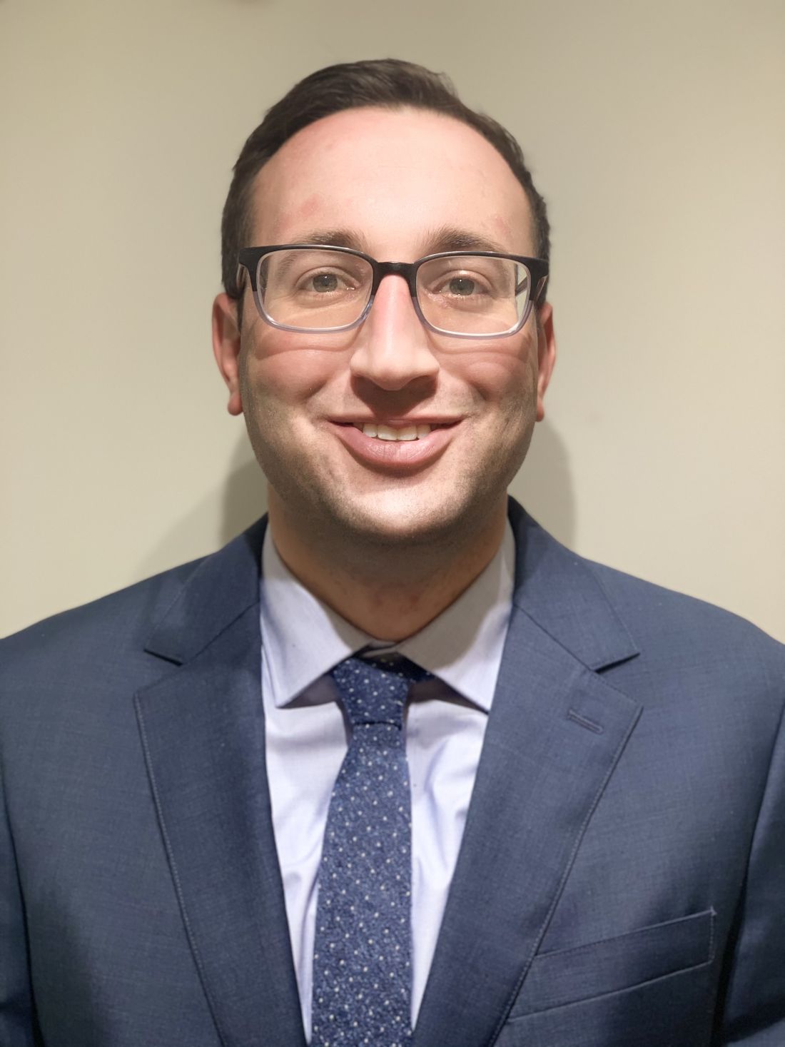
Andrew Badke, MD
Steering Committee Member
Respiratory care
COVID-19 pandemic bringing protocols to the forefront
COVID-19 has health care organizations threatened like never before. Staffing requirements, equipment necessities, and personnel training happen in a whirlwind.1 Information could change daily/hourly, and the need to protocolize guidelines became more evident each day.
While protocols have existed long before COVID-19, many institutions and organizations responded to the ever-changing pandemic by creating clinical practice guidelines (CPGs) to help not only their experienced staff members but also the non-traditional ICU caregivers thrust onto the front lines.3 Organizations worked on PPE protocols, respiratory care management, and ECMO guidelines to name a few.2,3 Protocols with algorithms and CPGs have been shown to reduce patient harm and improve standardization and communication.

A CPG is a general principle that guides the management of care, in which specific questions are posed, a literature review is completed, and the quality of the research evaluated. The questions are answered using the strength of the available research. CPG decision points are then based on the evidence or on the consensus of experts, resulting in a protocol that are descriptions of detailed behaviors to be followed in specific situations. These behaviors are provided in a list format or a flow diagram. Using a universal language for protocols with algorithms has aided many hospitals ensure effective care for patients and has even helped develop multidisciplinary relationships not present prior to the pandemic (onepagericu.com).
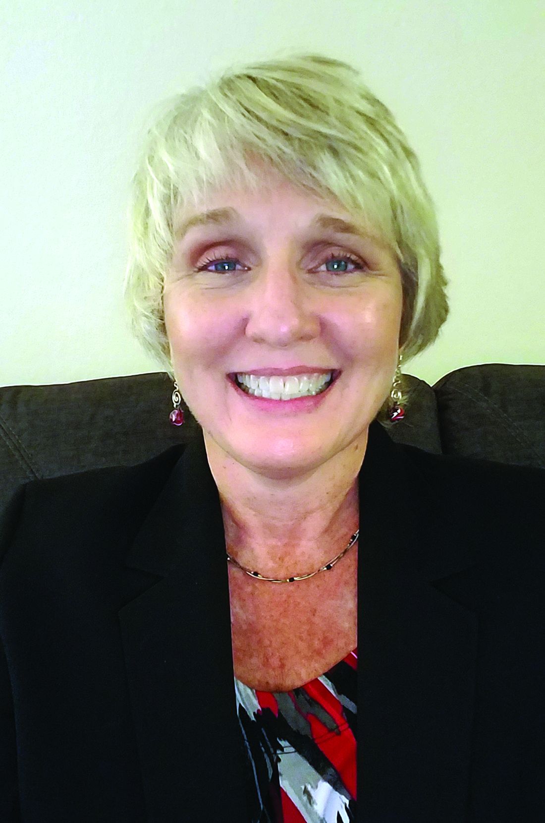
DeDe Gardner, RRT, DrPh, FCCP
Donna Tanner, RRT-ACCS, MBA, FCCP
Steering Committee Members
1. epub JAMA 2/2021.
2. WHO, Guidelines 1/2021.
3. CHEST, Clinical Resources.
4. Curr Treat Options Ped 2015,1:347
Sleep medicine
Time to move the dial: Sleep and burnout in health care workers during the COVID-19 pandemic
Although the interaction between sleep, mood disorders, and burnout is well established, many of us are still sleep-deprived. A cross-sectional study of over 800 health care workers during the pandemic stay-at-home orders in March 2020 reported that those working in-person had shorter sleep times and worse mood, while those with longer sleep times had improved mood (Conroy DA, et al. J Clin Sleep Med. 2021;17[2]:185). Even prior to COVID-19, many trainees were facing issues with sleep deprivation and burnout (Sharp M, et al. Chest. 2021;159[2]:733).
One year into the pandemic, we continue to face a unique set of hardships, exacerbating underlying sleep disorders such as insomnia, feelings of burnout, and mental health problems. An international team led by Dr. Joel Goh calculated the cost of burnout and its economic impact on the nation’s health care system and estimated this at $4.6 billion per year (Han S, et al. Ann Intern Med. 2019;170[11]:784). National medical organizations, including the National Academy of Medicine and the American Medical Association, have also placed greater emphasis on clinician well-being and resilience. Practical frameworks for creating wellness during the pandemic exist; however. senior-level executive champions are critical for implementation (Adibe B, et al. N Engl J Med Catalyst. Jun 2020). While the long-term impact remains unknown, the current state of sleep and mental health problems and the cost of burnout should be a warning to health systems and institutions to implement remedial interventions now.(“Taking action against burnout: A systems approach to professional well-being,” National Academies of Sciences, Engineering, and Medicine, October 2019.
Nancy H. Stewart, DO, MS, Steering Committee Member
Thoracic oncology
Impact of COVID-19 on lung cancer screening
Lung cancer is the leading cause of cancer-related death worldwide and COVID-19 is making this worse. Prior to the COVID-19 pandemic, despite evidence of improved mortality, the uptake of lung cancer screening (LCS) was quite low with only 4% of those eligible having undergone screening in 2015 (Jemal A, et al. JAMA Oncol. 2017;3[9]:1278).
As the COVID-19 pandemic unfolded, health care resources were re-allocated to critically ill patients and areas, and nonurgent care was postponed. Therefore, LCS programs were halted (Mazzone PJ, et al. Chest. 2020;158[1]:406). This led to concerns that fewer patients would undergo screening and more patients would experience delays in cancer diagnosis.
Using population-based modeling, researchers in England estimated the COVID-19 pandemic will result in decreased lung cancer survival and a subsequent increase in avoidable cancer deaths (Maringe C, et al. Lancet Oncol. 2020;21[8]:1023). And in fact, investigators in Spain found fewer new lung cancer diagnoses during the COVID-19 pandemic compared with the same time-period pre-pandemic, and those that were diagnosed were later stage disease (Reyes R, et al. IASCL World Conference. 2020. A3700).
As we learn more about COVID-19 and communities become vaccinated, it becomes critical to both resume LCS programs and improve participation. While the pandemic has hampered efforts to screening patients, it has also facilitated the uptake of new technologies such as telemedicine. In March 2020, due to the COVID-19 pandemic, the Centers for Medicare and Medicaid Services relaxed the rules for telehealth, and now covers shared decisions making (SDM) virtual visits for LCS (Centers for Medicare & Medicaid Services, “Telehealth Services.” ICN MLN901705, March 2020). This new tool, amongst others, could increase access to LCS, facilitate more widespread adoption of screening, and ultimately improve lung cancer outcomes.
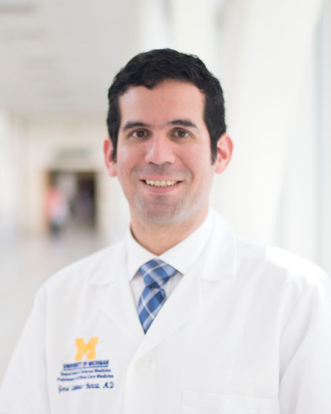
Max Wayne, MD, and Jose Cardenas-Garcia, MD
Steering Committee Members
Palliative and end-of-life care
Communicating serious news over video
Critical care consultation using telemedicine is increasingly prevalent. Having serious conversations regarding end-of life care over video can be extremely challenging. Here are some suggestions and sample phrases to make palliative-focused conversations more successful
Prior to initiating the conversation, communicate with the bedside team. Ensure they want you to discuss palliative options and make an outline of discussion topics together. Identify and include all important decision-makers. Family may need to be connected over a digital meeting platform such as Zoom© or WEBex© and arrange for interpreter services if needed.
Prepare the virtual meeting place ahead of time. Test the connection, and make sure the audiovisual quality is clear. Have the camera centrally positioned, and ensure adequate lighting to easily see facial expressions. Remove distracting background furniture, and clear your space of confidential material. Have a quiet area planned to avoid interruptions (J Gen Intern Med. 2020 Oct 27:1-4. doi: 10.1007/s11606-020-06278-z. Online ahead of print).
Open the conversation with introductions, and explore perceptions with open-ended questions: “So I know where to begin, tell me about your understanding of what has been happening?” Get a sense of the patient’s previous function, quality of life, and their values as an individual. Maintain good eye contact throughout. When ready to give an update, use simple language and avoid details: “Unfortunately, your condition is worse. You have not been responding to treatments as hoped. Your lungs are needing much more support, and I’m worried they are not going to get better.”Anticipate emotions, and provide empathetic responses: “I wish we had better news. This must be overwhelming for you” (Back, et al. Ann Int Med. 2020;172[11]:759). Finally, offer a recommendation. Most patients and families are interested in your advice and want guidance. Use the patient’s previously stated values to support your recommendation.
Andrew Badke, MD
Steering Committee Member
Respiratory care
COVID-19 pandemic bringing protocols to the forefront
COVID-19 has health care organizations threatened like never before. Staffing requirements, equipment necessities, and personnel training happen in a whirlwind.1 Information could change daily/hourly, and the need to protocolize guidelines became more evident each day.
While protocols have existed long before COVID-19, many institutions and organizations responded to the ever-changing pandemic by creating clinical practice guidelines (CPGs) to help not only their experienced staff members but also the non-traditional ICU caregivers thrust onto the front lines.3 Organizations worked on PPE protocols, respiratory care management, and ECMO guidelines to name a few.2,3 Protocols with algorithms and CPGs have been shown to reduce patient harm and improve standardization and communication.
A CPG is a general principle that guides the management of care, in which specific questions are posed, a literature review is completed, and the quality of the research evaluated. The questions are answered using the strength of the available research. CPG decision points are then based on the evidence or on the consensus of experts, resulting in a protocol that are descriptions of detailed behaviors to be followed in specific situations. These behaviors are provided in a list format or a flow diagram. Using a universal language for protocols with algorithms has aided many hospitals ensure effective care for patients and has even helped develop multidisciplinary relationships not present prior to the pandemic (onepagericu.com).
DeDe Gardner, RRT, DrPh, FCCP
Donna Tanner, RRT-ACCS, MBA, FCCP
Steering Committee Members
1. epub JAMA 2/2021.
2. WHO, Guidelines 1/2021.
3. CHEST, Clinical Resources.
4. Curr Treat Options Ped 2015,1:347
Sleep medicine
Time to move the dial: Sleep and burnout in health care workers during the COVID-19 pandemic
Although the interaction between sleep, mood disorders, and burnout is well established, many of us are still sleep-deprived. A cross-sectional study of over 800 health care workers during the pandemic stay-at-home orders in March 2020 reported that those working in-person had shorter sleep times and worse mood, while those with longer sleep times had improved mood (Conroy DA, et al. J Clin Sleep Med. 2021;17[2]:185). Even prior to COVID-19, many trainees were facing issues with sleep deprivation and burnout (Sharp M, et al. Chest. 2021;159[2]:733).
One year into the pandemic, we continue to face a unique set of hardships, exacerbating underlying sleep disorders such as insomnia, feelings of burnout, and mental health problems. An international team led by Dr. Joel Goh calculated the cost of burnout and its economic impact on the nation’s health care system and estimated this at $4.6 billion per year (Han S, et al. Ann Intern Med. 2019;170[11]:784). National medical organizations, including the National Academy of Medicine and the American Medical Association, have also placed greater emphasis on clinician well-being and resilience. Practical frameworks for creating wellness during the pandemic exist; however. senior-level executive champions are critical for implementation (Adibe B, et al. N Engl J Med Catalyst. Jun 2020). While the long-term impact remains unknown, the current state of sleep and mental health problems and the cost of burnout should be a warning to health systems and institutions to implement remedial interventions now.(“Taking action against burnout: A systems approach to professional well-being,” National Academies of Sciences, Engineering, and Medicine, October 2019.
Nancy H. Stewart, DO, MS, Steering Committee Member
Thoracic oncology
Impact of COVID-19 on lung cancer screening
Lung cancer is the leading cause of cancer-related death worldwide and COVID-19 is making this worse. Prior to the COVID-19 pandemic, despite evidence of improved mortality, the uptake of lung cancer screening (LCS) was quite low with only 4% of those eligible having undergone screening in 2015 (Jemal A, et al. JAMA Oncol. 2017;3[9]:1278).
As the COVID-19 pandemic unfolded, health care resources were re-allocated to critically ill patients and areas, and nonurgent care was postponed. Therefore, LCS programs were halted (Mazzone PJ, et al. Chest. 2020;158[1]:406). This led to concerns that fewer patients would undergo screening and more patients would experience delays in cancer diagnosis.
Using population-based modeling, researchers in England estimated the COVID-19 pandemic will result in decreased lung cancer survival and a subsequent increase in avoidable cancer deaths (Maringe C, et al. Lancet Oncol. 2020;21[8]:1023). And in fact, investigators in Spain found fewer new lung cancer diagnoses during the COVID-19 pandemic compared with the same time-period pre-pandemic, and those that were diagnosed were later stage disease (Reyes R, et al. IASCL World Conference. 2020. A3700).
As we learn more about COVID-19 and communities become vaccinated, it becomes critical to both resume LCS programs and improve participation. While the pandemic has hampered efforts to screening patients, it has also facilitated the uptake of new technologies such as telemedicine. In March 2020, due to the COVID-19 pandemic, the Centers for Medicare and Medicaid Services relaxed the rules for telehealth, and now covers shared decisions making (SDM) virtual visits for LCS (Centers for Medicare & Medicaid Services, “Telehealth Services.” ICN MLN901705, March 2020). This new tool, amongst others, could increase access to LCS, facilitate more widespread adoption of screening, and ultimately improve lung cancer outcomes.
Max Wayne, MD, and Jose Cardenas-Garcia, MD
Steering Committee Members
Reclaiming patient-centered care from the grip of COVID-19
Over a year has passed since the first case of COVID-19 was reported in the United States, with over 114 million cases now reported worldwide, and over 2.5 million deaths at the time of this writing (Dong E, et al. Lancet Infect Dis. doi: 10.1016/S1473-3099[20]30120-1). While our vaccination efforts here in the United States have provided a much-needed glimmer of hope, it has been bittersweet, as we recently surpassed the grim milestone of 500,000 COVID-19-related deaths.
The infectious nature of SARS-CoV-2, coupled with the lack of adequate PPE early in the pandemic, led to radical changes in most hospital visitor policies. Rather than welcoming families into the care setting as we have been accustomed, we were forced to restrict access. While well-intentioned, the impact of this on patients, their families – and as we later learned, ourselves – has been devastating. Patients found themselves alone in an unfamiliar environment, infected with a disease there was no effective treatment for, hearing dismal news regarding inpatient and ICU mortality rates on news networks, and families could not see for themselves how their loved ones were progressing in their hospital course.
The impact on patient-centered care
The impact of this pandemic on patients and health care providers alike cannot be overstated. Arguably, one of the greatest challenges created by COVID-19 has been its direct assault on the core values of patient-centered care that we have spent decades striving to promote and embody.
Since its identification as a quality gap by the Institute of Medicine in 2001, the definition of patient-centered care has been tweaked over the past 20 years (Institute of Medicine (IOM). Crossing the Quality Chasm: A New Health System for the 21st Century. Washington, D.C: National Academy Press; 2001). Most frameworks include the active participation of patients and their families as part of the health care team, encouraging and facilitating the presence of family members in the care setting, and focusing on patients’ physical comfort and emotional well-being as fundamental tenets of patient centeredness (NEJM Catalyst: What is Patient-Centered Care? Explore the definition, benefits, and examples of patient-centered care. How does patient-centered care translate to new delivery models? January 1,2017).
Families, the “F” in the ABCDEF Bundle, have been recognized as an integral part of care in the ICU setting (Ely EW. Crit Care Med. 2017;45[2]:321). While engagement of family members began with our recognition of their role in emotionally supporting patients and efforts to improve communication, we have also seen the impact of family participation on reducing ICU delirium through frequent re-orientation and encouragement of early mobility (McKenzie J, et al. Australas J Ageing. 2020;39:21). In fact, a recent study has suggested that family members could play an even more active role in detecting and assessing ICU delirium using objective assessment tools (Fiest K, et al. Crit Care Med. 2020;48[7]:954). Post-ICU PTSD has been well described in both ICU survivors as well as in their family members, with evidence that family participation in care of patients during their ICU stay leads to its reduction (Amass TH, et al. Crit Care Med. 2020[Feb];48[2]:176).
The emotional toll
Comforting patients and families in times of distress and suffering is something that comes naturally to many in critical care, and our training further improves our ability to do this effectively. No amount of training, however, could have prepared us for the degree and volume of suffering we bore witness to this past year and the resulting moral injury many are still dealing with. We were present for families’ most intimate moments, holding phones and tablets up to patients so their families could say their goodbyes, listening to the “I love yous,” “I’ll miss yous,” “I’m sorrys,” and “Please don’t gos.” Nurses held patients’ hands as they took their last breaths so they wouldn’t die alone and worked to move husbands and wives into the same room so they could be together in their final moments. Entrenched in each of our identities is the role of healer, and we found ourselves questioning our effectiveness in rising to meet suffering on a scale we had never seen before. Little did we understand that while our paradigms were reinforcing the benefits of patient-centered care for patients and their families, that framework was also serving to facilitate our role as healers – that without it, we all suffer.
Rising to the challenge
These unprecedented circumstances led to creative efforts to bridge some of these barriers. Health systems created photo lanyards that providers wore over their PPE so patients could identify their health care team and connect with them on a more human level. Video conferencing technology was brought to the patient bedside using smartphones and tablets to assist them in communicating with their families. Doctors and nurses coordinated multiple calls throughout the day to ensure families felt included in the care plans and were always abreast of any new developments.
All these initiatives were our way of attempting to alleviate some of the suffering we were witnessing, and in some ways felt complicit in. It is in hindsight that we can look back and question if we could have done things differently. We treated family as visitors, when in fact, they are fundamental members of the care team who play an active and critical role in patient care. This was, in part, driven by national unpreparedness when it came to PPE supplies, in addition to misinformation and inconsistent messaging early in the pandemic with regards to the mechanism of transmission of disease from various health organizations. While we did our best given the circumstances, we must not allow this experience to lead us away from the tenets we know to be essential to patient, family, and health care provider well-being.
All in health care met the call to action – nurses, physicians, advanced practice providers, respiratory therapists, nutritionists, pharmacists, physical therapists, patient transporters, environmental service workers, and all others who kept our hospitals and patient care facilities open through this pandemic and embarked on what amounted to a collective, global, ongoing “code-blue alert,” resuscitating patient after patient, hotspot after hotspot, region after region, and country after country. We expanded hospital bed capacities, created ICU beds where there were none, developed novel process protocols, and learned in real time what seemed to help (or not) in treating this novel disease, all while participating in incredible international scientific collaboration and information sharing that has contributed in getting the collective “us” through this first year of the pandemic. We did what we were trained and called to do.
Preparing for the future
There will inevitably be another public health crisis, and we must advocate for better preparedness next time, insisting on overall stronger public health systems and pandemic preparedness. We must address our PPE stores and supply chains. We must have disaster preparedness plans that go beyond the scope of mass casualty events and bioterrorism. Beyond physical recovery, we must tend to the factors that impact patients’ long-term recovery, with attention to emotional and psychological well-being. We must advocate for all of this now, while the memories are fresh and before the impact of this collective suffering begins to fade. It can never again be acceptable to exclude families from the health care setting. We must advocate for our patients and for the resources, systems, processes, and support that will allow us to do better.
Dr. Hegab is Associate Director, Pulmonary Hypertension Program, Medical Director, Pulmonary Embolism Response Team, Division of Pulmonary and Critical Care Medicine, Henry Ford Hospital; and Assistant Professor, Wayne State University School of Medicine, Detroit.

This month in the journal CHEST®
Editor’s picks
The relationship between asthma and cardiovascular disease: An examination of the Framingham offspring study. By Dr M. Pollevick, et al.
Projecting long-term health and economic burden of chronic obstructive pulmonary disease in the United States. By Dr. Z. Zafari, et al.
How I do it: Dosing fluids in early septic shock. By Dr. D. Chaudhuri, et al.
Essential components of an interstitial lung disease clinic: Results from a Delphi survey and patient focus group analysis. By Dr. B. A. Graney, et al.
Change: Leadership essentials for chest medicine professionals. By Dr. J. Stoller, et al.
Race correction and spirometry: Why history matters. By Dr. L. Braun.
Editor’s picks
Editor’s picks
The relationship between asthma and cardiovascular disease: An examination of the Framingham offspring study. By Dr M. Pollevick, et al.
Projecting long-term health and economic burden of chronic obstructive pulmonary disease in the United States. By Dr. Z. Zafari, et al.
How I do it: Dosing fluids in early septic shock. By Dr. D. Chaudhuri, et al.
Essential components of an interstitial lung disease clinic: Results from a Delphi survey and patient focus group analysis. By Dr. B. A. Graney, et al.
Change: Leadership essentials for chest medicine professionals. By Dr. J. Stoller, et al.
Race correction and spirometry: Why history matters. By Dr. L. Braun.
2020 Focused Updates to the Asthma Management Guidelines
National Asthma Education and Prevention Program (NAEPP) published its last Expert Panel Report in 2007. Since that time, substantial progress has been made in understanding the pathophysiology and treatment of asthma. A new report has provided a much-needed update in the evaluation and management of asthma. It focuses on several priority topics jointly decided upon by the National Heart, Lung, and Blood Institute (NHLBI) Advisory Council Asthma Expert Working Group, the National Asthma Education and Prevention Program (NAEPP) participant organizations, and the public in 2015. These topics include the role of fractional exhaled nitric oxide (FeNO), allergen mitigation, intermittent inhaled corticosteroids (ICS), long-acting muscarinic agents (LAMA), immunotherapy, and bronchial thermoplasty (BT) in asthma management. This document did not include the subsequent new developments in the role of biologics in asthma. The following is a summary of the recommendations made in the 2020 Focused Updates to the Asthma Management Guidelines.1

FeNO measurement is recommended to aid in asthma diagnosis and monitoring and to assist in ICS medication titration in individuals with asthma who are 5 years and older. The panel recommends that clinicians use FeNO levels, in conjunction with other relevant clinical data such as spirometry and asthma control questionnaires, for medical decision making. Similarly, when using FeNO to guide therapeutic changes in the ICS dose, the panel advises making changes based upon frequent measurements as a part of longitudinal assessment rather than one single measurement, as several factors can influence an FeNO measurement. Studies have demonstrated that a strategy that incorporates FeNO measurements into a treatment algorithm can reduce the risk of exacerbations; however, this has not been shown to reduce hospitalizations or quality of life.2

Allergen mitigation interventions, which can be used in individuals of all ages, are only recommended for those who have symptoms related to specific indoor aeroallergens exposure. This can be confirmed by skin testing or specific IgE in the appropriate clinical setting if specific allergen testing is not readily available. While most recommendations focus on using a multicomponent approach to allergen mitigation (ie, dust mite covers, HEPA filters, air purifiers, carpet removal, mold remediation, pest or pest removal, etc), pest removal was the only single-component approach that was deemed effective. Dust mite covers alone are unlikely to lead to significant improvement if not paired with additional mitigation strategies; however, note that there was low certainty about these recommendations. Ultimately, allergen mitigation should focus on addressing those identified triggers resulting in poor control of asthma. Simultaneously, the clinician should consider the resources and costs associated with some of these interventions.

The panel has recommended using ICS therapy for on-demand (prn) usage, even in those with mild persistent asthma, recognizing that earlier and more frequent on-demand ICS usage results in fewer exacerbations. While the recommendations slightly differ based upon the age group, in those >12 years with mild persistent asthma, recommendations are for either daily ICS + as-needed short-acting beta-agonist (SABA), or as-needed ICS and SABA use. As in the Global Initiative for Asthma (GINA) guidelines, the panel also recommends single maintenance and rescue therapy (SMART) using ICS-formoterol inhalers for moderate to severe asthma. SMART has also been shown to reduce the risk of exacerbation. The clinician needs to use ICS-LABA medications where formoterol is the LABA component due to its quick onset of action (within 5 minutes, hence allowing it to be used as a rescue). Shared decision-making must be utilized when considering cost, insurance formulary restrictions, and perhaps delayed insurer and pharmacy adoption of these guidelines, as patients are likely to use more than one canister in a month when utilizing SMART.3,4
LAMA is a pharmacologic class of long-acting inhaled bronchodilators. Guidelines addressed the role of LAMA in individuals aged 12 years and older. Three recommendations are made regarding the role of LAMA in this age group. In individuals with persistent, uncontrolled asthma while using ICS therapy, the guidelines recommend the addition of a LABA over LAMA therapy.5 LAMA can be added to ICS in individuals with uncontrolled asthma who cannot use LABA or are already on ICS-LABA maintenance therapy.
For those patients with mild to moderate allergic asthma, as defined by allergic sensitization via skin testing or in-vitro elevated serum IgE levels, the expert panel conditionally recommends subcutaneous immunotherapy (SCIT) as an adjunct treatment to standard pharmacotherapy. It is recommended only in those patients whose asthma remains controlled throughout initiation, build-up, and maintenance phases. SCIT should not be used for patients with severe asthma, and all attempts should be made to optimize asthma with standard therapy first. The risks and benefits of SCIT should be discussed with the specialist before starting therapy. Sublingual immunotherapy (SLIT) is not recommended for the treatment of asthma.
Regarding BT, the Expert Panel conditionally recommends against BT in individuals age 18 years and older with persistent asthma because of the small benefit to risk ratio and uncertain outcomes. Because there is a risk of worsening asthma control or inducing an exacerbation, it is advised that BT not be performed in individuals with an FEV <50%-60% or those with a history of life-threatening asthma. If BT is considered, it should be performed by an experienced specialist and should be done in conjunction with a clinical trial or registry to track its long-term safety and effectiveness.6 All efforts should be made to optimize asthma therapy and address comorbidities before pursuing BT.
This Expert Panel report provides a robust systematic review of the evidence that addresses key questions in the management of asthma. However, not providing any recommendations regarding the use of biologics was a significant gap. Further guidance regarding their role can be found in the GINA guidelines, and by the European Respiratory Society and American Thoracic Society, both of which were also published in 2020.7,8Dr. Adrish is Clinical Assistant Professor, Bronx Care Health System, New York; Dr. Patil is Assistant Professor, Department of Respiratory Sleep and Critical Care Medicine, Maharashtra University of Health Sciences (MUHS), India; Dr. Oberle is Assistant Professor of Medicine, Associate Medical Director, Duke Asthma, Allergy and Airway Center, Durham, NC.
References
1. Expert Panel Working Group of the National Heart, Lung, and Blood Institute (NHLBI) administered and coordinated National Asthma Education and Prevention Program Coordinating Committee (NAEPPCC), et al. 2020 Focused Updates to the Asthma Management Guidelines: A Report from the National Asthma Education and Prevention Program Coordinating Committee Expert Panel Working Group. J Allergy Clin Immunol. 2020 Dec;146(6):1217-1270. doi: 10.1016/j.jaci.2020.10.003. PMID: 33280709; PMCID: PMC7924476.
2. Zeiger RS, Schatz M, Zhang F, et al. Association of exhaled nitric oxide to asthma burden in asthmatics on inhaled corticosteroids. J Asthma. 2011;48:8-17.
3. Bacharier LB, Phillips BR, Zeiger RS, et al. Episodic use of an inhaled corticosteroid or leukotriene receptor antagonist in preschool children with moderate-to-severe intermittent wheezing. J Allergy Clin Immunol. 2008;122:1127-35.e8.
4. Zeiger RS, Mauger D, Bacharier LB, et al. Daily or intermittent budesonide in preschool children with recurrent wheezing. N Engl J Med. 2011;365:1990-2001.
5. Wechsler ME, Yawn BP, Fuhlbrigge AL, et al. Anticholinergic vs long-acting beta-agonist in combination with inhaled corticosteroids in black adults with asthma: The BELT randomized clinical trial. JAMA. 2015;314:1720-30.
6. Thomson NC, Rubin AS, Niven RM, et al. Long-term (5 year) safety of bronchial thermoplasty: Asthma Intervention Research (AIR) trial. BMC Pulm Med. 2011;11:8.
7. Global strategy for asthma management and prevention. 2020.
8. Holguin F, Cardet JC, Chung KF, et al. Management of severe asthma: a European Respiratory Society/American Thoracic Society guideline. Eur Respir J. 2020;55:1900588.
National Asthma Education and Prevention Program (NAEPP) published its last Expert Panel Report in 2007. Since that time, substantial progress has been made in understanding the pathophysiology and treatment of asthma. A new report has provided a much-needed update in the evaluation and management of asthma. It focuses on several priority topics jointly decided upon by the National Heart, Lung, and Blood Institute (NHLBI) Advisory Council Asthma Expert Working Group, the National Asthma Education and Prevention Program (NAEPP) participant organizations, and the public in 2015. These topics include the role of fractional exhaled nitric oxide (FeNO), allergen mitigation, intermittent inhaled corticosteroids (ICS), long-acting muscarinic agents (LAMA), immunotherapy, and bronchial thermoplasty (BT) in asthma management. This document did not include the subsequent new developments in the role of biologics in asthma. The following is a summary of the recommendations made in the 2020 Focused Updates to the Asthma Management Guidelines.1
FeNO measurement is recommended to aid in asthma diagnosis and monitoring and to assist in ICS medication titration in individuals with asthma who are 5 years and older. The panel recommends that clinicians use FeNO levels, in conjunction with other relevant clinical data such as spirometry and asthma control questionnaires, for medical decision making. Similarly, when using FeNO to guide therapeutic changes in the ICS dose, the panel advises making changes based upon frequent measurements as a part of longitudinal assessment rather than one single measurement, as several factors can influence an FeNO measurement. Studies have demonstrated that a strategy that incorporates FeNO measurements into a treatment algorithm can reduce the risk of exacerbations; however, this has not been shown to reduce hospitalizations or quality of life.2
Allergen mitigation interventions, which can be used in individuals of all ages, are only recommended for those who have symptoms related to specific indoor aeroallergens exposure. This can be confirmed by skin testing or specific IgE in the appropriate clinical setting if specific allergen testing is not readily available. While most recommendations focus on using a multicomponent approach to allergen mitigation (ie, dust mite covers, HEPA filters, air purifiers, carpet removal, mold remediation, pest or pest removal, etc), pest removal was the only single-component approach that was deemed effective. Dust mite covers alone are unlikely to lead to significant improvement if not paired with additional mitigation strategies; however, note that there was low certainty about these recommendations. Ultimately, allergen mitigation should focus on addressing those identified triggers resulting in poor control of asthma. Simultaneously, the clinician should consider the resources and costs associated with some of these interventions.
The panel has recommended using ICS therapy for on-demand (prn) usage, even in those with mild persistent asthma, recognizing that earlier and more frequent on-demand ICS usage results in fewer exacerbations. While the recommendations slightly differ based upon the age group, in those >12 years with mild persistent asthma, recommendations are for either daily ICS + as-needed short-acting beta-agonist (SABA), or as-needed ICS and SABA use. As in the Global Initiative for Asthma (GINA) guidelines, the panel also recommends single maintenance and rescue therapy (SMART) using ICS-formoterol inhalers for moderate to severe asthma. SMART has also been shown to reduce the risk of exacerbation. The clinician needs to use ICS-LABA medications where formoterol is the LABA component due to its quick onset of action (within 5 minutes, hence allowing it to be used as a rescue). Shared decision-making must be utilized when considering cost, insurance formulary restrictions, and perhaps delayed insurer and pharmacy adoption of these guidelines, as patients are likely to use more than one canister in a month when utilizing SMART.3,4
LAMA is a pharmacologic class of long-acting inhaled bronchodilators. Guidelines addressed the role of LAMA in individuals aged 12 years and older. Three recommendations are made regarding the role of LAMA in this age group. In individuals with persistent, uncontrolled asthma while using ICS therapy, the guidelines recommend the addition of a LABA over LAMA therapy.5 LAMA can be added to ICS in individuals with uncontrolled asthma who cannot use LABA or are already on ICS-LABA maintenance therapy.
For those patients with mild to moderate allergic asthma, as defined by allergic sensitization via skin testing or in-vitro elevated serum IgE levels, the expert panel conditionally recommends subcutaneous immunotherapy (SCIT) as an adjunct treatment to standard pharmacotherapy. It is recommended only in those patients whose asthma remains controlled throughout initiation, build-up, and maintenance phases. SCIT should not be used for patients with severe asthma, and all attempts should be made to optimize asthma with standard therapy first. The risks and benefits of SCIT should be discussed with the specialist before starting therapy. Sublingual immunotherapy (SLIT) is not recommended for the treatment of asthma.
Regarding BT, the Expert Panel conditionally recommends against BT in individuals age 18 years and older with persistent asthma because of the small benefit to risk ratio and uncertain outcomes. Because there is a risk of worsening asthma control or inducing an exacerbation, it is advised that BT not be performed in individuals with an FEV <50%-60% or those with a history of life-threatening asthma. If BT is considered, it should be performed by an experienced specialist and should be done in conjunction with a clinical trial or registry to track its long-term safety and effectiveness.6 All efforts should be made to optimize asthma therapy and address comorbidities before pursuing BT.
This Expert Panel report provides a robust systematic review of the evidence that addresses key questions in the management of asthma. However, not providing any recommendations regarding the use of biologics was a significant gap. Further guidance regarding their role can be found in the GINA guidelines, and by the European Respiratory Society and American Thoracic Society, both of which were also published in 2020.7,8Dr. Adrish is Clinical Assistant Professor, Bronx Care Health System, New York; Dr. Patil is Assistant Professor, Department of Respiratory Sleep and Critical Care Medicine, Maharashtra University of Health Sciences (MUHS), India; Dr. Oberle is Assistant Professor of Medicine, Associate Medical Director, Duke Asthma, Allergy and Airway Center, Durham, NC.
References
1. Expert Panel Working Group of the National Heart, Lung, and Blood Institute (NHLBI) administered and coordinated National Asthma Education and Prevention Program Coordinating Committee (NAEPPCC), et al. 2020 Focused Updates to the Asthma Management Guidelines: A Report from the National Asthma Education and Prevention Program Coordinating Committee Expert Panel Working Group. J Allergy Clin Immunol. 2020 Dec;146(6):1217-1270. doi: 10.1016/j.jaci.2020.10.003. PMID: 33280709; PMCID: PMC7924476.
2. Zeiger RS, Schatz M, Zhang F, et al. Association of exhaled nitric oxide to asthma burden in asthmatics on inhaled corticosteroids. J Asthma. 2011;48:8-17.
3. Bacharier LB, Phillips BR, Zeiger RS, et al. Episodic use of an inhaled corticosteroid or leukotriene receptor antagonist in preschool children with moderate-to-severe intermittent wheezing. J Allergy Clin Immunol. 2008;122:1127-35.e8.
4. Zeiger RS, Mauger D, Bacharier LB, et al. Daily or intermittent budesonide in preschool children with recurrent wheezing. N Engl J Med. 2011;365:1990-2001.
5. Wechsler ME, Yawn BP, Fuhlbrigge AL, et al. Anticholinergic vs long-acting beta-agonist in combination with inhaled corticosteroids in black adults with asthma: The BELT randomized clinical trial. JAMA. 2015;314:1720-30.
6. Thomson NC, Rubin AS, Niven RM, et al. Long-term (5 year) safety of bronchial thermoplasty: Asthma Intervention Research (AIR) trial. BMC Pulm Med. 2011;11:8.
7. Global strategy for asthma management and prevention. 2020.
8. Holguin F, Cardet JC, Chung KF, et al. Management of severe asthma: a European Respiratory Society/American Thoracic Society guideline. Eur Respir J. 2020;55:1900588.
CHEST Foundation looks to the future with 25th anniversary
With the confidence that comes from 25 years of strong guidance and inspired leadership, the CHEST Foundation is ready to step into a new role as conversation starters, access granters, and change makers. The Foundation will spend this anniversary year celebrating the past and sharing the bold future ahead with our community.

Leaders of the past
Founded on the promise of delivering grants and branching into education and outreach, the Foundation’s accomplishments are endless:
- Creating engaging tobacco cessation and educational programming.
- Launching the “Beyond Our Walls” campaign to support CHEST’s Simulation Center.
- Partnering with the Popovich family to secure a substantial ILD endowment.
- Providing COVID-19 microgrants aimed at community outreach.
- Launching a Listening Tours campaign to address health disparities.
- Producing a complimentary oxygen toolkit for patients across the United States.
Trailblazers of the future
The CHEST Foundation is rising to a new level of philanthropic work by – creating premier patient education tools, aggressively tackling health disparities in marginalized communities, awarding millions in community grants, and partnering with physicians to offer better resources to patients.
While we remember the journey here, it’s now time to blaze into the future. We hope you’ll join us by learning more about our anniversary, attending our virtual events, and getting involved with the Foundation.
Visit chestfoundation.org/25th-anniversary to learn more.
Title: Share Our Story on Social Media
Paragraph: Follow the hashtag #CHESTFoundation25 on Twitter, Instagram, and Facebook. We’ll be asking questions every month and would love to hear from you!
With the confidence that comes from 25 years of strong guidance and inspired leadership, the CHEST Foundation is ready to step into a new role as conversation starters, access granters, and change makers. The Foundation will spend this anniversary year celebrating the past and sharing the bold future ahead with our community.
Leaders of the past
Founded on the promise of delivering grants and branching into education and outreach, the Foundation’s accomplishments are endless:
- Creating engaging tobacco cessation and educational programming.
- Launching the “Beyond Our Walls” campaign to support CHEST’s Simulation Center.
- Partnering with the Popovich family to secure a substantial ILD endowment.
- Providing COVID-19 microgrants aimed at community outreach.
- Launching a Listening Tours campaign to address health disparities.
- Producing a complimentary oxygen toolkit for patients across the United States.
Trailblazers of the future
The CHEST Foundation is rising to a new level of philanthropic work by – creating premier patient education tools, aggressively tackling health disparities in marginalized communities, awarding millions in community grants, and partnering with physicians to offer better resources to patients.
While we remember the journey here, it’s now time to blaze into the future. We hope you’ll join us by learning more about our anniversary, attending our virtual events, and getting involved with the Foundation.
Visit chestfoundation.org/25th-anniversary to learn more.
Title: Share Our Story on Social Media
Paragraph: Follow the hashtag #CHESTFoundation25 on Twitter, Instagram, and Facebook. We’ll be asking questions every month and would love to hear from you!
Looking to Orlando for CHEST Annual Meeting 2021
Thinking about best option for attending CHEST 2021 – in-person or online? There are advantages to both.

For attendees who can’t travel because of restrictions, you will have access to all the learning that will take place from Oct 17-20 at CHEST 2021. You can view the sessions through live streaming and access them on demand. CHEST is building an even better delivery platform based on the highly successful online conference last year. Compete in the Players Hub and take part in simulations. We watched last year as participants shared images on social media, showing how they joined the conference. If online is the best option for you, CHEST 2021 will deliver all the learning whenever you can attend.
Joining us in Orlando provides you the opportunity to network with your colleagues, discuss and learn informally, stop by the poster presentations, and visit with exhibitors to hear what’s new to help you in your clinical practice.
Conference center and hotels
CHEST 2021 will be held at the Orange County Convention Center, which has 1.1 million square feet of meeting and exhibition space. This means ample room for social distancing and the ability to adhere to CDC safety protocols. We anticipate there will be changes in guidelines as vaccinations roll out across the country, but CHEST is planning based on procedures currently in place. And we are taking full advantage of all the square footage with wider pathways in the exhibit hall. The Orange County Convention Center is surrounded by hotels, four of them connecting directly to the convention center. Hilton Orlando will serve as the official conference hotel.
Visiting local attractions
You don’t go to Orlando without having a few destinations in mind. If you are planning to visit Disney World, Universal Studio, or SeaWorld, reservations are required. Each park has implemented a reservation system requiring guests and pass members to secure a specific day for their visit in advance. All ticket holders – including single day visitors, multi-day ticket holders, group ticket holders, complimentary ticket holders, seasonal and annual pass members and Fun Card holders – are required to make a reservation at each park before they visit. This is to limit the total number of people in the parks at one time. Same-day reservations may be possible but should not be counted on if visiting the parks is high on your list of things to do.
When it comes to dining and shopping, International Drive – which encompasses the Orange County Convention Center – has a diverse selection of restaurants and entertainment options, ensuring something for everyone. Whether it’s eating at the AAA Four Diamond restaurants at Rosen Shingle Creek or going casual and enjoying the authentically prepared and internationally inspired foods at the Wheelhouse in ICON Park, you’ll find something that satisfies.
Looking for something different? Try an airboat ride across the wetlands of central Florida. See alligators, turtles, birds, and more in their natural environment. Trips include day tours and night adventures. Or take a guided cruise through three of the seven lakes and two narrow canals on the tranquil Winter Park chain.
And, if a few hours in the sunshine chasing a little white ball are to your liking, just down the road from the convention center is a newly redesigned championship golf course by Arnold Palmer Design Company, the Shingle Creek Golf Club. Bring your clubs or rent them at the course.
Grab your friends and colleagues for some fun and try out a few of these places. Maybe even invite the family to join you before or after the conference, and enjoy the getaway.
Thinking about best option for attending CHEST 2021 – in-person or online? There are advantages to both.
For attendees who can’t travel because of restrictions, you will have access to all the learning that will take place from Oct 17-20 at CHEST 2021. You can view the sessions through live streaming and access them on demand. CHEST is building an even better delivery platform based on the highly successful online conference last year. Compete in the Players Hub and take part in simulations. We watched last year as participants shared images on social media, showing how they joined the conference. If online is the best option for you, CHEST 2021 will deliver all the learning whenever you can attend.
Joining us in Orlando provides you the opportunity to network with your colleagues, discuss and learn informally, stop by the poster presentations, and visit with exhibitors to hear what’s new to help you in your clinical practice.
Conference center and hotels
CHEST 2021 will be held at the Orange County Convention Center, which has 1.1 million square feet of meeting and exhibition space. This means ample room for social distancing and the ability to adhere to CDC safety protocols. We anticipate there will be changes in guidelines as vaccinations roll out across the country, but CHEST is planning based on procedures currently in place. And we are taking full advantage of all the square footage with wider pathways in the exhibit hall. The Orange County Convention Center is surrounded by hotels, four of them connecting directly to the convention center. Hilton Orlando will serve as the official conference hotel.
Visiting local attractions
You don’t go to Orlando without having a few destinations in mind. If you are planning to visit Disney World, Universal Studio, or SeaWorld, reservations are required. Each park has implemented a reservation system requiring guests and pass members to secure a specific day for their visit in advance. All ticket holders – including single day visitors, multi-day ticket holders, group ticket holders, complimentary ticket holders, seasonal and annual pass members and Fun Card holders – are required to make a reservation at each park before they visit. This is to limit the total number of people in the parks at one time. Same-day reservations may be possible but should not be counted on if visiting the parks is high on your list of things to do.
When it comes to dining and shopping, International Drive – which encompasses the Orange County Convention Center – has a diverse selection of restaurants and entertainment options, ensuring something for everyone. Whether it’s eating at the AAA Four Diamond restaurants at Rosen Shingle Creek or going casual and enjoying the authentically prepared and internationally inspired foods at the Wheelhouse in ICON Park, you’ll find something that satisfies.
Looking for something different? Try an airboat ride across the wetlands of central Florida. See alligators, turtles, birds, and more in their natural environment. Trips include day tours and night adventures. Or take a guided cruise through three of the seven lakes and two narrow canals on the tranquil Winter Park chain.
And, if a few hours in the sunshine chasing a little white ball are to your liking, just down the road from the convention center is a newly redesigned championship golf course by Arnold Palmer Design Company, the Shingle Creek Golf Club. Bring your clubs or rent them at the course.
Grab your friends and colleagues for some fun and try out a few of these places. Maybe even invite the family to join you before or after the conference, and enjoy the getaway.
Negative results when immunotherapy was added to chemoradiotherapy
The JAVELIN Head and Neck 100 trial randomly assigned 697 patients who had already undergone chemoradiotherapy for locally advanced, untreated disease to receive either avelumab or placebo.
This was the first phase 3 trial in which an immune checkpoint inhibitor was given concurrently with radiotherapy in addition to chemotherapy for any indication, noted the researchers. Checkpoint inhibitors have proved effective for recurrent or metastatic disease after standard options have failed. The current trial was one of the few trials to combine a checkpoint inhibitor with standard-of-care treatment in the first-line setting.
Not only did avelumab fail to improve outcomes, but there was also a trend for better progression-free survival (PFS) in the placebo arm, suggesting that avelumab may have had an “antagonistic effect,” the team reports.
Although not statistically significant, survival curves separated in favor of placebo. There was no obvious explanation for what happened, said lead investigator Nancy Lee, MD, of Memorial Sloan Kettering Cancer Center, New York.
“Be very careful when you are thinking about immunotherapy with fractionated radiotherapy,” Dr. Lee said in an interview.
The trial was terminated early for futility. The results were published online in The Lancet Oncology.
Possible detrimental effect?
The trial was conducted in patients with locally advanced squamous cell carcinoma of the oropharynx, hypopharynx, larynx, or oral cavity. Overall, 66% of patients had human papillomavirus (HPV)–negative disease, which is associated with poorer prognosis. The rest had HPV-positive disease.
Participants received either avelumab at 10 mg/kg intravenously or placebo along with cisplatin every 3 weeks and 70 Gy of intensity-modulated radiotherapy delivered in 35 fractions during the 9-week concurrent chemoradiotherapy phase of the trial. This was followed by avelumab or placebo maintenance for up to 12 months.
At a median follow-up of almost 15 months, the median PFS was not reached in the avelumab arm (118 events; 95% confidence interval, 16.9 months – not estimable) or in the placebo arm (106 events; 95% CI, 23 months – not estimable).
There was a hint of PFS benefit in a subgroup of 36 patients with tumors showing high PD-LI expression who were taking avelumab.
However, the overall results show that PFS (hazard ratio [HR], 1.21; 95% CI, .93-1.57; P = .920) and overall survival (HR, 1.31; 95% CI, 0.93–1.85; P = .937) trended in favor of placebo.
The findings cannot be explained by increased toxicity in the avelumab arm because there were no substantial safety differences in comparison with placebo, the researchers said.
Concurrent chemotherapy was probably not a problem either. There are robust data on the use of avelumab and other checkpoint inhibitors in combination with chemotherapy for numerous oncology indications. Two such drugs – pembrolizumab and nivolumab – are approved for recurrent or metastatic squamous cell head and neck cancer in combination with chemotherapy.
Dr. Lee suspects the findings could be due to a previously unrecognized negative interaction between avelumab and the high-volume fractionated radiotherapy used for locally advanced head and neck cancer.
“With chemoradiotherapy, we are killing T cells, but we are curing patients. What’s weird is why, when we combine it with immunotherapy, it looks like there’s a detrimental effect. That’s the mystery. We are looking [at tissue samples] to find out mechanistically or biologically why we saw what we saw,” she said.
Results from similar study eagerly awaited
A nearly identical phase 3 trial is underway. The KEYNOTE-412 trial is investigating the addition of pembrolizumab to chemoradiotherapy in the first-line treatment of locally advanced squamous cell head and neck cancer.
If this study also suggests worse survival with the immune checkpoint inhibitor, it’s unlikely there was something specific about avelumab that accounts for the JAVELIN findings, and “we can say we shouldn’t consider using immunotherapy with radiation at all,” Dr. Lee said.
“It could be that there is a detrimental effect of the combination of checkpoint inhibitors with definitive radiotherapy,” said Sjoukje Oosting, MD, PhD, medical oncologist at University Medical Center Groningen, the Netherlands. She was commenting after the JAVELIN results with avelumab were presented at a conference last year and said she now “eagerly awaits” the results with pembrolizumab.
For the time being, Dr. Lee said, “We have to be cautious.” Checkpoint inhibitors might still prove useful in some relationship to first-line chemoradiotherapy.
Studies are underway. One trial is investigating the adjuvant use of immunotherapy after upfront chemoradiotherapy for head and neck cancer.
The JAVELIN trial of avelumab was funded by Pfizer and Merck. The KEYNOTE-412 trial of pembrolizumab is funded by Merck. Dr. Lee is an adviser for and has received research funding from both companies. Dr. Oosting has received research grants from Celldex and Novartis.
A version of this article first appeared on Medscape.com.
Low-risk adenomas may not elevate risk of CRC-related death
Unlike high-risk adenomas (HRAs), low-risk adenomas (LRAs) have a minimal association with risk of metachronous colorectal cancer (CRC), and no relationship with odds of metachronous CRC-related mortality, according to a meta-analysis of more than 500,000 individuals.

These findings should impact surveillance guidelines and make follow-up the same for individuals with LRAs or no adenomas, reported lead author Abhiram Duvvuri, MD, of the division of gastroenterology and hepatology at the University of Kansas, Kansas City, and colleagues. Currently, the United States Multi-Society Task Force on Colorectal Cancer advises colonoscopy intervals of 3 years for individuals with HRAs, 7-10 years for those with LRAs, and 10 years for those without adenomas.
“The evidence supporting these surveillance recommendations for clinically relevant endpoints such as cancer and cancer-related deaths among patients who undergo adenoma removal, particularly LRA, is minimal, because most of the evidence was based on the surrogate risk of metachronous advanced neoplasia,” the investigators wrote in Gastroenterology.
To provide more solid evidence, the investigators performed a systematic review and meta-analysis, ultimately analyzing 12 studies with data from 510,019 individuals at a mean age of 59.2 years. All studies reported rates of LRA, HRA, or no adenoma at baseline colonoscopy, plus incidence of metachronous CRC and/or CRC-related mortality. With these data, the investigators determined incidence of metachronous CRC and CRC-related mortality for each of the adenoma groups and also compared these incidences per 10,000 person-years of follow-up across groups.
After a mean follow-up of 8.5 years, patients with HRAs had a significantly higher rate of CRC compared with patients who had LRAs (13.81 vs. 4.5; odds ratio, 2.35; 95% confidence interval, 1.72-3.20) or no adenomas (13.81 vs. 3.4; OR, 2.92; 95% CI, 2.31-3.69). Similarly, but to a lesser degree, LRAs were associated with significantly greater risk of CRC than that of no adenomas (4.5 vs. 3.4; OR, 1.26; 95% CI, 1.06-1.51).
Data on CRC- related mortality further supported these minimal risk profiles because LRAs did not significantly increase the risk of CRC-related mortality compared with no adenomas (OR, 1.15; 95% CI, 0.76-1.74). In contrast, HRAs were associated with significantly greater risk of CRC-related death than that of both LRAs (OR, 2.48; 95% CI, 1.30-4.75) and no adenomas (OR, 2.69; 95% CI, 1.87-3.87).
The investigators acknowledged certain limitations of their study. For one, there were no randomized controlled trials in the meta-analysis, which can introduce bias. Loss of patients to follow-up is also possible; however, the investigators noted that there was a robust sample of patients available for study outcomes all the same. There is also risk of comparability bias in that HRA and LRA groups underwent more colonoscopies; however, the duration of follow-up and timing of last colonoscopy were similar among groups. Lastly, it’s possible the patient sample wasn’t representative because of healthy screenee bias, but the investigators compared groups against general population to minimize that bias.
The investigators also highlighted several strengths of their study that make their findings more reliable than those of past meta-analyses. For one, their study is the largest of its kind to date, and involved a significantly higher number of patients with LRA and no adenomas. Also, in contrast with previous studies, CRC and CRC-related mortality were evaluated rather than advanced adenomas, they noted.
“Furthermore, we also analyzed CRC incidence and mortality in the LRA group compared with the general population, with the [standardized incidence ratio] being lower and [standardized mortality ratio] being comparable, confirming that it is indeed a low-risk group,” they wrote.
Considering these strengths and the nature of their findings, Dr. Duvvuri and colleagues called for a more conservative approach to CRC surveillance among individuals with LRAs, and more research to investigate extending colonoscopy intervals even further.
“We recommend that the interval for follow-up colonoscopy should be the same in patients with LRAs or no adenomas but that the HRA group should have a more frequent surveillance interval for CRC surveillance compared with these groups,” they concluded. “Future studies should evaluate whether surveillance intervals could be lengthened beyond 10 years in the no-adenoma and LRA groups after an initial high-quality index colonoscopy.”
One author disclosed affiliations with Erbe, Cdx Labs, Aries, and others. Dr. Duvvuri and the remaining authors disclosed no conflicts.
Unlike high-risk adenomas (HRAs), low-risk adenomas (LRAs) have a minimal association with risk of metachronous colorectal cancer (CRC), and no relationship with odds of metachronous CRC-related mortality, according to a meta-analysis of more than 500,000 individuals.
These findings should impact surveillance guidelines and make follow-up the same for individuals with LRAs or no adenomas, reported lead author Abhiram Duvvuri, MD, of the division of gastroenterology and hepatology at the University of Kansas, Kansas City, and colleagues. Currently, the United States Multi-Society Task Force on Colorectal Cancer advises colonoscopy intervals of 3 years for individuals with HRAs, 7-10 years for those with LRAs, and 10 years for those without adenomas.
“The evidence supporting these surveillance recommendations for clinically relevant endpoints such as cancer and cancer-related deaths among patients who undergo adenoma removal, particularly LRA, is minimal, because most of the evidence was based on the surrogate risk of metachronous advanced neoplasia,” the investigators wrote in Gastroenterology.
To provide more solid evidence, the investigators performed a systematic review and meta-analysis, ultimately analyzing 12 studies with data from 510,019 individuals at a mean age of 59.2 years. All studies reported rates of LRA, HRA, or no adenoma at baseline colonoscopy, plus incidence of metachronous CRC and/or CRC-related mortality. With these data, the investigators determined incidence of metachronous CRC and CRC-related mortality for each of the adenoma groups and also compared these incidences per 10,000 person-years of follow-up across groups.
After a mean follow-up of 8.5 years, patients with HRAs had a significantly higher rate of CRC compared with patients who had LRAs (13.81 vs. 4.5; odds ratio, 2.35; 95% confidence interval, 1.72-3.20) or no adenomas (13.81 vs. 3.4; OR, 2.92; 95% CI, 2.31-3.69). Similarly, but to a lesser degree, LRAs were associated with significantly greater risk of CRC than that of no adenomas (4.5 vs. 3.4; OR, 1.26; 95% CI, 1.06-1.51).
Data on CRC- related mortality further supported these minimal risk profiles because LRAs did not significantly increase the risk of CRC-related mortality compared with no adenomas (OR, 1.15; 95% CI, 0.76-1.74). In contrast, HRAs were associated with significantly greater risk of CRC-related death than that of both LRAs (OR, 2.48; 95% CI, 1.30-4.75) and no adenomas (OR, 2.69; 95% CI, 1.87-3.87).
The investigators acknowledged certain limitations of their study. For one, there were no randomized controlled trials in the meta-analysis, which can introduce bias. Loss of patients to follow-up is also possible; however, the investigators noted that there was a robust sample of patients available for study outcomes all the same. There is also risk of comparability bias in that HRA and LRA groups underwent more colonoscopies; however, the duration of follow-up and timing of last colonoscopy were similar among groups. Lastly, it’s possible the patient sample wasn’t representative because of healthy screenee bias, but the investigators compared groups against general population to minimize that bias.
The investigators also highlighted several strengths of their study that make their findings more reliable than those of past meta-analyses. For one, their study is the largest of its kind to date, and involved a significantly higher number of patients with LRA and no adenomas. Also, in contrast with previous studies, CRC and CRC-related mortality were evaluated rather than advanced adenomas, they noted.
“Furthermore, we also analyzed CRC incidence and mortality in the LRA group compared with the general population, with the [standardized incidence ratio] being lower and [standardized mortality ratio] being comparable, confirming that it is indeed a low-risk group,” they wrote.
Considering these strengths and the nature of their findings, Dr. Duvvuri and colleagues called for a more conservative approach to CRC surveillance among individuals with LRAs, and more research to investigate extending colonoscopy intervals even further.
“We recommend that the interval for follow-up colonoscopy should be the same in patients with LRAs or no adenomas but that the HRA group should have a more frequent surveillance interval for CRC surveillance compared with these groups,” they concluded. “Future studies should evaluate whether surveillance intervals could be lengthened beyond 10 years in the no-adenoma and LRA groups after an initial high-quality index colonoscopy.”
One author disclosed affiliations with Erbe, Cdx Labs, Aries, and others. Dr. Duvvuri and the remaining authors disclosed no conflicts.
FROM GASTROENTEROLOGY
Surveillance endoscopy in Barrett’s may perform better than expected
For patients with Barrett’s esophagus, surveillance endoscopy detects high-grade dysplasia (HGD) and esophageal adenocarcinoma (EAC) more often than previously reported, according to a retrospective analysis of more than 1,000 patients.

Neoplasia detection rate, defined as findings on initial surveillance endoscopy, was also lower than that observed in past studies, according to lead author Lovekirat Dhaliwal, MBBS, of Mayo Clinic, Rochester, Minn., and colleagues.
This study’s findings may help define quality control benchmarks for endoscopic surveillance of Barrett’s esophagus, the investigators wrote in Clinical Gastroenterology and Hepatology. Accurate metrics are needed, they noted, because almost 9 out of 10 patients with Barrett’s esophagus present with EAC outside of a surveillance program, which “may represent missed opportunities at screening.” At the same time, a previous study by the investigators and one from another group, have suggested that 25%-33% of HGD/EAC cases may go undetected by initial surveillance endoscopy.
“Dysplasia detection in [Barrett’s esophagus] is challenging because of its patchy distribution and often subtle appearance,” the investigators noted. “Lack of compliance with recommended biopsy guidelines is also well-documented.”
On the other hand, Dr. Dhaliwal and colleagues suggested that previous studies may not accurately portray community practice and, therefore, have limited value in determining quality control metrics. A 2019 review, for instance, reported a neoplasia detection rate of 7% among patients with Barrett’s esophagus, but this finding “is composed of data from largely referral center cohorts with endoscopy performed by experienced academic gastroenterologists,” they wrote, which may lead to overestimation of such detection.
To better characterize this landscape, the investigators conducted a retrospective analysis involving 1,066 patients with Barrett’s esophagus who underwent initial surveillance endoscopy between 1991 and 2019. Approximately three out of four surveillance endoscopies (77%) were performed by gastroenterologists, while the remaining were performed by nongastroenterologists, such as family practitioners or surgeons. About 60% of patients were adequately biopsied according to the Seattle protocol.
Analysis revealed that the neoplasia detection rate was 4.9% (95% confidence interval, 3.8%-6.4%), which is less than the previously reported rate of 7%. HGD was more common than EAC (33 cases vs. 20 cases). Out of 1,066 patients, 391 without neoplasia on initial endoscopy underwent repeat endoscopy within a year. Among these individuals, HGD or EAC was detected in eight patients, which suggests that 13% of diagnoses were missed on initial endoscopy, a rate well below the previously reported range of 25%-33%.
Technology challenged by technique
The neoplasia detection rate “appeared to increase significantly from 1991 to 2019 on univariate analysis (particularly after 2000), but this was not observed on multivariate analysis,” the investigators wrote. “This was despite the introduction of high definition monitors and high resolution endoscopes in subsequent years.
“This may suggest that in a low dysplasia prevalence setting, basic techniques such as careful white light inspection of the [Barrett’s esophagus] mucosa along with targeted and Seattle protocol biopsies may be more important,” they noted.
The importance of technique may be further supported by another finding: Gastroenterologists detected neoplasia almost four times as often as did nongastroenterologists (odds ratio, 3.6; P = .0154).
“This finding is novel and may be due to additional training in endoscopy, lesion recognition, and familiarity with surveillance guidelines in gastroenterologists,” the investigators wrote. “If this finding is replicated in other cohorts, it may support recommendations for the performance of surveillance by endoscopists trained in gastrointestinal endoscopy and well-versed in surveillance guidelines.
“[U]sing neoplasia detection as a quality metric coupled with outcome measures such as missed dysplasia rates could improve adherence to established biopsy protocols and improve the quality of care to patients,” they wrote. “Ultimately, this can be an opportunity to develop a high-value, evidence-based quality metric in [Barrett’s esophagus] surveillance.”
The authors acknowledged some limitations to their study. Its retrospective design meant no one biopsy protocol could be adopted across the entire study period; however, the results were “unchanged” when restricted to the period after introduction of the Seattle protocol in 2000. The study’s long period could have left results susceptible to changing guidelines, but the neoplasia detection rates remained relatively stable over time.
“Because prior reports consisted largely of tertiary care center cohorts, our findings may reflect the absence of referral bias and be more generalizable,” the investigators wrote.
The study was funded by the National Institute of Aging and the National Cancer Institute. The investigators disclosed relationships with Celgene, Nine Point Medical, Takeda, and others.
For patients with Barrett’s esophagus, surveillance endoscopy detects high-grade dysplasia (HGD) and esophageal adenocarcinoma (EAC) more often than previously reported, according to a retrospective analysis of more than 1,000 patients.
Neoplasia detection rate, defined as findings on initial surveillance endoscopy, was also lower than that observed in past studies, according to lead author Lovekirat Dhaliwal, MBBS, of Mayo Clinic, Rochester, Minn., and colleagues.
This study’s findings may help define quality control benchmarks for endoscopic surveillance of Barrett’s esophagus, the investigators wrote in Clinical Gastroenterology and Hepatology. Accurate metrics are needed, they noted, because almost 9 out of 10 patients with Barrett’s esophagus present with EAC outside of a surveillance program, which “may represent missed opportunities at screening.” At the same time, a previous study by the investigators and one from another group, have suggested that 25%-33% of HGD/EAC cases may go undetected by initial surveillance endoscopy.
“Dysplasia detection in [Barrett’s esophagus] is challenging because of its patchy distribution and often subtle appearance,” the investigators noted. “Lack of compliance with recommended biopsy guidelines is also well-documented.”
On the other hand, Dr. Dhaliwal and colleagues suggested that previous studies may not accurately portray community practice and, therefore, have limited value in determining quality control metrics. A 2019 review, for instance, reported a neoplasia detection rate of 7% among patients with Barrett’s esophagus, but this finding “is composed of data from largely referral center cohorts with endoscopy performed by experienced academic gastroenterologists,” they wrote, which may lead to overestimation of such detection.
To better characterize this landscape, the investigators conducted a retrospective analysis involving 1,066 patients with Barrett’s esophagus who underwent initial surveillance endoscopy between 1991 and 2019. Approximately three out of four surveillance endoscopies (77%) were performed by gastroenterologists, while the remaining were performed by nongastroenterologists, such as family practitioners or surgeons. About 60% of patients were adequately biopsied according to the Seattle protocol.
Analysis revealed that the neoplasia detection rate was 4.9% (95% confidence interval, 3.8%-6.4%), which is less than the previously reported rate of 7%. HGD was more common than EAC (33 cases vs. 20 cases). Out of 1,066 patients, 391 without neoplasia on initial endoscopy underwent repeat endoscopy within a year. Among these individuals, HGD or EAC was detected in eight patients, which suggests that 13% of diagnoses were missed on initial endoscopy, a rate well below the previously reported range of 25%-33%.
Technology challenged by technique
The neoplasia detection rate “appeared to increase significantly from 1991 to 2019 on univariate analysis (particularly after 2000), but this was not observed on multivariate analysis,” the investigators wrote. “This was despite the introduction of high definition monitors and high resolution endoscopes in subsequent years.
“This may suggest that in a low dysplasia prevalence setting, basic techniques such as careful white light inspection of the [Barrett’s esophagus] mucosa along with targeted and Seattle protocol biopsies may be more important,” they noted.
The importance of technique may be further supported by another finding: Gastroenterologists detected neoplasia almost four times as often as did nongastroenterologists (odds ratio, 3.6; P = .0154).
“This finding is novel and may be due to additional training in endoscopy, lesion recognition, and familiarity with surveillance guidelines in gastroenterologists,” the investigators wrote. “If this finding is replicated in other cohorts, it may support recommendations for the performance of surveillance by endoscopists trained in gastrointestinal endoscopy and well-versed in surveillance guidelines.
“[U]sing neoplasia detection as a quality metric coupled with outcome measures such as missed dysplasia rates could improve adherence to established biopsy protocols and improve the quality of care to patients,” they wrote. “Ultimately, this can be an opportunity to develop a high-value, evidence-based quality metric in [Barrett’s esophagus] surveillance.”
The authors acknowledged some limitations to their study. Its retrospective design meant no one biopsy protocol could be adopted across the entire study period; however, the results were “unchanged” when restricted to the period after introduction of the Seattle protocol in 2000. The study’s long period could have left results susceptible to changing guidelines, but the neoplasia detection rates remained relatively stable over time.
“Because prior reports consisted largely of tertiary care center cohorts, our findings may reflect the absence of referral bias and be more generalizable,” the investigators wrote.
The study was funded by the National Institute of Aging and the National Cancer Institute. The investigators disclosed relationships with Celgene, Nine Point Medical, Takeda, and others.
FROM CLINICAL GASTROENTEROLOGY AND HEPATOLOGY