User login
Products being developed for AKs therapy may appeal to patients
GRAND CAYMAN, CAYMAN ISLANDS – It’s unanimous: Patients with actinic keratoses (AKs) want them to go away quickly, painlessly, and pretty much invisibly. In fact, they’d rather risk developing cancer than deal with weeks of painful, red, oozing crusts.
But unless Ronco comes up with the AK-Away Wand, dermatologists and patients have to face facts, Theodore Rosen, MD, said at the Caribbean Dermatology Symposium, provided by Global Academy for Medical Education.
“Some AKs are going to just go away, and some are going to just sit there unchanging. Not all AKs are going to turn into squamous cell cancer. But you can’t tell which ones will, and because you can’t predict, they should all be treated. It’s our job to make patients care about this.”
That job starts with the very first conversation, said Dr. Rosen, professor of dermatology, Baylor University, Houston. “The way you frame the information at the very beginning is so important. You have to get the word ‘cancer’ in there.”
Most patients don’t fully grasp the serious threat that a transformed AK can pose, as illustrated by a survey of patients at the Milton S. Hershey Medical Center in Hershey, Pa.. The survey also highlighted the importance of the first discussion with the physician. Almost 550 dermatology clinic patients completed the survey, which presented five AK treatment decision scenarios, asking patients how likely they would be to pursue treatment in each situation (JAMA Dermatol. 2017;153[5]:421-6). Each scenario was factual, but the emphasis on facts varied. The first four questions characterized the lesions as sun damage and stressed the low incidence of malignant transformation (0.5%), and the large percentage that remain unchanged (75%) and spontaneously disappear (25%).
The last question was much simpler and more direct: “Actinic keratoses are precancers. Based on this statement, how likely are you to want treatment?”
“When AK was presented without the word ‘cancer’ in the description, there were lower proportions of individuals who said they would want to receive treatment [about 60%],” Dr. Rosen said. “Presenting AK as a precancer had the highest proportion of patients saying they would prefer treatment – about 92%.”
But current treatments aren’t ideal, at least from the standpoint of patients who prefer fast results with a minimum of erythema, oozing, crusting, and pain. Dr. Rosen looked into his crystal ball and saw a few encouraging treatment options coming down the drug development pike. To make it past regulatory hurdles, though, any new treatment has to hit the sweet spot of approximately 80% lesion clearance, with less than 40% recurrence at 1 year. Whether these investigational protocols can complete that journey remains to be seen.
VDA-1102
VDA-1102, in an ointment formulation, is based on a stress response chemical found in the jasmine plant. It contains a synthetic derivative of methyl jasmonate, a plant stress hormone found in jasmine. According to the patent record for VDA-1102, jasmonates are released in extreme ultraviolet radiation, osmotic shock, heat shock, and pathogen attack to initiate injury response and repair cascades.
The drug stops tumor growth by inhibiting glycolysis; it removes hexokinase 2 (HK2) from mitochondria. HK2 is found only in malignant cells; normal cells have the hexokinase 1 variant. Hexokinase is a key modulator of the transformation of adenosine triphosphate to adenosine diphosphate. As an HK2 modulator, VDA-1102 should, therefore, only induce apoptosis in the malignant cells, Dr. Rosen said.
“In preclinical studies in a hairless mouse model, they were approaching that 80% mark with lesion regression.” But the drug doesn’t induce necrosis or inflammation – a huge plus for patients. “There’s almost nothing in terms of redness, scaling, inflammation, or pain. This could be a really attractive addition to the AK toolkit. Improved aesthetics during treatment translates into improved patient willingness to undergo recurrent treatments. It may also be useful for treating large fields of AK, and in immunosuppressed patients.”
An Israeli company, Vidac Pharma, is conducting a phase 2b study of 150 patients with AK. The big question? Duration of effect – something that can’t be determined in the 21-week study. The company is aiming to launch a phase 3 trial next year.
KX-01
KX-01 (formerly KX2-391), being developed by Athenex, is a dual-action anticancer agent compounded into a 1% ointment. It inhibits both Src kinase and tubulin polymerization. Src regulates several signaling pathways in tumor cells, including proliferation, survival, migration, invasion, and angiogenesis. Tubulin formation is critical for cell replication: Without tubulin polymerization, mitotic spindles can’t form.
The drug passed two phase 3 studies (NCT03285477 and NCT03285490) with flying colors last year, clearing 100% of AK lesions by day 57 when used as field therapy on the head and neck. The studies comprised 702 subjects who applied the active ointment or vehicle once daily for 5 days.
“Local skin reactions were very low and resolved very quickly,” Dr. Rosen said. “But we don’t have any longterm data yet ... we need the 1-year clearance rate to see if it falls in that 40% sweet spot.”
Dr. Rosen disclosed being a consultant for Valeant (Ortho) and Cutanea Life Sciences.
Global Academy and this news organization are owned by the same parent company.
REPORTING FROM CARIBBEAN DERMATOLOGY SYMPOSIUM
Gametes for back pain, Alice in Wonderland syndrome, and liver-saving beer
A case of emission and injection
In what might win “Most Bizarre Attempt at Home Medicine” of 2019, a 33-year-old Irish man was hospitalized after injecting himself with his own semen … in his arm … multiple times … to reduce back pain. Whew. Does this count as holistic medicine?

This at-home remedy did not cure his back pain, shockingly enough. The patient instead developed a subcutaneous abscess after a year and a half of monthly intramuscular and intravenous injections, during which the semen has leaked into the soft tissues. He reported to a Dublin hospital after suffering severe back pain and a swollen arm, and eventually revealed to doctors his miracle cure.
The doctors did some Googling and found studies where rats and rabbits were injected with semen – possibly the research that inspired this trailblazer. Or, possibly, this was just an extreme case of reduce, reuse, and recycle.
In case you’re concerned, the man was given a course of more traditional medicine, and his back pain improved greatly. The patient chose to discharge himself before doctors could drain the “local collection” – perhaps he was proud of his work.
Down the rabbit hole
Imagine sitting at your computer when suddenly the icons begin to move off the screen and hover directly in front of your eyes. Your first thought might be that someone spiked your morning coffee with acid – and you’re not far off.

This curious occurrence happened to a 54-year-old man who was diagnosed with the rare perceptual disorder Alice in Wonderland syndrome (AIWS). AIWS causes people to develop a misperception of their body or surrounding space, and can be caused by a number of things, including migraine.
In this case, the man’s LSD-like visions were caused by a glioblastoma in the left temporal-occipital region of the brain. Tumors there can interfere with spatial perception, hence the temporary trip down the rabbit hole for this patient. After chemotherapy and radiation, the tumor was defeated, and the patient is back to feeling happier than the Mad Hatter at a tea party.
Must have been some party
On Dec. 25 in the Vietnamese province of Quang Tri, a 48-year-old man was taken to a hospital with a case of alcohol poisoning. Specifically, his body contained more than 1,000 times the recommended limit of methanol.

While the two types of alcohol, ethanol and methanol, are both toxic to the human body to some degree, the liver processes methanol differently and more slowly, making it far more dangerous than ethanol, the key ingredient in commercially available alcoholic beverages. Methanol is found in bootleg liquor and in such products as gasoline, paint, ink, and cleaning products. It can cause blindness, nervous system depression, and death.
However, there is a happy ending to this story. To save their patient’s life, his doctors hit upon an ingenious solution – one that would make Homer Simpson proud.
They administered cans of beer.
When the man was admitted, the doctors immediately gave him 3 cans’ worth, and then transfused an additional 12 at the rate of 1 can per hour. The liver will always prioritize processing ethanol over methanol. By feeding the patient a steady stream of relatively friendly and ethanol-rich beer, the doctors had enough time to perform dialysis and remove the methanol from the man’s system.
So, as Homer himself might declare, here’s to alcohol – truly the cause of, and solution to, all of life’s problems.
A mistake of the bloody type
Nurse: Mr. Smeggins, I need to clear up some of the answers on your new-patient information form.
Patient: I filled the whole thing out, didn’t I?
Nurse: You did, but a couple of your responses are less than helpful. You do realize that “Helvetica” is not a blood type, right?
Patient: I took a stab at it.
Nurse: You’re not the only one. It turns out that 43% of adults don’t know their blood type, and 62% don’t know their cholesterol level, according to a recent survey by Quest Diagnostics. The 1,004 respondents were more likely to know their bank account balances (75%) or their wifi passwords (74%).
Patient: Hey, that’s right! Mine is Earwiglover122.
Nurse: Great. And can I assume that you’re one of the 30% or so supposedly Web-savvy millennials (ages 20-37 years) who keep lab results in a filing cabinet at home?
Patient: Actually, I have a pile for stuff like that.
Nurse: Fine. Now about your other answers. When we asked about sex, we were not looking for “just last night.”

A case of emission and injection
In what might win “Most Bizarre Attempt at Home Medicine” of 2019, a 33-year-old Irish man was hospitalized after injecting himself with his own semen … in his arm … multiple times … to reduce back pain. Whew. Does this count as holistic medicine?
This at-home remedy did not cure his back pain, shockingly enough. The patient instead developed a subcutaneous abscess after a year and a half of monthly intramuscular and intravenous injections, during which the semen has leaked into the soft tissues. He reported to a Dublin hospital after suffering severe back pain and a swollen arm, and eventually revealed to doctors his miracle cure.
The doctors did some Googling and found studies where rats and rabbits were injected with semen – possibly the research that inspired this trailblazer. Or, possibly, this was just an extreme case of reduce, reuse, and recycle.
In case you’re concerned, the man was given a course of more traditional medicine, and his back pain improved greatly. The patient chose to discharge himself before doctors could drain the “local collection” – perhaps he was proud of his work.
Down the rabbit hole
Imagine sitting at your computer when suddenly the icons begin to move off the screen and hover directly in front of your eyes. Your first thought might be that someone spiked your morning coffee with acid – and you’re not far off.
This curious occurrence happened to a 54-year-old man who was diagnosed with the rare perceptual disorder Alice in Wonderland syndrome (AIWS). AIWS causes people to develop a misperception of their body or surrounding space, and can be caused by a number of things, including migraine.
In this case, the man’s LSD-like visions were caused by a glioblastoma in the left temporal-occipital region of the brain. Tumors there can interfere with spatial perception, hence the temporary trip down the rabbit hole for this patient. After chemotherapy and radiation, the tumor was defeated, and the patient is back to feeling happier than the Mad Hatter at a tea party.
Must have been some party
On Dec. 25 in the Vietnamese province of Quang Tri, a 48-year-old man was taken to a hospital with a case of alcohol poisoning. Specifically, his body contained more than 1,000 times the recommended limit of methanol.
While the two types of alcohol, ethanol and methanol, are both toxic to the human body to some degree, the liver processes methanol differently and more slowly, making it far more dangerous than ethanol, the key ingredient in commercially available alcoholic beverages. Methanol is found in bootleg liquor and in such products as gasoline, paint, ink, and cleaning products. It can cause blindness, nervous system depression, and death.
However, there is a happy ending to this story. To save their patient’s life, his doctors hit upon an ingenious solution – one that would make Homer Simpson proud.
They administered cans of beer.
When the man was admitted, the doctors immediately gave him 3 cans’ worth, and then transfused an additional 12 at the rate of 1 can per hour. The liver will always prioritize processing ethanol over methanol. By feeding the patient a steady stream of relatively friendly and ethanol-rich beer, the doctors had enough time to perform dialysis and remove the methanol from the man’s system.
So, as Homer himself might declare, here’s to alcohol – truly the cause of, and solution to, all of life’s problems.
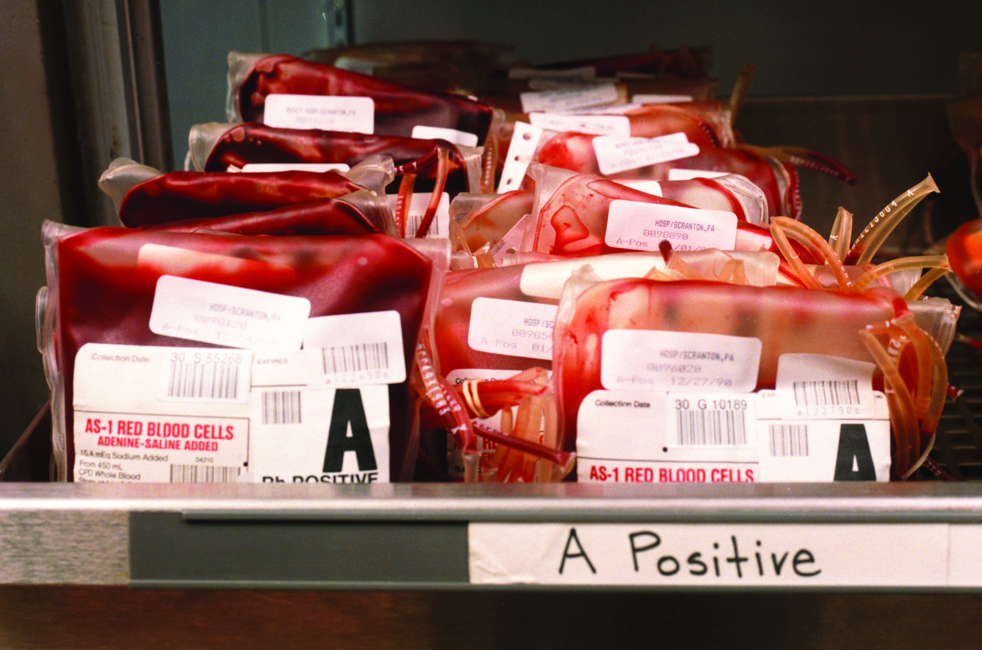
A mistake of the bloody type
Nurse: Mr. Smeggins, I need to clear up some of the answers on your new-patient information form.
Patient: I filled the whole thing out, didn’t I?
Nurse: You did, but a couple of your responses are less than helpful. You do realize that “Helvetica” is not a blood type, right?
Patient: I took a stab at it.
Nurse: You’re not the only one. It turns out that 43% of adults don’t know their blood type, and 62% don’t know their cholesterol level, according to a recent survey by Quest Diagnostics. The 1,004 respondents were more likely to know their bank account balances (75%) or their wifi passwords (74%).
Patient: Hey, that’s right! Mine is Earwiglover122.
Nurse: Great. And can I assume that you’re one of the 30% or so supposedly Web-savvy millennials (ages 20-37 years) who keep lab results in a filing cabinet at home?
Patient: Actually, I have a pile for stuff like that.
Nurse: Fine. Now about your other answers. When we asked about sex, we were not looking for “just last night.”
A case of emission and injection
In what might win “Most Bizarre Attempt at Home Medicine” of 2019, a 33-year-old Irish man was hospitalized after injecting himself with his own semen … in his arm … multiple times … to reduce back pain. Whew. Does this count as holistic medicine?
This at-home remedy did not cure his back pain, shockingly enough. The patient instead developed a subcutaneous abscess after a year and a half of monthly intramuscular and intravenous injections, during which the semen has leaked into the soft tissues. He reported to a Dublin hospital after suffering severe back pain and a swollen arm, and eventually revealed to doctors his miracle cure.
The doctors did some Googling and found studies where rats and rabbits were injected with semen – possibly the research that inspired this trailblazer. Or, possibly, this was just an extreme case of reduce, reuse, and recycle.
In case you’re concerned, the man was given a course of more traditional medicine, and his back pain improved greatly. The patient chose to discharge himself before doctors could drain the “local collection” – perhaps he was proud of his work.
Down the rabbit hole
Imagine sitting at your computer when suddenly the icons begin to move off the screen and hover directly in front of your eyes. Your first thought might be that someone spiked your morning coffee with acid – and you’re not far off.
This curious occurrence happened to a 54-year-old man who was diagnosed with the rare perceptual disorder Alice in Wonderland syndrome (AIWS). AIWS causes people to develop a misperception of their body or surrounding space, and can be caused by a number of things, including migraine.
In this case, the man’s LSD-like visions were caused by a glioblastoma in the left temporal-occipital region of the brain. Tumors there can interfere with spatial perception, hence the temporary trip down the rabbit hole for this patient. After chemotherapy and radiation, the tumor was defeated, and the patient is back to feeling happier than the Mad Hatter at a tea party.
Must have been some party
On Dec. 25 in the Vietnamese province of Quang Tri, a 48-year-old man was taken to a hospital with a case of alcohol poisoning. Specifically, his body contained more than 1,000 times the recommended limit of methanol.
While the two types of alcohol, ethanol and methanol, are both toxic to the human body to some degree, the liver processes methanol differently and more slowly, making it far more dangerous than ethanol, the key ingredient in commercially available alcoholic beverages. Methanol is found in bootleg liquor and in such products as gasoline, paint, ink, and cleaning products. It can cause blindness, nervous system depression, and death.
However, there is a happy ending to this story. To save their patient’s life, his doctors hit upon an ingenious solution – one that would make Homer Simpson proud.
They administered cans of beer.
When the man was admitted, the doctors immediately gave him 3 cans’ worth, and then transfused an additional 12 at the rate of 1 can per hour. The liver will always prioritize processing ethanol over methanol. By feeding the patient a steady stream of relatively friendly and ethanol-rich beer, the doctors had enough time to perform dialysis and remove the methanol from the man’s system.
So, as Homer himself might declare, here’s to alcohol – truly the cause of, and solution to, all of life’s problems.
A mistake of the bloody type
Nurse: Mr. Smeggins, I need to clear up some of the answers on your new-patient information form.
Patient: I filled the whole thing out, didn’t I?
Nurse: You did, but a couple of your responses are less than helpful. You do realize that “Helvetica” is not a blood type, right?
Patient: I took a stab at it.
Nurse: You’re not the only one. It turns out that 43% of adults don’t know their blood type, and 62% don’t know their cholesterol level, according to a recent survey by Quest Diagnostics. The 1,004 respondents were more likely to know their bank account balances (75%) or their wifi passwords (74%).
Patient: Hey, that’s right! Mine is Earwiglover122.
Nurse: Great. And can I assume that you’re one of the 30% or so supposedly Web-savvy millennials (ages 20-37 years) who keep lab results in a filing cabinet at home?
Patient: Actually, I have a pile for stuff like that.
Nurse: Fine. Now about your other answers. When we asked about sex, we were not looking for “just last night.”
#Patients looking for #clinicaltrials
I just hung up with a friend I haven’t seen in decades. Her father has advanced cancer, and while she does not have formal medical training, a passerby wouldn’t know it. Her questions are spot on, her resources are peer reviewed and validated, and her questions I’d more likely expect from trainees in a formal oncology training program than from the director of an elementary level tutoring service.
Her father is fortunately doing well, but she’s searching for the next plan for when the standard drugs ultimately fail. We know they will fail. She’s connected to patient advocacy groups, emailing physicians across the country, and looking into clinical trials with their exhaustive lists of exclusion criteria. She sees the logistic difficulties with trials far from home. She’s hit the key issues we face every day in clinical research, and she’s never stepped foot in a medical school lecture hall.
Amazingly her story is not unique. When cancer hits close to home is when these problems become very clear. This same story could easily have been retold as the narrative of former Vice President Joe Biden and his care for his son. Both my friend and Mr. Biden, in fact, asked me the same question: How do we get the cutting-edge science from major research centers out to the rest of the country?
The Cancer Moonshot initiative has done much to promote collaboration, but one major success has been in the Count Me In initiative, a partnership between the Biden Cancer Initiative, Emerson Collective, the Broad Institute, and the Dana-Farber Cancer Institute. Their goal is to gain access to thousands of patients, collect data on treatment and outcomes, and collect biological specimens. They are not alone, the MSK-IMPACT initiative – led by David B. Solit, MD, at my institution – aims to sequence rare cancers. Both programs have heavily leveraged social media to access and engage patients.
There are of course concerns. Coming from hundreds or thousands of different sites will mean the data will likely be heterogeneous in formatting and quality. How do we ensure the security of patient data? Can we rely on patients and family members to report accurately and without bias? We know there are challenges and upside to crowdsourced patient recruitment.
David Ginsburg, MD, Karl Desch, MD, and colleagues enrolled more than 1,000 students from the University of Michigan to participate in a study on blood clotting factors. This led to many important findings on the genetic basis for coagulopathies, but also was instructive in uncovering a worrisome aspect of online patient registration. The group recorded the time taken for registrants to read the consent form – including whether the participant clicked a hyperlink that was embedded. Nearly a quarter of participants accepted the terms of the 2,833-word document in less than 10 seconds, and less than 3% clicked the hyperlink (Ann Intern Med. 2011 Sep 6;155[5]:316-22).
Are these patients, who we are asking for their partnership and trust, really understanding to what they are agreeing?
Surely there is tremendous altruism on the part of these patients. Their hopes of helping the future of cancer care does have a real track record. Crowdsourcing efforts that were less far reaching in scope made substantial impact in discovering the genetic basis for polycythemia vera. The patients, contacted largely through printed newspaper ads, have helped millions of others. What will happen when we add in the power of social media will be exciting to see – and there is something else that comes with great power, but since I can’t seem to remember what that is, I’ll just search online.
Dr. Viny is with the Memorial Sloan-Kettering Cancer Center, N.Y., where he is an assistant attending physician on the leukemia service and is a clinical researcher in the Ross Levine Lab. Follow him on Twitter @TheDoctorIsVin.
Many misunderstand purpose of tumor profiling research
Although most cancer patients and parents of cancer patients understand that genomic tumor profiling research aims to improve care for future patients, many also believe that the process will benefit present treatment, according to a recent survey conducted at four academic treatment centers.
Misunderstandings were most common among less-educated individuals and those with little genetic knowledge, reported lead author Jonathan M. Marron, MD, MPH, of the Dana-Farber Cancer Institute in Boston and his colleagues.
Previous surveys have shown that “up to 60% of research participants demonstrate evidence of therapeutic misconception,” the investigators wrote in JCO Precision Oncology, referring to “the belief that the primary purpose of research is therapeutic in nature rather than acquisition of generalizable knowledge.”
“Although advances in targeted therapeutics generate great excitement, they may also blur the line between research and clinical care,” the investigators wrote. As such therapeutics become more common, so may misconceptions.
To evaluate current views of genomic tumor profiling research, the investigators surveyed 45 cancer patients and parents of cancer patients at four academic treatment centers. All patients were aged 30 years or younger at enrollment and undergoing tumor profiling; parents were asked to respond if patients were younger than 18 years.
The survey was divided into two sections: basic understanding and comprehensive understanding. To achieve basic understanding, a respondent needed to recognize that “the primary purpose was not to improve the patient’s treatment.” To achieve comprehensive understanding, the respondent needed to recognize four facts: “primary purpose was not to improve patient’s treatment,” “primary purpose was to improve treatment of future patients,” “there may not be direct medical benefit,” and “most likely result of participation was not increased likelihood of cure.”
Forty-four out of 45 survey participants responded. Of these, 30 (68%) demonstrated basic understanding, and 24 (55%) had comprehensive understanding. Respondents with higher education were more likely to answer correctly, with 81% showing basic understanding and 73% showing comprehensive understanding; among less-educated respondents, only half (50%) had basic understanding, and about 1 out of 4 (28%) had comprehensive understanding. Similar disparities were observed among respondents with more versus less genetic knowledge. Almost all respondents (93%) who thought that profiling would help present treatment also believed it would benefit future patients.
Taken as a whole, these findings suggest that therapeutic misconception in genomic tumor profiling research is relatively common, which echoes previous findings. The investigators recommended that clinicians anticipate these knowledge gaps and aim to overcome them.
“Interventional work to improve participant understanding of these complexities and nuances is necessary as sequencing moves from the laboratory to the clinic,” the investigators concluded. “Such work can guide pediatric oncologists in how to manage expectations and best counsel patients and families about the meaning and significance of clinical profiling results.”
The study was funded by Hyundai Hope on Wheels, the Friends for Life Foundation, the Gillmore Fund, National Institutes of Health, and others. The investigators reported financial affiliations with Merck, Millennium, Novartis, Roche, Amgen, and others.
SOURCE: Marron et al. JCO Precis Oncol. 2019 Jan 22. doi: 10.1200/PO.18.00176.
FROM JCO PRECISION ONCOLOGY
Key clinical point: Although most cancer patients and parents of cancer patients understand that genomic tumor profiling research aims to improve care for future patients, many also believe that the process will benefit present treatment.
Major finding: Fifty-five percent of respondents demonstrated comprehensive understanding the purpose of genomic tumor profiling research.
Study details: A survey of 45 cancer patients and parents of cancer patients conducted at four academic treatment centers.
Disclosures: The study was funded by Hyundai Hope on Wheels, the Friends for Life Foundation, the Gillmore Fund, National Institutes of Health, and others. The investigators reported financial affiliations with Merck, Millennium, Novartis, Roche, Amgen, and others.
Source: Marron et al. JCO Precis Oncol. 2019 Jan 22. doi: 10.1200/PO.18.00176.
Device approved to treat PDA in premature infants
weighing as little as 2 pounds.
PDA is a life-threatening opening between two blood vessels leading from the heart and commonly occurs in premature infants, with about one in five infants born prematurely having a hemodynamically significant PDA. The Amplatzer Piccolo Occluder is a self-expanding, wire mesh device that is minimally invasive and is the first device approved for use in very-low-birth-weight infants.
FDA approval was based on results of the ADO II AS trial, which evaluated the device in 50 patients with PDA who were older than 3 days. In addition, the safety and efficacy of the Amplatzer Piccolo Occluder was supported by a continued access protocol involving 150 more patients.
“This approval is a potentially life-saving advance for the very smallest premature infants that will help us treat these delicate babies who might otherwise not be able to survive,” said Evan Zahn, MD, principal investigator of ADO II AS and director of the congenital heart program at Cedars-Sinai’s Smidt Heart Institute in Los Angeles.
Find the full press release on the Abbott website.
Early lead exposure tied to greater psychopathology in adulthood
Lead exposure during childhood appears tied to a significant increase in the risk of psychopathology in adulthood, results of a multidecade, prospective cohort study show.
“These results suggest that early life lead exposure in the era of leaded gasoline experienced by individuals who are currently adults may have contributed to subtle, lifelong differences in emotion and behavior that are detectable at least up to 38 years of age,” Aaron Reuben and his coauthors wrote in JAMA Psychiatry.
The ongoing Dunedin longitudinal cohort study in New Zealand has followed 1,037 individuals born during 1972-1973. Of these individuals, 579 were tested for lead exposure at 11 years of age. The study assessed their mental health at 18, 21, 26, 32, and 38 years of age.
“Although follow-up studies of lead-tested children have reported the persistence of lead-related cognitive deficits well into adulthood, apart from antisocial outcomes, the long-term mental and behavioral health consequences of early life lead exposure have not been fully characterized,” wrote Mr. Reuben, a PhD student in the department of psychology and neuroscience at Duke University in Durham, N.C., and his coauthors.
Researchers saw that, for each 5-mcg/dL increase in childhood blood lead level, there was a significant 1.34-point increase in general psychopathology (P = 0.03), which was largely driven by a 1.41-point increase in internalizing (P = 0.02) and 1.30-point increase in thought-disorder symptoms (P = 0.04). Those associations were seen after adjustment for covariates, such as family socioeconomic status, maternal IQ, and family history of mental illness.
Adults who had higher lead exposure during childhood also were described by their informants – close friends or family members – as being significantly more neurotic, less agreeable, and less conscientious. However, they showed no significant differences in extroversion or in openness to experience, compared with those with less lead exposure.
“These results suggest that early-life lead exposure in the era of leaded gasoline experienced by individuals who are currently adults may have contributed to subtle, lifelong differences in emotion and behavior that are detectable at least up to 38 years of age,” the authors wrote.
They noted that the size of the effect was around one-third the size of the associations seen between psychopathology and other risk factors, such as family history of mental illness and childhood maltreatment. However, the effects of lead exposure on adult psychopathology were similar to its effects on IQ and stronger than the associations seen between lead exposure and criminal offending.
The researchers also examined how early these psychopathology symptoms could be detected with use of parent- and teacher-reported measures of antisocial behavior, hyperactivity, and internalizing from 11 years of age. This showed that individuals with higher lead exposure scored higher on these measures even at 11 years of age, “suggesting that
Mr. Reuben and his associates cited several limitations. One is that the study used a cohort that was predominantly white and born in the 1970s. Also, as an observational study, it does not establish causality between lead exposure and psychopathology.
Nevertheless, they wrote, the study results suggest that adult patients who were exposed to high levels of lead as children might benefit from increased screening and access to mental health services.
The Dunedin study is supported by the New Zealand Health Research Council and the New Zealand Ministry of Business, Innovation, and Employment. This study was supported by several entities, including the National Institute on Aging, the U.K. Medical Research Council, the National Institute of Child Health and Human Development, and the National Institute of Environmental Health Sciences. The authors reported no conflicts of interest.
SOURCE: Reuben A et al. JAMA Psychiatry. 2019 Jan 23. doi: 10.1001/jamapsychiatry.2018.4192.
FROM JAMA PSYCHIATRY
Key clinical point: Higher lead exposure in childhood is linked to psychopathology in adulthood.
Major finding: Children who experienced more lead exposure in childhood show more internalizing and thought-disorder symptoms in adulthood.
Study details: Longitudinal cohort study of 579 individuals.
Disclosures: The Dunedin study is supported by the New Zealand Health Research Council and the New Zealand Ministry of Business, Innovation, and Employment. This study was supported by several entities, including the National Institute on Aging, the U.K. Medical Research Council, the National Institute of Child Health and Human Development, and the National Institute of Environmental Health Sciences. The authors reported no conflicts of interest.
Source: Reuben A et al. JAMA Psychiatry. 2019 Jan 23. doi: 10.1001/jamapsychiatry.2018.4192.
Cost-of-care counseling can be done, but does it help?
Here’s a novel idea: Help patients figure out how large a bite health care will take from their life savings.
A pilot study of a randomized intervention showed that practice-based counseling of patients about the actual out-of-pocket costs of chemotherapy is both “feasible and acceptable.”
But the larger question about whether any of it does any good remains unanswered, reported Sheetal M. Kircher, MD, from Northwestern University in Chicago and her colleagues.
“The majority of participants found the FC [financial counseling] consultation helpful, but they felt the same about their finances after the intervention. Although it is reassuring that no participants felt worse about their cancer costs after the intervention, the variability in perceived value of the FC consultation highlights that the content of a financial intervention needs to be tailored to the patient’s individual needs,” they wrote in the Journal of Oncology Practice.
The investigators conducted a randomized trial with 95 patients at the Robert H. Lurie Comprehensive Cancer Center at Northwestern to study whether financial counseling about the estimated total costs to them of one cycle of chemotherapy could help patients weather financial distress.
The participants first completed a baseline assessment of their financial distress, health-related quality of life, and degree of health insurance literacy. They were then randomized to either standard of care or to a financial-counseling intervention consisting of education, financial-assistance screening, and a tool for estimating total billed charges and out-of-pocket costs. Participants in the experimental arm received a phone call and in-person visit from a financial counselor.
Financial distress was measured using the Comprehensive Score for Financial Toxicity (COST), InCharge Financial Distress/Financial Well-Being Scale, and a single European Organisation for Research and Treatment of Cancer (EORTC) question.
Patients in the standard-of-care arm had access to financial counselors on request, but they were not automatically assigned counseling.
Of the 95 patients randomized, 65 completed both the baseline and follow-up assessments. The investigators noted that participants tended to have more advanced disease; six patients in the standard-of-care arm and five in financial-counseling arm died before they could complete the follow-up assessment.
Of the 43 participants randomized to counseling, 42 (98%) completed the phone call, 20 (47%) completed the in-person visit, and 13 (30%) completed the entire intervention.
At the second, follow-up assessment, 83% of participants in the counseling arm said they were comfortable with answering questions about how the costs of treatment affected their pocketbooks, and 76% reported that they did not have difficulty understanding the estimation tool, and 88% said that the financial counselor helped them to understand their out-of-pocket costs “somewhat” or “a lot.”
However, when they were asked how they felt about paying for their care after completing the intervention, 76% said they felt about the same, 18% said they felt “a little better,” and only 6% said they felt “much better.”
In addition there were no statistically significant differences in any of the financial distress measures between the intervention and standard-of-care groups.
The investigators speculated that the study was underpowered to detect differences in COST (financial toxicity) scores.
High financial distress was significantly associated with worse emotional functioning (P = .0189), whereas being married or in a cohabiting relationship was associated with less financial distress (P = .0092).
“Future studies should aim to better understand patient perspectives and preferences around discussing transparent cost data and design system-level interventions to identify and assist patients efficiently with their specific financial concerns,” Dr. Kircher and her colleagues concluded.
The study was supported by the Robert H. Lurie Comprehensive Cancer Center and the Cancer Survivorship Institute of Northwestern University. Dr. Kircher disclosed stock or ownership interest in Penrose TherapeuTx. Five coauthors reported research funding, travel expenses, and/or honoraria from multiple other companies, as well as consulting/roles.
SOURCE: Kircher SM et al. J Oncol Pract. 2019 Jan 9. doi: 10.1200/JOP.18.00270.
FROM JOURNAL OF ONCOLOGY PRACTICE
Key clinical point: A practice-based, financial-counseling intervention did not significantly change how patients felt about the costs of cancer care, compared with the standard of care.
Major finding: Participants found the intervention acceptable, but there were no significant differences in measures of financial distress between the intervention and control arms.
Study details: Randomized study in 95 patients assigned to financial counseling or standard of care.
Disclosures: The study was supported by the Robert H. Lurie Comprehensive Cancer Center and the Cancer Survivorship Institute of Northwestern University. Dr. Kircher disclosed stock or ownership interest in Penrose TherapeuTx. Five coauthors reported research funding, travel expenses, and/or honoraria from multiple other companies, as well as consulting/roles.
Source: Kircher SM et al. J Oncol Pract. 2019 Jan 9. doi: 10.1200/JOP.18.00270.
Pembrolizumab bests chemo in PD-L1-positive esophageal cancer
SAN FRANCISCO – The immune checkpoint inhibitor pembrolizumab (Keytruda) may soon unseat chemotherapy as standard second-line therapy for certain advanced cancers of the esophagus or gastroesophageal junction, according to data from the global phase 3 KEYNOTE-181 trial.

“Patients with advanced esophageal cancer after first-line therapy have a poor prognosis and limited treatment options,” said lead investigator Takashi Kojima, MD, of the National Cancer Center Hospital East, Kashiwa, Japan. “Taxanes and irinotecan are commonly used after first-line chemotherapy; however, no overall survival benefit has been demonstrated for chemotherapy in a phase 3 study.”
The 628 patients in KEYNOTE-181 were randomly assigned to chemotherapy (paclitaxel, docetaxel, or irinotecan, left to investigator’s choice) or pembrolizumab, an antibody to programmed death 1 (PD-1). Currently, pembrolizumab is approved in the United States for use as third- or later-line therapy for gastric or gastroesophageal junction cancer that is positive for programmed death ligand 1 (PD-L1) as defined by a combined positive score (CPS) of 1 or greater, among many other indications.
Main trial results reported at the 2019 GI Cancers Symposium showed that among patients with high PD-L1 expression, defined by a CPS of 10 or higher, pembrolizumab reduced risk of death by about one-third, prolonging survival by 2.6 months. The difference met the predefined threshold for statistical significance.
There was a more modest, nonsignificant benefit among patients with tumors having squamous cell carcinoma histology and among the entire intention-to-treat population.
The rate of treatment-related adverse events of grade 3-5 was roughly half as high with pembrolizumab versus chemotherapy (18.2% vs. 40.9%).
“These data suggest that pembrolizumab should be considered a new standard of care for patients with PD-L1 CPS of 10 or greater metastatic esophageal cancer in the second-line setting,” Dr. Kojima concluded.
Implications for practice
“In the intention-to-treat population, the KEYNOTE-181 study failed to meet its primary endpoint of overall survival, so pembrolizumab is not indicated in unselected esophageal cancer patients,” said invited discussant Harry H. Yoon, MD, cochair of the Esophageal/Gastric Cancer Disease Group at the Mayo Clinic Cancer Center, Rochester, Minn.

For the patients with squamous histology, the negative findings are unlikely to be due to underpowering and may instead be related to the trial’s use of multiple primary endpoints, in his opinion. “Some may advocate using pembrolizumab off protocol [in this population], particularly for patients who cannot tolerate chemotherapy, because it is after all better tolerated than chemo. This can be a discussion point for guideline committees,” he said.
The results for the group with PD-L1 CPS scores of 10 or higher are statistically significant and clinically meaningful, as well as internally valid – with the caveat that patients were not stratified by PD-L1 status and some favorable risk factors were more common in the pembrolizumab group, according to Dr. Yoon. The 43% survival rate at 12 months translates to a number needed to treat of just four patients for one patient to be alive at that time point.
“A multivariate analysis could help clarify whether the positive results in the PD-L1 CPS 10-or-higher subgroup are explained by a higher frequency of favorable patient characteristics in the pembrolizumab arm,” he noted. “The strength of those results could influence guideline recommendations and implementation in clinical practice.”
Subgroup analyses suggested benefit was mainly seen in Asian patients, who tend to have higher prevalence of squamous tumors, Dr. Yoon said. Potential molecular differences at play here may be elucidated by ongoing research.
Ultimately, the findings in the PD-L1 CPS 10-or-higher group have potential implications for biomarker testing. “For the patient in front of me, I currently order PD-L1 and HER2 at first metastatic diagnosis in gastroesophageal adenocarcinomas,” he elaborated. “It’s reasonable to consider a practice change. This means ordering PD-L1 at first metastatic diagnosis in patients with squamous carcinoma of the esophagus. This would also mean some pathology labs may need to report a more detailed PD-L1 CPS score, if they don’t already.”
These findings also have potential implications for treatment. “For the second-line setting, for squamous carcinoma of the esophagus, esophageal adenocarcinomas, and Siewert 1 adenocarcinomas with a PD-L1 CPS of 10 or more, it’s reasonable to consider pembrolizumab,” Dr. Yoon noted. “This should be discussed within guideline committees, and the results will be submitted to regulatory authorities, who will have access to more detailed data. It’s possible that these recommendations could be modified in the near future.”
Study details
As KEYNOTE-181 had three primary endpoints (overall survival in each of three populations), P values required for statistical significance were defined accordingly. “The study was positive if one of the primary endpoints was met,” Dr. Kojima explained at the symposium, which was sponsored by the American Gastroenterological Association, the American Society for Clinical Oncology, the American Society for Radiation Oncology, and the Society of Surgical Oncology.
Some 35% of trial patients had a PD-L1 CPS of 10 or greater. In this population, median overall survival was 9.3 months with pembrolizumab and 6.7 months with chemotherapy. The hazard ratio was 0.69, with the P value (.0074) meeting that predefined for statistical significance in this population (less than or equal to .0085). The 12-month rate of overall survival was 43% and 20%, respectively.
About 64% of trial patients had squamous cell carcinoma histology. In this population, median overall survival was 8.2 months with pembrolizumab and 7.1 months with chemotherapy. The hazard ratio was 0.78, but the P value (.0095) did not meet that predefined for statistical significance in this group (less than or equal to .0077).
Finally, in the entire intention-to-treat population, median overall survival was identical, at 7.1 months, with pembrolizumab and with chemotherapy. The hazard ratio was 0.89 in favor of the antibody, but the P value (.0560) did not meet that predefined for statistical significance in this population (less than or equal to .0077).
A similar pattern was seen for other outcomes, with patients having PD-L1 CPS greater than or equal to 10 deriving greatest benefit from pembrolizumab over chemotherapy in terms of progression-free survival (hazard ratio, 0.73), response rate (21.5% vs. 6.1%), and median duration of response (9.3 vs. 7.7 months).
“Toxicity profiles were in line with previous reports of each treatment. No new safety signals were observed,” Dr. Kojima reported. Pembrolizumab was associated with a higher rate of immune-mediated and infusion reactions (23.2% vs. 7.4%), but lower rates of most gastrointestinal and hematologic adverse events.
Dr. Kojima disclosed ties to Oncolys BioPharma, Astellas, Amgen, MSD, Ono Pharmaceutical, and Shionogi. Merck Sharp & Dohme sponsored the trial.
SOURCE: Kojima T et al. GI Cancers Symposium Abstract 2, https://meetinglibrary.asco.org/record/169377/abstract.
SAN FRANCISCO – The immune checkpoint inhibitor pembrolizumab (Keytruda) may soon unseat chemotherapy as standard second-line therapy for certain advanced cancers of the esophagus or gastroesophageal junction, according to data from the global phase 3 KEYNOTE-181 trial.
“Patients with advanced esophageal cancer after first-line therapy have a poor prognosis and limited treatment options,” said lead investigator Takashi Kojima, MD, of the National Cancer Center Hospital East, Kashiwa, Japan. “Taxanes and irinotecan are commonly used after first-line chemotherapy; however, no overall survival benefit has been demonstrated for chemotherapy in a phase 3 study.”
The 628 patients in KEYNOTE-181 were randomly assigned to chemotherapy (paclitaxel, docetaxel, or irinotecan, left to investigator’s choice) or pembrolizumab, an antibody to programmed death 1 (PD-1). Currently, pembrolizumab is approved in the United States for use as third- or later-line therapy for gastric or gastroesophageal junction cancer that is positive for programmed death ligand 1 (PD-L1) as defined by a combined positive score (CPS) of 1 or greater, among many other indications.
Main trial results reported at the 2019 GI Cancers Symposium showed that among patients with high PD-L1 expression, defined by a CPS of 10 or higher, pembrolizumab reduced risk of death by about one-third, prolonging survival by 2.6 months. The difference met the predefined threshold for statistical significance.
There was a more modest, nonsignificant benefit among patients with tumors having squamous cell carcinoma histology and among the entire intention-to-treat population.
The rate of treatment-related adverse events of grade 3-5 was roughly half as high with pembrolizumab versus chemotherapy (18.2% vs. 40.9%).
“These data suggest that pembrolizumab should be considered a new standard of care for patients with PD-L1 CPS of 10 or greater metastatic esophageal cancer in the second-line setting,” Dr. Kojima concluded.
Implications for practice
“In the intention-to-treat population, the KEYNOTE-181 study failed to meet its primary endpoint of overall survival, so pembrolizumab is not indicated in unselected esophageal cancer patients,” said invited discussant Harry H. Yoon, MD, cochair of the Esophageal/Gastric Cancer Disease Group at the Mayo Clinic Cancer Center, Rochester, Minn.
For the patients with squamous histology, the negative findings are unlikely to be due to underpowering and may instead be related to the trial’s use of multiple primary endpoints, in his opinion. “Some may advocate using pembrolizumab off protocol [in this population], particularly for patients who cannot tolerate chemotherapy, because it is after all better tolerated than chemo. This can be a discussion point for guideline committees,” he said.
The results for the group with PD-L1 CPS scores of 10 or higher are statistically significant and clinically meaningful, as well as internally valid – with the caveat that patients were not stratified by PD-L1 status and some favorable risk factors were more common in the pembrolizumab group, according to Dr. Yoon. The 43% survival rate at 12 months translates to a number needed to treat of just four patients for one patient to be alive at that time point.
“A multivariate analysis could help clarify whether the positive results in the PD-L1 CPS 10-or-higher subgroup are explained by a higher frequency of favorable patient characteristics in the pembrolizumab arm,” he noted. “The strength of those results could influence guideline recommendations and implementation in clinical practice.”
Subgroup analyses suggested benefit was mainly seen in Asian patients, who tend to have higher prevalence of squamous tumors, Dr. Yoon said. Potential molecular differences at play here may be elucidated by ongoing research.
Ultimately, the findings in the PD-L1 CPS 10-or-higher group have potential implications for biomarker testing. “For the patient in front of me, I currently order PD-L1 and HER2 at first metastatic diagnosis in gastroesophageal adenocarcinomas,” he elaborated. “It’s reasonable to consider a practice change. This means ordering PD-L1 at first metastatic diagnosis in patients with squamous carcinoma of the esophagus. This would also mean some pathology labs may need to report a more detailed PD-L1 CPS score, if they don’t already.”
These findings also have potential implications for treatment. “For the second-line setting, for squamous carcinoma of the esophagus, esophageal adenocarcinomas, and Siewert 1 adenocarcinomas with a PD-L1 CPS of 10 or more, it’s reasonable to consider pembrolizumab,” Dr. Yoon noted. “This should be discussed within guideline committees, and the results will be submitted to regulatory authorities, who will have access to more detailed data. It’s possible that these recommendations could be modified in the near future.”
Study details
As KEYNOTE-181 had three primary endpoints (overall survival in each of three populations), P values required for statistical significance were defined accordingly. “The study was positive if one of the primary endpoints was met,” Dr. Kojima explained at the symposium, which was sponsored by the American Gastroenterological Association, the American Society for Clinical Oncology, the American Society for Radiation Oncology, and the Society of Surgical Oncology.
Some 35% of trial patients had a PD-L1 CPS of 10 or greater. In this population, median overall survival was 9.3 months with pembrolizumab and 6.7 months with chemotherapy. The hazard ratio was 0.69, with the P value (.0074) meeting that predefined for statistical significance in this population (less than or equal to .0085). The 12-month rate of overall survival was 43% and 20%, respectively.
About 64% of trial patients had squamous cell carcinoma histology. In this population, median overall survival was 8.2 months with pembrolizumab and 7.1 months with chemotherapy. The hazard ratio was 0.78, but the P value (.0095) did not meet that predefined for statistical significance in this group (less than or equal to .0077).
Finally, in the entire intention-to-treat population, median overall survival was identical, at 7.1 months, with pembrolizumab and with chemotherapy. The hazard ratio was 0.89 in favor of the antibody, but the P value (.0560) did not meet that predefined for statistical significance in this population (less than or equal to .0077).
A similar pattern was seen for other outcomes, with patients having PD-L1 CPS greater than or equal to 10 deriving greatest benefit from pembrolizumab over chemotherapy in terms of progression-free survival (hazard ratio, 0.73), response rate (21.5% vs. 6.1%), and median duration of response (9.3 vs. 7.7 months).
“Toxicity profiles were in line with previous reports of each treatment. No new safety signals were observed,” Dr. Kojima reported. Pembrolizumab was associated with a higher rate of immune-mediated and infusion reactions (23.2% vs. 7.4%), but lower rates of most gastrointestinal and hematologic adverse events.
Dr. Kojima disclosed ties to Oncolys BioPharma, Astellas, Amgen, MSD, Ono Pharmaceutical, and Shionogi. Merck Sharp & Dohme sponsored the trial.
SOURCE: Kojima T et al. GI Cancers Symposium Abstract 2, https://meetinglibrary.asco.org/record/169377/abstract.
REPORTING FROM THE 2019 GI CANCERS SYMPOSIUM
Key clinical point: .
Major finding: Among patients with a PD-L1 combined positive score of 10 or higher, median overall survival was 9.3 months with pembrolizumab and 6.7 months with chemotherapy (hazard ratio, 0.69; P = .0074).
Study details: A phase 3 randomized controlled trial among 628 patients having progression after first-line therapy for advanced cancer of the esophagus or GEJ (KEYNOTE-181).
Disclosures: Dr. Kojima disclosed ties to Oncolys BioPharma, Astellas, Amgen, MSD, Ono Pharmaceutical, and Shionogi. Merck Sharp & Dohme sponsored the trial.
Source: Kojima T et al. GI Cancers Symposium, Abstract 2.
Bedbugs in the Workplace


Product Update: Spectrum, Viera, YO, and SmartCurve
PREIMPLANTATION GENETIC TESTING
In a retrospective study published in Fertility and Sterility, Spectrum, a single-nucleotide polymorphism (SNP)-based PGT-A technology, was successful in screening all 24 chromosomes to provide comprehensive embryo aneuploidy results. Natera says that the study results showed that use of Spectrum PGT-A during IVF led to excellent implantation (70%), clinical pregnancy (71%), and live birth (65%) rates during single embryo transfer.
Spectrum evaluates the number of chromosomes in embryos to detect extra or missing chromosomes and screens for inherited genetic disorders to help provide the best chance of transferring a healthy embryo with the correct number of chromosomes.
FOR MORE INFORMATION, VISIT: https://www.natera.com/spectrum
PORTABLE BREAST ULTRASOUND

Viera is a wireless, handheld breast ultrasound scanner that Hologic says produces exceptional image quality. The scanner uses a 14-4 MHz linear transducer, contains 192 elements, and has 4 parallel software beamformers. It utilizes spatial compounding to reduce image noise and speckle. Presets are available for breast, dense breast, and interventional procedures with B, M, power Doppler, color Doppler, and needle enhancement modes. On-demand high-resolution images are transmitted wirelessly to smart devices and patient archive systems (PACS) in the office, exam room, or surgical suite, or to the Cloud for efficient documentation. Smart device platforms include iOS and Android devices using WiFi and Bluetooth connectivity. The system includes a 1.2-lb scanner, 2 rechargeable batteries, and a charger with global AC adapter.
FOR MORE INFORMATION, VISIT: https://www.vieraportableultrasound.com
HOME SPERM TEST
The customer downloads the smart-phone app and acquires the YO Kit. After collecting a semen sample, he uses a pipette from the kit to place semen on a slide, which is slipped into the Yo Clip. The clip slides onto the smartphone, which uses its camera to take a high-resolution video. Test results and the sperm video appear in about 2 minutes.
At $59.95, the YO Kit includes 2 tests, in case a second sample is desired.
FOR MORE INFORMATION, VISIT:
https://www.yospermtest.com
Continue to Improving the Mammography Experience…
IMPROVING THE MAMMOGRAPHY EXPERIENCE

SmartCurve is standard on Hologic’s new 3Dimensions™ mammography system and as an enhancement option to existing Hologic Selenia®Dimensions® systems.
FOR MORE INFORMATION, VISIT:https://www.smartcurvesystem.com
Share your thoughts! Send your Letter to the Editor to [email protected]. Please include your name and the city and state in which you practice.
PREIMPLANTATION GENETIC TESTING
In a retrospective study published in Fertility and Sterility, Spectrum, a single-nucleotide polymorphism (SNP)-based PGT-A technology, was successful in screening all 24 chromosomes to provide comprehensive embryo aneuploidy results. Natera says that the study results showed that use of Spectrum PGT-A during IVF led to excellent implantation (70%), clinical pregnancy (71%), and live birth (65%) rates during single embryo transfer.
Spectrum evaluates the number of chromosomes in embryos to detect extra or missing chromosomes and screens for inherited genetic disorders to help provide the best chance of transferring a healthy embryo with the correct number of chromosomes.
FOR MORE INFORMATION, VISIT: https://www.natera.com/spectrum
PORTABLE BREAST ULTRASOUND
Viera is a wireless, handheld breast ultrasound scanner that Hologic says produces exceptional image quality. The scanner uses a 14-4 MHz linear transducer, contains 192 elements, and has 4 parallel software beamformers. It utilizes spatial compounding to reduce image noise and speckle. Presets are available for breast, dense breast, and interventional procedures with B, M, power Doppler, color Doppler, and needle enhancement modes. On-demand high-resolution images are transmitted wirelessly to smart devices and patient archive systems (PACS) in the office, exam room, or surgical suite, or to the Cloud for efficient documentation. Smart device platforms include iOS and Android devices using WiFi and Bluetooth connectivity. The system includes a 1.2-lb scanner, 2 rechargeable batteries, and a charger with global AC adapter.
FOR MORE INFORMATION, VISIT: https://www.vieraportableultrasound.com
HOME SPERM TEST
The customer downloads the smart-phone app and acquires the YO Kit. After collecting a semen sample, he uses a pipette from the kit to place semen on a slide, which is slipped into the Yo Clip. The clip slides onto the smartphone, which uses its camera to take a high-resolution video. Test results and the sperm video appear in about 2 minutes.
At $59.95, the YO Kit includes 2 tests, in case a second sample is desired.
FOR MORE INFORMATION, VISIT:
https://www.yospermtest.com
Continue to Improving the Mammography Experience…
IMPROVING THE MAMMOGRAPHY EXPERIENCE
SmartCurve is standard on Hologic’s new 3Dimensions™ mammography system and as an enhancement option to existing Hologic Selenia®Dimensions® systems.
FOR MORE INFORMATION, VISIT:https://www.smartcurvesystem.com
Share your thoughts! Send your Letter to the Editor to [email protected]. Please include your name and the city and state in which you practice.