User login
All good things ...
The last few years have flown by for us as coeditors of the ACS Surgery News. It is often said that the perceived acceleration of time is a phenomenon of age as each year that goes by represents an ever decreasing percentage of one’s remaining time on earth.
As we age, we may come to feel that we have outlived our time and culture. What was certain yesterday is indeterminate today and likely completely wrong tomorrow. Among those things is the economic viability of print media. Some of you may remember the line from the old movie “Ghostbusters” (old: 1984!) in which Egon makes the statement, “Print is dead.” He was a few decades off, but even Gutenberg would have to admit that technology trumps almost everything when it comes to the written word.
So, ACS Surgery News comes to an end with this issue. We would like to believe that the editors past and present – Lazar Greenfield, Bing Rikkers, Karen Deveney, and Tyler Hughes – all aided you as surgeons in some small way. Our intent was always to inform and occasionally to entertain lightly.
The fact that the “Official Newspaper of the American College of Surgeons” is passing from the scene is, we hope, a reflection of technology and economics and not that our efforts were in vain. Behind the scenes were dozens of skilled reporters who did interviews and summarized papers. Our managing editor during our time as coeditors has been Therese Borden, who has been largely responsible for the quality and integrity of what was reported herein. If you have learned something unexpectedly in our newspaper, Therese has actually been the one behind the scenes making that accessible to you. Both of us are deeply grateful for her superb expertise and eternally positive attitude.
Of course, the American College of Surgeons was always the moving force behind this paper. We like to think that the value of the newspaper was largely because the college, with its dedication to surgery with skill and fidelity to all, gave us the credibility other such newspapers just don’t have. As editors, our primary goal has always been to report without concern for anything other than what is useful to the surgeon.
Although ACS has many other publications and products that serve the practicing surgeon, we do feel that ACS Surgery News provided seamless access for surgeons to learn about emerging techniques and ideas. The ACS leadership agrees. After considerable thought and discussion about possible digital replacements, we have agreed to use a platform that is already available to us and easy to use: the ACS Communities. A new Community, named the ACS Emerging News Community, is born.
We will continue as coeditors and offer a commentary every other month. The Editorial Board, the invaluable consultants to ACS Surgery News, will contribute short articles describing the best presentation that they have heard at a recent major surgical meeting and describe why it is important or summarize an article of importance from a recent major journal. Surgeons already on the General Surgery Community will receive the new community monthly unless they choose to unsubscribe. Community members can also respond to or query the authors of the articles and commentaries if they so desire. The ACS Emerging News Community will commence early in 2019, so look for it in your inbox then.
Many thanks to all of you for reading the ACS Surgery News when you had the time and special thanks to those who wrote in the paper or wrote to the paper to tell us how we were doing. As ACS Surgery News sunsets, we have no final words of great import that you can laminate and put in your wallets, purses, or on your computers. Whatever transpires in that as-yet-undiscovered country (the future), surgery will always boil down to those willing to care for a patient enough to cut to the cure with compassion, regardless of all other considerations. Good luck to you all. We’ll see you in the cloud.
Dr. Deveney is professor of surgery emerita in the department of surgery at Oregon Health & Science University, Portland. She is the coeditor of ACS Surgery News. Dr. Hughes is a clinical professor in the department of surgery and the director of medical education at the University of Kansas, Salina, and coeditor of ACS Surgery News.
Key takeaways regarding MPFS and QPP final rule posted
The American College of Surgeons (ACS) has posted a document that outlines key changes in the Centers for Medicare & Medicaid Services (CMS) final rule for the 2019 Medicare Physician Fee Schedule (MPFS) and the 2019 Quality Payment Program (QPP). The final rule and its effects on payment to surgeons will be described in greater detail in the January 2019 issue of the Bulletin, and the ACS will continue to develop resources to assist Fellows in meeting the requirements for these programs.
The document is available at bit.ly/2PH566U.
For more information, contact the Regulatory and Quality Affairs team, ACS Division of Advocacy and Health Policy, at [email protected].
ACS Introduces New Video: The Future. Through the Eyes of a Surgeon
As health care changes, it is critical that surgeons continue to have a strong voice and seat at the table in all patient care decisions. A video encouraging Fellows to become actively involved in helping the American College of Surgeons (ACS) take bold steps and speak with a unified voice on behalf of patients was released during Clinical Congress. The ACS encourages Fellows to share the video with colleagues and at your chapter meetings.
View the video on the ACS website at facs.org/member-services/through-the-eyes.
Call for nominations for ACS Officers-Elect and ACS Board of Regents
The American College of Surgeons (ACS) 2019 Nominating Committee of the Fellows (NCF) and the Nominating Committee of the Board of Governors (NCBG) will be selecting nominees for leadership positions in the College as follows.
Call for nominations for Officers-Elect
The 2019 NCF will select nominees for the three Officers-Elect positions of the ACS: President-Elect, First Vice-President-Elect, and Second Vice-President-Elect. The deadline for submitting nominations is February 22, 2019.
Criteria for consideration
The NCF will use the following guidelines when considering potential candidates:
- Nominees must be loyal members of the College who have demonstrated outstanding integrity and an unquestioned devotion to the highest principles of surgical practice.
- Nominees must have demonstrated leadership qualities, such as service and active participation on ACS committees or in other areas of the College.
- The ACS encourages consideration of women and underrepresented minorities for all leadership positions.
All nominations must include the following:
- A letter/letters of nomination
- A current curriculum vitae (CV)
- The name of one individual who can serve as a reference
In addition, nominations for President-Elect must include the following:
- A personal statement from the candidate detailing their ACS service and interest in the position
Further details
Entities such as surgical specialty societies, ACS Advisory Councils, ACS Committees, and ACS chapters that provide a letter of nomination must provide a description of their selection process and the total list of applicants reviewed.
Any attempt to contact or influence members of the NCF by a candidate or on behalf of a candidate will be viewed negatively and may result in disqualification. Applications submitted without the requested information will not be considered.
Nominations must be submitted to [email protected]. If you have any questions, contact Emily Kalata, staff liaison for the NCBG, at 312-202-5360 or [email protected].
Call for nominations for Board of Regents
The 2019 NCBG will select nominees for two pending vacancies on the Board of Regents to be filled at Clinical Congress 2019. The deadline for submitting nominations is February 22, 2019.
Criteria for consideration
The NCBG will use the following guidelines when considering potential candidates:
- Nominees must be loyal members of the College who have demonstrated outstanding integrity and an unquestioned devotion to the highest principles of surgical practice.
- Nominees must have demonstrated leadership qualities, such as service and active participation on ACS committees or in other areas of the College.
- The ACS encourages consideration of women and underrepresented minorities for all leadership positions.
- Only individuals who are currently and expected to remain in active surgical practice for their entire term may be nominated for election or reelection to the Board of Regents.
The NCBG recognizes the importance of the Board of Regents representing all who practice surgery in both academic and community practice, regardless of practice location or configuration. Nominations are open to surgeons of all specialties, but particular consideration will be given in this nomination cycle to the following specialties:
- Burn and critical care surgery
- Gastrointestinal surgery
- General surgery
- Surgical oncology
- Transplant surgery
- Trauma surgery
- Vascular surgery
All nominations must include the following:
- A letter of nomination
- A personal statement from the candidate detailing their ACS service and interest in the position
- A current CV
- The name of one individual who can serve as a reference
Further details
Entities such as surgical specialty societies, ACS Advisory Councils, ACS Committees, and ACS chapters who wish to provide a letter of nomination must provide at least two nominees, and a description of their selection process, along with the total list of applicants reviewed.
Any attempt to contact or influence members of the NCBG by a candidate or on behalf of a candidate will be viewed in a negative manner and may result in disqualification. Applications submitted without the requested information will not be considered.
Nominations may be submitted to [email protected]. If you have any questions, contact Emily Kalata, staff liaison for the NCBG, at 312-202-5360 or [email protected].
For information only, the current members of the Board of Regents who will be considered for reelection are (all MD, FACS): Anthony Atala, James W. Gigantelli, and Fabrizio Michelassi.
New Regents, B/G Executive Committee Members Elected
The Board of Governors (B/G) of the American College of Surgeons (ACS) elected two new members of the Board of Regents at the October 24 Annual Business Meeting of the Members.
Lena M. Napolitano, MD, FACS, FCCP, FCCM, is the Massey Foundation Professor of Surgery; founding division chief, acute care surgery; and director, surgical critical care, department of surgery, University of Michigan Health System, Ann Arbor. A Fellow of the ACS since 1995, Dr. Napolitano has been a tireless volunteer for the College and has served in several important leadership roles within the organization, including as Chair of the B/G.
Kenneth W. Sharp, MD, FACS, is professor of surgery and vice-chair, department of surgery, Vanderbilt University Medical Center, Nashville, TN, and is a highly regarded surgical educator and mentor. He became an ACS Fellow in 1987 and has subsequently served in many roles for the ACS, starting as the Young Surgeon Representative for the Tennessee Chapter in 1989 and rising to serve on the ACS B/G.
The B/G has elected Steven C. Stain, MD, FACS, Henry and Sally Schaffer Chair, department of surgery, Albany Medical Center, NY, to serve as its Chair; he previously was Vice-Chair. The newly elected Vice-Chair is Daniel L. Dent, MD, FACS, Distinguished Teaching Professor, general surgery residency program director, and professor of surgery, University of Texas Health School of Medicine, San Antonio; he previously was Secretary. The new Secretary is Ronald J. Weigel, MD, PhD, FACS, professor and chair of surgery, associate vice-president for UI Health Alliance, professor of surgery-surgical oncology and endocrine surgery, professor of biochemistry, professor of anatomy and cell biology, and professor of molecular physiology and biophysics, University of Iowa, Iowa City.
Other newly elected members of the B/G Executive Committee include Andre R. Campbell, MD, FACS, FACP, FCCM, professor of surgery, division of general surgery, director, surgery clerkship, and director, surgical critical care fellowship, University of California-San Francisco; Taylor Sohn Riall, MD, PhD, FACS, professor and chief, division of general surgery and surgical oncology, University of Arizona College of Medicine, Tucson; and Mika N. Sinanan, MD, PhD, FACS, a general surgeon, UW Medical Center and Seattle Cancer Care Alliance, and professor of general surgery and an adjunct professor of electrical engineering, University of Washington, Seattle.
Read more about the newly elected Regents, reelected Regents, and members of the B/G Executive Committee in the December Bulletin of the American College of Surgeons at www.bulletin.facs.org.
Valerie W. Rusch, MD, FACS, is 2018–2019 ACS President-Elect
Valerie W. Rusch, MD, FACS, an esteemed thoracic surgeon who practices in New York, NY, was elected to serve as the 2018−2019 President-Elect of the American College of Surgeons (ACS) at the October 24 Annual Business Meeting of Members. Dr. Rusch is vice-chair, clinical research, department of surgery; Miner Family Chair in Intrathoracic Cancers; attending surgeon, thoracic service, department of surgery, Memorial Sloan-Kettering Cancer Center; and professor of surgery, Weill Cornell Medical College. An ACS Fellow since 1986 and this year’s recipient of the ACS Distinguished Service Award (DSA), Dr. Rusch has led several prominent ACS bodies, including serving as Chair of the Board of Governors (2006−2008), Board of Regents (2015−2016), and several other ACS committees.
The First and Second Vice-Presidents-Elect also were elected at the meeting. The First Vice-President-Elect is John A. Weigelt, MD, DVM, FACS, who recently retired as the Milt & Lidy Lunda/Charles Aprahamian Professor of Trauma Surgery; professor and chief, division of trauma and critical care; and associate dean for quality, Medical College of Wisconsin; and a general surgeon and medical director of quality at Froedtert Memorial Lutheran Hospital, Milwaukee. Dr. Weigelt is a trauma, critical care, and acute care surgeon. Dr. Weigelt is now joining the faculty of Sanford Health System and the University of South Dakota, Sioux Falls, where he will be involved in the education programs for surgical residents and students. A Fellow since 1982 and the recipient of the 2015 DSA, Dr. Weigelt has been a leader of ACS Trauma Programs and is Medical Director, Surgical Education and Self-Assessment Program®.
The Second Vice-President-Elect is F. Dean Griffen, MD, FACS. Dr. Griffen is Albert Sklar Professor of Surgery at Louisiana State University Health Sciences Center (LSUHSC) Shreveport. Having served LSUHSC-Shreveport in several different capacities over the last 11 years (including acting chair of the department of surgery), he now practices general surgery at Ochsner LSU Health as clinical professor. For 35 years, Dr. Griffen was in private practice at the Highland Clinic, Shreveport, where he and his partners developed and introduced the double-stapling technique for low rectal reconstruction. A Fellow of the College since 1975 and the 2009 recipient of the DSA, Dr. Griffen has served the organization in a number of capacities.
To read more about the President and Vice-Presidents-Elect, read the December Bulletin of the American College of Surgeons
New PTSD prevention guidelines released
Hydrocortisone is only drug rated as an ‘intervention with emerging evidence of efficacy’
Barcelona – New evidence-based guidelines on posttraumatic stress disorder prevention and treatment from the International Society for Traumatic Stress Studies (ISTSS) highlight an uncomfortable truth: Namely, the basis for early formal intervention of any sort is sorely lacking.
“I’m acutely aware that a lot of people in the mental health field are not aware of the evidence base as it stands at the moment,” Jonathan I. Bisson, MD, said at the annual congress of the European College of Neuropsychopharmacology. “There’s something very human about trying to do something. I think we find it very hard to do nothing following a traumatic event.”
Dr. Bisson, a professor of psychiatry at Cardiff (Wales) University and the chair of the ISTSS guidelines committee, provided an advance look at the ISTSS guidelines, which have since been released.
Secondary prevention of PTSD can entail either blocking development of symptoms after exposure to trauma or treating early emergent PTSD symptoms. Dr. Bisson emphasized that, although multiple exciting prospects are on the horizon for secondary prevention, those interventions need further work before implementation. The ISTSS guidelines, based on the group’s meta-analyses of 361 randomized controlled trials, rated most of the diverse psychosocial, psychological, and pharmacologic interventions that have been proposed or are now actually being used in clinical practice as either “low effect,” “interventions with emerging evidence,” or “insufficient evidence to recommend.” Those interventions are not backed by sufficient evidence of efficacy to be ready for prime time use in clinical practice.
Morever, the potential for iatrogenic harm is very real.
to a trauma,” the psychiatrist observed. “It’s normal to cry after a bereavement, for example. But should we be pathologizing that, or is that the body’s way of actually bringing itself to terms with something that’s very extreme?
“So we’ve got to be careful in our efforts to shape emotional processing, which might do absolutely nothing – which I’d argue is a problem when we’ve got limited resources because we should be focusing those resources on things that make a difference. Or it could minimize or prevent prolonged distress or pathology, which is what we’re after. Or it could interfere with the adaptive acute stress response – and that’s a real problem and one we’ve got to be very careful about,” Dr. Bisson said. “So ‘primum non nocere’ – first do no harm – should be a principle we adhere to.”
Neurobiology of PTSD
The accepted view of the neurobiology of PTSD is that it represents a failure of the medial prefrontal/anterior cingulate network to regulate activity in the amygdala, with resultant hyperreactivity to threat. Enhanced negative feedback of cortisol occurs. The brain’s response to low cortisol is to increase levels of corticotropin-releasing factor, which has the unwanted consequence of increased locus coeruleus activity and noradrenaline release. The resultant adrenergic surge facilitates the laying down and consolidation of traumatic memories.
Also, low cortisol levels disinhibit retrieval of traumatic memories, so the affected individual thinks more about the trauma. All of this elicits an uncontrolled sympathetic response, so the patient remains in a constant state of hyperarousal characteristic of PTSD.
“In theory we should have some really simple ways to prevent PTSD from occurring if we get in there soon enough: reducing noradrenergic overactivity via alpha2-adrenergic receptor agonism with an agent such as clonidine; postsynaptic beta-adrenergic blocking with a drug such as propranolol; or alpha1-adrenergic receptor blocking, as with prazosin. All of these approaches reduce noradrenergic tone and therefore should be effective, in theory, to prevent PTSD.
“We should also be able to use indirect strategies to reduce noradrenergic overactivity: GABA agents like benzodiazepines, alcohol, and gabapentin oppose noradrenaline action in the amygdala. I’m not suggesting drinking all the time to prevent PTSD, but there’s a strong association in several studies, with about a 50% reduction in rates of PTSD in those who are intoxicated at the time of the trauma,” according to Dr. Bisson.
Unfortunately, to date, none of those pharmacologic approaches have been effective when studied in randomized trials.
One pharmacologic intervention
Only one drug, hydrocortisone, was rated an “intervention with emerging evidence of efficacy” for prevention of PTSD symptoms in adults when given within the first 3 months after a traumatic event. Three placebo-controlled, randomized trials have shown a positive effect.
“It should be said that most of the studies of hydrocortisone have been done in individuals following extreme physical illness, such as septic shock sufferers, so the generalizability is a bit of a question. Nevertheless, it’s the one agent that has meta-analytic evidence of being effective at preventing PTSD, although more research is needed,” Dr. Bisson said.
Results of randomized trials featuring those agents have been “really disappointing” in light of what seems a sound theoretic rationale, he continued.
“We’re really struggling from a pharmacologic perspective to know what to do. I would say we are still at the experimental stage, and there’s no real good evidence that we should give any medication to prevent PTSD,” Dr. Bisson said.
Early psychosocial interventions
The ISTSS guidelines rate only two single-session interventions for prevention as rising to the promising level of “emerging evidence” of clinically important benefit: single-session eye movement desensitization and reprocessing (EMDR), which in its multisession format is a well-established treatment with strong evidence of efficacy in established PTSD, and a program known as Group 512 PM, which combines group debriefing with group cohesion–building exercises.
“Group 512 PM was done in groups of Chinese army personnel helping in recovery efforts following a 2008 earthquake in China that killed 80,000 people. It resulted in nearly a 50% reduction in PTSD versus no debriefing. This cohesion training might be a clue to us as something to work on in the future,” Dr. Bisson said.
The ISTSS guidelines deem there is insufficient evidence to recommend single-session group debriefing, group stress management, heart stress management, group education, trauma-focused counselling, computerized visuospatial task, individual psychoeducation, or individual debriefing.
“In six randomized controlled trials over nearly the last 20 years, we see a strong signal that individual psychological debriefing isn’t effective. So, certainly, going into a room with an individual or a couple and talking about what they’ve been through in great detail and getting them to express their emotions and advising them that’s a normal reaction doesn’t seem to be enough. And rather worryingly, the people who tend to do worse with that sort of intervention are the people who’ve got the most symptoms when they started, so they’re the ones at highest risk of developing PTSD,” Dr. Bisson said.
Multisession prevention interventions such as brief dyadic therapy and self-guided Internet interventions are supported by emerging evidence. Less promising, and with insufficient evidence to recommend, according to the ISTSS, are brief interpersonal therapy, brief individual trauma processing therapy, telephone-based cognitive-behavioral therapy (CBT), and nurse-led intensive care recovery programs.
For multisession early treatment interventions for patients with emerging traumatic stress symptoms within the first 3 months, the new ISTSS guidelines recommend as standard therapy CBT with a trauma focus, EMDR, or cognitive therapy. Stepped or collaborative care is rated as having “low effect.” There is emerging evidence for structured writing interventions and Internet-based guided self-help. And there is insufficient evidence to recommend behavioral activation, Internet virtual reality therapy, telephone-based CBT with a trauma focus, computerized neurobehavioral training, or supportive counseling.
Treating adults with established PTSD
Pharmacotherapy, including fluoxetine, sertraline, paroxetine, and venlafaxine is rated in the guidelines as a low-effect treatment. Quetiapine has emerging evidence of efficacy. Everything else has insufficient evidence.
Psychological therapies such as EMDR, CBT with a trauma focus, prolonged exposure, cognitive therapy, and cognitive processing therapy received strong recommendations. In fact, those are the only interventions in the entire ISTSS guidelines that received a “strong recommendation” rating. A weaker “standard recommendation” is given to CBT without a trauma focus, narrative exposure therapy, present-centered therapy, group CBT with a trauma focus, and guided Internet-based therapy with a trauma focus. Interventions with emerging evidence of efficacy include virtual reality therapy, reconsolidation of traumatic memories, and couples CBT with a trauma focus.
Best-practice approach to prevention
“In my view, and what I tell people, is that after a traumatic event I think practical pragmatic support in an empathic manner is the best first step,” Dr. Bisson said. “And it doesn’t have to be provided by a mental health professional. In fact, your family and friends are the best people to provide that. And then, we watchfully wait to see if traumatic stress symptoms emerge. If they do, and particularly if their trajectory is going up, then at about 1 month, I would get in there and deliver a therapy, either CBT with a trauma focus, EMDR, or cognitive therapy with a trauma focus. All of those have a significant positive effect for this group.”
Although he restricted his talk to secondary prevention of PTSD in adults, the ISTSS guidelines also address early intervention in children and adolescents.
Dr. Bisson reported having no financial conflicts of interest regarding his presentation.
Hydrocortisone is only drug rated as an ‘intervention with emerging evidence of efficacy’
Hydrocortisone is only drug rated as an ‘intervention with emerging evidence of efficacy’
Barcelona – New evidence-based guidelines on posttraumatic stress disorder prevention and treatment from the International Society for Traumatic Stress Studies (ISTSS) highlight an uncomfortable truth: Namely, the basis for early formal intervention of any sort is sorely lacking.
“I’m acutely aware that a lot of people in the mental health field are not aware of the evidence base as it stands at the moment,” Jonathan I. Bisson, MD, said at the annual congress of the European College of Neuropsychopharmacology. “There’s something very human about trying to do something. I think we find it very hard to do nothing following a traumatic event.”
Dr. Bisson, a professor of psychiatry at Cardiff (Wales) University and the chair of the ISTSS guidelines committee, provided an advance look at the ISTSS guidelines, which have since been released.
Secondary prevention of PTSD can entail either blocking development of symptoms after exposure to trauma or treating early emergent PTSD symptoms. Dr. Bisson emphasized that, although multiple exciting prospects are on the horizon for secondary prevention, those interventions need further work before implementation. The ISTSS guidelines, based on the group’s meta-analyses of 361 randomized controlled trials, rated most of the diverse psychosocial, psychological, and pharmacologic interventions that have been proposed or are now actually being used in clinical practice as either “low effect,” “interventions with emerging evidence,” or “insufficient evidence to recommend.” Those interventions are not backed by sufficient evidence of efficacy to be ready for prime time use in clinical practice.
Morever, the potential for iatrogenic harm is very real.
to a trauma,” the psychiatrist observed. “It’s normal to cry after a bereavement, for example. But should we be pathologizing that, or is that the body’s way of actually bringing itself to terms with something that’s very extreme?
“So we’ve got to be careful in our efforts to shape emotional processing, which might do absolutely nothing – which I’d argue is a problem when we’ve got limited resources because we should be focusing those resources on things that make a difference. Or it could minimize or prevent prolonged distress or pathology, which is what we’re after. Or it could interfere with the adaptive acute stress response – and that’s a real problem and one we’ve got to be very careful about,” Dr. Bisson said. “So ‘primum non nocere’ – first do no harm – should be a principle we adhere to.”
Neurobiology of PTSD
The accepted view of the neurobiology of PTSD is that it represents a failure of the medial prefrontal/anterior cingulate network to regulate activity in the amygdala, with resultant hyperreactivity to threat. Enhanced negative feedback of cortisol occurs. The brain’s response to low cortisol is to increase levels of corticotropin-releasing factor, which has the unwanted consequence of increased locus coeruleus activity and noradrenaline release. The resultant adrenergic surge facilitates the laying down and consolidation of traumatic memories.
Also, low cortisol levels disinhibit retrieval of traumatic memories, so the affected individual thinks more about the trauma. All of this elicits an uncontrolled sympathetic response, so the patient remains in a constant state of hyperarousal characteristic of PTSD.
“In theory we should have some really simple ways to prevent PTSD from occurring if we get in there soon enough: reducing noradrenergic overactivity via alpha2-adrenergic receptor agonism with an agent such as clonidine; postsynaptic beta-adrenergic blocking with a drug such as propranolol; or alpha1-adrenergic receptor blocking, as with prazosin. All of these approaches reduce noradrenergic tone and therefore should be effective, in theory, to prevent PTSD.
“We should also be able to use indirect strategies to reduce noradrenergic overactivity: GABA agents like benzodiazepines, alcohol, and gabapentin oppose noradrenaline action in the amygdala. I’m not suggesting drinking all the time to prevent PTSD, but there’s a strong association in several studies, with about a 50% reduction in rates of PTSD in those who are intoxicated at the time of the trauma,” according to Dr. Bisson.
Unfortunately, to date, none of those pharmacologic approaches have been effective when studied in randomized trials.
One pharmacologic intervention
Only one drug, hydrocortisone, was rated an “intervention with emerging evidence of efficacy” for prevention of PTSD symptoms in adults when given within the first 3 months after a traumatic event. Three placebo-controlled, randomized trials have shown a positive effect.
“It should be said that most of the studies of hydrocortisone have been done in individuals following extreme physical illness, such as septic shock sufferers, so the generalizability is a bit of a question. Nevertheless, it’s the one agent that has meta-analytic evidence of being effective at preventing PTSD, although more research is needed,” Dr. Bisson said.
Results of randomized trials featuring those agents have been “really disappointing” in light of what seems a sound theoretic rationale, he continued.
“We’re really struggling from a pharmacologic perspective to know what to do. I would say we are still at the experimental stage, and there’s no real good evidence that we should give any medication to prevent PTSD,” Dr. Bisson said.
Early psychosocial interventions
The ISTSS guidelines rate only two single-session interventions for prevention as rising to the promising level of “emerging evidence” of clinically important benefit: single-session eye movement desensitization and reprocessing (EMDR), which in its multisession format is a well-established treatment with strong evidence of efficacy in established PTSD, and a program known as Group 512 PM, which combines group debriefing with group cohesion–building exercises.
“Group 512 PM was done in groups of Chinese army personnel helping in recovery efforts following a 2008 earthquake in China that killed 80,000 people. It resulted in nearly a 50% reduction in PTSD versus no debriefing. This cohesion training might be a clue to us as something to work on in the future,” Dr. Bisson said.
The ISTSS guidelines deem there is insufficient evidence to recommend single-session group debriefing, group stress management, heart stress management, group education, trauma-focused counselling, computerized visuospatial task, individual psychoeducation, or individual debriefing.
“In six randomized controlled trials over nearly the last 20 years, we see a strong signal that individual psychological debriefing isn’t effective. So, certainly, going into a room with an individual or a couple and talking about what they’ve been through in great detail and getting them to express their emotions and advising them that’s a normal reaction doesn’t seem to be enough. And rather worryingly, the people who tend to do worse with that sort of intervention are the people who’ve got the most symptoms when they started, so they’re the ones at highest risk of developing PTSD,” Dr. Bisson said.
Multisession prevention interventions such as brief dyadic therapy and self-guided Internet interventions are supported by emerging evidence. Less promising, and with insufficient evidence to recommend, according to the ISTSS, are brief interpersonal therapy, brief individual trauma processing therapy, telephone-based cognitive-behavioral therapy (CBT), and nurse-led intensive care recovery programs.
For multisession early treatment interventions for patients with emerging traumatic stress symptoms within the first 3 months, the new ISTSS guidelines recommend as standard therapy CBT with a trauma focus, EMDR, or cognitive therapy. Stepped or collaborative care is rated as having “low effect.” There is emerging evidence for structured writing interventions and Internet-based guided self-help. And there is insufficient evidence to recommend behavioral activation, Internet virtual reality therapy, telephone-based CBT with a trauma focus, computerized neurobehavioral training, or supportive counseling.
Treating adults with established PTSD
Pharmacotherapy, including fluoxetine, sertraline, paroxetine, and venlafaxine is rated in the guidelines as a low-effect treatment. Quetiapine has emerging evidence of efficacy. Everything else has insufficient evidence.
Psychological therapies such as EMDR, CBT with a trauma focus, prolonged exposure, cognitive therapy, and cognitive processing therapy received strong recommendations. In fact, those are the only interventions in the entire ISTSS guidelines that received a “strong recommendation” rating. A weaker “standard recommendation” is given to CBT without a trauma focus, narrative exposure therapy, present-centered therapy, group CBT with a trauma focus, and guided Internet-based therapy with a trauma focus. Interventions with emerging evidence of efficacy include virtual reality therapy, reconsolidation of traumatic memories, and couples CBT with a trauma focus.
Best-practice approach to prevention
“In my view, and what I tell people, is that after a traumatic event I think practical pragmatic support in an empathic manner is the best first step,” Dr. Bisson said. “And it doesn’t have to be provided by a mental health professional. In fact, your family and friends are the best people to provide that. And then, we watchfully wait to see if traumatic stress symptoms emerge. If they do, and particularly if their trajectory is going up, then at about 1 month, I would get in there and deliver a therapy, either CBT with a trauma focus, EMDR, or cognitive therapy with a trauma focus. All of those have a significant positive effect for this group.”
Although he restricted his talk to secondary prevention of PTSD in adults, the ISTSS guidelines also address early intervention in children and adolescents.
Dr. Bisson reported having no financial conflicts of interest regarding his presentation.
EXPERT ANALYSIS FROM THE ECNP CONGRESS
Heavy drinkers have a harder time keeping the weight off
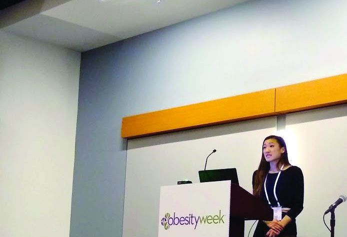
NASHVILLE – Advising patients in a comprehensive weight loss intervention to moderate their alcohol consumption did not change how much they drank over the long term. At the same time, abstinent patients kept off more weight over time than those who were classified as heavy drinkers, in a new analysis of data from a multicenter trial.
Abstinent individuals lost just 1.6% more of their body weight after 4 years than those who drank (P = .003), a figure with “uncertain clinical significance,” Ariana Chao, PhD, said at the meeting presented by the Obesity Society and the American Society for Metabolic and Bariatric Surgery. “The results should be taken in the context of the potential – though controversial – benefits of light to moderate alcohol consumption,” she added.
Alcohol contains 7.1 kcal/g, and “calories from alcohol usually add, rather than substitute, for food intake,” said Dr. Chao. Alcohol’s disinhibiting effects are thought to contribute to increased food intake and the making of less healthy food choices. However, existing research has shown inconsistent findings about the relationship between alcohol consumption and body weight, she said.
Reducing or completely cutting out alcoholic beverage consumption is common advice for those trying to lose weight, but whether this advice is followed, and whether it makes a difference over the long term, has been an open question, said Dr. Chao.
She and her collaborators at the University of Pennsylvania, Philadelphia, used data from Look AHEAD, “a multicenter, randomized, clinical trial that compared an intensive lifestyle intervention (ILI) to a diabetes support and education (DSE) control group,” for 5,145 people with overweight or obesity and type 2 diabetes, explained Dr. Chao and her coinvestigators.
Dr. Chao and her colleagues looked at the effect that the lifestyle intervention had on alcohol consumption. Additionally, to see how drinkers and nondrinkers fared over the long term, they examined the interaction between alcohol consumption and weight loss at year 4, hypothesizing that individuals who received ILI would have a greater decrease in their alcohol consumption by year 4 than those who received DSE. The investigators had a second hypothesis that, among the ILI cohort, greater alcohol consumption would be associated with less weight loss over the 4 years studied.
To measure alcohol consumption, participants completed a questionnaire at baseline and annually thereafter. The questionnaire asked whether participants had consumed any alcoholic beverages in the past week, and how many drinks per week of wine, beer, or liquor per week were typical for those who did consume alcohol.
Respondents were grouped into four categories according to their baseline alcohol consumption: nondrinkers, light drinkers (fewer than 7 drinks weekly for men and 4 for women), moderate drinkers (7-14 drinks weekly for men and 4-7 for women), and heavy drinkers (more than 14 drinks weekly for men and 7 for women).
At baseline, 38% of participants reported being abstinent from alcohol, and about 54% reported being light drinkers. Moderate drinkers made up 6%, and 2% reported falling into the heavy drinking category. Females were more likely than males to be nondrinkers.
Heavy drinkers took in significantly more calories than nondrinkers at baseline (2,397 versus 1,907 kcal/day; P less than .001).
Individuals who had consistently been heavy drinkers throughout the study lost less weight than any other group, dropping just 2.4% of their body weight at year 4, compared with their baseline weight. Those who were abstinent from alcohol fared the best, losing 5.1% of their initial body weight (P = .04 for difference). “Heavy drinking is a risk factor for suboptimal long-term weight loss,” said Dr. Chao.
Even those who were consistent light drinkers lost a bit less than those who were abstinent, keeping off 4.2% of their baseline body weight at 4 years (P = .04).
Look AHEAD included individuals aged 45-76 years with type 2 diabetes mellitus and a body mass index of at least 25 kg/m2, or 27 kg/m2 for those on insulin. Excluded were those with hemoglobin A1c of at least 11%, blood pressure of at least 160/100 mm Hg, and triglycerides over 600 mg/dL. A total of 4,901 patients had complete data available in the public access data set and were included in the present analysis. Dr. Chao and her colleagues used statistical techniques to adjust for baseline differences among participants.
The three-part ILI in Look AHEAD began by encouraging a low-calorie diet of 1,200-1,500 kcal/day for those weighing under 250 pounds, and 1,500-1,800 kcal/day for those who were heavier at baseline. Advice was to consume a balanced diet with less than 30% fat, less than 10% saturated fat, and at least 15% protein.
Patients were advised to strive for 10,000 steps per day, with 175 minutes of moderate-intensity exercise each week. Exercise was unsupervised.
Behavioral modification techniques included goal-setting, stimulus control, self-monitoring, and ideas for problem solving and relapse prevention. The intervention used motivational interviewing techniques.
With regard to alcohol, the ILI group was given information about the number of calories in various alcoholic beverages and advised to reduce the amount of alcohol consumed, in order to reduce calories.
The DSE group participated in three group sessions annually, and received general information about nutrition, exercise, and general support.
A potentially important limitation of the study was that alcohol consumption was assessed by self-report and a request for annual recall of typical drinking habits. An audience member from the United Kingdom commented that she found the overall rate of reported alcohol consumption to be “shockingly low,” compared with what her patients report drinking in England. The average United States resident drinks 2.3 gallons of alcohol, or 494 standard drinks, annually, according to the National Institute on Alcohol Abuse and Alcoholism, said Dr. Chao.
The midlife age range of participants, their diabetes diagnosis, and the fact that depressive symptoms were overall low limits generalizability of the findings, said Dr. Chao, adding that psychosocial factors, other health conditions, and current or past alcohol use disorder could also cause some residual confounding of the data.
Dr. Chao has received research support from Shire Pharmaceuticals.
SOURCE: Chao A et al. Obesity Week 2018, Abstract T-OR-2017.
REPORTING FROM OBESITY WEEK 2018
Key clinical point: After 4 years of an intervention program, heavy drinkers had the smallest net loss in body weight.
Major finding: Heavy drinkers kept off less than half as much weight as teetotalers (2.4% versus 5.1% of baseline weight, P = .04).
Study details: Analysis of public data from Look AHEAD, a multicenter randomized trial of intensive lifestyle intervention for weight loss that enrolled 5,145 people.
Disclosures: Dr. Chao reported receiving research funding from Shire Pharmaceuticals.
Source: Chao A et al. Obesity Week 2018, Abstract T-OR-2017.
Combination immunotherapy ups survival in ILD patients with anti-MDA5–positive dermatomyositis
CHICAGO – Early treatment with combined high-dose glucocorticoids, tacrolimus, and intravenous cyclophosphamide therapy significantly improves survival vs. step-up therapy in interstitial lung disease patients with anti–melanoma differentiation–associated gene 5 (anti-MDA5)–positive dermatomyositis, according to findings from a prospective, multicenter study.
However, the combination therapy was associated with a high risk of cytomegalovirus reactivation and other opportunistic infections that warrants careful monitoring of treated patients, Hideaki Tsuji, MD, reported at the annual meeting of the American College of Rheumatology.
ILD accompanied by anti-MDA5–positive dermatomyositis (DM) is often intractable and associated with high mortality in Japanese patients. Case reports have suggested improved outcomes with combined immunosuppressive therapy, but a standard treatment has not been established, said Dr. Tsuji of Kyoto University.
“Therefore, we evaluated the efficacy and safety of combined immunosuppressive therapy for anti-MDA5–positive DM with ILD in a prospective single-arm study,” he said, adding that early administration, a short interval of intravenous cyclophosphamide, use of plasmapheresis as an additional therapy, and control of opportunistic infections may contribute to the improved outcomes seen with the regimen in this study.
The primary endpoint of 6-month survival was reached by 24 (89%) of 27 patients treated with the combination regimen for 52 weeks, compared with 5 (33%) of 15 historical controls who received high-dose glucocorticoids followed by step-wise addition of immunosuppressants. At 12 months, the survival rates were 85% and 33%, respectively, Dr. Tsuji said.
Additionally, anti-MDA5 titer, serum ferritin level, C-reactive protein level, lactate dehydrogenase, and KL-6 level gradually decreased over the 52 months, and percent vital capacity increased with combination vs. step-up therapy, he noted.
Cytomegalovirus reactivation occurred in 90% of combination regimen patients vs. 33% of controls over the 52-week study period, he said, adding that pneumocystic pneumonia and sepsis also occurred in combination regimen group patients, and were associated with death in four patients.
When the 23 surviving patients in the combination regimen group were compared with the 4 in the group who died, it was noted that the deceased patients were significantly more likely to have cutaneous ulcers (75% vs. 13%), higher mean C-reactive protein level (2.7 vs. 0.77 mg/dL), and higher creatine kinase level (644.3 vs. 219.3 IU/L), respectively, before treatment, he said.
Study subjects were Japanese adults with new-onset anti-MDA5–positive dermatomyositis with interstitial lung disease (ILD) who were enrolled between July 2014 and September 2017.
They were treated with 1 mg/kg/day of prednisolone for 4 weeks with reduced doses thereafter, 500-1,000 mg/m2 of IV cyclophosphamide every 2 weeks for six cycles then every 4 weeks for up to a total of 10-15 treatments, and 10-12 ng/mL of tacrolimus (12-hour trough). Plasmapheresis was allowed in patients who progressed and needed oxygenation after the regimen was initiated, and it was administered in nine patients (31%) in the combination regimen group vs. one (7%) of the historical controls.
Given the different frequencies of rapidly progressive ILD in Asian vs. Western countries (39%-71% vs. 22%-57%, respectively), it is unclear whether the results seen in this study can be extrapolated to patients from the United States and Europe. Therefore, it is necessary to analyze the efficacy of the regimen in those patient populations, Dr. Tsuji said, also noting that future studies should evaluate risk-based modifications of the regimen to identify the optimal treatment for individuals based on factors such as age, respiratory dysfunction, hyperferritinemia, and treatment delay.
Dr. Tsuji reported having no disclosures.
SOURCE: Tsuji H et al. Arthritis Rheumatol. 2018;70(Suppl 10), Abstract 838.
REPORTING FROM THE ACR ANNUAL MEETING
Key clinical point:
Major finding: 6-month survival was 89% vs. 33% with combination immunotherapy vs. step-up therapy.
Study details: A prospective, multicenter study of 27 patients and 15 historical controls.
Disclosures: Dr. Tsuji reported having no disclosures.
Source: Tsuji H et al. Arthritis Rheumatol. 2018;70(Suppl 10), Abstract 838.
New worldwide atopic dermatitis survey brings big surprises
PARIS – A major worldwide survey of the 12-month prevalence of atopic dermatitis (AD) across the course of life provides new insights into global disease trends, Jonathan I. Silverberg, MD, PhD, reported at the annual congress of the European Academy of Dermatology and Venereology.
Among the most important takeaways from this Internet-based survey of more than 273,645 infants, children, and adults in 18 countries across five continents conducted in 2017 was that “global atopic dermatitis prevalence appears to be higher in adults, at 10%, than in younger cohorts, where it’s 4%-8%, which I think is quite provocative and requires further study and confirmation,” said Dr. Silverberg, a dermatologist at Northwestern University in Chicago.
“Let’s keep in mind that there’s this accepted dogma in the literature than atopic dermatitis is somehow only a childhood disorder – it doesn’t affect adults. Well, these data tell a very different story because we’re actually seeing overall highest prevalences throughout the world occurring in adulthood,” based on the U.K. Working Party’s Diagnostic Criteria for Atopic Dermatitis (Br J Dermatol. 1994 Sep;131[3]:383-96).
This is the biggest epidemiologic survey ever to examine the 12-month prevalence and severity of AD around the world for both adults and children. Survey respondents included 172,627 adults aged 18 years and older, 34,212 adolescents aged 12-17 years, 54,806 children aged 2-11 years, and more than 12,000 infants.
Key findings from the study include the following:
- AD prevalence rates varied widely from country to country around the world, as well as by age groups (see graphic).
- The highest rate in adults was observed in China. South Korea had the highest rates in both children and adolescents. The top AD rates in infancy occurred in France and the United Kingdom.
- Rates across the age spectrum were consistently lowest in Israel and Switzerland.

“These kinds of patterns raise fascinating questions about the potential risk factors or protective factors that happen in different countries. There are some startling differences in terms of the different regions,” Dr. Silverberg observed. “Certain regions of the world really stand out as having much higher prevalences, particularly China and South Korea, and then as you get into the adult years, Brazil and Mexico, which I think are areas that, at least in the global atopic dermatitis epidemiology community, are not quite as well recognized as being hot spots for atopic dermatitis.”
Indeed, the 12-month prevalence rate of AD among adults was 14% in Mexico and 12% in Brazil, as compared with 13% in Saudi Arabia, 11% in Australia and Spain, 10% in Canada and the United Kingdom, and 9% in the United States.
The prevalence was generally lowest in infants, then jumped substantially within countries during the childhood years, declined slightly in adolescents, and then peaked in adulthood.
AD severity was assessed using PO-SCORAD, the Patient-Oriented Scoring AD measure. Most affected individuals had moderate AD as defined by a PO-SCORAD score of 25-50. Across the age spectrum, the highest proportion of infants with AD who had moderate disease was in China, with 72%. In Taiwan, 63% of children with AD had moderate disease, as did 68% of adolescents and an equal proportion of adults.
In the United Kingdom, 49% percent of infants with AD had severe disease, making that country the world leader in the youngest age group. Severe AD was most common among Turkish children, where 30% of kids with the skin disease had a PO-SCORAD score greater than 50. In Brazil, 31% of adolescents with AD had severe disease, the world’s highest rate in that age group. Among adults with AD, the world’s highest rate of severe disease was 25%, which was seen in the United States, Brazil, and Saudi Arabia.
Across the age spectrum, Japan had a consistently lower-end, overall, 12-month AD prevalence rate of 5%. Germany, Italy, and France had overall rates of 6%, 7%, and 8%, respectively. The rate was 9% in the United States and Canada, and it was 10% in Australia.
Dr. Silverberg performed validation analyses using the Patient-Oriented Eczema Measure (POEM) and diagnostic criteria similar to the earlier landmark International Study of Asthma and Allergies in Childhood, or ISAAC (Lancet. 1998 Apr 25;351[9111]:1225-32). This was a huge study that excluded the United States, leaving a hole in the epidemiologic picture of the disease that the new survey fills. The validation analyses were supportive of the main findings based on the U.K. Working Party criteria.
Dr. Silverberg reported serving as a consultant to Pfizer, which sponsored the global epidemiologic survey, as well as to roughly a dozen other pharmaceutical companies.
SOURCE: Silverberg JI. EADV Congress, Abstract FC01.01.
PARIS – A major worldwide survey of the 12-month prevalence of atopic dermatitis (AD) across the course of life provides new insights into global disease trends, Jonathan I. Silverberg, MD, PhD, reported at the annual congress of the European Academy of Dermatology and Venereology.
Among the most important takeaways from this Internet-based survey of more than 273,645 infants, children, and adults in 18 countries across five continents conducted in 2017 was that “global atopic dermatitis prevalence appears to be higher in adults, at 10%, than in younger cohorts, where it’s 4%-8%, which I think is quite provocative and requires further study and confirmation,” said Dr. Silverberg, a dermatologist at Northwestern University in Chicago.
“Let’s keep in mind that there’s this accepted dogma in the literature than atopic dermatitis is somehow only a childhood disorder – it doesn’t affect adults. Well, these data tell a very different story because we’re actually seeing overall highest prevalences throughout the world occurring in adulthood,” based on the U.K. Working Party’s Diagnostic Criteria for Atopic Dermatitis (Br J Dermatol. 1994 Sep;131[3]:383-96).
This is the biggest epidemiologic survey ever to examine the 12-month prevalence and severity of AD around the world for both adults and children. Survey respondents included 172,627 adults aged 18 years and older, 34,212 adolescents aged 12-17 years, 54,806 children aged 2-11 years, and more than 12,000 infants.
Key findings from the study include the following:
- AD prevalence rates varied widely from country to country around the world, as well as by age groups (see graphic).
- The highest rate in adults was observed in China. South Korea had the highest rates in both children and adolescents. The top AD rates in infancy occurred in France and the United Kingdom.
- Rates across the age spectrum were consistently lowest in Israel and Switzerland.

“These kinds of patterns raise fascinating questions about the potential risk factors or protective factors that happen in different countries. There are some startling differences in terms of the different regions,” Dr. Silverberg observed. “Certain regions of the world really stand out as having much higher prevalences, particularly China and South Korea, and then as you get into the adult years, Brazil and Mexico, which I think are areas that, at least in the global atopic dermatitis epidemiology community, are not quite as well recognized as being hot spots for atopic dermatitis.”
Indeed, the 12-month prevalence rate of AD among adults was 14% in Mexico and 12% in Brazil, as compared with 13% in Saudi Arabia, 11% in Australia and Spain, 10% in Canada and the United Kingdom, and 9% in the United States.
The prevalence was generally lowest in infants, then jumped substantially within countries during the childhood years, declined slightly in adolescents, and then peaked in adulthood.
AD severity was assessed using PO-SCORAD, the Patient-Oriented Scoring AD measure. Most affected individuals had moderate AD as defined by a PO-SCORAD score of 25-50. Across the age spectrum, the highest proportion of infants with AD who had moderate disease was in China, with 72%. In Taiwan, 63% of children with AD had moderate disease, as did 68% of adolescents and an equal proportion of adults.
In the United Kingdom, 49% percent of infants with AD had severe disease, making that country the world leader in the youngest age group. Severe AD was most common among Turkish children, where 30% of kids with the skin disease had a PO-SCORAD score greater than 50. In Brazil, 31% of adolescents with AD had severe disease, the world’s highest rate in that age group. Among adults with AD, the world’s highest rate of severe disease was 25%, which was seen in the United States, Brazil, and Saudi Arabia.
Across the age spectrum, Japan had a consistently lower-end, overall, 12-month AD prevalence rate of 5%. Germany, Italy, and France had overall rates of 6%, 7%, and 8%, respectively. The rate was 9% in the United States and Canada, and it was 10% in Australia.
Dr. Silverberg performed validation analyses using the Patient-Oriented Eczema Measure (POEM) and diagnostic criteria similar to the earlier landmark International Study of Asthma and Allergies in Childhood, or ISAAC (Lancet. 1998 Apr 25;351[9111]:1225-32). This was a huge study that excluded the United States, leaving a hole in the epidemiologic picture of the disease that the new survey fills. The validation analyses were supportive of the main findings based on the U.K. Working Party criteria.
Dr. Silverberg reported serving as a consultant to Pfizer, which sponsored the global epidemiologic survey, as well as to roughly a dozen other pharmaceutical companies.
SOURCE: Silverberg JI. EADV Congress, Abstract FC01.01.
PARIS – A major worldwide survey of the 12-month prevalence of atopic dermatitis (AD) across the course of life provides new insights into global disease trends, Jonathan I. Silverberg, MD, PhD, reported at the annual congress of the European Academy of Dermatology and Venereology.
Among the most important takeaways from this Internet-based survey of more than 273,645 infants, children, and adults in 18 countries across five continents conducted in 2017 was that “global atopic dermatitis prevalence appears to be higher in adults, at 10%, than in younger cohorts, where it’s 4%-8%, which I think is quite provocative and requires further study and confirmation,” said Dr. Silverberg, a dermatologist at Northwestern University in Chicago.
“Let’s keep in mind that there’s this accepted dogma in the literature than atopic dermatitis is somehow only a childhood disorder – it doesn’t affect adults. Well, these data tell a very different story because we’re actually seeing overall highest prevalences throughout the world occurring in adulthood,” based on the U.K. Working Party’s Diagnostic Criteria for Atopic Dermatitis (Br J Dermatol. 1994 Sep;131[3]:383-96).
This is the biggest epidemiologic survey ever to examine the 12-month prevalence and severity of AD around the world for both adults and children. Survey respondents included 172,627 adults aged 18 years and older, 34,212 adolescents aged 12-17 years, 54,806 children aged 2-11 years, and more than 12,000 infants.
Key findings from the study include the following:
- AD prevalence rates varied widely from country to country around the world, as well as by age groups (see graphic).
- The highest rate in adults was observed in China. South Korea had the highest rates in both children and adolescents. The top AD rates in infancy occurred in France and the United Kingdom.
- Rates across the age spectrum were consistently lowest in Israel and Switzerland.

“These kinds of patterns raise fascinating questions about the potential risk factors or protective factors that happen in different countries. There are some startling differences in terms of the different regions,” Dr. Silverberg observed. “Certain regions of the world really stand out as having much higher prevalences, particularly China and South Korea, and then as you get into the adult years, Brazil and Mexico, which I think are areas that, at least in the global atopic dermatitis epidemiology community, are not quite as well recognized as being hot spots for atopic dermatitis.”
Indeed, the 12-month prevalence rate of AD among adults was 14% in Mexico and 12% in Brazil, as compared with 13% in Saudi Arabia, 11% in Australia and Spain, 10% in Canada and the United Kingdom, and 9% in the United States.
The prevalence was generally lowest in infants, then jumped substantially within countries during the childhood years, declined slightly in adolescents, and then peaked in adulthood.
AD severity was assessed using PO-SCORAD, the Patient-Oriented Scoring AD measure. Most affected individuals had moderate AD as defined by a PO-SCORAD score of 25-50. Across the age spectrum, the highest proportion of infants with AD who had moderate disease was in China, with 72%. In Taiwan, 63% of children with AD had moderate disease, as did 68% of adolescents and an equal proportion of adults.
In the United Kingdom, 49% percent of infants with AD had severe disease, making that country the world leader in the youngest age group. Severe AD was most common among Turkish children, where 30% of kids with the skin disease had a PO-SCORAD score greater than 50. In Brazil, 31% of adolescents with AD had severe disease, the world’s highest rate in that age group. Among adults with AD, the world’s highest rate of severe disease was 25%, which was seen in the United States, Brazil, and Saudi Arabia.
Across the age spectrum, Japan had a consistently lower-end, overall, 12-month AD prevalence rate of 5%. Germany, Italy, and France had overall rates of 6%, 7%, and 8%, respectively. The rate was 9% in the United States and Canada, and it was 10% in Australia.
Dr. Silverberg performed validation analyses using the Patient-Oriented Eczema Measure (POEM) and diagnostic criteria similar to the earlier landmark International Study of Asthma and Allergies in Childhood, or ISAAC (Lancet. 1998 Apr 25;351[9111]:1225-32). This was a huge study that excluded the United States, leaving a hole in the epidemiologic picture of the disease that the new survey fills. The validation analyses were supportive of the main findings based on the U.K. Working Party criteria.
Dr. Silverberg reported serving as a consultant to Pfizer, which sponsored the global epidemiologic survey, as well as to roughly a dozen other pharmaceutical companies.
SOURCE: Silverberg JI. EADV Congress, Abstract FC01.01.
REPORTING FROM THE EADV CONGRESS
Key clinical point: Worldwide, the 12-month prevalence of atopic dermatitis (AD) varies substantially but is unexpectedly highest in adults.
Major finding: The global 12-month prevalence of AD in adults is 10%, substantially higher than in infants, children, or adolescents.
Study details: This was an Internet survey of 273,654 subjects conducted in 2017 in 18 countries on five continents.
Disclosures: The presenter reported serving as a consultant to Pfizer, the study sponsor, as well as to roughly a dozen other pharmaceutical companies.
Source: Silverberg JI. EADV Congress, Abstract FC01.01.
