User login
ARISTOPHANES: Apixaban edges other DOACS for octogenarians
The findings come from an analysis of insurance claims data from more than 50,000 U.S. patients – the largest observational study to date to compare these three direct-acting oral anticoagulants (DOACs) in octogenarians with nonvalvular atrial fibrillation, Steven B. Deitelzweig, MD, said at the American Heart Association scientific sessions.
“The results may help clinicians evaluate the risk-benefit ratio of the DOACs” in this population, said Dr. Deitelzweig, vice president for medical affairs at Ochsner Medical Center in New Orleans.
He noted that the results were consistent with prior reports from observational data and registries, as well as the results in a recent analysis commissioned by the Agency for Healthcare Research and Quality. “We see a consistent message that apixaban always has less risk for major bleeding, and at least comparable efficacy” when compared with other DOACs, he said in a video interview.
And for the foreseeable future, this sort of data will need to suffice for clinicians trying to decide which DOAC to use because “I know of no head-to-head trials, nor do I anticipate any head-to-head trials” that could provide a more definitive comparison of the DOACs, Dr. Deitelzweig said.
The data came from a large number of patients – about 38% of the U.S. population – which boosts the generalizability of the finding. “I think our data are useful” for helping to make treatment decisions, he concluded.
The analysis he reported came from the ARISTOPHANES (Anticoagulants for Reduction in Stroke: Observational Pooled Analysis on Health Outcomes and Experience of Patients) study, which pooled U.S. insurance claims data from several sources. For the octogenarian study, Dr. Deitelzweig and his associates used data from about 123 million U.S. residents collected between January 2012 and September 2015 by Medicare and three different commercial insurance databases. The overall level of beneficiary overlap between these four data sources was less than 0.5%.
The researchers identified patients with nonvalvular atrial fibrillation who started anticoagulant treatment with a DOAC and were at least 80 years old. This included 19,752 patients started on apixaban (Eliquis), 6,741 started on dabigatran (Pradaxa), and 27,217 started on rivaroxaban (Xarelto). A majority of the patients were at least 84 years old.
The analysis used propensity-score matching to compare similar patients and to minimize the impact of potentially confounding differences among the patients in each treatment subgroup. During a median follow-up of 7-9 months, the incidence of stroke or systemic embolism was 35% lower in the apixaban-treated patients, compared with those who received dabigatran, and 28% lower in the apixaban patients, compared with those treated with rivaroxaban, both statistically significant differences, Dr. Deitelzweig reported. The incidence of major bleeding episodes was 40% lower with apixaban than with dabigatran and 50% lower with apixaban, compared with rivaroxaban, also statistically significant differences.
When the analysis compared dabigatran with rivaroxaban it showed no statistically significant difference for the efficacy endpoint, but dabigatran produced 23% fewer major bleeds than rivaroxaban, a statistically significant difference.
These findings jibed with a recently published analysis from Dr. Deitelzweig and his associates that used data from all adults with nonvalvular atrial fibrillation started on an oral anticoagulant in an expanded ARISTOPHANES database for 2012-2015 that included more than 180 million U.S. beneficiaries. After propensity-score matching, this created subgroups of about 58,000 patients started on apixaban, nearly 27,000 started on dabigatran, and more than 83,000 started on rivaroxaban. The patients averaged about 73 years old. Again, with about 7-9 months of follow-up, very similar outcomes occurred. Patients on apixaban had significantly fewer strokes and systemic embolic events as well as significantly fewer major bleeds compared with patients treated with one of the other DOACs (Stroke. 2018 Dec;49[12]:2933-44).
The study was funded by Bristol-Myers Squibb and Pfizer, the companies that market apixaban (Eliquis). Dr. Deitelzweig is a consultant to and speaker on behalf of Bristol-Myers Squibb and Pfizer. He is also a consultant to or speaker on behalf of Boehringer Ingelheim, Daiichi-Sankyo, Janssen, and Portola Pharmaceuticals.
SOURCE: Deitelzweig SB et al. Circulation. 2018 Nov 6;138(suppl 1):A14900.
REPORTING FROM THE AHA SCIENTIFIC SESSIONS
Key clinical point: Apixaban surpassed two other direct-acting oral anticoagulants in a large, observational database.
Major finding: The adjusted stroke or systemic embolism rate with apixaban was 35% less than dabigatran and 28% less than rivaroxaban.
Study details: A retrospective analysis of observational data collected in insurance claims from 53,710 U.S. octogenarians.
Disclosures: The study was funded by Bristol-Myers Squibb and Pfizer, the companies that market apixaban (Eliquis). Dr. Deitelzweig is a consultant to and speaker on behalf of Bristol-Myers Squibb and Pfizer. He is also a consultant to or speaker on behalf of Boehringer Ingelheim, Daiichi-Sankyo, Janssen, and Portola Pharmaceuticals.
Source: Deitelzweig SB et al. Circulation. 2018 Nov 6;138[suppl 1]:A14900.

Program decreased seizure frequency for people with epilepsy
NEW ORLEANS – A self-management program that focused on medication adherence, sleep, nutrition, and stress reduction was associated with decreased seizures and improved quality of life for adults with epilepsy.
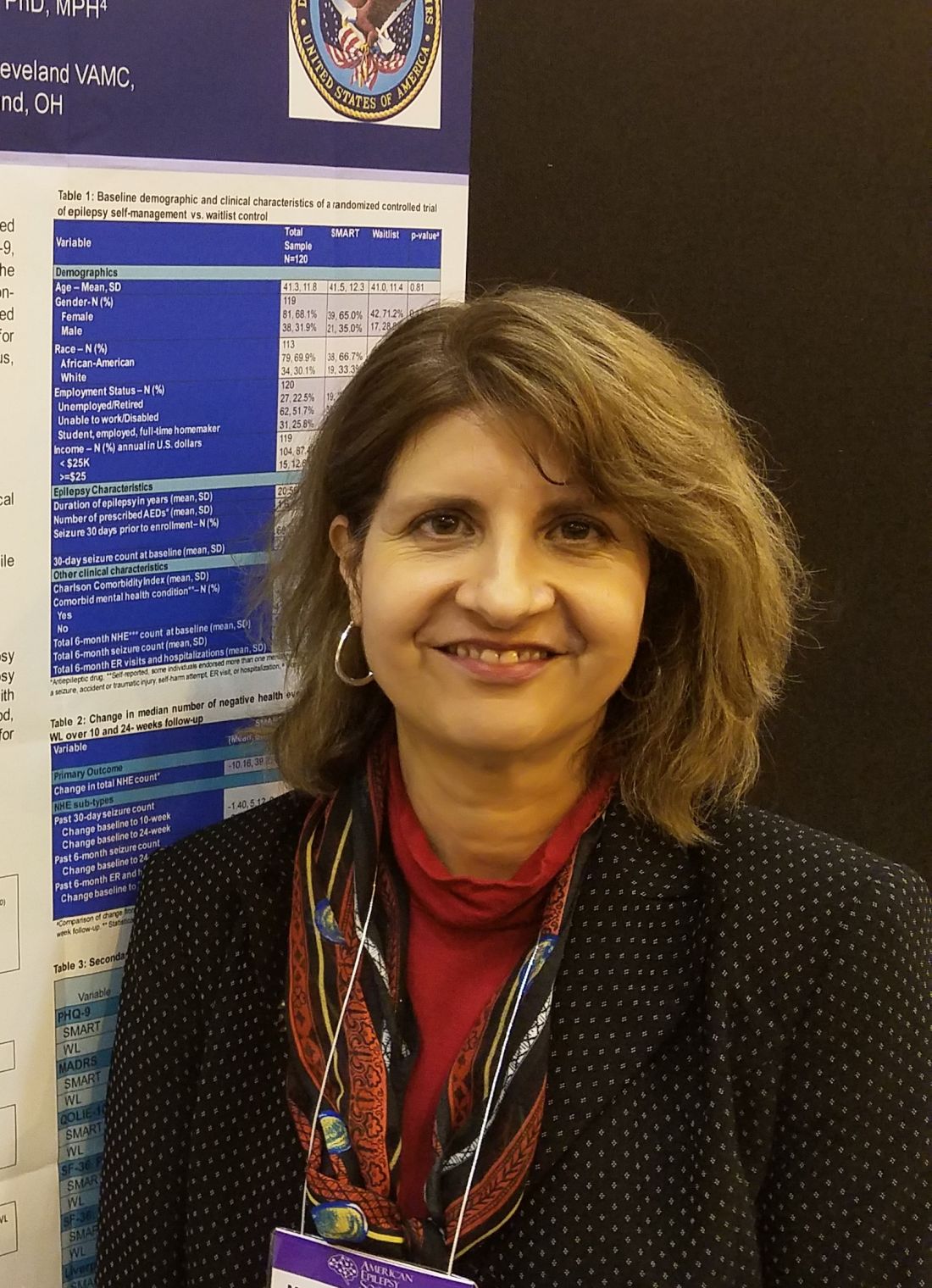
SMART (Self‐management for people with epilepsy and a history of negative health events) also was associated with improved depression scores and overall quality of life measures in participants, compared with a wait-listed control group, Martha Sajatovic, MD, said at the annual meeting of the American Epilepsy Society.
“I believe what we’re seeing is a result of improved self-management,” said Dr. Sajatovic, the Willard Brown Chair in Neurological Outcomes Research at Case Western Reserve University, Cleveland. “This is multimodal, including better medication adherence, which in turn is related to better communication with the clinician. For example, if patients are not sleeping well or their medicine makes them nauseated or they experience sexual dysfunction, we encourage them to talk to their docs about what they can live with, and what they can’t.”
Presented as a poster during the meeting, the SMART study was also published in Epilepsia.
SMART is an 8-week online educational program delivered by a nurse educator and a “peer educator,” a person with epilepsy who has had at least three negative health events. The first session is an in-person visit during which the team gets acquainted and discusses goals. The remaining sessions are self-paced and delivered on computer tablets provided by the investigators.
SMART didn’t just focus on the physical issues of living with epilepsy, Dr. Sajatovic said in an interview. Sessions also discussed the stigma still associated with the disorder, and myths that unnecessarily inflate perceptions. Discussions include goal setting, epilepsy complications and how to manage them, the importance of good sleep hygiene, problem-solving skills, nutrition and substance abuse, exercise, and how to deal with medication side effects.
“One thing we really stressed was sharing information in a way that was accessible to all patients and fostered self-motivation,” she said. “Most of our participants had never been in a program like this before. It was very empowering for many.”
The researchers chose participants who were socioeconomically challenged for this project; 88% made less than $25,000 per year and 74% were unemployed. The mean age of participants was 41 years, 70% were black, and most had been living with epilepsy at least half of their life. About 70% lived alone, and 70% had experienced at least one seizure within the month before enrolling. Mental health comorbidities were common; 69% had depression, 32% had anxiety, and 13% had PTSD.
The study enrolled 120 people, who were evenly divided between the intervention group and the wait-list group. The primary outcome was the change in total negative health events from baseline to the study’s end. Negative health events were seizures and ED or hospital admissions for any other causes including attempts at self-harm, falls, and accidents.
Secondary outcomes included changes in depression scores as measured by the Montgomery-Åsberg Depression Rating Scale and the 9-item Patient Health Questionnaire. Quality of life was measured using the 10-item Quality of Life in Epilepsy; functional status was measured using the 36-Item Short-Form Health Survey.
At baseline, the total mean 6-month negative health events count was 15, with 13 events being seizures. The other events were hospital or ED visits for other reasons.
At the end of the study, the intervention group experienced a significant mean decrease of 10 fewer negative health events, compared with a decrease of 2 in the wait-listed group. This was largely driven by a mean of 7.8 fewer seizures in the active group, compared with a decrease of about 1.0 in the wait-listed group. The 6-month ER and hospitalization counts did not significantly change.
Among the secondary outcomes, depression, overall health, and quality of life all improved significantly in the intervention group, compared with the wait-listed group. The intervention group also had significant decreases in depression measures and improvements in daily function measures, Dr. Sajatovic said.
“It was so gratifying to see this. Most of our participants had never been in a program like this before. It was a chance for them to take control of their epilepsy, instead of simply having it control them,” she said.
This study was supported by a grant from the Centers for Disease Control and Prevention. Dr. Sajatovic had no financial disclosures related to this presentation.
SOURCE: Sajatovic M et al.
NEW ORLEANS – A self-management program that focused on medication adherence, sleep, nutrition, and stress reduction was associated with decreased seizures and improved quality of life for adults with epilepsy.
SMART (Self‐management for people with epilepsy and a history of negative health events) also was associated with improved depression scores and overall quality of life measures in participants, compared with a wait-listed control group, Martha Sajatovic, MD, said at the annual meeting of the American Epilepsy Society.
“I believe what we’re seeing is a result of improved self-management,” said Dr. Sajatovic, the Willard Brown Chair in Neurological Outcomes Research at Case Western Reserve University, Cleveland. “This is multimodal, including better medication adherence, which in turn is related to better communication with the clinician. For example, if patients are not sleeping well or their medicine makes them nauseated or they experience sexual dysfunction, we encourage them to talk to their docs about what they can live with, and what they can’t.”
Presented as a poster during the meeting, the SMART study was also published in Epilepsia.
SMART is an 8-week online educational program delivered by a nurse educator and a “peer educator,” a person with epilepsy who has had at least three negative health events. The first session is an in-person visit during which the team gets acquainted and discusses goals. The remaining sessions are self-paced and delivered on computer tablets provided by the investigators.
SMART didn’t just focus on the physical issues of living with epilepsy, Dr. Sajatovic said in an interview. Sessions also discussed the stigma still associated with the disorder, and myths that unnecessarily inflate perceptions. Discussions include goal setting, epilepsy complications and how to manage them, the importance of good sleep hygiene, problem-solving skills, nutrition and substance abuse, exercise, and how to deal with medication side effects.
“One thing we really stressed was sharing information in a way that was accessible to all patients and fostered self-motivation,” she said. “Most of our participants had never been in a program like this before. It was very empowering for many.”
The researchers chose participants who were socioeconomically challenged for this project; 88% made less than $25,000 per year and 74% were unemployed. The mean age of participants was 41 years, 70% were black, and most had been living with epilepsy at least half of their life. About 70% lived alone, and 70% had experienced at least one seizure within the month before enrolling. Mental health comorbidities were common; 69% had depression, 32% had anxiety, and 13% had PTSD.
The study enrolled 120 people, who were evenly divided between the intervention group and the wait-list group. The primary outcome was the change in total negative health events from baseline to the study’s end. Negative health events were seizures and ED or hospital admissions for any other causes including attempts at self-harm, falls, and accidents.
Secondary outcomes included changes in depression scores as measured by the Montgomery-Åsberg Depression Rating Scale and the 9-item Patient Health Questionnaire. Quality of life was measured using the 10-item Quality of Life in Epilepsy; functional status was measured using the 36-Item Short-Form Health Survey.
At baseline, the total mean 6-month negative health events count was 15, with 13 events being seizures. The other events were hospital or ED visits for other reasons.
At the end of the study, the intervention group experienced a significant mean decrease of 10 fewer negative health events, compared with a decrease of 2 in the wait-listed group. This was largely driven by a mean of 7.8 fewer seizures in the active group, compared with a decrease of about 1.0 in the wait-listed group. The 6-month ER and hospitalization counts did not significantly change.
Among the secondary outcomes, depression, overall health, and quality of life all improved significantly in the intervention group, compared with the wait-listed group. The intervention group also had significant decreases in depression measures and improvements in daily function measures, Dr. Sajatovic said.
“It was so gratifying to see this. Most of our participants had never been in a program like this before. It was a chance for them to take control of their epilepsy, instead of simply having it control them,” she said.
This study was supported by a grant from the Centers for Disease Control and Prevention. Dr. Sajatovic had no financial disclosures related to this presentation.
SOURCE: Sajatovic M et al.
REPORTING FROM AES 2018
Key clinical point: Patients with epilepsy had fewer seizures and improved quality of life after 6 months of participation in a program that teaches self-management techniques.
Major finding: The intervention group had a mean of 7.8 fewer seizures, compared with their baseline count during the 6-month study.
Study details: The prospective study randomized 120 people to either the intervention group or a wait-list group.
Disclosures: This study was supported by a grant from the Centers for Disease Control and Prevention. Dr. Sajatovic reported no financial disclosures related to this presentation.
Source: Sajatovic M et al. AES
Patients with epilepsy may develop tolerance to CBD-enriched oil
NEW ORLEANS – according to a study presented at the annual meeting of the American Epilepsy Society.
“CBD is a good option for children and adults with certain kinds of epilepsy, but as with antiepileptic drugs, it can become less effective over time, and the dose may need to be increased to manage the seizures,” said Shimrit Uliel-Sibony, MD, lead author of the study and head of the pediatric epilepsy service at Tel Aviv Sourasky Medical Center’s Dana-Dwek Children’s Hospital.
Prior studies have found that the efficacy of cannabinoids may wane when used for pain management. Efficacy also declines in animals with seizures.
To assess the tolerance rate of cannabinoids in the treatment of children and adults with epilepsy, researchers in Israel conducted a prospective review of 92 consecutive patients with treatment-resistant epilepsy. Patients were aged 1-37 years (mean age, 11.8 years) and were treated with cannabis oil extract during March 1, 2014–Dec. 31, 2017. The researchers defined tolerance as the need to increase the dose by at least 30% following a reduction in efficacy, or a more than 30% reduction in treatment response.
The patients had various forms of epilepsy (e.g., Dravet syndrome, Lennox-Gastaut syndrome, and epilepsy caused by stroke) and used cannabis oil extract for an average of 19.8 months. Of the 84 patients included in the tolerance analysis, 21 patients (25%) developed tolerance after an average of 7.3 months (range, 1-24 months) at an average dose of 12.6 mg/kg per day. After patients with tolerance received an increased dose, 4 patients returned to their previous response levels, and 10 patients had a response that was “satisfying but less than [the] prior response level,” Dr. Uliel-Sibony and colleagues said.
About a third of patients discontinued treatment because of side effects or lack of efficacy. Side effects included sleepiness, nausea, decreased appetite, and vomiting. In addition, seizures worsened in two patients, and one patient had signs of psychosis; treatment was stopped immediately in those three patients.
The investigators had no disclosures and received no funding for this study.
SOURCE: Uliel-Sibony S et al., AES 2018, Abstract 2.233.
REPORTING FROM AES 2018
Key clinical point: Cannabis oil extract may become less effective, and the dose may need to be increased to manage seizures.
Major finding: About a quarter of patients who received cannabis oil extract developed tolerance.
Study details: Prospective review of 92 consecutive patients with treatment-resistant epilepsy.
Disclosures: The investigators had no disclosures and received no funding for this study.
Source: Uliel-Sibony S et al. AES 2018, Abstract 2.233.
Ibrutinib outperforms bendamustine and rituximab in older CLL patients
SAN DIEGO – Ibrutinib alone or in combination with rituximab resulted in superior progression-free survival (PFS) when compared with bendamustine plus rituximab in the randomized, phase 3 Alliance A041202 trial of older patients with previously untreated chronic lymphocytic leukemia (CLL).
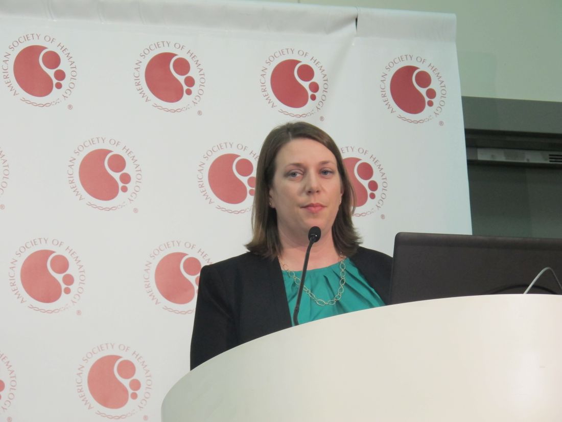
“There was no difference in progression-free survival between ibrutinib and ibrutinib plus rituximab,” said Dr. Woyach of the Ohio State University, Columbus.
Median PFS in this study was 43 months in the BR arm, and was not reached in either of the ibrutinib-containing arms, she said. No significant differences in overall survival (OS) were seen among the treatment arms, which may have been because of short follow-up and the fact that patients in the BR arm were allowed to cross over to ibrutinib if they progressed on treatment.
Participants in the international, multicenter trial – a project of the National Cancer Institute National Clinical Trials Network – were 547 adults aged 65 years or older (median, 71 years) with previously untreated, symptomatic CLL. They were enrolled from 219 sites across the United States and Canada between Dec. 9, 2013, and May 16, 2016.
The three arms were well matched with respect to baseline characteristics except for a slightly higher number of patients with complex karyotypes in the IR arm, Dr. Woyach said.
Treatment in the BR arm included bendamustine 90 mg/m2 on days 1 and 2 of each 28-day cycle plus rituximab at a dose of 375 mg/m2 on day 0 of cycle 1 then 500 mg/m2 on day 1 of cycles 2-6. Patients in the ibrutinib arms received 420 mg daily until disease progression either with or without rituximab at 375 mg/m2 weekly for 4 weeks starting at cycle 2 day 1 and then given on day 1 of cycles 3-6.
Grade 3-5 treatment-emergent hematologic adverse events (AEs) occurred in 61%, 41%, and 38% of patients in the BR, ibrutinib, and IR arms, respectively. Neutropenia and thrombocytopenia occurred more often in the BR than in the ibrutinib arms. Nonhematologic AEs occurred in 63%, 74%, and 74%, respectively, with an overall greater incidence in the ibrutinib arms. Infections and sudden deaths were numerically but not significantly higher in the ibrutinib arms.
“We undertook this study to determine the most effective therapy for older patients with CLL,” Dr. Woyach said, explaining that while older patients make up the majority of patients with CLL, they are typically underrepresented in trials.
At the start of the study, BR was widely used and the Bruton’s tyrosine kinase inhibitor ibrutinib was “just entering the clinic,” she noted.
“Despite now-widespread use in the [front-line setting] following FDA approval for this indication in 2016, the efficacy of ibrutinib versus standard chemoimmunotherapy has not previously been investigated,” she said.
Since adding rituximab has been shown to improve PFS and OS when added to chemotherapy in CLL, she and her colleagues also looked at whether this was the case with ibrutinib as well.
“This is the only phase 3 trial designed to answer this question,” she noted, adding that the findings justify the use of ibrutinib as a standard-of-care treatment for CLL patients aged 65 years and older.
David P. Steensma, MD, of Dana-Farber Cancer Institute in Boston, who moderated the press briefing, agreed. “I think this really does indicate that ibrutinib as front-line therapy, which many clinicians have been doing, is a very reasonable practice.”
Dr. Woyach added, however, that while ibrutinib represents a major therapeutic advance, its cost and its toxicities in older patients are a concern that warrant close monitoring and development of strategies to reduce the need for long-term continuous treatment.
Additional phase 3 studies set to open soon will compare ibrutinib in combination with venetoclax and obinutuzumab with standard ibrutinib
Dr. Woyach reported having no disclosures. Dr. Steensma reported receiving research funding from, and/or serving as a consultant, board member, or adviser for Takeda Pharmaceutical, Syros Pharmaceuticals, Otsuka Pharmaceutical, Onconova Therapeutics, Novartis, Kura Oncology, Janssen, H3 Biosciences, Celgene, Amphivena Therapeutics, and Acceleron Pharma.
SOURCE: Woyach JA et al. ASH 2018, Abstract 6.
SAN DIEGO – Ibrutinib alone or in combination with rituximab resulted in superior progression-free survival (PFS) when compared with bendamustine plus rituximab in the randomized, phase 3 Alliance A041202 trial of older patients with previously untreated chronic lymphocytic leukemia (CLL).
“There was no difference in progression-free survival between ibrutinib and ibrutinib plus rituximab,” said Dr. Woyach of the Ohio State University, Columbus.
Median PFS in this study was 43 months in the BR arm, and was not reached in either of the ibrutinib-containing arms, she said. No significant differences in overall survival (OS) were seen among the treatment arms, which may have been because of short follow-up and the fact that patients in the BR arm were allowed to cross over to ibrutinib if they progressed on treatment.
Participants in the international, multicenter trial – a project of the National Cancer Institute National Clinical Trials Network – were 547 adults aged 65 years or older (median, 71 years) with previously untreated, symptomatic CLL. They were enrolled from 219 sites across the United States and Canada between Dec. 9, 2013, and May 16, 2016.
The three arms were well matched with respect to baseline characteristics except for a slightly higher number of patients with complex karyotypes in the IR arm, Dr. Woyach said.
Treatment in the BR arm included bendamustine 90 mg/m2 on days 1 and 2 of each 28-day cycle plus rituximab at a dose of 375 mg/m2 on day 0 of cycle 1 then 500 mg/m2 on day 1 of cycles 2-6. Patients in the ibrutinib arms received 420 mg daily until disease progression either with or without rituximab at 375 mg/m2 weekly for 4 weeks starting at cycle 2 day 1 and then given on day 1 of cycles 3-6.
Grade 3-5 treatment-emergent hematologic adverse events (AEs) occurred in 61%, 41%, and 38% of patients in the BR, ibrutinib, and IR arms, respectively. Neutropenia and thrombocytopenia occurred more often in the BR than in the ibrutinib arms. Nonhematologic AEs occurred in 63%, 74%, and 74%, respectively, with an overall greater incidence in the ibrutinib arms. Infections and sudden deaths were numerically but not significantly higher in the ibrutinib arms.
“We undertook this study to determine the most effective therapy for older patients with CLL,” Dr. Woyach said, explaining that while older patients make up the majority of patients with CLL, they are typically underrepresented in trials.
At the start of the study, BR was widely used and the Bruton’s tyrosine kinase inhibitor ibrutinib was “just entering the clinic,” she noted.
“Despite now-widespread use in the [front-line setting] following FDA approval for this indication in 2016, the efficacy of ibrutinib versus standard chemoimmunotherapy has not previously been investigated,” she said.
Since adding rituximab has been shown to improve PFS and OS when added to chemotherapy in CLL, she and her colleagues also looked at whether this was the case with ibrutinib as well.
“This is the only phase 3 trial designed to answer this question,” she noted, adding that the findings justify the use of ibrutinib as a standard-of-care treatment for CLL patients aged 65 years and older.
David P. Steensma, MD, of Dana-Farber Cancer Institute in Boston, who moderated the press briefing, agreed. “I think this really does indicate that ibrutinib as front-line therapy, which many clinicians have been doing, is a very reasonable practice.”
Dr. Woyach added, however, that while ibrutinib represents a major therapeutic advance, its cost and its toxicities in older patients are a concern that warrant close monitoring and development of strategies to reduce the need for long-term continuous treatment.
Additional phase 3 studies set to open soon will compare ibrutinib in combination with venetoclax and obinutuzumab with standard ibrutinib
Dr. Woyach reported having no disclosures. Dr. Steensma reported receiving research funding from, and/or serving as a consultant, board member, or adviser for Takeda Pharmaceutical, Syros Pharmaceuticals, Otsuka Pharmaceutical, Onconova Therapeutics, Novartis, Kura Oncology, Janssen, H3 Biosciences, Celgene, Amphivena Therapeutics, and Acceleron Pharma.
SOURCE: Woyach JA et al. ASH 2018, Abstract 6.
REPORTING FROM ASH 2018
Key clinical point: In chronic lymphocytic leukemia patients aged 65 years and older, progression-free survival is better with ibrutinib than with bendamustine and rituximab.
Major finding: The 2-year progression-free survival was 74%, 87%, and 88% with bendamustine and rituximab, ibrutinib, and ibrutinib and rituximab, respectively.
Study details: A randomized, phase 3 study of 547 previously untreated patients with CLL.
Disclosures: Dr. Woyach reported having no disclosures. Dr. Steensma reported receiving research funding from, and/or serving as a consultant, board member, or adviser for Takeda Pharmaceutical, Syros Pharmaceuticals, Otsuka Pharmaceutical, Onconova Therapeutics, Novartis, Kura Oncology, Janssen, H3 Biosciences, Celgene, Amphivena Therapeutics, and Acceleron Pharma.
Source: Woyach JA et al. ASH 2018, Abstract 6.
Opioids appear safe for sickle cell pain
SAN DIEGO – Inpatient deaths from opioid overdose in the U.S. population has risen dramatically in recent years, but this is essentially a “never event” among patients with sickle cell disease (SCD).
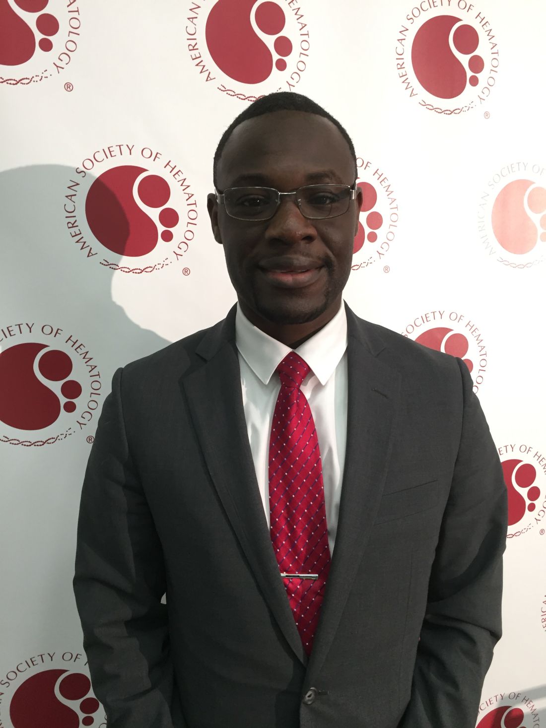
These findings come from an analysis of hospital discharge data in the National Inpatient Sample from 1998 to 2013. Oladimeji Akinola Akinboro, MBBS, who led the study, said the findings suggest that the current patterns of opioid use in SCD patients are safe and that opioids should not be withheld when they are appropriate.
From 1998 to 2013, the rate of inpatient deaths from opioid overdose in the United States rose 350% overall and approximately 8% annually. In contrast, deaths among SCD patients were flat, with a rate at or near zero throughout the same time period.
Over the 16-year period, there were just nine deaths reported among SCD patients because of opioids in the inpatient setting, Dr. Akinboro of Boston University Medical Center reported at the annual meeting of the American Society of Hematology.
While the reasons behind the difference were not explored in the study, Dr. Akinboro suggested that the sickle cell community has greater experience with opioids and that patients and physicians typically have long-standing clinical relationships that make mitigation of opioid misuse easier to manage.
Opioid-related hospitalizations, however, were comparable among the general U.S. population and SCD patients. And for both groups the rates remained relatively steady throughout the study period, with the exception of a drop among SCD patients in 2002, he noted.
The study also revealed age-related trends in hospitalizations overall. Hospitalizations among sickle cell patients were stable from 1998 to 2013. However, when the researchers broke the data down by age they found that adults aged 18-44 years had an increase in hospitalizations, with the steepest rise in patients aged 65 years and older.
In total, there were more than 1.7 million hospitalizations among SCD patients in the United States from 1998 to 2013. The rate declined by 9.9% each year from 1998 to 2002, then remained flat at around 27 per 100,000 persons from 2002 to 2013.
However, for adults aged 18-44 years, hospitalizations increased from 43 per 100,000 persons in 2002 to 71 per 100,000 persons in 2013 – an annual increase of 3.8%. Patients aged 65 years and older saw their rate of hospitalization increase from 2.7 to 5.4 per 100,000 persons from 1998 to 2013 – a 6.5% increase for each year.
The study did not explore the causes behind the age-related trends but Dr. Akinboro suggested it may be because of fragmentation of adult SCD care and age- and pain-related medical comorbidities.
The researchers reported having no relevant financial disclosures.
SOURCE: Akinboro OA et al. ASH 2018, Abstract 315.
SAN DIEGO – Inpatient deaths from opioid overdose in the U.S. population has risen dramatically in recent years, but this is essentially a “never event” among patients with sickle cell disease (SCD).
These findings come from an analysis of hospital discharge data in the National Inpatient Sample from 1998 to 2013. Oladimeji Akinola Akinboro, MBBS, who led the study, said the findings suggest that the current patterns of opioid use in SCD patients are safe and that opioids should not be withheld when they are appropriate.
From 1998 to 2013, the rate of inpatient deaths from opioid overdose in the United States rose 350% overall and approximately 8% annually. In contrast, deaths among SCD patients were flat, with a rate at or near zero throughout the same time period.
Over the 16-year period, there were just nine deaths reported among SCD patients because of opioids in the inpatient setting, Dr. Akinboro of Boston University Medical Center reported at the annual meeting of the American Society of Hematology.
While the reasons behind the difference were not explored in the study, Dr. Akinboro suggested that the sickle cell community has greater experience with opioids and that patients and physicians typically have long-standing clinical relationships that make mitigation of opioid misuse easier to manage.
Opioid-related hospitalizations, however, were comparable among the general U.S. population and SCD patients. And for both groups the rates remained relatively steady throughout the study period, with the exception of a drop among SCD patients in 2002, he noted.
The study also revealed age-related trends in hospitalizations overall. Hospitalizations among sickle cell patients were stable from 1998 to 2013. However, when the researchers broke the data down by age they found that adults aged 18-44 years had an increase in hospitalizations, with the steepest rise in patients aged 65 years and older.
In total, there were more than 1.7 million hospitalizations among SCD patients in the United States from 1998 to 2013. The rate declined by 9.9% each year from 1998 to 2002, then remained flat at around 27 per 100,000 persons from 2002 to 2013.
However, for adults aged 18-44 years, hospitalizations increased from 43 per 100,000 persons in 2002 to 71 per 100,000 persons in 2013 – an annual increase of 3.8%. Patients aged 65 years and older saw their rate of hospitalization increase from 2.7 to 5.4 per 100,000 persons from 1998 to 2013 – a 6.5% increase for each year.
The study did not explore the causes behind the age-related trends but Dr. Akinboro suggested it may be because of fragmentation of adult SCD care and age- and pain-related medical comorbidities.
The researchers reported having no relevant financial disclosures.
SOURCE: Akinboro OA et al. ASH 2018, Abstract 315.
REPORTING FROM ASH 2018
Key clinical point:
Major finding: While the rate of inpatient death from opioid overdose rose 350% among the general U.S. population from 1998 to 2013, it remained essentially at zero among patients with sickle cell disease.
Study details: A retrospective analysis of discharge diagnoses from the National Inpatient Sample from 1998 to 2013.
Disclosures: The researchers reported having no relevant financial disclosures.
Source: Akinboro OA et al. ASH 2018, Abstract 315.
SUDEP risk may change over time
, based on study results presented at the annual meeting of the American Epilepsy Society.
Based on 3 years of data collected from over 12,000 people with epilepsy, 27.0% who had been at high risk (three or more generalized tonic-clonic seizures [GTCs] per year) at baseline moved out of the high-risk category. In addition, 32.5% at medium risk (one to two GTCs per year) at baseline changed categories. Finally, 7.0% in the low-risk category (no GTC seizures in the last year) at baseline moved to a higher-risk category.
“An individual’s risk [of SUDEP] is not set in stone,” said Neishay Ayub, MD, of Beth Israel Deaconess Medical Center, Boston, who presented the data at the meeting. “Our findings support the recommendation that for people with epilepsy who have ongoing generalized tonic-clonic seizures, the goal of treatment is to reduce GTCs and thereby lower SUDEP risk.”
A 2017 practice guideline summary from the American Academy of Neurology and the American Epilepsy Society identified the presence and frequency of GTCs and absence of seizure freedom as risk factors for SUDEP. Using these measures, Dr. Ayub and colleagues sought to stratify patients according to their risk of SUDEP and monitor how risk changed over time. They collected information about more than 1.4 million seizures that occurred from December 2007 to February 2018 in 12,402 users of the electronic diary Seizure Tracker.
For each user, the researchers calculated the number of generalized seizures for each year since the initial seizure diary entry. They categorized each user as being at low, medium, or high risk of SUDEP during each year. Low risk was defined as no generalized seizures in a year. Medium risk was defined as one or two generalized seizures in a year. High risk was defined as three or more generalized seizures in a year.
“The next step would be to see if we can confirm this patient-reported data with an objective study to determine when seizures did or did not occur,” said Daniel Goldenholz, MD, PhD, also of Beth Israel Deaconess Medical Center and senior author of the study. “For example, assessing information using new FDA [Food and Drug Administration]-approved wearable seizure tracker devices could give us a more comprehensive picture.”
The study was funded by the Harvard School of Public Health, Boston.
SOURCE: Ayub N et al. AES 2018, Abstract 2.158.
REPORTING FROM AES 2018
Key clinical point: Yearly patient risk assessments for sudden unexpected death in epilepsy are advisable.
Major finding: About 7% of people with no generalized tonic-clonic seizures in the last year at baseline moved to a higher-risk category.
Study details: An analysis of self-reported seizures by 12,402 users of Seizure Tracker.
Disclosures: The Harvard School of Public Health, Boston, funded the study.
Source: Ayub N et al. AES 2018, Abstract 2.158.
New PCNSL guidelines emphasize importance of patient fitness

New guidelines on the diagnosis and management of patients with primary central nervous system lymphoma (PCNSL) emphasize prompt diagnosis, aggressive treatment whenever possible, and multidisciplinary team support.
A unique aspect for hematologic cancers, the guidelines note, is that appropriate treatment for PCNSL requires input from neurology specialists.
And the guidelines recommend methotrexate-based treatment only be administered at centers experienced in delivering intensive chemotherapy.
Christopher P. Fox, MD, of the Nottingham University Hospitals NHS Trust in Nottingham, U.K., and his colleagues on behalf of the British Society for Haematology published the guidelines in BJH.
The authors incorporated findings from studies published since the society’s last comprehensive PCNSL guidelines were issued more than a decade ago.
The new guidelines provide recommendations for diagnosis and imaging, primary treatment of PCNSL, consolidation chemotherapy, follow-up, management of relapsed/refractory disease, and neuropsychological assessments.
Highlights include:
- People with suspected PCNSL must receive quick and coordinated attention from a multidisciplinary team of neurologists, hematologist-oncologists, and ocular specialists
- Histological diagnoses in addition to imaging findings should be performed
- Corticosteroids should be avoided or discontinued before biopsy, as even a short course of steroids can impede diagnosis
- Aggressive induction treatment should be chosen based on the patient’s fitness
- Patients should be offered entry into clinical trials whenever possible
- Universal screening for eye involvement should be conducted.
Primary treatment
Dr. Fox and his colleagues say definitive treatment for PCNSL—induction of remission followed by consolidation—should start within 2 weeks of diagnosis, and a treatment regimen should be chosen according to a patient’s physiological fitness, not age.
The fittest patients, who have better organ function and fewer comorbidities, should be eligible for intensive combination immuno-chemotherapy incorporating high-dose methotrexate (HD-MTX)—optimally, four cycles of HD-MTX, cytarabine, thiotepa, and rituximab.
Those deemed unfit for this regimen should be offered induction treatment with HD-MTX, rituximab, and procarbazine, the guidelines say.
If patients cannot tolerate HD-MTX, oral chemotherapy, whole-brain radiotherapy (WBRT), or corticosteroids may be used.
The authors do not recommend intrathecal chemotherapy alongside systemic CNS-directed therapy.
Response should be assessed with contrast-enhanced magnetic resonance imaging (MRI) routinely after every two cycles of HD-MTX-based therapy and at the end of remission induction.
Consolidation chemotherapy
Consolidation therapy should be initiated after induction for all patients with non-progressive disease. High-dose thiotepa-based chemotherapy with autologous stem cell transplant (ASCT) is the recommended first-line option for consolidation.
Patients ineligible for high-dose therapy followed by ASCT who have residual disease after induction therapy should be considered for WBRT. This is also the case for patients with residual disease after thiotepa-based ASCT.
However, Dr. Fox and his colleagues say WBRT consolidation is “contentious” for patients in complete response after HD-MTX regimens but ineligible for ASCT. The authors suggest carefully balancing potential improvement in progression-free survival against risks of neurocognitive toxicity.
Response to consolidation, again measured with contrast-enhanced MRI, should be carried out between 1 and 2 months after therapy is completed, and patients should be referred for neuropsychological testing to assess cognitive function.
Patients with relapsed or refractory disease should be approached with maximum urgency—the guidelines offer an algorithm for retreatment options—and offered clinical trial entry wherever possible.
Some coauthors, including the lead author, disclosed receiving fees from pharmaceutical manufacturers Adienne and/or F. Hoffman-La Roche.
MEDALIST: Erythroid maturation agent reduced transfusion burden in MDS
SAN DIEGO – A novel erythroid maturation agent significantly reduced transfusion burden versus placebo in patients with anemia caused by myelodysplastic syndromes (MDS) and ringed sideroblasts, results of a randomized, phase 3 trial demonstrate.
Luspatercept was “very well tolerated” and responses were durable, with approximately 40% of patients remaining transfusion free after 1 year of therapy, said Alan F. List, MD, of Moffitt Cancer Center, Tampa.
“Luspatercept is a potential new therapy that we think could be very effective in patients with lower-risk MDS with ringed sideroblasts who are red blood cell transfusion–dependent,” said Dr. List, senior author of the MEDALIST trial, said in a press conference at the annual meeting of the American Society of Hematology.
The first-in-class erythroid maturation agent is being developed as a treatment for anemia related to MDS and beta-thalassemia, Dr. List said.
In a separate randomized, placebo-controlled, phase 3 study presented at ASH 2018, Maria Domenica Cappellini, MD, of the University of Milan, reported that, in beta-thalassemia patients who were transfusion dependent, luspatercept treatment resulted in a statistically significant reductions in transfusion burden versus placebo, and was generally well tolerated.
Luspatercept is a soluble receptor chimera that binds to an array of ligands in the transforming growth factor–beta superfamily, which is known to be important in suppressing erythropoiesis in patients with MDS, Dr. List said.
The MDS study included patients with very low–, low-, or intermediate-risk disease and ringed sideroblasts who were RBC transfusion–dependent and were refractory to, unresponsive to, or ineligible for first-line treatment with an erythropoiesis-stimulating agent (ESA).
A total of 153 patients were randomly allocated to luspatercept 1.0 mg/kg, administered subcutaneously every 21 days for at least 24 weeks, while 76 were randomized to placebo every 21 days. The primary end point was the proportion of patients achieving RBC transfusion independence for at least 8 weeks during the first 24 weeks of treatment.
A total of 37.9% of luspatercept-treated patients achieved that primary endpoint, compared with 13.2% of placebo-treated patient (P less than .0001), Dr. List reported. The luspatercept-treated patients also had a 52.9% rate of erythroid response, compared with 11.8% in the placebo group (P less than .0001).
There were no differences in treatment-emergent adverse events, severe adverse events, or frequency of progression of acute myeloid leukemia. “This was a very clean drug and a very safe drug,” he said.
The decision to study luspatercept in patients with ringed sideroblasts was based on results of a large, phase 2 European study showing a higher response rate in that subset of MDS patients, according to Dr. List.
That study also included a small number of patients who had not previously received an ESA. Currently underway is a phase 3 trial looking at luspatercept in ESA-naive, lower-risk MDS patients with anemia who require RBC transfusions.
Luspatercept would be a useful therapy to have in clinic for patients with ring sideroblasts, who represent about 25% of patients overall, according to MDS expert David Steensma, MD, of Dana-Farber Cancer Institute and Harvard Medical School, Boston.
“It’s been 12 years since we had an FDA [Food and Drug Administration]-approved drug in MDS, and there have been seven in acute myeloid leukemia in the last year and a half, so it’s our turn, I think,” said Dr. Steensma, who moderated the press conference.
Dr. List reported research funding from Celgene.
SOURCE: List AF et al. ASH 2018, Abstract 1.
REPORTING FROM ASH 2018
Key clinical point: Luspatercept, a novel erythroid maturation agent, significantly reduced transfusion burden versus placebo in patients with anemia caused by myelodysplastic syndromes and ringed sideroblasts.
Major finding: The proportion of patients achieving RBC transfusion independence for at least 8 weeks during the first 24 weeks of treatment was 37.9% for luspatercept and 13.2% for placebo (P less than .0001).
Study details: A randomized, phase 3 trial including 220 lower-risk myelodysplastic syndromes patients with ringed sideroblasts who were RBC transfusion–dependent.
Disclosures: Dr. List reported research funding from Celgene.
Source: List AF et al. ASH 2018, abstract 1.
FLYER: Four cycles of R-CHOP as good as six in low-risk DLBCL
SAN DIEGO – A shortened regimen of four cycles of rituximab plus CHOP chemotherapy was noninferior in efficacy to the standard six cycles of R-CHOP in patients aged under age 60 years with favorable-risk diffuse large B-cell lymphoma (DLBCL), and the truncated regimen was associated with about a one-third reduction in nonhematologic adverse events, investigators in the FLYER trial reported.

Among 588 evaluable patients aged younger than 60 years with favorable-prognosis diffuse DLBCL, there were no significant differences in either progression-free survival (PFS), event-free survival, or overall survival (OS) between patients who were randomly assigned to therapy with four cycles of R-CHOP (rituximab, cyclophosphamide, doxorubicin, vincristine, and prednisone), compared with patients assigned to six cycles, reported Viola Poeschel, MD, of Saarland University in Homburg, Germany.
“Six cycles of R-CHOP led to a higher toxicity with respect to leukocytopenia and anemia, both of any grades and also of grades 3 to 4, compared to four cycles of R-CHOP,” she said in a briefing at the annual meeting of the American Society of Hematology.
For younger patients with favorable-prognosis DLBCL – defined as an age-adjusted International Prognostic Index score of 0 and low tumor burden (less than 7.5 cm) – four cycles of R-CHOP can be a new standard of care, Dr. Poeschel said.
The investigators were prompted to look at the question of a shorter R-CHOP regimen by the results of the MInT trial, in which a subpopulation of favorable-prognosis DLBCL had a 3-year PFS rate of 89% (Lancet Oncol. 2006 May;7[5]379-91). The FLYER trial was designed as a noninferiority study to see whether in a similar group of patients reducing the number of R-CHOP cycles could maintain efficacy while reducing toxicity.
At a median follow-up of 66 months, the PFS rate, the primary endpoint, was 94% in the R-CHOP 6 group, compared with 96% for R-CHOP 4.
“As the lower limit of the 95% confidence interval of our experimental arm was 94%, it is shown that it is definitely noninferior to the standard arm, six cycles of R-CHOP,” Dr. Poeschel said.
Similarly, the rate of 3-year OS was 98% in the six-cycle group, compared with 99% in the four-cycle group, and the survival curves were virtually superimposable out to more than 10 years of follow-up.
R-CHOP 6 was associated with more frequent hematologic adverse events, compared with R-CHOP 4, with leukopenia of any grade occurring in 237 versus 171 patients, respectively, and grade 3 or 4 events occurring in 110 versus 80 patients, respectively.
Any grade anemia occurred in 172 patients assigned to six cycles versus 107 assigned to four cycles. Rates of grade 3-4 anemia and thrombocytopenia of any grade or of grade 3-4 were similar between the groups.
Nonhematologic adverse events of any grade or of grade 3 or 4 that were more frequent with R-CHOP 6 versus R-CHOP 4 included all events considered together, paresthesias, nausea, infection, vomiting, and mucositis.
As noted before, the total number of nonhematologic adverse events was reduced by about one-third.

“We are certainly always looking for ways to make treatments easier for our patients to reduce adverse effects, and certainly for this subgroup of patients it appears that we can make their treatment shorter and have less burden but equivalent efficacy,” commented David Steensma, MD, from the Dana-Farber Cancer Institute/Harvard Cancer Center in Boston, who moderated the briefing.
Dr. Steensma and Dr. Poeschel both cautioned that the results of the study pertain only to those patients with DLBCL who are younger and have favorable-prognosis disease.
“We can’t extend it to other subtypes of large cell lymphoma, but that’s always a laudable goal, so I think this will immediately influence clinical practice,” Dr. Steensma said.
The study was sponsored by the German High-Grade Non-Hodgkin’s Lymphoma Study Group. Dr. Poeschel reporteed travel grants from Roche and Amgen. Dr. Steensma reported no disclosures relevant to the study.
SOURCE: Poeschel V et al. ASH 2018, Abstract 781.
SAN DIEGO – A shortened regimen of four cycles of rituximab plus CHOP chemotherapy was noninferior in efficacy to the standard six cycles of R-CHOP in patients aged under age 60 years with favorable-risk diffuse large B-cell lymphoma (DLBCL), and the truncated regimen was associated with about a one-third reduction in nonhematologic adverse events, investigators in the FLYER trial reported.
Among 588 evaluable patients aged younger than 60 years with favorable-prognosis diffuse DLBCL, there were no significant differences in either progression-free survival (PFS), event-free survival, or overall survival (OS) between patients who were randomly assigned to therapy with four cycles of R-CHOP (rituximab, cyclophosphamide, doxorubicin, vincristine, and prednisone), compared with patients assigned to six cycles, reported Viola Poeschel, MD, of Saarland University in Homburg, Germany.
“Six cycles of R-CHOP led to a higher toxicity with respect to leukocytopenia and anemia, both of any grades and also of grades 3 to 4, compared to four cycles of R-CHOP,” she said in a briefing at the annual meeting of the American Society of Hematology.
For younger patients with favorable-prognosis DLBCL – defined as an age-adjusted International Prognostic Index score of 0 and low tumor burden (less than 7.5 cm) – four cycles of R-CHOP can be a new standard of care, Dr. Poeschel said.
The investigators were prompted to look at the question of a shorter R-CHOP regimen by the results of the MInT trial, in which a subpopulation of favorable-prognosis DLBCL had a 3-year PFS rate of 89% (Lancet Oncol. 2006 May;7[5]379-91). The FLYER trial was designed as a noninferiority study to see whether in a similar group of patients reducing the number of R-CHOP cycles could maintain efficacy while reducing toxicity.
At a median follow-up of 66 months, the PFS rate, the primary endpoint, was 94% in the R-CHOP 6 group, compared with 96% for R-CHOP 4.
“As the lower limit of the 95% confidence interval of our experimental arm was 94%, it is shown that it is definitely noninferior to the standard arm, six cycles of R-CHOP,” Dr. Poeschel said.
Similarly, the rate of 3-year OS was 98% in the six-cycle group, compared with 99% in the four-cycle group, and the survival curves were virtually superimposable out to more than 10 years of follow-up.
R-CHOP 6 was associated with more frequent hematologic adverse events, compared with R-CHOP 4, with leukopenia of any grade occurring in 237 versus 171 patients, respectively, and grade 3 or 4 events occurring in 110 versus 80 patients, respectively.
Any grade anemia occurred in 172 patients assigned to six cycles versus 107 assigned to four cycles. Rates of grade 3-4 anemia and thrombocytopenia of any grade or of grade 3-4 were similar between the groups.
Nonhematologic adverse events of any grade or of grade 3 or 4 that were more frequent with R-CHOP 6 versus R-CHOP 4 included all events considered together, paresthesias, nausea, infection, vomiting, and mucositis.
As noted before, the total number of nonhematologic adverse events was reduced by about one-third.
“We are certainly always looking for ways to make treatments easier for our patients to reduce adverse effects, and certainly for this subgroup of patients it appears that we can make their treatment shorter and have less burden but equivalent efficacy,” commented David Steensma, MD, from the Dana-Farber Cancer Institute/Harvard Cancer Center in Boston, who moderated the briefing.
Dr. Steensma and Dr. Poeschel both cautioned that the results of the study pertain only to those patients with DLBCL who are younger and have favorable-prognosis disease.
“We can’t extend it to other subtypes of large cell lymphoma, but that’s always a laudable goal, so I think this will immediately influence clinical practice,” Dr. Steensma said.
The study was sponsored by the German High-Grade Non-Hodgkin’s Lymphoma Study Group. Dr. Poeschel reporteed travel grants from Roche and Amgen. Dr. Steensma reported no disclosures relevant to the study.
SOURCE: Poeschel V et al. ASH 2018, Abstract 781.
REPORTING FROM ASH 2018
Key clinical point: Four cycles of R-CHOP was noninferior to six cycles in younger patients with favorable-prognosis diffuse large B-cell lymphoma.
Major finding: R-CHOP 4 was noninferior to R-CHOP 6 for the primary progression-free survival endpoint.
Study details: A randomized trial in 588 patients with favorable-prognosis diffuse large B-cell lymphoma.
Disclosures: The study was sponsored by the German High-Grade Non-Hodgkin’s Lymphoma Study Group. Dr. Poeschel reporteed travel grants from Roche and Amgen. Dr. Steensma reported no disclosures relevant to the study.
Source: Poeschel V et al. ASH 2018, Abstract 781.
Stem cell transplant after CAR T cells may reduce B-ALL relapse risk
SAN DIEGO – A hematopoietic cell transplant following chimeric antigen receptor (CAR) T-cell therapy for B-cell acute lymphocytic leukemia (B-ALL) may reduce late relapse risk in certain patients, a retrospective analysis suggests.
Corinne Summers, MD, of Seattle Children’s Hospital, and her colleagues evaluated the potential benefits of allogeneic hematopoietic cell transplant (HCT) in 50 pediatric and young adult B-ALL patients who had sustained leukemic remission after receiving SCRI-CAR19v1, a CD19-specific CAR T-cell product.
Leukemia-free survival was significantly improved for patients with no history of HCT who received CD19 CAR T-cell therapy followed by consolidative HCT, Dr. Summers reported at the annual meeting of the American Society of Hematology.
However, the benefits of consolidative HCT are unclear for patients with a history of HCT, Dr. Summers said at the meeting, noting that larger studies are needed.
In her video interview at ASH 2018, Dr. Summers talked more about the challenges of late leukemic relapse and the potential role of HCT after CAR T-cell therapy.
Dr. Summers reported no disclosures related to her presentation.
REPORTING FROM ASH 2018