User login
Even short-term steroids can be problematic
Clinical Question: What is the frequency of short-term corticosteroid prescriptions and adverse events associated with their use?
Background: Long-term corticosteroid use is usually avoided given risks of complications. Less is known about the risk and frequency of short-term corticosteroid use.
Study Design: Retrospective cohort study and self-controlled case series.
Setting: National U.S. dataset of private insurance claims.

Synopsis: Data from 1,548,945 adults (aged 18-64 years) showed that 21.1% of adults received a prescription for short-term corticosteroids. Within 30 days of filling corticosteroids, incident rate ratios (IRR) were increased for sepsis (5.3; 95% confidence interval, 3.8-7.4), venous thromboembolism (3.3; 95% CI, 2.78-3.99), and fracture (1.87; 95% CI, 1.69-2.07).
Short-term corticosteroids were frequently prescribed for indications with little evidence of benefit, such as upper respiratory conditions, spinal conditions, and allergies. For these conditions, patients should be educated about the risks of short-term corticosteroid use and alternative treatments should be considered. This study only evaluated for these three adverse reactions and excluded the elderly, so these findings likely underestimate the adverse effects of short-term corticosteroids.
Bottom Line: Corticosteroids are frequently prescribed for short courses and were associated with increased rates of sepsis, venous thromboembolism, and fracture.
Citation: Waljee AK, Rogers MA, Lin P, et al. Short term use of oral corticosteroids and related harms among adults in the United States: Population based cohort study. BMJ. 2017;357:j1415.
Dr. Gray is assistant professor in the University of Kentucky division of hospital medicine and the Lexington VA Medical Center.
Clinical Question: What is the frequency of short-term corticosteroid prescriptions and adverse events associated with their use?
Background: Long-term corticosteroid use is usually avoided given risks of complications. Less is known about the risk and frequency of short-term corticosteroid use.
Study Design: Retrospective cohort study and self-controlled case series.
Setting: National U.S. dataset of private insurance claims.
Synopsis: Data from 1,548,945 adults (aged 18-64 years) showed that 21.1% of adults received a prescription for short-term corticosteroids. Within 30 days of filling corticosteroids, incident rate ratios (IRR) were increased for sepsis (5.3; 95% confidence interval, 3.8-7.4), venous thromboembolism (3.3; 95% CI, 2.78-3.99), and fracture (1.87; 95% CI, 1.69-2.07).
Short-term corticosteroids were frequently prescribed for indications with little evidence of benefit, such as upper respiratory conditions, spinal conditions, and allergies. For these conditions, patients should be educated about the risks of short-term corticosteroid use and alternative treatments should be considered. This study only evaluated for these three adverse reactions and excluded the elderly, so these findings likely underestimate the adverse effects of short-term corticosteroids.
Bottom Line: Corticosteroids are frequently prescribed for short courses and were associated with increased rates of sepsis, venous thromboembolism, and fracture.
Citation: Waljee AK, Rogers MA, Lin P, et al. Short term use of oral corticosteroids and related harms among adults in the United States: Population based cohort study. BMJ. 2017;357:j1415.
Dr. Gray is assistant professor in the University of Kentucky division of hospital medicine and the Lexington VA Medical Center.

Axillary thermometry is the best choice for newborns
NASHVILLE, TENN. – Axillary thermometry outperformed both rectal and temporal artery thermometry in 205 newborns aged 12-72 hours in a study performed at the University of North Carolina at Chapel Hill.
The infants had two temperatures taken by each method over a period of 15 minutes, for a total of six readings per child and 1,230 measurements overall. Axillary thermometry proved both accurate and reliable. Rectal thermometry was accurate but less reliable, and temporal thermometry was reliable but less accurate.

Lead investigator Ketan Nadkarni, MD, a 3rd-year pediatrics resident, and his colleagues wanted to compare the three methods head-to-head to make sure axillary thermometers were okay to use in the nursery, and to see if it really was necessary to tell parents to use rectal thermometers; many are reluctant to use them. Plus, “there’s been a lot of controversy” in pediatrics “over the best way to measure temperature,” Dr. Nadkarni said at the Pediatric Hospital Medicine annual meeting.
“With our data, we think axillary is what we should continue to use in the newborn nursery,” he said. Some attending physicians still are hesitant to recommend axillary thermometers to new parents, but “all of the nurses are aware of” the study findings “and a lot of the residents are, too, so I think we are starting to move” in that direction.
The study had some unexpected findings as well: “The biggest surprise was how wide the distribution of rectal temperatures was. The distribution” around the mean “was way larger than we had thought, so [rectal thermometry was] not very reliable at all. Our study surprisingly exhibited suboptimal performance in terms of reliability,” for rectal thermometry, he said at the meeting, which was sponsored by the Society of Hospital Medicine, the American Academy of Pediatrics, and the Academic Pediatric Association.
Specifically, the average distance of any given rectal measurement from the mean rectal temperature of 98.3º F was 0.45º F. The second rectal temperature in the study sometimes varied a half a degree or more from the first taken shortly before, in the same infant.
The average distance of an axillary temperature from the axillary mean of 98.32º F was 0.32º F; for temporal thermometry it was 0.34º F from a mean of 98.55º F.
Another surprise was that temporal thermometry overestimated temperature by an average of about a quarter of a degree, compared with rectal readings. Even small overestimates could lead to unnecessary sepsis work-ups; “the last thing we want is to hospitalize these kids when they don’t need to be,” Dr. Nadkarni said.
The mean axillary and rectal temperatures, meanwhile, were only 0.02º F apart, which was not statistically significant. “Axillary was absolutely interchangeable with rectal in terms of accuracy,” he said.
The children were born at 37 weeks’ gestation or later, and were excluded if they had a temperature of 100.4º F or higher by any method. Rectal and axillary temperatures were taken with a Welch Allyn SureTemp Plus 690. Temple temperatures were taken with an Exergen TAT-2000c.
The investigators plan to run a similar trial in the ED with children up to 3 months old.
There was no external funding for the work, and Dr. Nadkarni had no relevant financial disclosures.
NASHVILLE, TENN. – Axillary thermometry outperformed both rectal and temporal artery thermometry in 205 newborns aged 12-72 hours in a study performed at the University of North Carolina at Chapel Hill.
The infants had two temperatures taken by each method over a period of 15 minutes, for a total of six readings per child and 1,230 measurements overall. Axillary thermometry proved both accurate and reliable. Rectal thermometry was accurate but less reliable, and temporal thermometry was reliable but less accurate.
Lead investigator Ketan Nadkarni, MD, a 3rd-year pediatrics resident, and his colleagues wanted to compare the three methods head-to-head to make sure axillary thermometers were okay to use in the nursery, and to see if it really was necessary to tell parents to use rectal thermometers; many are reluctant to use them. Plus, “there’s been a lot of controversy” in pediatrics “over the best way to measure temperature,” Dr. Nadkarni said at the Pediatric Hospital Medicine annual meeting.
“With our data, we think axillary is what we should continue to use in the newborn nursery,” he said. Some attending physicians still are hesitant to recommend axillary thermometers to new parents, but “all of the nurses are aware of” the study findings “and a lot of the residents are, too, so I think we are starting to move” in that direction.
The study had some unexpected findings as well: “The biggest surprise was how wide the distribution of rectal temperatures was. The distribution” around the mean “was way larger than we had thought, so [rectal thermometry was] not very reliable at all. Our study surprisingly exhibited suboptimal performance in terms of reliability,” for rectal thermometry, he said at the meeting, which was sponsored by the Society of Hospital Medicine, the American Academy of Pediatrics, and the Academic Pediatric Association.
Specifically, the average distance of any given rectal measurement from the mean rectal temperature of 98.3º F was 0.45º F. The second rectal temperature in the study sometimes varied a half a degree or more from the first taken shortly before, in the same infant.
The average distance of an axillary temperature from the axillary mean of 98.32º F was 0.32º F; for temporal thermometry it was 0.34º F from a mean of 98.55º F.
Another surprise was that temporal thermometry overestimated temperature by an average of about a quarter of a degree, compared with rectal readings. Even small overestimates could lead to unnecessary sepsis work-ups; “the last thing we want is to hospitalize these kids when they don’t need to be,” Dr. Nadkarni said.
The mean axillary and rectal temperatures, meanwhile, were only 0.02º F apart, which was not statistically significant. “Axillary was absolutely interchangeable with rectal in terms of accuracy,” he said.
The children were born at 37 weeks’ gestation or later, and were excluded if they had a temperature of 100.4º F or higher by any method. Rectal and axillary temperatures were taken with a Welch Allyn SureTemp Plus 690. Temple temperatures were taken with an Exergen TAT-2000c.
The investigators plan to run a similar trial in the ED with children up to 3 months old.
There was no external funding for the work, and Dr. Nadkarni had no relevant financial disclosures.
AT PHM 2017
Key clinical point:
Major finding: The average distance of an axillary temperature from the axillary mean of 98.32º F was only 0.32º F, while the average distance of any given rectal measurement from the mean rectal temperature of 98.3º F was 0.45º F, and for temporal thermometry it was 0.34º F from a mean of 98.55º F.
Data source: Head-to-head thermometry study in more than 200 infants aged 12-72 hours.
Disclosures: There was no outside funding, and Dr. Nadkarni had no relevant financial disclosures.
All guts and glory – esophagus, stomach, small intestine
Prakash Gyawali, MD, led off the session with a lecture on functional heartburn, which he defined as burning retrosternal discomfort not relieved by antisecretory therapy, in a patient for whom endoscopy, esophageal pH monitoring, and manometry have revealed no evidence of gastroesophageal reflux disease (GERD), eosinophilic esophagitis (EoE), or major esophageal motility disorders. When performing pH monitoring to assess for functional heartburn, Dr. Gyawali advised that the test be done with patients off of proton pump inhibitors (PPIs). Despite similarity to the heartburn sensation of GERD, Dr. Gyawali pointed out how functional heartburn has features resembling irritable bowel syndrome, such as its association with anxiety and depression, and its response to neuromodulators (e.g., antidepressants). He discussed how new impedance-based esophageal metrics such as the postswallow-induced peristaltic wave index and measurement of mean nocturnal baseline impedance might be used to confirm a diagnosis of functional heartburn. Although neuromodulators remain the mainstay of management for functional heartburn, Dr. Gyawali discussed encouraging preliminary results of studies on psychotherapy, hypnotherapy, and alternative therapies such as acupuncture.
Rhonda Souza, MD, AGAF, discussed EoE, focusing especially on the controversial condition of PPI-responsive esophageal eosinophilia (PPI-REE) in which patients have typical EoE symptoms and histology, with no evidence of GERD by endoscopy or esophageal pH monitoring, yet they respond to PPI therapy. She discussed two possible explanations for PPI-REE: 1) the patients have subclinical GERD that responds to PPI antisecretory effects, or 2) the patients have EoE that responds to PPI anti-inflammatory effects. Dr. Souza reviewed data from her laboratory showing that omeprazole can block the secretion of eotaxin-3, a potent eosinophil chemoattractant in esophageal epithelial cells stimulated with allergic (Th2) cytokines in vitro. This potential anti-inflammatory PPI effect is entirely independent of any effect on gastric acid inhibition. After reviewing recent clinical and esophageal transcriptome data, Dr. Souza concluded that PPI-REE is probably just a subset of EoE, not an independent disorder.
John Inadomi, MD, AGAF, discussed Barrett’s esophagus and esophageal adenocarcinoma. He pointed out the inadequacy of current screening programs, noting studies showing that less than 10% of patients found to have esophageal adenocarcinoma had a prior diagnosis of Barrett’s esophagus. He estimated the annual incidence of adenocarcinoma at 0.12%-0.5% for patients with nondysplastic Barrett’s metaplasia. He discussed how current American College of Gastroenterology guidelines call for screening men who have chronic GERD symptoms with at least two other Barrett’s cancer risk factors (age greater than 50 years, white race, central obesity, cigarette smoking, family history of Barrett’s esophagus), and noted how the very low risk of esophageal adenocarcinoma in women (similar to the risk of breast cancer in men) supports the ACG recommendation not to screen women routinely for Barrett’s esophagus. Dr. Inadomi recommended endoscopic eradication as the preferred treatment for patients with confirmed dysplasia of any grade. He also noted that the removal of nodular lesions by endoscopic mucosal resection or endoscopic submucosal dissection is a crucial part of endoscopic eradication therapy.
In a lecture titled “The truth about PPIs,” Byron Cryer, MD, reviewed a number of potential adverse effects of PPIs, including Clostridium difficile–associated diarrhea, bone fractures, kidney disease, dementia, myocardial infarction, and interactions with clopidogrel. He pointed out that most of these putative adverse effects have been identified as modest increases in risks noted in observational studies, and the quality of this evidence is considered low or very low. In contrast, the benefits of PPIs for patients with complicated GERD and for patients at risk for NSAID complications have been established in high-quality, randomized controlled trials. Dr. Cryer concluded that, when PPIs are prescribed appropriately, their benefits likely outweigh their risks. However, he also noted that PPIs frequently are prescribed inappropriately, in which case they have no benefit and their potential for risk assumes greater importance.
Sheila Crowe, MD, AGAF, delivered the last lecture of the session, discussing celiac disease and gluten sensitivity. She reviewed recent data suggesting a role for infection with reovirus in triggering the development of celiac disease, and she noted that tissue transglutaminase IgA remains the best test to screen for the condition. She discussed the controversial topic of nonceliac gluten sensitivity, in which patients report symptoms or health alterations that they perceive to be the result of gluten ingestion. She pointed out difficulties in establishing an unequivocal diagnosis of nonceliac gluten sensitivity and, for patients without celiac disease, she highlighted a number of potential drawbacks of a gluten-free diet, including its expense, higher fat and sugar content, and increased levels of toxic metals such as arsenic and mercury.
Dr. Spechler is chief of the division of gastroenterology and co-director of the Center for Esophageal Diseases, Baylor University Medical Center at Dallas; he is an investigator/professor and co-director of the Center for Esophageal Research, Baylor Scott and White Research Institute, Dallas. This is a summary provided by the moderator of one of the AGA Postgraduate Courses held at DDW 2017.
Hot topics in 2017
The 2017 Postgraduate Course started out with four hot topics that dominated the year – opioid dependence, a cure for hepatitis C, and understanding and then manipulating the microbiome. From David Dickerson, MD, we learned that abdominal pain is complex and with an evolving classification scheme. Ignoring the biopsychosocial aspects and origins of pain is a sure way to lead to addiction and “pain behavior.” He reviewed the opioid guidelines that involve a comprehensive approach to therapy – setting functional goals, assessing the risks and benefits, and using the lowest necessary doses of short-acting agents for a defined period of time and then reassessing. In patients with chronic pain and opioid dependence, the gastroenterologist should seek the help of a chronic pain specialist. We should also refer for nonpharmacologic therapy such as cognitive behavioral therapy and biofeedback.

Norah Terrault, MD, MPH, discussed management of chronic hepatitis C (HCV) after the cure, achievable in more than 95% of patients, which has resulted in a sharp decline in listings for liver transplant. A cure is defined as undetectable HCV RNA 12 weeks after completion of therapy. So what happens next? If the patient is at risk for reinfection, they should have HCV RNA testing annually or if their liver enzymes increase. Otherwise, if their pretreatment fibrosis is low stage, no further monitoring is needed and they can follow up with their primary care provider. Intermediate-stage fibrosis should be monitored for progression. Advanced-stage fibrosis needs long-term follow-up for hepatocellular carcinoma and variceal surveillance. Modifiable risk factors, i.e., metabolic fatty liver and alcohol abuse, should be identified with appropriate counseling provided.
We went back to the microbiome for our last talk: Larry Brandt, MD, AGAF, discussed FMT for Clostridium difficile infection (CDI). Patients should be considered for FMT if they have more than 3 recurrences of mild to moderate CDI and failure to respond to standard therapy; more than 2 episodes of CDI resulting in hospitalization and significant morbidity; moderate CDI with no response after 1 week of standard therapy; and severe CDI with no response to standard therapy within 48 hours. Serious adverse events associated with FMT include infections and perhaps new-onset immune-mediated disease such as Sjogren’s, rheumatoid arthritis, and idiopathic thrombocytopenic purpura. It is hoped that the NIH-sponsored AGA national registry for FMT will help better define outcomes and adverse events over the next 10 years.
Dr. Mahadevan is professor of clinical medicine at UCSF Medical Center, San Francisco. This is a summary provided by the moderator of one of the AGA Postgraduate Courses held at DDW 2017.
The 2017 Postgraduate Course started out with four hot topics that dominated the year – opioid dependence, a cure for hepatitis C, and understanding and then manipulating the microbiome. From David Dickerson, MD, we learned that abdominal pain is complex and with an evolving classification scheme. Ignoring the biopsychosocial aspects and origins of pain is a sure way to lead to addiction and “pain behavior.” He reviewed the opioid guidelines that involve a comprehensive approach to therapy – setting functional goals, assessing the risks and benefits, and using the lowest necessary doses of short-acting agents for a defined period of time and then reassessing. In patients with chronic pain and opioid dependence, the gastroenterologist should seek the help of a chronic pain specialist. We should also refer for nonpharmacologic therapy such as cognitive behavioral therapy and biofeedback.
Norah Terrault, MD, MPH, discussed management of chronic hepatitis C (HCV) after the cure, achievable in more than 95% of patients, which has resulted in a sharp decline in listings for liver transplant. A cure is defined as undetectable HCV RNA 12 weeks after completion of therapy. So what happens next? If the patient is at risk for reinfection, they should have HCV RNA testing annually or if their liver enzymes increase. Otherwise, if their pretreatment fibrosis is low stage, no further monitoring is needed and they can follow up with their primary care provider. Intermediate-stage fibrosis should be monitored for progression. Advanced-stage fibrosis needs long-term follow-up for hepatocellular carcinoma and variceal surveillance. Modifiable risk factors, i.e., metabolic fatty liver and alcohol abuse, should be identified with appropriate counseling provided.
We went back to the microbiome for our last talk: Larry Brandt, MD, AGAF, discussed FMT for Clostridium difficile infection (CDI). Patients should be considered for FMT if they have more than 3 recurrences of mild to moderate CDI and failure to respond to standard therapy; more than 2 episodes of CDI resulting in hospitalization and significant morbidity; moderate CDI with no response after 1 week of standard therapy; and severe CDI with no response to standard therapy within 48 hours. Serious adverse events associated with FMT include infections and perhaps new-onset immune-mediated disease such as Sjogren’s, rheumatoid arthritis, and idiopathic thrombocytopenic purpura. It is hoped that the NIH-sponsored AGA national registry for FMT will help better define outcomes and adverse events over the next 10 years.
Dr. Mahadevan is professor of clinical medicine at UCSF Medical Center, San Francisco. This is a summary provided by the moderator of one of the AGA Postgraduate Courses held at DDW 2017.
Latest U.S. alcohol use data critiqued
The findings of a recent study in JAMA Psychiatry suggest dramatic increases – approaching 50% – in the prevalence of alcohol use disorders in the United States. But the study’s methodology has come under scrutiny: The data sets that the researchers used might be too different for reliable comparison.
The JAMA Psychiatry research, led by Bridget F. Grant, PhD, of the National Institute on Alcohol Abuse and Alcoholism, analyzed data from two waves of the National Epidemiologic Survey on Alcohol and Related Conditions (NESARC), a cross-sectional, federal survey administered during 2001-2002, known as Wave 1, and during 2012-2013, known as Wave 3 (JAMA Psychiatry. 2017 Aug 9. doi: 10.1001/jamapsychiatry.2017.2161). The findings were stark: In addition to a sharp increase in the rate of 12-month alcohol use and high-risk drinking, Dr. Grant and her associates found that the rate of DSM-IV alcohol use disorder climbed from 8.5% of the population during the first wave to 12.7% of the population during the third.
“I would urge caution in drawing conclusions based on only two time points,” Dr. Grucza said. Comparing those two waves of data is inherently problematic, he explained.
“The NESARC made important changes between 2001-2002 and 2012-2013 – and several of these changes could be expected to lead to higher rates of disclosure,” said Dr. Grucza, a professor of psychiatry at Washington University in St. Louis. “This would lead to apparent increases in a variety of things – for example, they saw a 100% increase in prevalence of marijuana use among adults. So, the problem isn’t with the 2012-2013 NESARC, per se, but in the comparison between the 2012-2013 NESARC and the 2001-2002 NESARC.”
The changes include differences in the way each wave of the data was collected. The 2001-2002 survey was conducted using U.S. Census Bureau employees and didn’t offer participants incentives for survey completion. The 2012-2013 NESARC, however, was conducted through a private contractor; in addition, participants provided biological samples and received modest cash incentives for completing the survey.
“We can only speculate, but [the collection of DNA through samples of saliva] might make the participants think that their drug use would be known – and that they might therefore disclose it, anyway,” Dr. Grucza said. “I would guess that they would have been assured that this wasn’t the case during the informed consent process, but that process tends to be long, and people don’t pay attention to the whole informed consent document.”
The NESARC findings differed substantially from those found by the National Survey on Drug Use and Health (NSDUH), a federal survey of people aged 12 or older released annually by the Substance Abuse and Mental Health Services Administration. The NSDUH gathers data first in a screening phase and then in an interview phase; data are gathered in the interview phase through computer-assisted, self-administered interviews. Dr. Grucza said countless studies “suggest people respond more faithfully when disclosing to a computer as opposed to a live interviewer.”
According to the NSDUH, the rate of past-month use of alcohol fell slightly in 2015 from the previous year’s rate and was comparable to the estimates in 2005-2013.
However, Dr. Grant pointed out that substantial changes made to the questions on the 2015 NSDUH also make comparisons to previous years problematic. As a result, she said, the “NSDUH is not able to estimate the trends during the time period analyzed in the NESARC,” Dr. Grant said. “We stand by the reliability and validity of our survey data.”
For his part, Robert L. DuPont, MD, said it’s important to look at the data broadly. “The bigger picture is that alcohol is the most commonly used addictive substance by Americans, and there is evidence of increased problems resulting from drinking. For example, alcohol liver disease and cirrhosis of the liver are rising in adults and have been described as one of a group of ‘diseases of despair,’ along with suicide and drug overdose deaths,” said Dr. DuPont, the first director of the National Institute on Drug Abuse and the president of the Institute for Behavior and Health in Rockville, Md. “The fundamental message is that alcohol (and drug use) patterns show complexity that has significant public health importance. Alcohol use and related problems are more complex than a bumper sticker stating that they are either ‘up’ or ‘down.’ ”
Dr. Grucza, Dr. Grant, and Dr. DuPont had no conflicts to disclose.
The full scope of GI advances
What distinguished this year’s course offering was the overall approach and philosophy to utilize educational processes and educational theory resulting in an educational program that adhered to the AGA’s commitment to high-quality, evidence-based, and theory-driven programming.
As a first step in planning the course, we performed a needs assessment. By identifying what learners need to know, we endeavored to develop the ideal course. Our course directors, supported by the AGA staff, reviewed past course evaluations, and in particular, the comments related to suggestions for future programs. We also reviewed and discussed with experts the emerging trend topics and need-to-know areas in GI and hepatology. In doing so, an outline of topics was created, which was subsequently approved by AGA Institute’s Education and Training Committee.

Objectives
At the completion of this course the attendee will be able to:
1. Identify new strategies in the evaluation and management of GI and hepatobiliary problems
2. Recognize medical, surgical, and technological advances in the field of GI and hepatology
3. Apply new strategies for evaluation, therapeutic options, and technology to the optimal care of patients
It is challenging to craft large audience educational experiences so that they also address adult learning principles. We know that adult learners benefit from experiences that are relevant, are problem-centered (rather than content oriented), promote active learning, and provide feedback to the learner. We therefore requested that each session begin with a brief case. Having clinical examples helps learners frame the disease process, and can help demonstrate the importance of learning the material. Finally, all participants were given the opportunity to review each session, and the course in its entirety, to help us improve future programming.
Lunch sessions promoted active learning with the opportunity for interaction, and we also included case-based breakout sessions. Not only was CME accreditation provided, but Maintenance of Certification (MOC) credit was also available.
This educational offering provided a setting to hear from leaders in GI and hepatology, and for learners to gain new insights to take home and apply to the care of patients. The sections that follow provide brief summaries of the sessions from the course written by the moderators.
Please visit http://pgcourse.gastro.org/home to access the content from DDW.
Dr. Rose is a professor of medicine, the Senior Associate Dean for Education, University of Connecticut School of Medicine, Farmington, and the 2017 AGA Postgraduate Course Director. This is a summary provided by the moderator of one of the AGA Postgraduate Courses held at DDW 2017.
What distinguished this year’s course offering was the overall approach and philosophy to utilize educational processes and educational theory resulting in an educational program that adhered to the AGA’s commitment to high-quality, evidence-based, and theory-driven programming.
As a first step in planning the course, we performed a needs assessment. By identifying what learners need to know, we endeavored to develop the ideal course. Our course directors, supported by the AGA staff, reviewed past course evaluations, and in particular, the comments related to suggestions for future programs. We also reviewed and discussed with experts the emerging trend topics and need-to-know areas in GI and hepatology. In doing so, an outline of topics was created, which was subsequently approved by AGA Institute’s Education and Training Committee.
Objectives
At the completion of this course the attendee will be able to:
1. Identify new strategies in the evaluation and management of GI and hepatobiliary problems
2. Recognize medical, surgical, and technological advances in the field of GI and hepatology
3. Apply new strategies for evaluation, therapeutic options, and technology to the optimal care of patients
It is challenging to craft large audience educational experiences so that they also address adult learning principles. We know that adult learners benefit from experiences that are relevant, are problem-centered (rather than content oriented), promote active learning, and provide feedback to the learner. We therefore requested that each session begin with a brief case. Having clinical examples helps learners frame the disease process, and can help demonstrate the importance of learning the material. Finally, all participants were given the opportunity to review each session, and the course in its entirety, to help us improve future programming.
Lunch sessions promoted active learning with the opportunity for interaction, and we also included case-based breakout sessions. Not only was CME accreditation provided, but Maintenance of Certification (MOC) credit was also available.
This educational offering provided a setting to hear from leaders in GI and hepatology, and for learners to gain new insights to take home and apply to the care of patients. The sections that follow provide brief summaries of the sessions from the course written by the moderators.
Please visit http://pgcourse.gastro.org/home to access the content from DDW.
Dr. Rose is a professor of medicine, the Senior Associate Dean for Education, University of Connecticut School of Medicine, Farmington, and the 2017 AGA Postgraduate Course Director. This is a summary provided by the moderator of one of the AGA Postgraduate Courses held at DDW 2017.
Amyloid PET May Influence Care of Patients With Cognitive Problems
LONDON—Amyloid PET imaging may change the care plan for a large proportion of patients with mild cognitive impairment (MCI) or dementia, according to interim results presented at the 2017 Alzheimer’s Association International Conference. The most common change following PET imaging may involve pharmaceutical treatment. The study could influence the reimbursement of PET imaging for appropriately selected patients.
Amyloid PET imaging detects amyloid plaques in the brains of living people. The technique can clarify the diagnosis of patients with cognitive impairment of uncertain cause. In a 2013 National Coverage Decision, the Centers for Medicare and Medicaid Services stated that they would not reimburse clinicians for amyloid PET imaging because “the evidence is insufficient to conclude that the use of PET amyloid-beta imaging is reasonable and necessary for the diagnosis or treatment of illness or injury or to improve the functioning of … Medicare beneficiaries with dementia or neurodegenerative disease.”
Examining Care Plans Before and After Imaging
Gil D. Rabinovici, MD, Professor of Neurology at the University of California, San Francisco, and colleagues initiated the Imaging Dementia Evidence for Amyloid Scanning (IDEAS) study with the goals of assessing the effect of amyloid PET on the patient care plan at three months, and assessing the scan’s effect on health outcomes at one year. More than 1,100 participating clinicians across the United States plan to enroll 18,500 Medicare beneficiaries with MCI or dementia of uncertain etiology.

In the study, referring clinicians document their care plans for their patients before the patients undergo amyloid imaging. After patients undergo imaging, the clinicians receive the results and make recommendations accordingly. Patients return for a follow-up visit 90 days later. Finally, the clinicians record what management changes have been implemented.
Dr. Rabinovici reported the results of a prespecified interim analysis of the first 3,979 patients enrolled in the study. The analysis was intended to determine the feasibility of detecting a 30% change in the patient management plan. The composite end point includes changes in the use of medications specifically indicated for Alzheimer’s disease, changes in other neurologic medications, and finally, counseling about safety and future planning.
Most Patients’ Care Plans Changed
Approximately 64% of participants had MCI, and about 36% had dementia. The population’s mean age was 75. Approximately 51% of patients were women. In 76% of participants, the suspected cause of cognitive impairment was Alzheimer’s disease. PET scans were positive for 54.3% of patients with MCI and 70.5% of patients with dementia.
The care plan changed for 67.6% of participants, and the percentage change was similar between patients with MCI (67.8%) and those with dementia (65.9%). The most common change was in the use of Alzheimer’s-disease-specific medications (about 48% in the MCI and dementia groups), followed by the use of other neurologic medications (36.0% for the MCI group and 32.2% for the dementia group) and counseling (23.9% in the MCI group and 15.9% in the dementia group). “Each patient might have had changes in two, or even all three, of these components, and this would be counted as only one change in the composite for that patient,” said Dr. Rabinovici.
PET led to a more precise diagnosis and treatment plan for participants, he added. Among patients with a positive amyloid PET scan, the rate of Alzheimer’s disease diagnosis increased from 78.5% to 95.2%. In patients with a negative scan, the rate of Alzheimer’s diagnosis decreased from 73.0% to 14.5%. “Many patients would have otherwise been diagnosed with Alzheimer’s disease, and yet there was no biologic evidence of amyloid plaques in the brain,” said Dr. Rabinovici.
These changes in diagnosis led to appropriate treatment. For patients with a positive scan, the use of Alzheimer’s-disease-specific drugs increased from 50.9% to 83.8%. For patients with a negative scan, the use of these drugs decreased from 39.1% to 30.8%.
“The pivotal trials for cholinesterase inhibitors and memantine in Alzheimer’s disease did not require a biomarker, so we do not really know that only amyloid-positive patients benefit,” said Dr. Rabinovici. “Also, patients with Lewy body dementia have been shown to benefit from cholinesterase inhibitors, and [they] can be negative on amyloid PET.”
Imaging May Dispel Uncertainty
“These are partial results reflecting one-third of the sample,” said Dr. Rabinovici. Recruitment is ongoing and may be complete by early 2018. After enrollment is complete, a year of follow-up will be required to determine whether amyloid PET improves participants’ outcomes. The researchers may finish the study earlier than they had anticipated, and final results pertaining to changes in management may be available in one year.
“One thing that this study is not capturing is the meaning of diagnosis,” said Dr. Rabinovici. “Patients want to know what is going on in their brain. They want to understand what the cause of cognitive impairment is…. A lot of times, the uncertainty is worse than the certainty, even when the information is bad news.”
—Erik Greb
Suggested Reading
Jack CR Jr, Barrio JR, Kepe V. Cerebral amyloid PET imaging in Alzheimer’s disease. Acta Neuropathol. 2013; 126(5):643-657.
Johnson KA, Minoshima S, Bohnen NI, et al. Appropriate use criteria for amyloid PET: a report of the Amyloid Imaging Task Force, the Society of Nuclear Medicine and Molecular Imaging, and the Alzheimer’s Association. Alzheimers Dement. 2013;9(1):e-1-16.
Vandenberghe R, Adamczuk K, Van Laere K. The interest of amyloid PET imaging in the diagnosis of Alzheimer’s disease. Curr Opin Neurol. 2013;26(6):646-655.
LONDON—Amyloid PET imaging may change the care plan for a large proportion of patients with mild cognitive impairment (MCI) or dementia, according to interim results presented at the 2017 Alzheimer’s Association International Conference. The most common change following PET imaging may involve pharmaceutical treatment. The study could influence the reimbursement of PET imaging for appropriately selected patients.
Amyloid PET imaging detects amyloid plaques in the brains of living people. The technique can clarify the diagnosis of patients with cognitive impairment of uncertain cause. In a 2013 National Coverage Decision, the Centers for Medicare and Medicaid Services stated that they would not reimburse clinicians for amyloid PET imaging because “the evidence is insufficient to conclude that the use of PET amyloid-beta imaging is reasonable and necessary for the diagnosis or treatment of illness or injury or to improve the functioning of … Medicare beneficiaries with dementia or neurodegenerative disease.”
Examining Care Plans Before and After Imaging
Gil D. Rabinovici, MD, Professor of Neurology at the University of California, San Francisco, and colleagues initiated the Imaging Dementia Evidence for Amyloid Scanning (IDEAS) study with the goals of assessing the effect of amyloid PET on the patient care plan at three months, and assessing the scan’s effect on health outcomes at one year. More than 1,100 participating clinicians across the United States plan to enroll 18,500 Medicare beneficiaries with MCI or dementia of uncertain etiology.
In the study, referring clinicians document their care plans for their patients before the patients undergo amyloid imaging. After patients undergo imaging, the clinicians receive the results and make recommendations accordingly. Patients return for a follow-up visit 90 days later. Finally, the clinicians record what management changes have been implemented.
Dr. Rabinovici reported the results of a prespecified interim analysis of the first 3,979 patients enrolled in the study. The analysis was intended to determine the feasibility of detecting a 30% change in the patient management plan. The composite end point includes changes in the use of medications specifically indicated for Alzheimer’s disease, changes in other neurologic medications, and finally, counseling about safety and future planning.
Most Patients’ Care Plans Changed
Approximately 64% of participants had MCI, and about 36% had dementia. The population’s mean age was 75. Approximately 51% of patients were women. In 76% of participants, the suspected cause of cognitive impairment was Alzheimer’s disease. PET scans were positive for 54.3% of patients with MCI and 70.5% of patients with dementia.
The care plan changed for 67.6% of participants, and the percentage change was similar between patients with MCI (67.8%) and those with dementia (65.9%). The most common change was in the use of Alzheimer’s-disease-specific medications (about 48% in the MCI and dementia groups), followed by the use of other neurologic medications (36.0% for the MCI group and 32.2% for the dementia group) and counseling (23.9% in the MCI group and 15.9% in the dementia group). “Each patient might have had changes in two, or even all three, of these components, and this would be counted as only one change in the composite for that patient,” said Dr. Rabinovici.
PET led to a more precise diagnosis and treatment plan for participants, he added. Among patients with a positive amyloid PET scan, the rate of Alzheimer’s disease diagnosis increased from 78.5% to 95.2%. In patients with a negative scan, the rate of Alzheimer’s diagnosis decreased from 73.0% to 14.5%. “Many patients would have otherwise been diagnosed with Alzheimer’s disease, and yet there was no biologic evidence of amyloid plaques in the brain,” said Dr. Rabinovici.
These changes in diagnosis led to appropriate treatment. For patients with a positive scan, the use of Alzheimer’s-disease-specific drugs increased from 50.9% to 83.8%. For patients with a negative scan, the use of these drugs decreased from 39.1% to 30.8%.
“The pivotal trials for cholinesterase inhibitors and memantine in Alzheimer’s disease did not require a biomarker, so we do not really know that only amyloid-positive patients benefit,” said Dr. Rabinovici. “Also, patients with Lewy body dementia have been shown to benefit from cholinesterase inhibitors, and [they] can be negative on amyloid PET.”
Imaging May Dispel Uncertainty
“These are partial results reflecting one-third of the sample,” said Dr. Rabinovici. Recruitment is ongoing and may be complete by early 2018. After enrollment is complete, a year of follow-up will be required to determine whether amyloid PET improves participants’ outcomes. The researchers may finish the study earlier than they had anticipated, and final results pertaining to changes in management may be available in one year.
“One thing that this study is not capturing is the meaning of diagnosis,” said Dr. Rabinovici. “Patients want to know what is going on in their brain. They want to understand what the cause of cognitive impairment is…. A lot of times, the uncertainty is worse than the certainty, even when the information is bad news.”
—Erik Greb
Suggested Reading
Jack CR Jr, Barrio JR, Kepe V. Cerebral amyloid PET imaging in Alzheimer’s disease. Acta Neuropathol. 2013; 126(5):643-657.
Johnson KA, Minoshima S, Bohnen NI, et al. Appropriate use criteria for amyloid PET: a report of the Amyloid Imaging Task Force, the Society of Nuclear Medicine and Molecular Imaging, and the Alzheimer’s Association. Alzheimers Dement. 2013;9(1):e-1-16.
Vandenberghe R, Adamczuk K, Van Laere K. The interest of amyloid PET imaging in the diagnosis of Alzheimer’s disease. Curr Opin Neurol. 2013;26(6):646-655.
Intraoperative ketamine makes no dent in postop delirium or pain
Postoperative delirium remains a problem without an effective solution, wrote Michael S. Avidan, MBBCh, FCASA, of Washington University, Saint Louis, and his colleagues (Lancet 2017;390[10091]:267-75).
Recent guidelines published by the American Pain Society, the American Society of Regional Anesthesia and Pain Medicine, and the American Society of Anesthesiologists’ Committee on Regional Anesthesia, Executive Committee, and Administrative Council include ketamine as a recommended component of multimodal pain therapy for several commonly performed surgeries. “Before recommending widespread administration of an intraoperative bolus of subanaesthetic ketamine, demonstrating that ketamine decreases either delirium or pain, or both, without incurring adverse effects in a large, pragmatic trial was warranted,” the researchers said.
In the PODCAST (Prevention of Delirium and Complications Associated With Surgical Treatments) trial, the researchers randomized 672 patients over the age of 60 undergoing major open surgery under general anesthesia (such as open cardiac or noncardiac surgery, urological surgery, gynecologic surgery, or intra-abdominal surgery) to 0.5 mg/kg ketamine (227), 1.0 mg/kg ketamine (223), or placebo (222). The ketamine or placebo was given after anesthesia and before surgical incision.
Overall, no difference in the incidence of delirium occurred between patients in the combined ketamine groups (19.5%) and the placebo group (19.8%), and there was no significant difference in delirium across all three treatment groups.
No differences in pain based on visual analog scale scores were observed across the three groups, and overall adverse event rates were similar as well: approximately 40.8% in the 1.0-mg ketamine group, 39.6% in the 0.5-mg ketamine group, and 36.9% in the placebo group.
The study findings were limited by several factors, including a study population potentially too small to show an effect of ketamine on delirium, and a lack of data on other variables that might contribute to delirium and pain, the researchers noted. However, the results suggest that “despite present evidence and guidelines, the administration of a subanaesthetic ketamine dose during surgery is not useful for preventing postoperative delirium (primary outcome) or reducing postoperative pain and minimising opioid consumption (related secondary outcomes),” and appears to increase postoperative hallucinations and nightmares to an extent that might be prohibitive, they said.
The National Institutes of Health and Cancer Center Support funded the study. The researchers had no financial conflicts to disclose.
FROM THE LANCET
Key clinical point: Ketamine failed to reduce postoperative delirium in older adults.
Major finding: No difference was observed in the incidence of postoperative delirium between patients given ketamine before surgical incision and patients on placebo.
Data source: The Prevention of Delirium and Complications Associated With Surgical Treatments study, a randomized, multicenter trial of 672 adults older than 60 years.
Disclosures: The National Institutes of Health and Cancer Center Support funded the study. The researchers had no financial conflicts to disclose.
Mental health courts: Is recidivism what counts?
In 2004, the federal government authorized $50 million for state initiatives to coordinate crime control efforts between the criminal justice and mental health systems. Some of that money was dedicated toward expansion of state mental health courts in an attempt to reduce the number of people with serious mental illness in jails and prisons.
Unlike traditional criminal courts, a mental health court is a nonadversarial problem-solving program that brings together all the stakeholders involved in a defendant’s criminal case and mental health care. A criminal defendant who agrees to participate in mental health court must participate in regular status hearings during which a judge reviews the defendant’s adherence to a court-imposed treatment plan and monitors his progress while on probation. The judge considers input from the prosecutor and the defense attorney, and also from the court’s social worker or case manager. A defendant who agrees to mental health court involvement is often placed on probation rather than sentenced to incarceration and is usually incarcerated for a shorter time pending trial.

Although mental health courts have been in existence for 20 years, data about program efficacy have been mixed. Outcome studies have been hampered by lack of truly blind subject assignment, inadequate controls, and selection bias because many mental health courts exclude defendants charged with serious violent crimes or sex offenses. Also, mental health courts are voluntary programs with significant dropout rates. Nevertheless, proponents of mental health courts believe that they prevent crime, reduce violent offending, and serve to stabilize people with serious mental illness in the community.
In a study published recently in Psychiatric Services (2017 Aug 15. doi: 10.1176/appi.ps.201700107), researchers from North Carolina University, Raleigh, addressed the effect of mental health court involvement on criminal recidivism. Lowder et al. looked at 17 outcome studies of mental health court participants published between 2004 and 2015. These studies included 16,129 participants followed for a minimum of 1 year.
Most of the included studies defined recidivism as a new arrest. For the purpose of the meta-analysis, researchers also included any incarceration, conviction, or new charge. Each study also included a comparison group of defendants processed through usual criminal procedures. Included studies generally looked at reoffending in the year after entry into the program or in the year following exit from the program.
The meta-analysis found a small but significant reduction in criminal recidivism, although the degree of this effect varied considerably between studies, and the effect disappeared completely when only moderate and high-quality studies were considered.
These results should not surprise anyone. Crime is a complex human behavior mediated by more than just psychiatric issues. Poverty, stressful life events, and substance abuse all play a role. And as an outcome variable, an arrest is not a sensitive measure because many crimes go unreported. Defendants with lower rates of reoffending may simply have more forgiving victims; for people with mental illness, that victim is often a family member.
Fortunately, crime prevention is not the sole justification for mental health programs. There are also the intangible social benefits that aren’t measured in these studies – benefits like improved quality of life, restored relationships, and the ability to participate more fully in all the joys that life has to offer. For people with serious mental illness who graduate from mental health court, that’s the outcome that counts.
Dr. Hanson is a forensic psychiatrist and coauthor of “Committed: The Battle over Involuntary Psychiatric Care” (Baltimore: Johns Hopkins University Press, 2016). The opinions expressed are those of the author only and do not represent those of any of Dr. Hanson’s employers or consultees, including the Maryland Department of Health and Mental Hygiene and the Maryland Division of Correction.
In 2004, the federal government authorized $50 million for state initiatives to coordinate crime control efforts between the criminal justice and mental health systems. Some of that money was dedicated toward expansion of state mental health courts in an attempt to reduce the number of people with serious mental illness in jails and prisons.
Unlike traditional criminal courts, a mental health court is a nonadversarial problem-solving program that brings together all the stakeholders involved in a defendant’s criminal case and mental health care. A criminal defendant who agrees to participate in mental health court must participate in regular status hearings during which a judge reviews the defendant’s adherence to a court-imposed treatment plan and monitors his progress while on probation. The judge considers input from the prosecutor and the defense attorney, and also from the court’s social worker or case manager. A defendant who agrees to mental health court involvement is often placed on probation rather than sentenced to incarceration and is usually incarcerated for a shorter time pending trial.
Although mental health courts have been in existence for 20 years, data about program efficacy have been mixed. Outcome studies have been hampered by lack of truly blind subject assignment, inadequate controls, and selection bias because many mental health courts exclude defendants charged with serious violent crimes or sex offenses. Also, mental health courts are voluntary programs with significant dropout rates. Nevertheless, proponents of mental health courts believe that they prevent crime, reduce violent offending, and serve to stabilize people with serious mental illness in the community.
In a study published recently in Psychiatric Services (2017 Aug 15. doi: 10.1176/appi.ps.201700107), researchers from North Carolina University, Raleigh, addressed the effect of mental health court involvement on criminal recidivism. Lowder et al. looked at 17 outcome studies of mental health court participants published between 2004 and 2015. These studies included 16,129 participants followed for a minimum of 1 year.
Most of the included studies defined recidivism as a new arrest. For the purpose of the meta-analysis, researchers also included any incarceration, conviction, or new charge. Each study also included a comparison group of defendants processed through usual criminal procedures. Included studies generally looked at reoffending in the year after entry into the program or in the year following exit from the program.
The meta-analysis found a small but significant reduction in criminal recidivism, although the degree of this effect varied considerably between studies, and the effect disappeared completely when only moderate and high-quality studies were considered.
These results should not surprise anyone. Crime is a complex human behavior mediated by more than just psychiatric issues. Poverty, stressful life events, and substance abuse all play a role. And as an outcome variable, an arrest is not a sensitive measure because many crimes go unreported. Defendants with lower rates of reoffending may simply have more forgiving victims; for people with mental illness, that victim is often a family member.
Fortunately, crime prevention is not the sole justification for mental health programs. There are also the intangible social benefits that aren’t measured in these studies – benefits like improved quality of life, restored relationships, and the ability to participate more fully in all the joys that life has to offer. For people with serious mental illness who graduate from mental health court, that’s the outcome that counts.
Dr. Hanson is a forensic psychiatrist and coauthor of “Committed: The Battle over Involuntary Psychiatric Care” (Baltimore: Johns Hopkins University Press, 2016). The opinions expressed are those of the author only and do not represent those of any of Dr. Hanson’s employers or consultees, including the Maryland Department of Health and Mental Hygiene and the Maryland Division of Correction.
Safety issues not that unusual in medical offices
More than a quarter of medical offices report that they are contacted by pharmacies on a daily or weekly basis to clarify or correct prescriptions, according to the Agency for Healthcare Research and Quality.
That safety issue – reported by 27% of outpatient medical offices – was the most common among those included in the Medical Office Survey on Patient Safety Culture from November 2013 to November 2015, the AHRQ said in its annual National Healthcare Quality and Disparities Report. Another 12% of respondents said that such contact with a pharmacy was a monthly occurrence.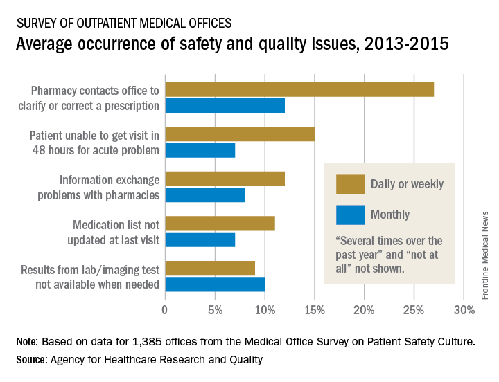
Information exchange problems with pharmacies occurred daily or weekly in 12% of offices and monthly in 8%, while the occurrence of medication lists not being updated at the last visit was 11% daily/weekly and 7% monthly. Additionally, 9% of offices reported that results from laboratory or imaging tests were not available when needed on a daily/weekly basis and 10% on a monthly basis, the AHRQ reported.
“Lack of access to care and lack of access to timely and accurate medical information and test results may contribute to patient safety events such as missed or delayed diagnoses, medication errors, failure to order appropriate diagnostic or laboratory tests, incorrect interpretation of tests, and inadequate follow-up on results,” the AHRQ said in the report.