User login
Link Between Intestinal Fungi and Alcoholic Liver Disease Grows Stronger
Alcoholic liver disease (ALD) has been associated with bacterial overgrowth in the intestines, as well as a shift in the types of bacteria found there. But until now, according to the National Institute on Alcohol Abuse and Alcoholism, little was actually known about the role of intestinal fungi in ALD. A University of California-San Diego and J. Craig Venter Institute in Rockville, Maryland study offers more evidence to support the connection.
In the study, the researchers found that fungi flourished in the intestines of mice with chronic alcohol exposure, and that fungal overgrowth exacerbated alcohol-induced liver disease. Treatment with amphotericin B reduced levels of liver injury and fat accumulation.
In small preliminary studies with humans, the researchers also linked intestinal fungi to ALD. People with alcohol use disorder and various stages of liver disease tended to have an overgrowth of a specific fungal species, as well as less fungal diversity, compared with healthy control subjects. Moreover, the more prevalent the fungal overgrowth in people with ALD, the higher the risk of death.
The researchers say that if further study confirms that fungi are involved in the worsening of ALD, it may be possible to slow disease progression by adjusting the balance of fungal species in the intestine.
Health Canada approves new use for brentuximab vedotin

Health Canada has issued a non-conditional marketing authorization for brentuximab vedotin (Adcetris).
This means the drug is now approved for use as consolidation after autologous stem cell transplant (ASCT) in patients with Hodgkin lymphoma (HL) who have an increased risk of relapse or progression.
Brentuximab vedotin previously received approval with conditions in Canada to treat HL patients who relapse after ASCT or HL patients who are not ASCT candidates and relapse after at least 2 multi-agent chemotherapy regimens.
Brentuximab vedotin also has conditional approval in Canada to treat patients with systemic anaplastic large-cell lymphoma who relapse after at least 1 multi-agent chemotherapy regimen.
Brentuximab vedotin is an antibody-drug conjugate consisting of an anti-CD30 monoclonal antibody attached by a protease-cleavable linker to a microtubule disrupting agent, monomethyl auristatin E.
Brentuximab vedotin has marketing authorization in 67 countries for the treatment of relapsed or refractory HL and systemic anaplastic large-cell lymphoma.
Seattle Genetics and Takeda are jointly developing brentuximab vedotin. Seattle Genetics has US and Canadian commercialization rights, and Takeda has rights to commercialize the drug in the rest of the world.
AETHERA trial
Health Canada’s decision to extend the marketing authorization of brentuximab vedotin is based on results from the phase 3 AETHERA trial.
The trial was designed to compare brentuximab vedotin to placebo, both administered for up to 16 cycles (approximately 1 year) every 3 weeks following ASCT. Results from the trial were published in The Lancet in March 2015.
The study enrolled 329 HL patients at risk of relapse or progression—165 on the brentuximab vedotin arm and 164 on the placebo arm.
Patients were eligible for enrollment if they had a history of primary refractory HL, relapsed within a year of receiving frontline chemotherapy, and/or had disease outside of the lymph nodes at the time of pre-ASCT relapse.
Brentuximab vedotin conferred a significant increase in progression-free survival over placebo, with a hazard ratio of 0.57 (P=0.001). The median progression-free survival was 43 months for patients who received brentuximab vedotin and 24 months for those who received placebo.
The most common adverse events (≥20%), of any grade and regardless of causality, in the brentuximab vedotin arm were neutropenia (78%), peripheral sensory neuropathy (56%), thrombocytopenia (41%), anemia (27%), upper respiratory tract infection (26%), fatigue (24%), peripheral motor neuropathy (23%), nausea (22%), cough (21%), and diarrhea (20%).
The most common adverse events (≥20%), of any grade and regardless of causality, in the placebo arm were neutropenia (34%), upper respiratory tract infection (23%), and thrombocytopenia (20%).
In all, 67% of patients on the brentuximab vedotin arm experienced peripheral neuropathy. Of those patients, 85% had resolution (59%) or partial improvement (26%) in symptoms at the time of their last evaluation, with a median time to improvement of 23 weeks (range, 0.1-138). ![]()
Survey reveals change in public perception of clinical trials

More Americans are viewing clinical trials in a positive light, according to a survey commissioned by Research!America and the Association of Clinical Research Organizations (ACRO).
The results of the survey, which included 1000 participants and was conducted this month, reveal a shift in public attitudes about clinical trials since a similar survey was commissioned in 2013.
Thirty-seven percent of respondents involved in the current survey said they would “very likely” enroll in a clinical trial if their doctor recommended it. This represents an 11% increase from 2013.
Eighty-four percent of current survey respondents said they are willing to share personal health information, assuming that appropriate privacy protections are in place, so researchers can better understand diseases and develop new ways to prevent, treat, and cure them. This is a 10% increase from 2013.
When asked how much they admire people who volunteer for clinical trials, 46% of current respondents said “a great deal,” which is a 9% increase from 2013.
“More and more Americans appear to recognize the value of clinical trials—a very positive sign—but stubborn barriers to participation remain in place,” said Mary Woolley, president and CEO of Research!America.
“Development of incentives to drive more discussions between patients and healthcare professionals about the importance of participating in trials could encourage both ill and healthy individuals to view this as a routine health behavior.”
“We are pleased to have joined with Research!America in commissioning this important survey and gratified that the public’s knowledge of and attitudes toward clinical trials have moved in positive directions since 2013,” said Doug Peddicord, executive director of ACRO.
“The option to participate in a clinical trial, when appropriate, should be a routine part of the healthcare encounter, and ACRO will continue to work with Research!America and others to spread that message to doctors and patients alike.”
Survey results
The survey of 1000 US adults was conducted by Zogby Analytics for Research!America and ACRO in July 2017. The margin of error is +/- 3.1 percentage points.
Ninety percent of survey respondents said clinical trials are important to advancing science, and 87% said trials are important to improving the nation’s health. Seventy-five percent said taking part in clinical trials is as valuable to the US healthcare system as giving blood.
Eighty-six percent of respondents said healthcare professionals should discuss clinical trials with patients diagnosed with a disease as part of their standard care.
And 44% of respondents said clinical trial participation should be a routine health behavior, whether a person is healthy or ill, similar to getting an annual checkup with a healthcare provider.
Men (48%) were significantly more likely than women (39%) to say trial participation should be routine. A larger percentage of 18- to 29-year-olds (53%) and 30- to 49-year olds (48%) were in favor of routine trial participation, compared to people ages 50 to 64 (38%) and those 65 and older (34%).
Eighty percent of respondents said they had heard of a clinical trial, and 18% said they or someone in their family had participated in one.
Seventy-four percent of respondents said they would participate in a clinical trial if asked by someone they trust.
However, respondents were split on whether it’s important for everyone to take part in a clinical trial if asked. Forty-four percent said it is important, 45% disagreed, and 12% were not sure.
More than half of respondents (55%) said people don’t participate in clinical trials because of lack of awareness and information, 43% said it’s because trials are viewed as “too risky,” 41% said it’s due to a lack of information about the process, 38% said it’s due to a lack of trust, and 34% said it’s due to the risk of adverse health effects.
Nearly two-thirds of respondents (64%) said a doctor or healthcare provider is a reliable source for clinical trial information.
Respondents said doctors and other healthcare providers (44%), followed by the government (23%), have the greatest responsibility in educating the public about clinical trials. However, 74% said no healthcare professional has ever talked to them about medical research.
Seventy-two percent of respondents said they would be likely to use technology such as apps and monitoring devices to share their personal health data for clinical research.
Eighty-eight percent said clinical trial participants should have access to trial results. And 47% said they would like having clinical trial information/data/results delivered through their phone. ![]()
Predicting response to treatment in AML, MDS

Researchers say they have determined which patients will respond to treatment with SY-1425, a retinoic acid receptor alpha (RARα) agonist.
The team discovered a subset of patients with acute myeloid leukemia (AML) who had a super-enhancer associated with the RARA gene, which is predictive of response to SY-1425.
The researchers also identified a subset of patients with myelodysplastic syndromes (MDS) who had high expression of the RARA gene.
And experiments showed that RARA-high MDS had a similar response to SY-1425 as that seen in AML driven by the RARA super-enhancer.
Ravindra Majeti MD, PhD, of Stanford University School of Medicine in California, and colleagues reported these findings in Cancer Discovery. Employees of Syros Pharmaceuticals, the company developing SY-1425, were also involved in this research.
In collaboration with the Majeti lab, Syros used its gene control platform to analyze 66 AML patients’ tumor samples. In this way, the researchers identified 6 distinct patient subsets based on super-enhancer profiles, including 1 enriched for a super-enhancer associated with the RARA gene.
The team found that super-enhancer profiles were strongly associated with survival outcomes, often independent of known genetic mutations in AML.
The RARA super-enhancer was associated with high expression of the RARA gene, which codes for a transcription factor targeted by SY-1425.
The RARA super-enhancer was predictive of response to SY-1425. In AML cells with high RARA expression, SY-1425 reduced proliferation and promoted differentiation.
Moreover, SY-1425 decreased tumor burden and prolonged survival in patient-derived xenograft models of AML with high RARA expression. However, there was no effect on AML cells or models with low RARA expression.
The researchers said SY-1425 induced profound transcriptional changes promoting cell differentiation in AML cells with high RARA expression, but the drug produced little to no transcriptional changes in AML cells with low RARA expression.
DHRS3 was the most strongly and rapidly induced gene in response to treatment with SY-1425. This led to the identification of DHRS3 induction as an early indicator of whether SY-1425 is affecting the targeted biology in defined subsets of AML and MDS patients. It is therefore used as a pharmacodynamic marker in the ongoing phase 2 trial of SY-1425.
In this trial, researchers are assessing the safety and efficacy of SY-1425 as a single agent in 4 AML and MDS patient populations, as well as testing SY-1425 in combination with azacitidine in newly diagnosed AML patients who are not suitable candidates for standard chemotherapy. ![]()
PEARL score for COPD exacerbations
TITLE: PEARL score predicts COPD readmissions
CLINICAL QUESTION: Which prognostic score is best at predicting 90-day readmission and mortality for patients admitted with an acute exacerbation of chronic obstructive pulmonary disease (AECOPD)?
STUDY DESIGN: Prospective study with three separate cohorts: derivation, internal validation, and external validation.
SETTING: Six hospitals in the United Kingdom.
SYNOPSIS: 2,417 patients were included and 936 were readmitted or died within 90 days of index admission. Patients with expected survival for less than 1 year for reasons other than COPD were excluded. The indices retained in the final PEARL score were: Previous admissions for AECOPD of 2 or more (2 points), extended medical research council (MRC) dyspnea score of 4, 5a or 5b (1, 2, or 3 points), age of 80 or older (1 point), clinical diagnoses of right-sided heart failure (1 point) and/or left-sided heart failure on echocardiogram (1 point). Higher scores were associated with a shorter time to death or readmission. The performance of PEARL was superior to all alternative scoring systems. The major limitation to this study is that it did not differentiate between respiratory and other causes of readmission.
BOTTOM LINE: The PEARL score can be calculated for patients hospitalized for AECOPD to predict their 90-day readmission rate and/or mortality risk.
CITATION: Echevarria C, Steer J, Heslop-Marshall K, Stenton SC, Hickey PM, Hughes R, et al. The PEARL score predicts 90-day readmission or death after hospitalization for acute exacerbation of COPD. Thorax. 2017; doi: 10.1136/thoraxjnl-2016-209298.
Dr. Ayoubieh is assistant professor in the division of hospital medicine at the University of New Mexico.
Tips for managing dermatology procedures in kids
CHICAGO – True complications in pediatric dermatologic surgery probably aren’t that frequent, but no solid data on the topic exist in the medical literature.
“An appropriate and thorough perioperative evaluation and planning may limit complications,” Harper N. Price, MD, said at the World Congress of Pediatric Dermatology.

The first step is to make the child comfortable in the office or operating room (OR) setting; this can include approaching children slowly unless you know them well. “Sit at their level, because coming up very fast and being over ... them is intimidating,” she advised. “Make sure you include the child in the conversation you’re having; it elicits more trust and belief in what’s going to happen. You want to explain what’s going to happen in a friendly manner. I think sometimes we have residents who are new to pediatrics that come in and say, ‘We’re going to cut this out,’ and the next thing you know, the child’s in tears. Describe what the procedure is going to be like in words that they can understand, and whatever you do, do not lie about what’s going to happen.”
Dr. Price also makes it a point to cover surgical trays before they’re wheeled in. “They don’t need to see needles and sharp objects,” she said. “Even afterward, bloody gauze can be scary to kids.” Positioning the patient properly also is important. “We’ll wrap young children up in a swaddle,” she said. “In my opinion, you should not be forcefully restraining an older child. They need to cooperate and it needs to be a safe procedure, otherwise, you should consider doing it in the operating room. I never enlist a parent to hold or restrain a child.”
One key to managing pain during dermatologic procedures in children comes down to anticipation: What kinds of distractions might the child need? What preoperative analgesia will be required? What postoperative pain medications should be used? “We know that certain procedures in children might be more painful, such as nail procedures, ablative laser procedures, and large excisions with extensive undermining,” Dr. Price said. “Pain is subjective and differs from child to child in the way it’s experienced, so you need to consider the child’s age, coping style, and temperament, and what their history of pain is like. We know that inadequate pain control in children has a negative impact and a negative implication on their future health care interventions, as well as their reactions to further pain.”
Parental involvement can sometimes help. “I like a parent to stay in the room if I’m doing a procedure in the office, as long as they agree to stay seated,” she said. “It may make your office staff more anxious, and it may make parents more anxious, too, so it’s something to think about.” There is some evidence that having a parent present during an in-office procedure increases parental satisfaction as well.
In an effort to minimize pain and anxiety before in-office procedures, Dr. Price and her associates at Phoenix Children’s Hospital often use instant ice packs. “They get cold really fast, they’re cheap, and you don’t have to run to a refrigerator to get ice,” she said. Other beneficial measures include topical anesthetics and breathing techniques, such as having the child blow on a pinwheel, blow bubbles, or perform diaphragmatic breathing. Using distractions – stuffed animals, picture books, or video games on a tablet – can also help. “If the child is going to the OR, using preoperative midazolam can help relax the child, especially if they’re having repeated procedures,” Dr. Price said. Oral sucrose solution in infants, especially in young infants, provides about 5-8 minutes of temporary analgesia and can be placed on their pacifier or their tongue, she added, noting that ethyl chloride spray can also be helpful prior to injections.
During the procedure itself, counter-stimulatory methods can be helpful; this can include handheld devices that use a combination of vibration, ice, and distraction methods. “Buffer your lidocaine and don’t inject cold lidocaine; that hurts a lot more,” she recommended. “Inject slowly; inject deep. If you have a painful procedure and you’re in an OR setting where you give Marcaine [bupivacaine], put that in at the end of the procedure for short-term postoperative pain relief.” After the procedure, it’s better not to apologize for causing pain or if the procedure didn’t go well. “Give positive incentives like stickers and stuffed animals, and use a dressing wrap with bright colors,” she said. “We often doctor up stuffed animals in the OR so when [the children] wake up, they have something fun to look at.”
Postoperatively, the best way to prevent pain is to recommend limited physical activity. “Children become active quickly after a procedure, and then they hurt,” Dr. Price said. “For extremity wounds, consider ice and elevation. I like bulky dressings to prevent trauma, to remind the families that they’ve had a procedure done. They can usually keep them on for several days.”
Surgical site infections are uncommon, but if they do occur, it’s usually between postoperative days 4 and 10. “The biggest indicator of an infection in my opinion is pain,” she said. “If they’re having a lot of pain, I would be concerned. Causes may be the presence of bacteria on the skin or mucosa or improper wound care at home.”
The risk factors for surgical site infections in children are not well defined in dermatologic surgery, Dr. Price added, “but we know that if you’re going to be operating in the diaper area, that’s a place where you’re going to have a high risk of infection. Preoperative hair removal – if you shave the scalp before surgery creating small nicks – could [introduce] bacteria. And it’s likely that the overall health of the patient may impact their risk of infection. You want to know the difference between normal wound healing and an infection. Culture it. If you’re worried, you may want to start empiric antibiotics. If you have a severe infection, something with necrosis, fluctuance, or dehiscence, you might want to consider partially opening that wound and letting it drain and heal in by secondary intention.”
Measures to prevent postoperative infections include perioperative counseling to restrict excessive activity to prevent trauma, bleeding, and dehiscence; use of bulky dressings, and explicit wound care instructions. “My nurse calls [the patient’s family] the day after a procedure, and I usually have them come in for a wound check, even if there are no sutures to remove, just to make sure things look OK,” she said.
Suture reactions are another potential complication of dermatologic surgery in children. The incidence is unknown, but suture reactions usually occur around 6 weeks postoperatively and tend to happen more often in older children. “Excessive reactions, while uncommon, can lead to an increased risk of dehiscence, infection, and delayed healing,” Dr. Price said. Small caliber monofilament sutures are less reactive than large caliber, multifilament sutures, she added, while synthetic and nonabsorbable sutures are less reactive than natural materials such as silk and surgical gut. Dr. Price favors using poliglecaprone, polyglactin 910, and polypropylene.
Tips for minimizing suture reactions include the following: Use the smallest caliber suture appropriate for the wound; avoid buried sutures too close to the surface of the skin; use a smaller caliber suture at the end of excisions, where there tends to be less tension; and keep knots small and flat at the apexes of excision. “Manage suture reactions with reassurance,” she said. “The nice thing is that these often heal fine without any delay. When possible, remove the offending suture material. A lot of times, I’ll use sterile forceps. At home, I’ll have [parents] massage the area with warm compresses to try to extrude the suture. But, if you wait long enough, it usually comes out.”
Dr. Price reported having no financial disclosures.
[email protected]
CHICAGO – True complications in pediatric dermatologic surgery probably aren’t that frequent, but no solid data on the topic exist in the medical literature.
“An appropriate and thorough perioperative evaluation and planning may limit complications,” Harper N. Price, MD, said at the World Congress of Pediatric Dermatology.
The first step is to make the child comfortable in the office or operating room (OR) setting; this can include approaching children slowly unless you know them well. “Sit at their level, because coming up very fast and being over ... them is intimidating,” she advised. “Make sure you include the child in the conversation you’re having; it elicits more trust and belief in what’s going to happen. You want to explain what’s going to happen in a friendly manner. I think sometimes we have residents who are new to pediatrics that come in and say, ‘We’re going to cut this out,’ and the next thing you know, the child’s in tears. Describe what the procedure is going to be like in words that they can understand, and whatever you do, do not lie about what’s going to happen.”
Dr. Price also makes it a point to cover surgical trays before they’re wheeled in. “They don’t need to see needles and sharp objects,” she said. “Even afterward, bloody gauze can be scary to kids.” Positioning the patient properly also is important. “We’ll wrap young children up in a swaddle,” she said. “In my opinion, you should not be forcefully restraining an older child. They need to cooperate and it needs to be a safe procedure, otherwise, you should consider doing it in the operating room. I never enlist a parent to hold or restrain a child.”
One key to managing pain during dermatologic procedures in children comes down to anticipation: What kinds of distractions might the child need? What preoperative analgesia will be required? What postoperative pain medications should be used? “We know that certain procedures in children might be more painful, such as nail procedures, ablative laser procedures, and large excisions with extensive undermining,” Dr. Price said. “Pain is subjective and differs from child to child in the way it’s experienced, so you need to consider the child’s age, coping style, and temperament, and what their history of pain is like. We know that inadequate pain control in children has a negative impact and a negative implication on their future health care interventions, as well as their reactions to further pain.”
Parental involvement can sometimes help. “I like a parent to stay in the room if I’m doing a procedure in the office, as long as they agree to stay seated,” she said. “It may make your office staff more anxious, and it may make parents more anxious, too, so it’s something to think about.” There is some evidence that having a parent present during an in-office procedure increases parental satisfaction as well.
In an effort to minimize pain and anxiety before in-office procedures, Dr. Price and her associates at Phoenix Children’s Hospital often use instant ice packs. “They get cold really fast, they’re cheap, and you don’t have to run to a refrigerator to get ice,” she said. Other beneficial measures include topical anesthetics and breathing techniques, such as having the child blow on a pinwheel, blow bubbles, or perform diaphragmatic breathing. Using distractions – stuffed animals, picture books, or video games on a tablet – can also help. “If the child is going to the OR, using preoperative midazolam can help relax the child, especially if they’re having repeated procedures,” Dr. Price said. Oral sucrose solution in infants, especially in young infants, provides about 5-8 minutes of temporary analgesia and can be placed on their pacifier or their tongue, she added, noting that ethyl chloride spray can also be helpful prior to injections.
During the procedure itself, counter-stimulatory methods can be helpful; this can include handheld devices that use a combination of vibration, ice, and distraction methods. “Buffer your lidocaine and don’t inject cold lidocaine; that hurts a lot more,” she recommended. “Inject slowly; inject deep. If you have a painful procedure and you’re in an OR setting where you give Marcaine [bupivacaine], put that in at the end of the procedure for short-term postoperative pain relief.” After the procedure, it’s better not to apologize for causing pain or if the procedure didn’t go well. “Give positive incentives like stickers and stuffed animals, and use a dressing wrap with bright colors,” she said. “We often doctor up stuffed animals in the OR so when [the children] wake up, they have something fun to look at.”
Postoperatively, the best way to prevent pain is to recommend limited physical activity. “Children become active quickly after a procedure, and then they hurt,” Dr. Price said. “For extremity wounds, consider ice and elevation. I like bulky dressings to prevent trauma, to remind the families that they’ve had a procedure done. They can usually keep them on for several days.”
Surgical site infections are uncommon, but if they do occur, it’s usually between postoperative days 4 and 10. “The biggest indicator of an infection in my opinion is pain,” she said. “If they’re having a lot of pain, I would be concerned. Causes may be the presence of bacteria on the skin or mucosa or improper wound care at home.”
The risk factors for surgical site infections in children are not well defined in dermatologic surgery, Dr. Price added, “but we know that if you’re going to be operating in the diaper area, that’s a place where you’re going to have a high risk of infection. Preoperative hair removal – if you shave the scalp before surgery creating small nicks – could [introduce] bacteria. And it’s likely that the overall health of the patient may impact their risk of infection. You want to know the difference between normal wound healing and an infection. Culture it. If you’re worried, you may want to start empiric antibiotics. If you have a severe infection, something with necrosis, fluctuance, or dehiscence, you might want to consider partially opening that wound and letting it drain and heal in by secondary intention.”
Measures to prevent postoperative infections include perioperative counseling to restrict excessive activity to prevent trauma, bleeding, and dehiscence; use of bulky dressings, and explicit wound care instructions. “My nurse calls [the patient’s family] the day after a procedure, and I usually have them come in for a wound check, even if there are no sutures to remove, just to make sure things look OK,” she said.
Suture reactions are another potential complication of dermatologic surgery in children. The incidence is unknown, but suture reactions usually occur around 6 weeks postoperatively and tend to happen more often in older children. “Excessive reactions, while uncommon, can lead to an increased risk of dehiscence, infection, and delayed healing,” Dr. Price said. Small caliber monofilament sutures are less reactive than large caliber, multifilament sutures, she added, while synthetic and nonabsorbable sutures are less reactive than natural materials such as silk and surgical gut. Dr. Price favors using poliglecaprone, polyglactin 910, and polypropylene.
Tips for minimizing suture reactions include the following: Use the smallest caliber suture appropriate for the wound; avoid buried sutures too close to the surface of the skin; use a smaller caliber suture at the end of excisions, where there tends to be less tension; and keep knots small and flat at the apexes of excision. “Manage suture reactions with reassurance,” she said. “The nice thing is that these often heal fine without any delay. When possible, remove the offending suture material. A lot of times, I’ll use sterile forceps. At home, I’ll have [parents] massage the area with warm compresses to try to extrude the suture. But, if you wait long enough, it usually comes out.”
Dr. Price reported having no financial disclosures.
[email protected]
AT WCPD 2017
CDC refocuses Zika testing recommendations in pregnancy
, including those who may have been exposed before pregnancy through travel or sexual contact.
In updated guidance released July 24, the Centers for Disease Control and Prevention cited a combination of factors behind the change in recommendations, including the declining prevalence of Zika virus across the Americas and a high likelihood of false positives associated with the use of a common serologic assay (MMWR Morb Mortal Wkly Rep. ePub 2017 Jul 24. doi: 10.15585/mmwr.mm6629e1).
Positive IgM results can also occur after previous exposure to other flaviviruses besides Zika, Dr. Oduyebo and her colleagues noted.
The CDC now recommends that pregnant women with likely continuing – not previous – exposure to the Zika virus and those with symptoms suggestive of Zika virus disease be tested. Those higher-risk groups should receive nucleic acid testing (NAT).
The new guidance presents two updated testing algorithms, one for each group.
Any pregnant woman with symptoms suggestive of Zika should be tested “as soon as possible through 12 weeks after symptom onset,” the CDC said, with both NAT (serum and urine) and IgM serology testing.
Women with likely ongoing exposure to Zika – such as those living in or traveling to an area of mosquito-borne Zika transmission or those whose partners are living in or traveling to such an area – should be tested up to three times during the pregnancy using NAT serum and urine tests. IgM testing is not recommended for that group.
All pregnant women should be asked about their potential Zika exposures before and during the current pregnancy, the CDC said. That discussion, which covers potential travel and partner exposures along with questions about symptoms, should be repeated at every prenatal visit.
While routine testing of asymptomatic women without ongoing exposure is not recommended, patient preferences, clinical judgment, and a “balanced assessment of risks and expected outcomes” should guide decisions about testing, according to the CDC.
FROM MMWR
Racial differences in dementia risk persist from midlife to oldest old
LONDON – The racial and ethnic patterns of dementia risk seen in older adults appear to hold steady even in the oldest old, with blacks about 30% more likely than are whites to develop the disorder even well into their 90s.
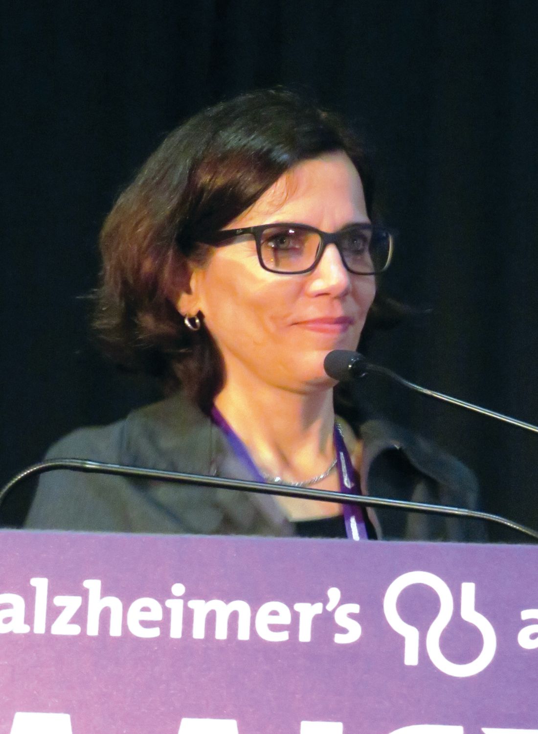
The magnitude of disparity remained consistent even after researchers controlled for traditional risk factors such as access to health care, cardiovascular risk, stroke, and education, Maria Corrada, ScD, said at the Alzheimer’s Association International Conference.
“These are the first estimates of dementia in a diverse cohort of subjects 90 years or older,” said Dr. Corrada, a professor of epidemiology at the University of California, Irvine. “The racial and ethnic differences in dementia incidence didn’t appear to be due to these factors that we have come to expect. These estimates can provide us with an important foundation for understanding the burden of racial disparities in the oldest old, which is the fastest-growing segment of our population in the U.S. and in many other countries.”
Most of the cohort (72%) was white; blacks comprised 16%, Latinos 4%, and Asians 7%. Not surprisingly, most were women (65%). The mean age at baseline was 93 years. Overall, 50% had at least a high school education, but that percentage was higher in whites (56%) and lower in blacks and Latinos (33% and 24%, respectively).
Midlife obesity was present in 42% overall, but this was significantly higher in blacks and Latinos (63%), and lower in whites and Asians (38% and 24%). Midlife hypertension was present in 38% overall. This was highest in blacks (63%), followed by Latinos (42%) and whites (35%). Only 7% of Asians had high blood pressure in midlife. About 40% of each group had experienced a late-life stroke, with the exception of Asians, with a stroke incidence of just 7%. Diabetes was present in 24% overall, in 20% of whites, and about a third of the other groups.
Over the follow-up period, dementia developed in 33% of subjects, at a mean age of 95 years, which did not vary among groups. The incidence of dementia was lowest in whites (32%) and Asians (31%). It was most frequent in blacks (39%), followed by Latinos (35%). The age-adjusted, 5-year incidence rate was 10% overall. It was lowest in Asians (9%) and whites (9.7%), followed by Latinos (10.6%); it was highest in blacks (12%).
Dr. Corrada then conducted a series of five multivariate regression analyses to examine the effect of various risk factors on dementia. The first three controlled for age alone; for age and education; and for age, education, and midlife risk factors of obesity, hypertension, and cholesterol levels. In every model, blacks were 30% more likely to develop dementia over the follow-up period than were whites. Model 4 controlled for age, education, and the late-life risk factors of stroke, depression, ischemic heart disease, heart failure, and heart attack. Model 5 controlled for age, education, and both the late- and midlife risk factors.
Again, no matter which model was used, blacks faced the same 30% increased risk, compared with whites. “The differences remained very consistent,” Dr. Corrada said. “The patterns of racial and ethnic disparity seen in younger elderly continued well after age 90.”
Dr. Corrada had no financial conflicts of interest.
[email protected]
On Twitter @alz_gal
LONDON – The racial and ethnic patterns of dementia risk seen in older adults appear to hold steady even in the oldest old, with blacks about 30% more likely than are whites to develop the disorder even well into their 90s.
The magnitude of disparity remained consistent even after researchers controlled for traditional risk factors such as access to health care, cardiovascular risk, stroke, and education, Maria Corrada, ScD, said at the Alzheimer’s Association International Conference.
“These are the first estimates of dementia in a diverse cohort of subjects 90 years or older,” said Dr. Corrada, a professor of epidemiology at the University of California, Irvine. “The racial and ethnic differences in dementia incidence didn’t appear to be due to these factors that we have come to expect. These estimates can provide us with an important foundation for understanding the burden of racial disparities in the oldest old, which is the fastest-growing segment of our population in the U.S. and in many other countries.”
Most of the cohort (72%) was white; blacks comprised 16%, Latinos 4%, and Asians 7%. Not surprisingly, most were women (65%). The mean age at baseline was 93 years. Overall, 50% had at least a high school education, but that percentage was higher in whites (56%) and lower in blacks and Latinos (33% and 24%, respectively).
Midlife obesity was present in 42% overall, but this was significantly higher in blacks and Latinos (63%), and lower in whites and Asians (38% and 24%). Midlife hypertension was present in 38% overall. This was highest in blacks (63%), followed by Latinos (42%) and whites (35%). Only 7% of Asians had high blood pressure in midlife. About 40% of each group had experienced a late-life stroke, with the exception of Asians, with a stroke incidence of just 7%. Diabetes was present in 24% overall, in 20% of whites, and about a third of the other groups.
Over the follow-up period, dementia developed in 33% of subjects, at a mean age of 95 years, which did not vary among groups. The incidence of dementia was lowest in whites (32%) and Asians (31%). It was most frequent in blacks (39%), followed by Latinos (35%). The age-adjusted, 5-year incidence rate was 10% overall. It was lowest in Asians (9%) and whites (9.7%), followed by Latinos (10.6%); it was highest in blacks (12%).
Dr. Corrada then conducted a series of five multivariate regression analyses to examine the effect of various risk factors on dementia. The first three controlled for age alone; for age and education; and for age, education, and midlife risk factors of obesity, hypertension, and cholesterol levels. In every model, blacks were 30% more likely to develop dementia over the follow-up period than were whites. Model 4 controlled for age, education, and the late-life risk factors of stroke, depression, ischemic heart disease, heart failure, and heart attack. Model 5 controlled for age, education, and both the late- and midlife risk factors.
Again, no matter which model was used, blacks faced the same 30% increased risk, compared with whites. “The differences remained very consistent,” Dr. Corrada said. “The patterns of racial and ethnic disparity seen in younger elderly continued well after age 90.”
Dr. Corrada had no financial conflicts of interest.
[email protected]
On Twitter @alz_gal
AT AAIC 2017
Key clinical point:
Major finding: Blacks were 30% more likely to develop dementia than were whites, even after researchers controlled for age, education, and mid- and late-life health–related risk factors.
Data source: The 5-year prospective study comprised more than 2,000 subjects aged 90 years and older.
Disclosures: The presenter had no financial disclosures.
Lupus classification criteria need input from dermatologists
Editor’s note: This commentary relates to the story, “ New classification system for systemic lupus erythematosus moves forward .”
The ACR/EULAR committee that is developing new classification criteria for systemic lupus erythematosus (SLE) has done the field a service by releasing its draft version in a presentation at the recent EULAR meeting. Releasing the draft version facilitates comments before the new classification criteria become finalized.
Many in the derm-rheum field, ourselves included, classify patients with skin-predominant lupus as lupus, but the new draft classification would place a significant percentage of these patients outside of lupus.
The presentation by Dr. Johnson at EULAR stated, “... a patient can’t be classified on skin findings alone. There is concern that skin findings by themselves may not be lupus, but something else, and some people even consider that cutaneous and systemic lupus are two different things.”
Abundant data indicate instead that lupus is a spectrum that includes skin-predominant lupus. For example, the histology is identical between discoid lupus erythematosus whether or not there is SLE. Moreover, we and others have published significant rates of progression of cutaneous lupus erythematosus (CLE) to SLE. By not viewing CLE in the lupus spectrum, we have a false sense that the patients won’t progress to SLE, yet many of them do.
Importantly, patients respond similarly to therapies when they have either CLE or SLE, so removing this subset of lupus hurts their inclusion in trials and access to new treatments.
When criteria are devised by one group without input from experts who see a specific subset of the disease, that is also a problem. We went down that path in dermatomyositis and missed a lot of patients with the disease when criteria were devised that said the patient had to have muscle involvement. Those criteria have now finally been revised as the ACR/EULAR myositis criteria.
We and others from the derm-rheum community would be happy to speak with the ACR/EULAR committee about these concerns.
Victoria P. Werth, MD, is professor of medicine and dermatology at the University of Pennsylvania, Philadelphia. Joseph F. Merola, MD, is codirector of the Center for Skin and Related Musculoskeletal Diseases at Brigham and Women’s Hospital, Boston. Andrew G. Franks, MD, is a clinical professor in the departments of medicine and dermatology at New York University. Benjamin F. Chong, MD, is an assistant professor of dermatology at the University of Texas, Dallas.
Lupus classification criteria effort is going in the wrong direction
Editor’s note: This commentary relates to the story, “New classification system for systemic lupus erythematosus moves forward.”
While work to develop a new set of lupus classification criteria more suitable for clinical research is important, the Lupus Foundation of America believes the current work is going in the wrong direction.
Increasingly, key opinion leaders understand lupus to be a spectrum of disease, and there is ample justification of this from scientific evidence (Nat Rev Rheumatol. 2015;11[7]:385-6) The criteria being worked on by ACR and EULAR draws upon an archaic concept with the musty name of “systemic lupus erythematosus,” which relies on a 19th century approach to categorizing disease by counting the signs and symptoms instead of by modern concepts of pathophysiology and prognostic severity. This imposes a homogeneity on the population that simply does not exist. Attempts to re-order obsolescent arrays of organs and autoantibodies to classify lupus will be futile, especially if the aim is to improve the rationale for clinical trial recruitment. Recent clinical trials and modern immunologic methods have already demonstrated, beyond a doubt, that subsets of patients, definable by gene expression patterns or state of the art pharmacodynamic responses, do or do not respond to individual targeted treatments. We now know that patients who require different treatments may well share many of their symptoms in many of the same organs, and this fact defies the outmoded, abacus-based approach to disease classification.
We are concerned that redefining SLE by weighting all disease in one organ as more or less impactful than all disease in another organ flies in the face of current scientific knowledge. If this is the direction in which the effort is going, there is the potential for a negative impact on drug development, clinical care, and access to treatment. The term itself, “SLE,” interferes with selecting appropriate lupus patients for participation in trials. Many people have moderate and even severe lupus syndromes who do not meet enough criteria to be labeled “SLE” (for example, immune thrombocytopenia, hemolytic anemia, discoid lupus, or subacute cutaneous lupus, which can cause severe rashes covering wide areas of the body). In this iteration, assigning less weight to cutaneous lupus as currently proposed is not just problematic, it will set the field back.
By not viewing cutaneous lupus as part of the lupus spectrum, we develop a false sense that this subset of people with lupus will not progress to “SLE,” yet many of them do. Even those who do not later develop features in other organs besides the skin may have more severe disease than other patients who do. Minimizing the “score” for cutaneous lupus will lead to individuals who carry a significant burden of disease being barred from trials, and from access to the treatments they need, once approved.
Conversely, people who do meet criteria for “SLE” under any algorithm, past or present, may have a range of severity, from very severe to very mild. The very mild patients (who may have, in their lifetime met the multiorgan criteria) are entering trials in large numbers and contributing to the high placebo responses which have stopped many promising investigational treatments from further development. Additionally, the common misuse of the current classification criteria as diagnostic criteria has become an unacceptable norm in the lupus community. This leads us to believe that new criteria will continue to be misused, further disenfranchising a huge segment of the population who have lupus from access to state-of-the-art research and care.
Advancing the development new classification criteria deserves a wider discussion among the field’s stakeholders, particularly those with expertise in clinical trial outcomes and the clinical care of the full spectrum of lupus patients.
Sandra C. Raymond is CEO and President, Leslie M. Hanrahan is VP of Education and Research, and Joan Merrill, MD, is the Chief Adviser for Clinical Development at the Lupus Foundation of America. Dr. Merrill is also the Oklahoma Medical Research Foundation Professor of Medicine at the University of Oklahoma, Oklahoma City.