User login
RIV winners celebrated for their creative use of data
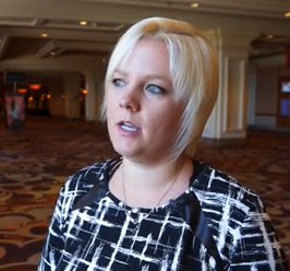
This year’s RIV innovation winners reflect a nascent trend of applying informatics to quality improvement and patient safety initiatives.
“One striking thing is that all three winners used either EHR or Big Data and large collaboratives to achieve their goals,” Margaret Fang, MD, MPH, FHM, program chair for HM17’s scientific abstracts competition, and moderator of the winners’ panel, said in an interview. This year’s winners included a sleep-promoting “nudge” system that Dr. Fang said she expects will help improve sleep and lower rates of delirium and a source code that connects disparate data systems for daily updates on where quality can be improved. The third winner used what Dr. Fang, a hospitalist and the medical director of the anticoagulation clinic at the University of California, San Francisco, called a “classic quality improvement collaborative,” which simplifies the decision tree around venous thromboembolism (VTE) prophylaxis for better patient outcomes.
Calling uninterrupted sleep the “sine qua non” of patient care, RIV award recipient Vineet Arora, MD, an associate professor of medicine at the University of Chicago, described the rationale for her RIV award-winning SIESTA (Sleep for Inpatients: Empowering Staff to Act) program. She and her colleagues surveyed hospitalists, nurses, residents, and patients to determine the most common sleep disrupters in their institution and devised “nudges” to alter how staff performed various tasks that otherwise might interfere with patient sleep. Rather than use overt incentives, nudges are changes in what Dr. Arora called the “choice architecture” of people’s behavior.
Based on survey feedback, Dr. Arora and her colleagues worked with their electronic health record (EHR) vendor to consolidate the performance of certain tasks that were affecting patient sleep. Reminders were added to daily nursing huddles to prompt them to look for ways they could decrease patient interruptions, and empowerment coaching was offered to nurses to encourage patient advocacy when physicians had given orders that would interfere with patients’ sleep.
When tested and measured over the course of a year, SIESTA’s EHR innovations resulted in six fewer nighttime disruptions than before the intervention, compared with controls, a statistically significant difference. The nursing-based interventions resulted in one less nocturnal interruption on average, also a significant change.
“If every patient were admitted into a SIESTA unit, 84% would say they were not disrupted by medications, compared to 57%. For interruptions for vitals, it would be 17% vs. 41%,” Dr. Arora said. In terms of the Hospital Consumer Assessment of Healthcare Providers and Systems) data, this translates into as much as a 25th-percentile performance improvement for hospitals in related domains, according to Dr. Arora.
Nader Najafi, MD, an assistant clinical professor of medicine at UCSF, and his colleagues created Murmur, an open-source code data aggregator, which can be customized to solve a variety of quality improvement issues. RIV award winner Dr. Najafi applied the code to determine how systems failures in their institution were contributing to avoidable inpatient days, for example. At a daily appointed time, Murmur would determine which staff members were scheduled to work that day. Each provider would then receive a brief, customized survey about patients for that day on their cell phone. The data were then collected to create instant reports of where the delays in discharge were occurring.
Testing by gastroenterologists was pinpointed as a “huge source of delays, something we had never been able to quantify before, “ Dr. Najafi said. This led to brainstorming sessions with the department for solutions.
To reduce rates of hospital-associated VTE, 35 California hospitals with varying numbers of beds and locations collaborated on a project led by RIV award recipient Ian Jenkins, MD, SFHM, a health sciences clinical professor at the University of California, San Diego. Key components of the intervention were mentoring at the sites by VTE prophylaxis experts, group webinars in best practices, and a “measure-vention.” Teams were taught how to rate patient risk for VTE and apply specific protocols according to risk rating using the SHM-mentored implementation model. Real-time monitoring of the intervention was used to make any necessary adjustments. When before-and-after data were compared, following the 18-month period during which the intervention was measured, Dr. Jenkins said an average of 330 VTEs were averted annually. “We found the results very gratifying,” said Dr. Jenkins.
“These projects all reflect a broader trend in hospital medicine where we are using the wealth of data we have now for quality improvement and for outcomes research,” Dr. Fang said in the interview.
There were no relevant disclosures.
VIDEO: Hospitalists can help improve antibiotic stewardship
Hospitalists can – and should – help curb unnecessary antibiotic use, according to an expert who spoke at HM17.
Nearly three-quarters of patients who have been diagnosed with community acquired pneumonia are receiving antibiotics for longer periods than necessary, either because the severity of their illness doesn’t warrant them or because they do not have pneumonia, according to Valerie M. Vaughn, MD, a research scientist in the division of hospital medicine and the Patient Safety Enhancement Program at Michigan Medicine, Ann Arbor.
“As hospitalists, we have a role to play in antibiotic stewardship,” Dr. Vaughn said in this interview recorded at the meeting.
The video associated with this article is no longer available on this site. Please view all of our videos on the MDedge YouTube channel
VIDEO: How informatics can help your hospital prevent infections
Hospitalists have a powerful tool to help them fight outbreaks of Clostridium difficile and other infectious agents: electronic health record data.
Sara Murray, MD, an assistant professor of medicine at the University of California, San Francisco, and her colleagues, used EHR data to map temporal and spatial coordinates to determine where patients in their hospital were at highest risk for C. difficile. Patients who’d had a CT scan on a particular machine in the emergency department within 24 hours of an infected person having been scanned there had a threefold higher risk of infection, they found. This information helped the hospital’s infection control team to create a more effective sterilization plan for that specific machine.
“The takeaway is that we should be leveraging our EHR data to inform our quality improvement efforts,” Dr. Murray said in this video interview, recorded during HM17.
The video associated with this article is no longer available on this site. Please view all of our videos on the MDedge YouTube channel

Twice-daily tofacitinib induces ulcerative colitis remission
A 10-mg dose of tofacitinib twice daily was significantly more effective than placebo for inducing remission in ulcerative colitis patients, based on data from a group of three randomized trials totaling approximately 1,500 adults. The findings were published online May 3 in the New England Journal of Medicine (2017;376:1723-36).
The series of OCTAVE trials (Oral Clinical Trials for Tofacitinib in Ulcerative Colitis) included adults with moderately to severely active ulcerative colitis (UC). Patients were randomized to 10 mg of tofacitinib, 5 mg tofacitinib, or placebo. The studies were conducted over a 4-year period, at 144 sites for OCTAVE 1, 169 sites for OCTAVE 2, and 297 sites for OCTAVE Sustain.

In both OCTAVE 1 and OCTAVE 2, the remission rates at 8 weeks were significantly higher in the 10-mg tofacitinib groups, compared with the placebo groups (18.5% vs. 8.2%, respectively; 16.6% vs. 3.6%, respectively). The rate of remission at 52 weeks was significantly higher in the 5-mg and 10-mg tofacitinib groups (34.3% and 40.6%, respectively) than in the placebo group (11.1%) in the OCTAVE Sustain trial.

“Pharmacokinetic results in the OCTAVE trials did not indicate a decrease in plasma tofacitinib concentrations during the course of treatment at any given dose in individual patients. These results are consistent with the previously established physicochemical characteristics and clearance mechanisms of tofacitinib,” noted William J. Sandborn, MD, of the University of California, San Diego, and his colleagues.
In the OCTAVE 1 trial, serious adverse events occurred in 4.2% and 8.0% of patients in the 10-mg and placebo groups, respectively. In the OCTAVE 2 trial, they occured in 3.4% and 4.1% of the 10-mg and placebo groups, respectively. The rate of serious adverse events in the OCTAVE Sustain trial was 5.1%, 5.6%, and 6.6% in the 10-mg, 5-mg, and placebo groups, respectively. Tofacitinib was associated with increased lipid levels, as well as higher rates of overall infection and herpes zoster infection, compared with placebo.
The study was supported by Pfizer. Lead author Dr. Sandborn and several coauthors disclosed financial relationships with multiple companies including Pfizer.
AGA Resource
AGA offers materials to help you manage patients with Crohn’s disease and ulcerative colitis, and integrate patient education into your practice, including multimedia publications that are accessible and informative. Learn more at http://www.gastro.org/ibd
“This report is the culmination of an international effort,” over a 4-year period, wrote Sonia Friedman, MD, of Harvard University in an accompanying editorial.
“This study has all the elements of a high-quality trial: a large, international cohort of patients and investigators; reasonable enrollment criteria that would apply to many of my own patients with moderate-to-severe ulcerative colitis; rigorous and unbiased scoring for response, remission, and mucosal healing; fair adjudication of adverse events; and meticulous reporting of all meaningful outcomes and laboratory test results,” she said. Tofacitinib has proven its efficacy – its exact role will be determined by additional research, she noted.
“Only a continued combination of human ingenuity, worldwide cooperation, and enthusiastic funding will allow investigators to further explore the mechanisms by which JAK inhibition ameliorates inflammation in patients with ulcerative colitis and ... identify the specific subsets of patients who will most likely benefit from this new therapy,” Dr. Friedman emphasized (N. Engl. J. Med. 2017;376:1792-3).
Dr. Friedman is affiliated with Harvard University, Boston, Mass., and Brigham and Women’s Hospital Center for Crohn’s and Colitis, Chestnut Hill, Mass. She disclosed receiving personal fees from Boston University.
A 10-mg dose of tofacitinib twice daily was significantly more effective than placebo for inducing remission in ulcerative colitis patients, based on data from a group of three randomized trials totaling approximately 1,500 adults. The findings were published online May 3 in the New England Journal of Medicine (2017;376:1723-36).
The series of OCTAVE trials (Oral Clinical Trials for Tofacitinib in Ulcerative Colitis) included adults with moderately to severely active ulcerative colitis (UC). Patients were randomized to 10 mg of tofacitinib, 5 mg tofacitinib, or placebo. The studies were conducted over a 4-year period, at 144 sites for OCTAVE 1, 169 sites for OCTAVE 2, and 297 sites for OCTAVE Sustain.
In both OCTAVE 1 and OCTAVE 2, the remission rates at 8 weeks were significantly higher in the 10-mg tofacitinib groups, compared with the placebo groups (18.5% vs. 8.2%, respectively; 16.6% vs. 3.6%, respectively). The rate of remission at 52 weeks was significantly higher in the 5-mg and 10-mg tofacitinib groups (34.3% and 40.6%, respectively) than in the placebo group (11.1%) in the OCTAVE Sustain trial.
“Pharmacokinetic results in the OCTAVE trials did not indicate a decrease in plasma tofacitinib concentrations during the course of treatment at any given dose in individual patients. These results are consistent with the previously established physicochemical characteristics and clearance mechanisms of tofacitinib,” noted William J. Sandborn, MD, of the University of California, San Diego, and his colleagues.
In the OCTAVE 1 trial, serious adverse events occurred in 4.2% and 8.0% of patients in the 10-mg and placebo groups, respectively. In the OCTAVE 2 trial, they occured in 3.4% and 4.1% of the 10-mg and placebo groups, respectively. The rate of serious adverse events in the OCTAVE Sustain trial was 5.1%, 5.6%, and 6.6% in the 10-mg, 5-mg, and placebo groups, respectively. Tofacitinib was associated with increased lipid levels, as well as higher rates of overall infection and herpes zoster infection, compared with placebo.
The study was supported by Pfizer. Lead author Dr. Sandborn and several coauthors disclosed financial relationships with multiple companies including Pfizer.
AGA Resource
AGA offers materials to help you manage patients with Crohn’s disease and ulcerative colitis, and integrate patient education into your practice, including multimedia publications that are accessible and informative. Learn more at http://www.gastro.org/ibd
FROM THE NEW ENGLAND JOURNAL OF MEDICINE
Key clinical point: Tofacitinib, a JAK inhibitor, was a significantly more effective induction and maintenance therapy for patients with moderate to severe ulcerative colitis compared with placebo.
Major finding: Tofacitinib dosed at 10 mg twice daily yielded a remission rate of 41% at 52 weeks, compared with 11% in a placebo group.
Data source: The OCTAVE series of three randomized trials totaled approximately 1,500 adults with moderate to severely active ulcerative colitis.
Disclosures: The study was supported by Pfizer. Lead author Dr. Sandborn and several coauthors disclosed financial relationships with multiple companies, including Pfizer.

Twice-daily tofacitinib induces ulcerative colitis remission
A 10-mg dose of tofacitinib twice daily was significantly more effective than placebo for inducing remission in ulcerative colitis patients, based on data from a group of three randomized trials totaling approximately 1,500 adults. The findings were published online May 3 in the New England Journal of Medicine (2017;376:1723-36).
The series of OCTAVE trials (Oral Clinical Trials for Tofacitinib in Ulcerative Colitis) included adults with moderately to severely active ulcerative colitis (UC). Patients were randomized to 10 mg of tofacitinib, 5 mg tofacitinib, or placebo. The studies were conducted over a 4-year period, at 144 sites for OCTAVE 1, 169 sites for OCTAVE 2, and 297 sites for OCTAVE Sustain.
In both OCTAVE 1 and OCTAVE 2, the remission rates at 8 weeks were significantly higher in the 10-mg tofacitinib groups, compared with the placebo groups (18.5% vs. 8.2%, respectively; 16.6% vs. 3.6%, respectively). The rate of remission at 52 weeks was significantly higher in the 5-mg and 10-mg tofacitinib groups (34.3% and 40.6%, respectively) than in the placebo group (11.1%) in the OCTAVE Sustain trial.
In addition, rates of mucosal healing were greater in the tofacitinib group than in the placebo group at 8 weeks and 52 weeks.
In the OCTAVE 1 trial, serious adverse events occurred in 4.2% and 8.0% of patients in the 10-mg and placebo groups, respectively. In the OCTAVE 2 trial, they occured in 3.4% and 4.1% of the 10-mg and placebo groups, respectively. The rate of serious adverse events in the OCTAVE Sustain trial was 5.1%, 5.6%, and 6.6% in the 10-mg, 5-mg, and placebo groups, respectively. Tofacitinib was associated with increased lipid levels, as well as higher rates of overall infection and herpes zoster infection, compared with placebo.
The study was supported by Pfizer. Lead author Dr. Sandborn and several coauthors disclosed financial relationships with multiple companies including Pfizer.
“This report is the culmination of an international effort,” over a 4-year period, wrote Sonia Friedman, MD, of Harvard University in an accompanying editorial.
“This study has all the elements of a high-quality trial: a large, international cohort of patients and investigators; reasonable enrollment criteria that would apply to many of my own patients with moderate-to-severe ulcerative colitis; rigorous and unbiased scoring for response, remission, and mucosal healing; fair adjudication of adverse events; and meticulous reporting of all meaningful outcomes and laboratory test results,” she said. Tofacitinib has proven its efficacy – its exact role will be determined by additional research, she noted.
“Only a continued combination of human ingenuity, worldwide cooperation, and enthusiastic funding will allow investigators to further explore the mechanisms by which JAK inhibition ameliorates inflammation in patients with ulcerative colitis and ... identify the specific subsets of patients who will most likely benefit from this new therapy,” Dr. Friedman emphasized (N. Engl. J. Med. 2017;376:1792-3).
Dr. Friedman is affiliated with Harvard University, Boston, Mass., and Brigham and Women’s Hospital Center for Crohn’s and Colitis, Chestnut Hill, Mass. She disclosed receiving personal fees from Boston University.
A 10-mg dose of tofacitinib twice daily was significantly more effective than placebo for inducing remission in ulcerative colitis patients, based on data from a group of three randomized trials totaling approximately 1,500 adults. The findings were published online May 3 in the New England Journal of Medicine (2017;376:1723-36).
The series of OCTAVE trials (Oral Clinical Trials for Tofacitinib in Ulcerative Colitis) included adults with moderately to severely active ulcerative colitis (UC). Patients were randomized to 10 mg of tofacitinib, 5 mg tofacitinib, or placebo. The studies were conducted over a 4-year period, at 144 sites for OCTAVE 1, 169 sites for OCTAVE 2, and 297 sites for OCTAVE Sustain.
In both OCTAVE 1 and OCTAVE 2, the remission rates at 8 weeks were significantly higher in the 10-mg tofacitinib groups, compared with the placebo groups (18.5% vs. 8.2%, respectively; 16.6% vs. 3.6%, respectively). The rate of remission at 52 weeks was significantly higher in the 5-mg and 10-mg tofacitinib groups (34.3% and 40.6%, respectively) than in the placebo group (11.1%) in the OCTAVE Sustain trial.
In addition, rates of mucosal healing were greater in the tofacitinib group than in the placebo group at 8 weeks and 52 weeks.
In the OCTAVE 1 trial, serious adverse events occurred in 4.2% and 8.0% of patients in the 10-mg and placebo groups, respectively. In the OCTAVE 2 trial, they occured in 3.4% and 4.1% of the 10-mg and placebo groups, respectively. The rate of serious adverse events in the OCTAVE Sustain trial was 5.1%, 5.6%, and 6.6% in the 10-mg, 5-mg, and placebo groups, respectively. Tofacitinib was associated with increased lipid levels, as well as higher rates of overall infection and herpes zoster infection, compared with placebo.
The study was supported by Pfizer. Lead author Dr. Sandborn and several coauthors disclosed financial relationships with multiple companies including Pfizer.
FROM THE NEW ENGLAND JOURNAL OF MEDICINE
Key clinical point: Tofacitinib, a JAK inhibitor, was a significantly more effective induction and maintenance therapy for patients with moderate to severe ulcerative colitis compared with placebo.
Major finding: Tofacitinib dosed at 10 mg twice daily yielded a remission rate of 41% at 52 weeks, compared with 11% in a placebo group.
Data source: The OCTAVE series of three randomized trials totaled approximately 1,500 adults with moderate to severely active ulcerative colitis.
Disclosures: The study was supported by Pfizer. Lead author Dr. Sandborn and several coauthors disclosed financial relationships with multiple companies, including Pfizer.
SJS, TEN occur less frequently in children than adults
Although incidences of Stevens-Johnson syndrome (SJS) and toxic epidermal necrolysis (TEN) are lower in children than in adults, the increased costs, lengths of stay, and mortality still pose a substantial health burden, said Derek Y. Hsu, of Northwestern University, Chicago, and his associates.
Using data from the 2009-2012 Nationwide Inpatient Sample of 1,687,172 pediatric admissions, “estimated frequencies per million children ranged from 4.3 to 5.8 for SJS, 0.6 to 1.4 for SJS/TEN, and 0 to 0.7 for TEN,” the study researchers reported. In adults, those numbers are 9.3, 1.9, and 1.6 per million adults per year, according to a 2016 study by the authors (J Invest Dermatol. 2016 Jul;136[7]:1387-97).
Pediatric SJS, SJS/TEN, and TEN mean hospital costs were $24,947, $63,787, and $102,243, respectively, compared with $10,496 for the control group.
The mean length of stay for patients with SJS, SJS/TEN, and TEN was 9.4 days, 15.7 days, and 20.4 days, compared with 4.6 days in children without these disorders, respectively, and they most often were discharged to their home or to other self-care.
“One in 10 children with SJS, SJS/TEN, and TEN underwent mechanical ventilation,” Mr. Hsu and his associates reported.
Mortality was 0% for SJS, 4% for SJS/TEN, and 16% for TEN.
Read more at the Journal of the American Academy of Dermatology (2017 May;76[5]:811-7).
Foresight and hard work – not inevitability – shaped HM, CEO says
So, the changing currents in medicine made it inevitable that the hospital medicine movement – and SHM – would have happened no matter what, right?
Not so much, said Laurence Wellikson, MD, MHM, chief executive officer of the Society of Hospital Medicine.
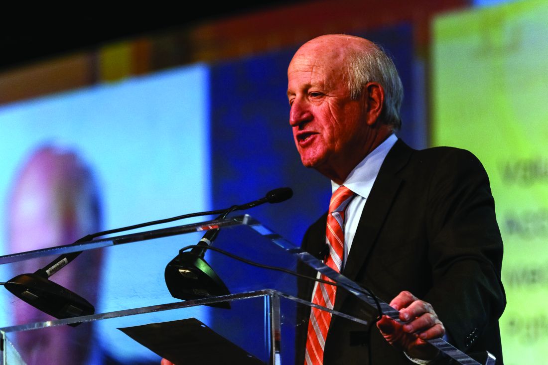
“How did Netflix pop up and not Blockbuster? Why did Sears not become Amazon?” he asked. “I think that SHM is very proud that we – very early on, when there were just a couple hundred hospitalists – saw the potential for this specialty.” At the same time, he said, “many of the other medical associations that were hundreds of years old not only did not see the potential for our specialty but, in many ways, were [also] somewhat contrary to the kinds of things that all of you have been doing over the last 20 years.”
He acknowledged that there were some “winds at our back” – it was clear that many physicians were wanting to leave the hospital – but he said the hospital medicine movement and the society nonetheless had to push up against resistance to change.
The movement continues to grow, he noted. As proof, there are 57,000 hospitalists, according to American Hospital Association surveys, and 15,000 SHM members.
In a note of caution, he recognized the many roles hospitalists are being asked to fill – from patient-safety projects to perioperative work – and said “hospitalists are more likely to say ‘yes’ when they should probably say ‘maybe.’ ” At the same time, he praised hospitalists for their openness to new ways of thinking and their willingness to partner.
Despite uncertainty, he said the future of health care is right up SHM’s alley.
“The future fits what hospital medicine is all about: We’re about value, we’re about creating the new future,” he said. “We’re about being accountable for what we do, being measured, and improving off those measures. ... We understand that we need to give up some of our autonomy and be in an integrated process to benefit from the expertise and the energies of other people.”
He touted the vital role that hospitalists will play.
“As we stand here today, we are not just large. We are not just the fastest-growing medical specialty of all time,” he said. “We are the answer to many of the questions going forward.”
So, the changing currents in medicine made it inevitable that the hospital medicine movement – and SHM – would have happened no matter what, right?
Not so much, said Laurence Wellikson, MD, MHM, chief executive officer of the Society of Hospital Medicine.
“How did Netflix pop up and not Blockbuster? Why did Sears not become Amazon?” he asked. “I think that SHM is very proud that we – very early on, when there were just a couple hundred hospitalists – saw the potential for this specialty.” At the same time, he said, “many of the other medical associations that were hundreds of years old not only did not see the potential for our specialty but, in many ways, were [also] somewhat contrary to the kinds of things that all of you have been doing over the last 20 years.”
He acknowledged that there were some “winds at our back” – it was clear that many physicians were wanting to leave the hospital – but he said the hospital medicine movement and the society nonetheless had to push up against resistance to change.
The movement continues to grow, he noted. As proof, there are 57,000 hospitalists, according to American Hospital Association surveys, and 15,000 SHM members.
In a note of caution, he recognized the many roles hospitalists are being asked to fill – from patient-safety projects to perioperative work – and said “hospitalists are more likely to say ‘yes’ when they should probably say ‘maybe.’ ” At the same time, he praised hospitalists for their openness to new ways of thinking and their willingness to partner.
Despite uncertainty, he said the future of health care is right up SHM’s alley.
“The future fits what hospital medicine is all about: We’re about value, we’re about creating the new future,” he said. “We’re about being accountable for what we do, being measured, and improving off those measures. ... We understand that we need to give up some of our autonomy and be in an integrated process to benefit from the expertise and the energies of other people.”
He touted the vital role that hospitalists will play.
“As we stand here today, we are not just large. We are not just the fastest-growing medical specialty of all time,” he said. “We are the answer to many of the questions going forward.”
Conway says health care payment, quality reform to continue
Keynote speaker Patrick Conway, MD, MSc, MHM, told hospitalists at HM17 Wednesday that, while there is a seemingly endless stream of punditry about the fate of the Affordable Care Act, health care will continue its trajectory to higher value, lower costs, and improved quality for patients.
“Health system transformation, innovation, delivery system reform, accountability, the work that you all do each and every day ... is a bipartisan ideal,” he said. The work “on value, the work on accountability, the work on bundled payments ... will continue and will continue to be important to you and the patients you serve.”
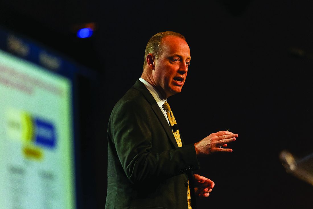
He also echoed Tuesday’s keynote address from Karen DeSalvo, MD, former Acting Assistant Secretary for Health in the U.S. Department of Health & Human Services, that hospital medicine needs to look at health care more holistically to help work on social issues. Dr. Conway, who still moonlights as a pediatric academic hospitalist on weekends, knows the problem first-hand as he often sees children on Medicaid who have multiple chronic conditions.
“I can tell you our system still does not have a highly reliable, whole health system for those children and their families,” he said. “Every weekend, I have a family that I can’t discharge because they don’t have the social and home-based supports for them to go home. So, they literally sit in the hospital until Monday. That makes no sense for our overall health system.”
Dr. Conway said that the gravitation away from fee-for-service toward alternative payment models would ideally lead to better patient outcomes, more coordinated care, and financial savings. He urged hospitalists to continue to help design new payment and care-delivery systems.
“You know what you’re passionate about and where you want to drive better care,” he said. “If the army of people in this room and all the places you are working [at] are the driver of better quality, better safety, coordinated care for patients ... that’s what it’s all about.”
Keynote speaker Patrick Conway, MD, MSc, MHM, told hospitalists at HM17 Wednesday that, while there is a seemingly endless stream of punditry about the fate of the Affordable Care Act, health care will continue its trajectory to higher value, lower costs, and improved quality for patients.
“Health system transformation, innovation, delivery system reform, accountability, the work that you all do each and every day ... is a bipartisan ideal,” he said. The work “on value, the work on accountability, the work on bundled payments ... will continue and will continue to be important to you and the patients you serve.”
He also echoed Tuesday’s keynote address from Karen DeSalvo, MD, former Acting Assistant Secretary for Health in the U.S. Department of Health & Human Services, that hospital medicine needs to look at health care more holistically to help work on social issues. Dr. Conway, who still moonlights as a pediatric academic hospitalist on weekends, knows the problem first-hand as he often sees children on Medicaid who have multiple chronic conditions.
“I can tell you our system still does not have a highly reliable, whole health system for those children and their families,” he said. “Every weekend, I have a family that I can’t discharge because they don’t have the social and home-based supports for them to go home. So, they literally sit in the hospital until Monday. That makes no sense for our overall health system.”
Dr. Conway said that the gravitation away from fee-for-service toward alternative payment models would ideally lead to better patient outcomes, more coordinated care, and financial savings. He urged hospitalists to continue to help design new payment and care-delivery systems.
“You know what you’re passionate about and where you want to drive better care,” he said. “If the army of people in this room and all the places you are working [at] are the driver of better quality, better safety, coordinated care for patients ... that’s what it’s all about.”

Hidradenitis suppurativa diagnosis typically delayed in children
WASHINGTON – Children with hidradenitis suppurativa (HS) may suffer with symptoms for an average of 7 years before they are diagnosed, according to pediatric dermatologist Anna Yasmine Kirkorian, MD.
Data from a 2015 study showed that 73% of pediatric patients with HS were diagnosed more than 2 years after the onset of symptoms, said Dr. Kirkorian of the department of dermatology at Children’s National Health System and George Washington University, Washington. (Br J Dermatol. 2015 Dec;173[6]:1546-9).
Genetics can play a role in HS, likely via mutations in the gamma-secretase protein that leads to epidermal differentiation and immune regulation, Dr. Kirkorian said. Most of her patients with HS are black, and a recent study described a gamma-secretase mutation in a black family of a proband and four family members, she noted (JAMA Dermatol. 2015 Jun;151[6]:668-70). Gamma-secretase mutations also have been identified in Han Chinese populations, she said.

HS also is associated with precocious puberty. However, defining the age of onset of puberty can be difficulty because pubertal onset may vary between different ethnicities, noted Dr. Kirkorian. “Prepubertal children presenting with HS warrant an endocrinologic evaluation,” she said.
Dr. Kirkorian added that more research is needed to pinpoint the possible genetic component of HS and to identify genetic susceptibility that could lead to targeted treatment strategies.
The optimal treatment plan for pediatric HS is multimodal and addresses the comorbidities common with the condition, she said, and she predicted that specialized clinic or treatment centers that bring together areas, including psychiatry, wound care, pain management, surgery, endocrinology, and genetics, will evolve to serve these patients. To support these collaborative efforts, Dr. Kirkorian is a member of the Pediatric Dermatology Research Alliance (PeDRA), an organization formed to accelerate research on skin diseases in children.
The symposium was sponsored by AbbVie. Dr. Kirkorian had no financial conflicts to disclose. She is on the editorial board of Dermatology News.
WASHINGTON – Children with hidradenitis suppurativa (HS) may suffer with symptoms for an average of 7 years before they are diagnosed, according to pediatric dermatologist Anna Yasmine Kirkorian, MD.
Data from a 2015 study showed that 73% of pediatric patients with HS were diagnosed more than 2 years after the onset of symptoms, said Dr. Kirkorian of the department of dermatology at Children’s National Health System and George Washington University, Washington. (Br J Dermatol. 2015 Dec;173[6]:1546-9).
Genetics can play a role in HS, likely via mutations in the gamma-secretase protein that leads to epidermal differentiation and immune regulation, Dr. Kirkorian said. Most of her patients with HS are black, and a recent study described a gamma-secretase mutation in a black family of a proband and four family members, she noted (JAMA Dermatol. 2015 Jun;151[6]:668-70). Gamma-secretase mutations also have been identified in Han Chinese populations, she said.
HS also is associated with precocious puberty. However, defining the age of onset of puberty can be difficulty because pubertal onset may vary between different ethnicities, noted Dr. Kirkorian. “Prepubertal children presenting with HS warrant an endocrinologic evaluation,” she said.
Dr. Kirkorian added that more research is needed to pinpoint the possible genetic component of HS and to identify genetic susceptibility that could lead to targeted treatment strategies.
The optimal treatment plan for pediatric HS is multimodal and addresses the comorbidities common with the condition, she said, and she predicted that specialized clinic or treatment centers that bring together areas, including psychiatry, wound care, pain management, surgery, endocrinology, and genetics, will evolve to serve these patients. To support these collaborative efforts, Dr. Kirkorian is a member of the Pediatric Dermatology Research Alliance (PeDRA), an organization formed to accelerate research on skin diseases in children.
The symposium was sponsored by AbbVie. Dr. Kirkorian had no financial conflicts to disclose. She is on the editorial board of Dermatology News.

From idea to abstract, RIV posters highlight novel thinking
Masih Shinwa, MD, stood beside a half-circle of judges Tuesday night at SHM’s annual Research, Innovations and Clinical Vignettes (RIV) poster competition and argued why his entry, already a finalist, should win. And to think, his poster, “Please ‘THINK’ Before You Order: A Multidisciplinary Approach to Decreasing Overutilization of Daily Labs,” was born simply of a group of medical students who incredulously said they were amazed patients would be woken in the middle of the night for laboratory tests.
Eighteen months later, the poster drew hundreds of questions from passersby on how a team approach could help generate fewer labs and chemistry testing. Now, the work could serve as a potential guide for other hospitals seeking to reduce unnecessary tests, a focal point of SHM and the ABIM Foundation’s Choosing Wisely campaign.
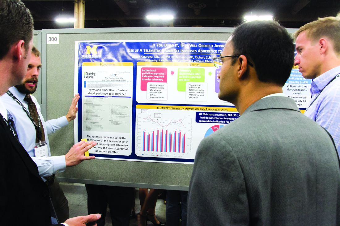
“This is a way to make it national,” he said. “You may have affected the lives of the patients in your hospital, but unless you attend these types of national meetings, it’s hard to get that perspective across [the country].”
That level of personal and professional collaboration is the purpose of the RIV, one of the best-attended events of SHM’s annual meeting. The event has become so popular that submissions this year tallied 1,712, nearly triple the number of submissions at the 2010 event.
“One of the amazing things is everyone has their own poster. They are doing their work,” said Margaret Fang, MD, MPH, FHM, program chair for HM17’s scientific abstracts competition, the formal name for the poster contest. “But then they start up conversations with the people next to them. ... seeing the organic networking and the discussions that arise from that is really exciting. RIV really serves as a way of connecting people together who might not have known the other person was doing that kind of work.”
Dr. Shinwa said that the specialty’s focus on research is important. He said it positions the field to be leaders, not just for patient care, but for hospitals and institutions.
“We are physicians. Our role is taking care of patients,” he said. “[But] knowing that there are people who are not just focusing on taking care of specific patients, but are actually there to improve the entire system and the process, that is really gratifying.”
Masih Shinwa, MD, stood beside a half-circle of judges Tuesday night at SHM’s annual Research, Innovations and Clinical Vignettes (RIV) poster competition and argued why his entry, already a finalist, should win. And to think, his poster, “Please ‘THINK’ Before You Order: A Multidisciplinary Approach to Decreasing Overutilization of Daily Labs,” was born simply of a group of medical students who incredulously said they were amazed patients would be woken in the middle of the night for laboratory tests.
Eighteen months later, the poster drew hundreds of questions from passersby on how a team approach could help generate fewer labs and chemistry testing. Now, the work could serve as a potential guide for other hospitals seeking to reduce unnecessary tests, a focal point of SHM and the ABIM Foundation’s Choosing Wisely campaign.
“This is a way to make it national,” he said. “You may have affected the lives of the patients in your hospital, but unless you attend these types of national meetings, it’s hard to get that perspective across [the country].”
That level of personal and professional collaboration is the purpose of the RIV, one of the best-attended events of SHM’s annual meeting. The event has become so popular that submissions this year tallied 1,712, nearly triple the number of submissions at the 2010 event.
“One of the amazing things is everyone has their own poster. They are doing their work,” said Margaret Fang, MD, MPH, FHM, program chair for HM17’s scientific abstracts competition, the formal name for the poster contest. “But then they start up conversations with the people next to them. ... seeing the organic networking and the discussions that arise from that is really exciting. RIV really serves as a way of connecting people together who might not have known the other person was doing that kind of work.”
Dr. Shinwa said that the specialty’s focus on research is important. He said it positions the field to be leaders, not just for patient care, but for hospitals and institutions.
“We are physicians. Our role is taking care of patients,” he said. “[But] knowing that there are people who are not just focusing on taking care of specific patients, but are actually there to improve the entire system and the process, that is really gratifying.”